4 Steps to Manage Your Diabetes for Life

Man and a woman cooking together

This publication has been reviewed by NDEP for plain language principles. Learn more about our review process.

Actions you can take

The check mark marks in this booklet show actions you can take to manage your diabetes.


  • check mark Help your health care team make a diabetes care plan that will work for you.

  • check mark Learn to make wise choices for your diabetes care each day.


Step 1: Learn about diabetes.

What is diabetes?

There are three main types of diabetes:

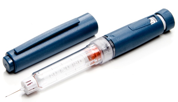
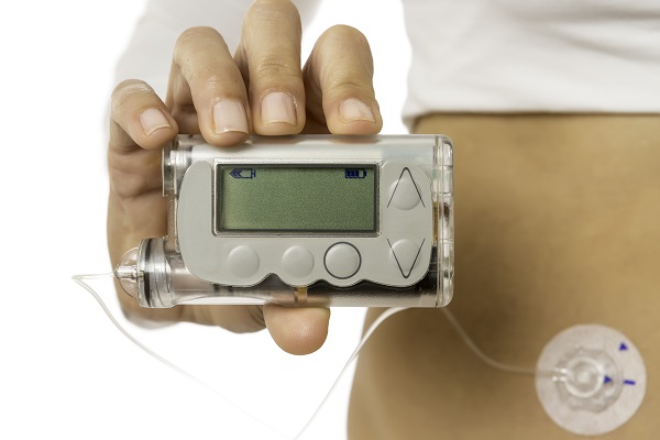
  • Type 1 diabetes – Your body does not make insulin. This is a problem because you need insulin to take the sugar (glucose) from the foods you eat and turn it into energy for your body. You need to take insulin every day to live.
  • Type 2 diabetes – Your body does not make or use insulin well. You may need to take pills or insulin to help control your diabetes. Type 2 is the most common type of diabetes.
  • Gestational (jest-TAY-shun-al) diabetes – Some women get this kind of diabetes when they are pregnant. Most of the time, it goes away after the baby is born. But even if it goes away, these women and their children have a greater chance of getting diabetes later in life.

You are the most important member of your health care team.

Older man with a younger woman talking to a doctor

You are the one who manages your diabetes day by day. Talk to your doctor about how you can best care for your diabetes to stay healthy. Some others who can help are:

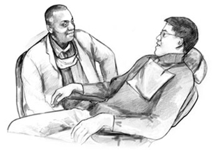
  • dentist
  • diabetes doctor
  • diabetes educator
  • dietitian
  • eye doctor
  • foot doctor
  • friends and family
  • mental health counselor
  • nurse
  • nurse practitioner
  • pharmacist
  • social worker

How to learn more about diabetes.

  • Take classes to learn more about living with diabetes. To find a class, check with your health care team, hospital, or area health clinic. You can also search online.
  • Join a support group — in-person or online — to get peer support with managing your diabetes.
  • Read about diabetes online. Go to National Diabetes Education Program.

Take diabetes seriously.

A man talks to his doctor

You may have heard people say they have “a touch of diabetes” or that their “sugar is a little high.” These words suggest that diabetes is not a serious disease. That is not correct. Diabetes is serious, but you can learn to manage it.

People with diabetes need to make healthy food choices, stay at a healthy weight, move more every day, and take their medicine even when they feel good. It’s a lot to do. It’s not easy, but it’s worth it!

Why take care of your diabetes?

Taking care of yourself and your diabetes can help you feel good today and in the future. When your blood sugar (glucose) is close to normal, you are likely to:

  • have more energy
  • be less tired and thirsty
  • need to pass urine less often
  • heal better
  • have fewer skin or bladder infections

You will also have less chance of having health problems caused by diabetes such as:

  • heart attack or stroke
  • eye problems that can lead to trouble seeing or going blind
  • pain, tingling, or numbness in your hands and feet, also called nerve damage
  • kidney problems that can cause your kidneys to stop working
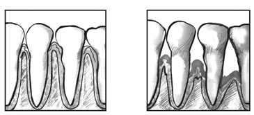
  • teeth and gum problems

check markActions you can take
  • check mark Ask your health care team what type of diabetes you have.

  • check mark Learn where you can go for support.

  • check mark Learn how caring for your diabetes helps you feel good today and in the future.


Step 2: Know your diabetes ABCs.

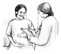
A woman speaking with her doctor

Talk to your health care team about how to manage your A1C, Blood pressure, and Cholesterol. This can help lower your chances of having a heart attack, stroke, or other diabetes problems.

A for the A1C test (A-one-C).

What is it?

The A1C is a blood test that measures your average blood sugar level over the past three months. It is different from the blood sugar checks you do each day.

Why is it important?

You need to know your blood sugar levels over time. You don’t want those numbers to get too high. High levels of blood sugar can harm your heart, blood vessels, kidneys, feet, and eyes.

What is the A1C goal?

The A1C goal for many people with diabetes is below 7. It may be different for you. Ask what your goal should be.

B for Blood pressure.

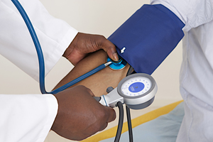
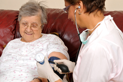
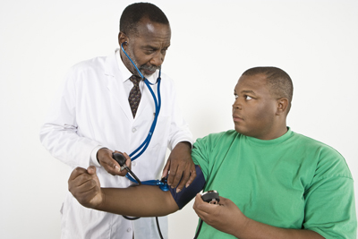
A nurse takes the blood pressure of an elderly lady

What is it?

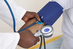
Blood pressure is the force of your blood against the wall of your blood vessels.

Why is it important?

If your blood pressure gets too high, it makes your heart work too hard. It can cause a heart attack, stroke, and damage your kidneys and eyes.

What is the blood pressure goal?

The blood pressure goal for most people with diabetes is below 140/90. It may be different for you. Ask what your goal should be.

C for Cholesterol (ko-LESS-tuh-ruhl).

What is it?

There are two kinds of cholesterol in your blood: LDL and HDL.

LDL or “bad” cholesterol can build up and clog your blood vessels. It can cause a heart attack or stroke.

HDL or “good” cholesterol helps remove the “bad” cholesterol from your blood vessels.

What are the LDL and HDL goals?

Ask what your cholesterol numbers should be. Your goals may be different from other people. If you are over 40 years of age, you may need to take a statin drug for heart health.

check markActions you can take

  • check mark Ask your health care team:
    • what your A1C, blood pressure, and cholesterol numbers are and what they should be. Your ABC goals will depend on how long you have had diabetes, other health problems, and how hard your diabetes is to manage.
    • what you can do to reach your ABC goals
  • check markWrite down your numbers on the record at the back of this booklet to track your progress.

Step 3: Learn how to live with diabetes.

An older asian man doing work in his garden

It is common to feel overwhelmed, sad, or angry when you are living with diabetes. You may know the steps you should take to stay healthy, but have trouble sticking with your plan over time. This section has tips on how to cope with your diabetes, eat well, and be active.

Cope with your diabetes.

  • Stress can raise your blood sugar. Learn ways to lower your stress. Try deep breathing, gardening, taking a walk, meditating, working on your hobby, or listening to your favorite music.
  • Ask for help if you feel down. A mental health counselor, support group, member of the clergy, friend, or family member who will listen to your concerns may help you feel better.

Eat well.

  • Make a diabetes meal plan with help from your health care team.
  • Choose foods that are lower in calories, saturated fat, trans fat, sugar, and salt.
  • Eat foods with more fiber, such as whole grain cereals, breads, crackers, rice, or pasta.
  • Choose foods such as fruits, vegetables, whole grains, bread and cereals, and low-fat or skim milk and cheese.
  • Drink water instead of juice and regular soda.
  • A plate of food with proportions listed
  • When eating a meal, fill half of your plate with fruits and vegetables, one quarter with a lean protein, such as beans, or chicken or turkey without the skin, and one quarter with a whole grain, such as brown rice or whole wheat pasta.

Be active.

A family walking
  • Set a goal to be more active most days of the week. Start slow by taking 10 minute walks, 3 times a day.
  • Twice a week, work to increase your muscle strength. Use stretch bands, do yoga, heavy gardening (digging and planting with tools), or try push-ups.
  • Stay at or get to a healthy weight by using your meal plan and moving more.

Know what to do every day.

  • Take your medicines for diabetes and any other health problems even when you feel good. Ask your doctor if you need aspirin to prevent a heart attack or stroke. Tell your doctor if you cannot afford your medicines or if you have any side effects.
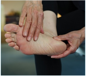
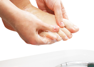
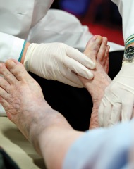
  • Check your feet every day for cuts, blisters, red spots, and swelling. Call your health care team right away about any sores that do not go away.
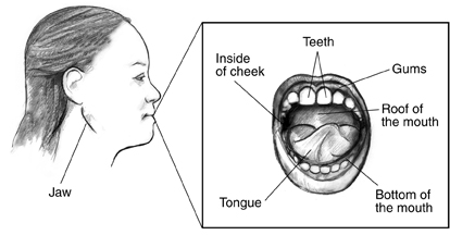
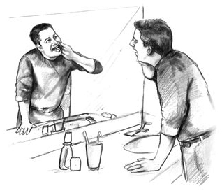
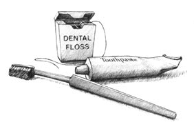
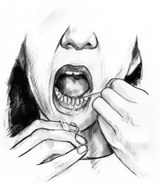
  • Brush your teeth and floss every day to keep your mouth, teeth, and gums healthy.
  • Stop smoking. Ask for help to quit. Call 1-800-QUITNOW (1-800-784-8669).
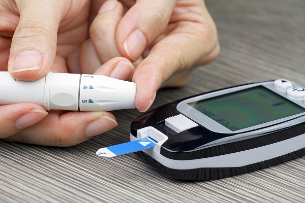
  • Keep track of your blood sugar. You may want to check it one or more times a day. Use the card at the back of this booklet to keep a record of your blood sugar numbers. Be sure to talk about it with your health care team.
  • Check your blood pressure if your doctor advises and keep a record of it.

Talk to your health care team.

  • Ask your doctor if you have any questions about your diabetes.
  • Report any changes in your health.

check markActions you can take
  • check markAsk for a healthy meal plan.
  • check markAsk about ways to be more active.
  • check markAsk how and when to test your blood sugar and how to use the results to manage your diabetes.
  • check markUse these tips to help with your self-care.
  • check markDiscuss how your diabetes plan is working for you each time you visit your health care team.

Step 4: Get routine care to stay healthy.

See your health care team at least twice a year to find and treat any problems early.

At each visit, be sure you have a:

  • blood pressure check
  • foot check
  • weight check
  • review of your self-care plan

Two times each year, have an:

  • A1C test. It may be checked more often if it is over 7.

Once each year, be sure you have a:

  • cholesterol test
  • complete foot exam
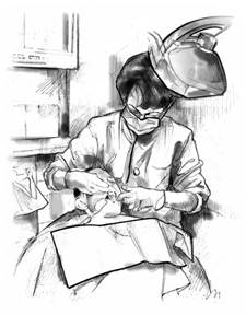
  • dental exam to check teeth and gums
  • dilated eye exam to check for eye problems
  • flu shot
  • urine and a blood test to check for kidney problems

At least once in your lifetime, get a:

  • pneumonia (nu-mo-nya) shot
  • hepatitis B (HEP-uh-TY-tiss) shot

Medicare and diabetes.

If you have Medicare, check to see how your plan covers diabetes care. Medicare covers some of the costs for:

  • diabetes education
  • diabetes supplies
  • diabetes medicine
  • visits with a dietitian
  • special shoes, if you need them

check markActions you can take

  • check markAsk your health care team about these and other tests you may need. Ask what your results mean.

  • check markWrite down the date and time of your next visit.

  • check markUse the card at the back of this booklet to keep a record of your diabetes care.

  • check markIf you have Medicare, check your plan.


Things to Remember:

  • You are the most important member of your health care team.
  • Follow the four steps in this booklet to help you learn how to manage your diabetes.
  • Learn how to reach your diabetes ABC goals.
  • Ask your health care team for help.

My Diabetes Care Record: Page 1

How to use the record.

First read the shaded bar across the page. This tells you:

  • the name of the test or check-up
  • how often to get the test or check-up
  • what your personal goal is (for A1C, blood pressure, and cholesterol)

Then, write down the date and results for each test or check-up you get. Take this card with you on your health care visits. Show it to your health care team. Talk about your goals and how you are doing.

A1C – At least twice each year My goal: ______
Date


Result


Blood Pressure (BP) – At each visit My goal: ______
Date


Result


Cholesterol – Once each year My goal: ______
Date


Result


My Diabetes Care Record: Page 2

How to use the record.

Use this page to write down the date and results of each test, exam, or shot.

Each Visit Date Result
Foot check

Review self-care plan

Weight check

Review medicines

Once a Year Date Result
Dental exam

Dilated eye exam

Complete foot exam

Flu shot

Kidney check

At Least Once Date Result
Pneumonia shot

Hepatitis B shot

Self Checks of Blood Sugar

How to use this card.

This card has three sections. Each section tells you when to check your blood sugar: before each meal, 1 to 2 hours after each meal, and at bedtime. Each time you check your blood sugar, write down the date, time, and results. Take this card with you on your health care visits. Show it to your health care team. Talk about your goals and how you are doing.


Date Time Result
My blood sugar before meals:
Usual goal 80 to 130*
My goal: ________


My blood sugar 1-2 hours after meals:
Usual goal below 180*
My goal: ________


My blood sugar at bedtime:
Usual goal 110 to 150*
My goal: ________


* Your blood sugar goals may be different if you are an older adult (over 65) and have had diabetes a long time. They may be different if you have other health problems like heart disease, or your blood sugar often gets too low.

The U.S. Department of Health and Human Services’ National Diabetes Education Program (NDEP) is jointly sponsored by the National Institutes of Health (NIH) and the Centers for Disease Control and Prevention (CDC) with the support of more than 200 partner organizations.

This content is provided as a service of the National Institute of Diabetes and Digestive and Kidney Diseases (NIDDK), part of the National Institutes of Health. The NIDDK translates and disseminates research findings through its clearinghouses and education programs to increase knowledge and understanding about health and disease among patients, health professionals, and the public. Content produced by the NIDDK is carefully reviewed by NIDDK scientists and other experts.

Abdominal Adhesions

What are abdominal adhesions?

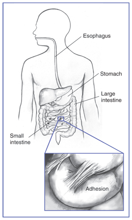
Abdominal adhesions are bands of fibrous tissue that can form between abdominal tissues and organs. Normally, internal tissues and organs have slippery surfaces, preventing them from sticking together as the body moves. However, abdominal adhesions cause tissues and organs in the abdominal cavity to stick together.

Drawing of the gastrointestinal tract showing the esophagus, stomach, and large intestine. Inset shows abdominal adhesions on the small intestine.
Abdominal adhesions are bands of fibrous tissue that can form between abdominal tissues and organs.

What is the abdominal cavity?

The abdominal cavity is the internal area of the body between the chest and hips that contains the lower part of the esophagus, stomach, small intestine, and large intestine. The esophagus carries food and liquids from the mouth to the stomach, which slowly pumps them into the small and large intestines. Abdominal adhesions can kink, twist, or pull the small and large intestines out of place, causing an intestinal obstruction. Intestinal obstruction, also called a bowel obstruction, results in the partial or complete blockage of movement of food or stool through the intestines.

What causes abdominal adhesions?

Abdominal surgery is the most frequent cause of abdominal adhesions. Surgery-related causes include

  • cuts involving internal organs
  • handling of internal organs
  • drying out of internal organs and tissues
  • contact of internal tissues with foreign materials, such as gauze, surgical gloves, and stitches
  • blood or blood clots that were not rinsed away during surgery

Abdominal adhesions can also result from inflammation not related to surgery, including

  • appendix rupture
  • radiation treatment
  • gynecological infections
  • abdominal infections

Rarely, abdominal adhesions form without apparent cause.

How common are abdominal adhesions and who is at risk?

Of patients who undergo abdominal surgery, 93 percent develop abdominal adhesions.1 Surgery in the lower abdomen and pelvis, including bowel and gynecological operations, carries an even greater chance of abdominal adhesions. Abdominal adhesions can become larger and tighter as time passes, sometimes causing problems years after surgery.

What are the symptoms of abdominal adhesions?

In most cases, abdominal adhesions do not cause symptoms. When symptoms are present, chronic abdominal pain is the most common.

What are the complications of abdominal adhesions?

Abdominal adhesions can cause intestinal obstruction and female infertility—the inability to become pregnant after a year of trying.

Abdominal adhesions can lead to female infertility by preventing fertilized eggs from reaching the uterus, where fetal development takes place. Women with abdominal adhesions in or around their fallopian tubes have an increased chance of ectopic pregnancy—a fertilized egg growing outside the uterus. Abdominal adhesions inside the uterus may result in repeated miscarriages—a pregnancy failure before 20 weeks.

How are abdominal adhesions and intestinal obstructions diagnosed?

Abdominal adhesions cannot be detected by tests or seen through imaging techniques such as x-rays or ultrasound. Most abdominal adhesions are found during surgery performed to examine the abdomen. However, abdominal x-rays, a lower gastrointestinal (GI) series, and computerized tomography (CT) scans can diagnose intestinal obstructions.

  • Abdominal x-rays use a small amount of radiation to create an image that is recorded on film or a computer. An x-ray is performed at a hospital or an outpatient center by an x-ray technician, and the images are interpreted by a radiologist—a doctor who specializes in medical imaging. An x-ray does not require anesthesia. The person will lie on a table or stand during the x-ray. The x-ray machine is positioned over the abdominal area. The person will hold his or her breath as the picture is taken so that the picture will not be blurry. The person may be asked to change position for additional pictures.
  • A lower GI series is an x-ray exam that is used to look at the large intestine. The test is performed at a hospital or an outpatient center by an x-ray technician, and the images are interpreted by a radiologist. Anesthesia is not needed. The health care provider may provide written bowel prep instructions to follow at home before the test. The person may be asked to follow a clear liquid diet for 1 to 3 days before the procedure. A laxative or an enema may be used before the test. A laxative is medication that loosens stool and increases bowel movements. An enema involves flushing water or laxative into the rectum using a special squirt bottle.

    For the test, the person will lie on a table while the radiologist inserts a flexible tube into the person’s anus. The large intestine is filled with barium, making signs of underlying problems show up more clearly on x-rays.
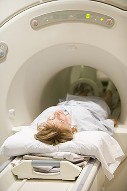
  • CT scans use a combination of x-rays and computer technology to create images. The procedure is performed at a hospital or an outpatient center by an x-ray technician, and the images are interpreted by a radiologist. Anesthesia is not needed. A CT scan may include the injection of a special dye, called contrast medium. The person will lie on a table that slides into a tunnel-shaped device where the x-rays are taken.

How are abdominal adhesions and intestinal obstructions treated?

Abdominal adhesions that do not cause symptoms generally do not require treatment. Surgery is the only way to treat abdominal adhesions that cause pain, intestinal obstruction, or fertility problems. More surgery, however, carries the risk of additional abdominal adhesions. People should speak with their health care provider about the best way to treat their abdominal adhesions.

Complete intestinal obstructions usually require immediate surgery to clear the blockage. Most partial intestinal obstructions can be managed without surgery.

How can abdominal adhesions be prevented?

Abdominal adhesions are difficult to prevent; however, certain surgical techniques can minimize abdominal adhesions.

Laparoscopic surgery decreases the potential for abdominal adhesions because several tiny incisions are made in the lower abdomen instead of one large incision. The surgeon inserts a laparoscope—a thin tube with a tiny video camera attached—into one of the small incisions. The camera sends a magnified image from inside the body to a video monitor. Patients will usually receive general anesthesia during this surgery.

If laparoscopic surgery is not possible and a large abdominal incision is required, at the end of surgery a special filmlike material can be inserted between organs or between the organs and the abdominal incision. The filmlike material, which looks similar to wax paper and is absorbed by the body in about a week, hydrates organs to help prevent abdominal adhesions.

Other steps taken during surgery to reduce abdominal adhesions include

  • using starch- and latex-free gloves
  • handling tissues and organs gently
  • shortening surgery time
  • using moistened drapes and swabs
  • occasionally applying saline solution

Eating, Diet, and Nutrition

Researchers have not found that eating, diet, and nutrition play a role in causing or preventing abdominal adhesions. A person with a partial intestinal obstruction may relieve symptoms with a liquid or low-fiber diet, which is more easily broken down into smaller particles by the digestive system.

Points to Remember

  • Abdominal adhesions are bands of fibrous tissue that can form between abdominal tissues and organs. Abdominal adhesions cause tissues and organs in the abdominal cavity to stick together.
  • Abdominal surgery is the most frequent cause of abdominal adhesions. Of patients who undergo abdominal surgery, 93 percent develop abdominal adhesions.
  • In most cases, abdominal adhesions do not cause symptoms. When symptoms are present, chronic abdominal pain is the most common.
  • A complete intestinal obstruction is life threatening and requires immediate medical attention and often surgery.
  • Abdominal adhesions cannot be detected by tests or seen through imaging techniques such as x-rays or ultrasound. However, abdominal x-rays, a lower gastrointestinal (GI) series, and computerized tomography (CT) scans can diagnose intestinal obstructions.
  • Surgery is the only way to treat abdominal adhesions that cause pain, intestinal obstruction, or fertility problems.

References

Clinical Trials

The National Institute of Diabetes and Digestive and Kidney Diseases (NIDDK) and other components of the National Institutes of Health (NIH) conduct and support research into many diseases and conditions.

What are clinical trials, and are they right for you?

Clinical trials are part of clinical research and at the heart of all medical advances. Clinical trials look at new ways to prevent, detect, or treat disease. Researchers also use clinical trials to look at other aspects of care, such as improving the quality of life for people with chronic illnesses. Find out if clinical trials are right for you.

What clinical trials are open?

Clinical trials that are currently open and are recruiting can be viewed at www.ClinicalTrials.gov.

This content is provided as a service of the National Institute of Diabetes and Digestive and Kidney Diseases (NIDDK), part of the National Institutes of Health. The NIDDK translates and disseminates research findings through its clearinghouses and education programs to increase knowledge and understanding about health and disease among patients, health professionals, and the public. Content produced by the NIDDK is carefully reviewed by NIDDK scientists and other experts.

The NIDDK would like to thank:
James M. Becker, M.D., F.A.C.S.; Arthur F. Stucchi, Ph.D.; Karen L. Reed, Ph.D.

Acquired Cystic Kidney Disease

What is acquired cystic kidney disease?

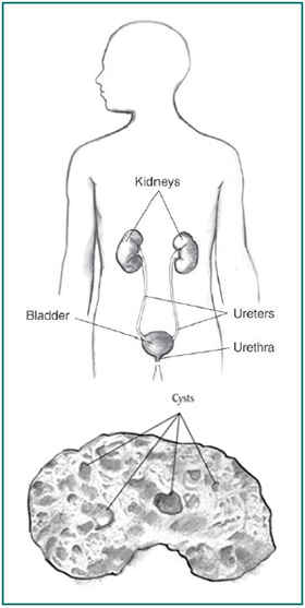
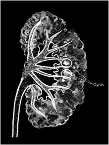
Acquired cystic kidney disease happens when a person's kidneys develop fluid-filled sacs, called cysts, over time. Acquired cystic kidney disease is not the same as polycystic kidney disease (PKD), another disease that causes the kidneys to develop multiple cysts.

Acquired cystic kidney disease occurs in children and adults who have

  • chronic kidney disease (CKD)—a condition that develops over many years and may lead to end-stage kidney disease, or ESRD. The kidneys of people with CKD gradually lose their ability to filter wastes, extra salt, and fluid from the blood properly.
  • end-stage kidney disease—total and permanent kidney failure that requires a kidney transplant or blood-filtering treatments called dialysis.

The cysts are more likely to develop in people who are on kidney dialysis. The chance of developing acquired cystic kidney disease increases with the number of years a person is on dialysis. However, the cysts are caused by CKD or kidney failure, not dialysis treatments.

More information is provided in the NIDDK health topics, kidney failure and dialysis.

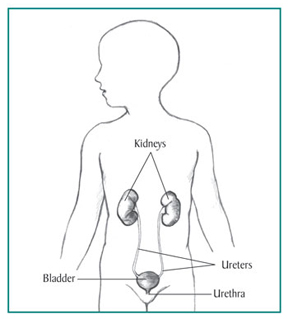
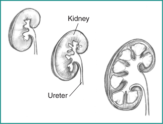
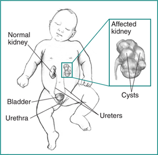
Drawing of a male torso with kidneys, ureters, bladder, and urethra labeled, and a drawing below of a kidney with sacs of fluid labeled as
Acquired cystic kidney disease happens when a person's kidneys develop fluid-filled sacs called cysts.

What are the differences between acquired cystic kidney disease and polycystic kidney disease?

Acquired cystic kidney disease differs from PKD in several ways. Unlike acquired cystic kidney disease, PKD is a genetic, or inherited, disorder that can cause complications such as high blood pressure and problems with blood vessels in the brain and heart.

The following chart lists the differences:

People with Polycystic Kidney Disease

  • are born with a gene that causes the disease
  • have enlarged kidneys
  • develop cysts in the liver and other parts of the body

People with Acquired Cystic Kidney Disease

  • do not have a disease-causing gene
  • have kidneys that are normal-sized or smaller
  • do not form cysts in other parts of the body

In addition, for people with PKD, the presence of cysts marks the onset of their disease, while people with acquired cystic kidney disease already have CKD when they develop cysts.

More information is provided in the NIDDK health topic, Polycystic Kidney Disease.

How common is acquired cystic kidney disease?

Acquired cystic kidney disease becomes more common the longer a person has CKD.

  • About 7 to 22 percent of people with CKD already have acquired cystic kidney disease before starting dialysis treatments.
  • Almost 60 percent of people on dialysis for 2 to 4 years develop acquired cystic kidney disease.1
  • About 90 percent of people on dialysis for 8 years develop acquired cystic kidney disease.1

What causes acquired cystic kidney disease?

Researchers do not fully understand what causes cysts to grow in the kidneys of people with CKD. The fact that these cysts occur only in the kidneys and not in other parts of the body, as in PKD, indicates that the processes that lead to cyst formation take place primarily inside the kidneys.2

What are the signs and symptoms of acquired cystic kidney disease?

A person with acquired cystic kidney disease often has no symptoms. However, the complications of acquired cystic kidney disease can have signs and symptoms.

What are the complications of acquired cystic kidney disease?

People with acquired cystic kidney disease may develop the following complications:

  • an infected cyst, which can cause fever and back pain.
  • blood in the urine, which can signal that a cyst in the kidney is bleeding.
  • tumors in the kidneys. People with acquired cystic kidney disease are more likely than people in the general population to have cancerous kidney tumors. However, the chance of cancer spreading is lower in people with acquired cystic kidney disease than that of other kidney cancers not associated with acquired cystic kidney disease, and the long-term outlook is better.1

How is acquired cystic kidney disease diagnosed?

A health care provider may diagnose a person with acquired cystic kidney disease based on

  • medical history
  • imaging tests

Medical History

Taking a medical history may help a health care provider diagnose acquired cystic kidney disease. A health care provider may suspect acquired cystic kidney disease if a person who has been on dialysis for several years develops symptoms such as fever, back pain, or blood in the urine.

Imaging Tests

To confirm the diagnosis, the health care provider may order one or more imaging tests. A radiologist—a doctor who specializes in medical imaging—interprets the images from these tests, and the patient does not need anesthesia.

  • Ultrasound uses a device, called a transducer, that bounces safe, painless sound waves off organs to create an image of their structure. A specially trained technician performs the procedure in a health care provider's office, an outpatient center, or a hospital. The images can show cysts in the kidneys as well as the kidneys' size and shape.
  • Computerized tomography (CT) scans use a combination of x-rays and computer technology to create images. For a CT scan, a nurse or technician may give the patient a solution to drink and an injection of a special dye, called contrast medium. CT scans require the patient to lie on a table that slides into a tunnel-shaped device where an x-ray technician takes the x-rays. An x-ray technician performs the procedure in an outpatient center or a hospital. CT scans can show cysts and tumors in the kidneys.
  • Magnetic resonance imaging (MRI) is a test that takes pictures of the body's internal organs and soft tissues without using x-rays. A specially trained technician performs the procedure in an outpatient center or a hospital. Although the patient does not need anesthesia, a health care provider may give people with a fear of confined spaces light sedation, taken by mouth. An MRI may include the injection of contrast medium. With most MRI machines, the patient will lie on a table that slides into a tunnel-shaped device that may be open-ended or closed at one end. Some machines allow the patient to lie in a more open space. During an MRI, the patient, although usually awake, must remain perfectly still while the technician takes the images, which usually takes only a few minutes. The technician will take a sequence of images from different angles to create a detailed picture of the kidneys. During the test, the patient will hear loud mechanical knocking and humming noises from the machine.

Sometimes a health care provider may discover acquired cystic kidney disease during an imaging exam for another condition. Images of the kidneys may help the health care provider distinguish acquired cystic kidney disease from PKD.

How is acquired cystic kidney disease treated?

If acquired cystic kidney disease is not causing complications, a person does not need treatment. A health care provider will treat infections with antibiotics—medications that kill bacteria. If large cysts are causing pain, a health care provider may drain the cyst using a long needle inserted into the cyst through the skin.

When a surgeon transplants a new kidney into a patient's body to treat kidney failure, acquired cystic kidney disease in the damaged kidneys, which usually remain in place after a transplant, often disappears.

A surgeon may perform an operation to remove tumors or suspected tumors. In rare cases, a surgeon performs an operation to stop cysts from bleeding.

Eating, Diet, and Nutrition

No specific diet will prevent or delay acquired cystic kidney disease. In general, a diet designed for people on hemodialysis or peritoneal dialysis reduces the amount of wastes that accumulate in the body between dialysis sessions.

More information is provided in the NIDDK health topics, Eat Right to Feel Right on Hemodialysis and Nutrition for Advanced Chronic Kidney Disease in Adults.

Points to Remember

  • Acquired cystic kidney disease happens when a person's kidneys develop fluid-filled sacs, called cysts, over time.
  • Acquired cystic kidney disease occurs in children and adults who have
    • chronic kidney disease (CKD)
    • end-stage kidney disease (ESRD)
  • People with acquired cystic kidney disease may develop the following complications:
    • an infected cyst, which can cause fever and back pain
    • blood in the urine, which can signal that a cyst in the kidney is bleeding
    • tumors in the kidneys
  • To confirm the diagnosis, the health care provider may order one or more imaging tests:
    • Ultrasound
    • Computerized tomography (CT) scan
    • Magnetic resonance imaging (MRI)
  • If acquired cystic kidney disease is not causing complications, a person does not need treatment.
  • A health care provider will treat infections with antibiotics—medications that kill bacteria.
  • If large cysts are causing pain, a health care provider may drain the cyst using a long needle inserted into the cyst through the skin.
  • A surgeon may perform an operation to remove tumors or suspected tumors. In rare cases, a surgeon performs an operation to stop cysts from bleeding.

References

Clinical Trials

The National Institute of Diabetes and Digestive and Kidney Diseases (NIDDK) and other components of the National Institutes of Health (NIH) conduct and support research into many diseases and conditions.

What are clinical trials, and are they right for you?

Clinical trials are part of clinical research and at the heart of all medical advances. Clinical trials look at new ways to prevent, detect, or treat disease. Researchers also use clinical trials to look at other aspects of care, such as improving the quality of life for people with chronic illnesses. Find out if clinical trials are right for you.

What clinical trials are open?

Clinical trials that are currently open and are recruiting can be viewed at www.ClinicalTrials.gov.

This content is provided as a service of the National Institute of Diabetes and Digestive and Kidney Diseases (NIDDK), part of the National Institutes of Health. The NIDDK translates and disseminates research findings through its clearinghouses and education programs to increase knowledge and understanding about health and disease among patients, health professionals, and the public. Content produced by the NIDDK is carefully reviewed by NIDDK scientists and other experts.

The NIDDK would like to thank:
Catherine Kelleher, M.D., University of Colorado Health Sciences Center

Albuminuria: Albumin in the Urine

What is albuminuria?

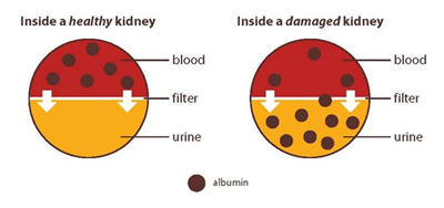
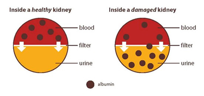
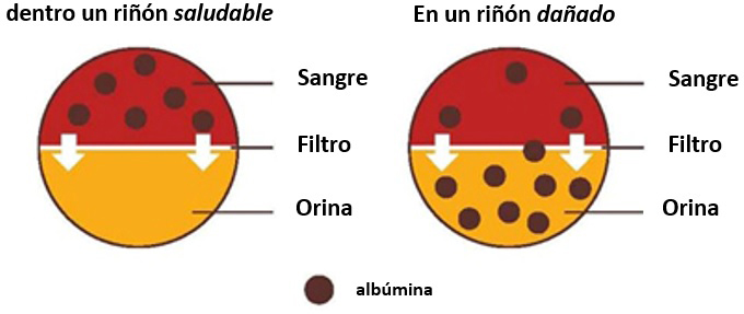
Albuminuria is a sign of kidney disease and means that you have too much albumin in your urine. Albumin is a protein found in the blood. A healthy kidney doesn’t let albumin pass from the blood into the urine. A damaged kidney lets some albumin pass into the urine. The less albumin in your urine, the better.

Sometimes albuminuria is also called proteinuria.

A diagram illustrating a healthy kidney with albumin only found in blood, and a damaged kidney that has albumin in both blood and urine.
A healthy kidney doesn’t let albumin pass into the urine. A damaged kidney lets some albumin pass into the urine.

More information is provided in the NIDDK health topics, Diabetic Kidney Disease and High Blood Pressure and Kidney Disease.

Why is urine albumin important?

Measurement of urine albumin is an important tool for

  • diagnosing kidney disease
  • monitoring the progression of kidney disease

Health care providers regularly test people for albuminuria as part of a routine medical exam and will closely monitor urine albumin in people with kidney disease.

A urine albumin level that stays the same or goes down may mean that treatments are working. Treatment that lowers the urine albumin level may lower the chances that kidney disease will progress to kidney failure.

People who have diabetes, high blood pressure, heart disease, or a family history of kidney failure are at risk for kidney disease. Talk with your health care provider about how often you should get a urine test for albumin.

How is albuminuria detected?

A health care provider often tests for albuminuria using a urine dipstick test followed by a urine albumin and creatinine measurement.

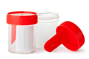
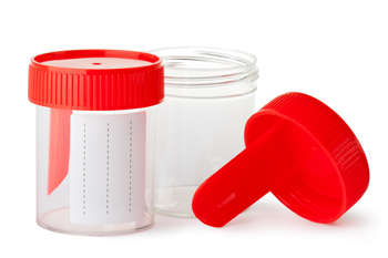
You will be asked to collect a urine sample in a special container in your health care provider’s office or a commercial facility. The office or facility tests the sample onsite or sends it to a lab for analysis.

Dipstick test for albumin. A dipstick test performed on a urine sample can detect the presence of albumin in the urine. For the test, a nurse or technician places a dipstick, a strip of chemically treated paper, into the urine. The dipstick changes color if albumin is present in the urine.

Albumin and creatinine measurement. A health care provider uses this measurement to determine the ratio between the albumin and creatinine in the urine and to estimate the amount of albumin excreted in 24 hours. Creatinine is a waste product that is filtered in the kidneys and excreted in the urine. Health care providers consider a urine albumin-to-creatinine ratio above 30 mg/g higher than normal.

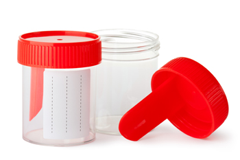
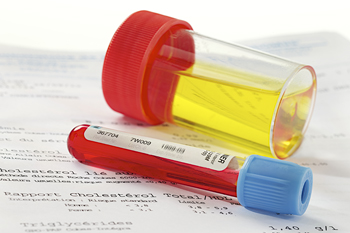
Photo of two containers that store urine to be tested for albumin.
If you have kidney disease or are at risk for kidney disease, talk with your health care provider about how often you should get a urine test for albumin.

How can albuminuria be reduced?

You may be able to reduce the amount of albumin in your urine by taking medicines that lower blood pressure called ACE inhibitors or ARBs. The names of these medicines end in -pril or -sartan.

You may also be able to protect your kidneys and reduce albuminuria by working with a registered dietitian who can help you plan meals and change your eating habits. The meal plan may help you

A dietitian discusses healthy eating habits and how to plan meals with a patient.
Meet with a dietitian who can help you plan meals and change your eating habits.

Clinical Trials

The National Institute of Diabetes and Digestive and Kidney Diseases (NIDDK) and other components of the National Institutes of Health (NIH) conduct and support research into many diseases and conditions.

What are clinical trials, and are they right for you?

Clinical trials are part of clinical research and at the heart of all medical advances. Clinical trials look at new ways to prevent, detect, or treat disease. Researchers also use clinical trials to look at other aspects of care, such as improving the quality of life for people with chronic illnesses. Find out if clinical trials are right for you.

What clinical trials are open?

Clinical trials that are currently open and are recruiting can be viewed at www.ClinicalTrials.gov.

This content is provided as a service of the National Institute of Diabetes and Digestive and Kidney Diseases (NIDDK), part of the National Institutes of Health. The NIDDK translates and disseminates research findings through its clearinghouses and education programs to increase knowledge and understanding about health and disease among patients, health professionals, and the public. Content produced by the NIDDK is carefully reviewed by NIDDK scientists and other experts.

Alimentación y nutrición durante la hemodiálisis

Cómo usar esta publicación

Cuando comienza a someterse a la hemodiálisis, debe hacer muchos cambios en su vida. Será más sano si presta atención a los alimentos que come. Esta publicación le ayudará a escoger los alimentos adecuados.

Imprima esta publicación y úsela con un dietista para aprender a comer bien para sentirse bien durante el tratamiento de hemodiálisis. Lea una sección a la vez. Luego, haga el ejercicio para cada sección junto con su dietista.

Una vez que haya completado cada ejercicio, guarde una copia de esta publicación para recordar qué alimentos puede comer y cuáles debe evitar.

El nombre de mi dietista es: ________________

Teléfono: ________________

¿Cómo afectan los alimentos a la hemodiálisis?

Los alimentos le proporcionan energía y ayudan al cuerpo a repararse. Los alimentos se deshacen en el estómago y los intestinos. La sangre recoge los nutrientes de los alimentos digeridos y los transporta a todas las células del cuerpo. Estas células toman los nutrientes de la sangre y vierten al flujo sanguíneo los productos de desecho. Cuando sus riñones estaban sanos, funcionaban todo el tiempo para eliminar los desechos de la sangre. Los desechos salían de su cuerpo cuando usted orinaba. Otros desechos se eliminan con las evacuaciones.

Ahora que sus riñones han dejado de funcionar, la hemodiálisis elimina los desechos de la sangre. Pero entre sesiones de diálisis, los desechos pueden acumularse en la sangre y enfermarle. Puede disminuir la cantidad de desechos cuidando lo que come y bebe. Un buen plan de comidas puede mejorar la diálisis y su salud.

Ilustración de una paciente hablando con un nutricionista. La nutricionista tiene un libro abierto y esta apuntando hacia una foto en la pirámide alimentaria.
Hable con un dietista para aprender a comer bien durante su tratamiento de hemodiálisis.

En su clínica hay un dietista que le ayudará a planificar sus comidas. Un dietista se especializa en alimentos y nutrición. Un dietista con formación especial en la salud de los riñones se llama dietista renal.

¿Qué debo saber sobre los líquidos?

Usted ya sabe que debe prestar atención a cuánto líquido bebe. Todo alimento que esté líquido a temperatura ambiente también contiene agua. Algunos ejemplos son la sopa, la gelatina (Jell-O) y el helado. Muchas frutas y vegetales también contienen mucha agua. Entre ellas se incluyen los melones, las uvas, las manzanas, las naranjas, los tomates, la lechuga y el apio. Todos estos alimentos aumentan su consumo de líquido.

Controle su sed

La mejor manera de disminuir el consumo de líquidos es reducir la sed causada por la sal que come. Evite los alimentos salados como las papitas y los pretzels. Elija productos con bajo contenido de sodio.

Puede mantener bajo su consumo de líquidos bebiendo en tazas o vasos más pequeños. Congele el jugo en una cubitera para hacer hielo y cómalo como si fuera una paleta helada. (¡Cuente esa paleta helada dentro de sus líquidos permitidos!) El dietista le puede dar otros consejos para controlar su sed.

Los líquidos pueden acumularse entre sesiones de diálisis causando hinchazón y aumento de peso. El líquido adicional afecta su presión arterial y podría hacer que a su corazón le cueste más trabajo funcionar. Podría padecer graves problemas cardíacos por sobrecargar su cuerpo de líquidos.

Su "peso seco" es su peso luego de una sesión de diálisis, cuando se ha eliminado todo el líquido de sobra de su cuerpo. Si deja que se acumule demasiado líquido entre sesiones, es más difícil bajar hasta su peso seco adecuado. Su peso seco puede cambiar durante un período de 3 a 6 semanas. Hable con su médico regularmente acerca de cuál debería ser su peso seco.

Mi peso seco debería ser _______________.

Ilustración de varios alimentos que contienen agua, entre los que se incluyen uvas, apio, helado, una naranja, gelatina y sopa. Los alimentos aparecen en la parte izquierda del dibujo. En la parte derecha hay un jarrón de agua. Un signo de igual que se encuentra en la mitad indica que ingerir los alimentos representados en la ilustración es lo mismo que beber agua.
Muchos alimentos contienen agua.

Hable con un dietista

Pese a estar sometido a hemodiálisis, es posible que sus riñones todavía puedan eliminar algo de líquido. O puede que no eliminen nada de líquido. Es por eso que para cada paciente se recomienda una diferente cantidad diaria de líquidos. Hable con su dietista sobre cuánto líquido usted puede consumir al día.

Puedo consumir _________ onzas de líquido al día.

Planifique 1 día de porciones de líquidos:

Puedo consumir _________ onza(s) de
________________ en el desayuno.

Puedo consumir _________ onza(s) de
______________durante la mañana.

Puedo consumir _________ onza(s) de
________________ en el almuerzo.

Puedo consumir _________ onza(s) de
______________durante la tarde.

Puedo consumir _________ onza(s) de
________________ en la cena.

Puedo consumir _________ onza(s) de
______________ durante la noche.

TOTAL: _________ onzas (debe ser igual a la misma cantidad anotada donde dice "Puedo consumir _______ onzas de líquido al día").

NOTA: 1 onza = .03 litros/30 mL

Ilustración de una balanza de alimentos, una taza graduada, tazas de medidas y cucharas sobre una mesada de cocina.
Lleve un registro detallado de sus líquidos y demás alimentos.

¿Qué debo saber sobre el potasio?

El potasio es un mineral presente en muchos alimentos, en especial en la leche, las frutas y los vegetales. Este afecta la regularidad del ritmo del corazón. Los riñones sanos conservan la cantidad adecuada de potasio en la sangre para que el corazón siga latiendo a un ritmo regular. Los niveles de potasio pueden subir entre sesiones de diálisis y afectar su ritmo cardíaco. Comer demasiado potasio puede ser muy peligroso para el corazón. Hasta puede llegar a provocar la muerte.

Ilustración de papas cortadas en cubo hirviendo en una olla. Detrás de la olla hay una papa cortada en cubos en una tabla de picar.
Puede eliminar parte del potasio de las papas al cortarlas o picarlas y luego hervirlas en agua.

Para controlar sus niveles de potasio en la sangre, evite alimentos con altos contenidos de potasio como aguacates, bananas/plátanos, kiwis y frutas deshidratadas. Además, coma porciones más pequeñas de otros alimentos con alto contenido de potasio. Por ejemplo, coma media pera en lugar de una pera entera. Coma sólo porciones muy pequeñas de naranjas y melones.

Reduciendo parte del potasio al cortar y hervir las papas

Puede eliminar parte del potasio de las papas al cortarlas o picarlas y luego hervirlas en agua. El dietista le dará información más específica sobre el contenido de potasio de los alimentos.

Hable con un dietista

Elabore un plan de comidas que disminuya el potasio de su dieta. Comience marcando los alimentos con alto contenido de potasio que come hoy en día usando la siguiente lista. El dietista puede ayudarle a agregar otros alimentos a la lista.

Alimentos con alto contenido de potasio:
damascos/chabacanos
aguacates
bananas/plátanos
remolachas
coles de bruselas
cantalupo/bardado
almejas
dátiles
higos
kiwis
habas
melones
leche
nectarinas
jugo de naranja
naranjas
cacahuates/maní
peras (frescas)
papas
jugo de ciruelas pasas
ciruelas pasas
pasas de uva
sardinas
espinaca
tomates
calabaza de invierno
yogur
Otros: ___________________________________________

Cambios

Hable con un dietista sobre los alimentos que puede comer en lugar de los alimentos con alto contenido de potasio.

En lugar de _______________, comeré ________________.

En lugar de _______________, comeré ________________.

En lugar de _______________, comeré ________________.

En lugar de _______________, comeré ________________.

¿Qué debo saber sobre el fósforo?

El fósforo es un mineral presente en muchos alimentos. El exceso de fósforo en la sangre quita el calcio de los huesos. La pérdida de calcio debilitará los huesos y aumentará la probabilidad de sufrir fracturas. Además, demasiado fósforo puede provocarle comezón en la piel. Los alimentos como la leche y el queso, los frijoles/habichuelas secos, los chícharos/guisantes, las sodas/gaseosas, las nueces y la mantequilla de maní/cacahuate tienen alto contenido de fósforo. Por lo general, se limita a las personas en diálisis a beber sólo 1/2 taza de leche por día. El dietista renal le dará información más específica sobre el fósforo.

Probablemente deba tomar un aglutinante de fosfatos como Renagel, PhosLo, Tums o carbonato de calcio para controlar su nivel de fósforo en la sangre entre sesiones de diálisis. Estos medicamentos actúan como esponjas que absorben, o aglutinan, el fósforo mientras está en el estómago. Al estar aglutinado, el fósforo no llega a la sangre y se elimina del cuerpo en las heces.

Ilustración de dos aglutinantes de fosfatos sobre una mesa. Una botella se etiqueta
Tomar un aglutinante de fosfatos ayuda a controlar el nivel de fósforo en la sangre.

¿Qué debo saber sobre la proteína?

Antes de someterse a diálisis, su médico quizá le dijo que siguiera una dieta baja en proteína. Someterse a diálisis cambia esto. A la mayoría de las personas en diálisis se les anima a comer tanta proteína de alta calidad como pueda. La proteína le ayuda a usted a conservar la masa muscular y a reparar los tejidos. Cuanto mejor nutrido esté, más sano estará. También tendrá una mayor resistencia a las infecciones y se recuperará más rápido de las cirugías.

El cuerpo descompone la proteína en un producto de desecho llamado urea. La acumulación de urea en la sangre es un síntoma de que se ha enfermado gravemente. Comer principalmente proteína de alta calidad es importante porque ésta produce menos desecho. La proteína de alta calidad proviene de la carne, el pescado, la carne de aves y los huevos (en especial las claras de huevos).

Ilustración de una carne de ave y un pescado cocido en un plato. Una pizca de perejil decora el pescado.
La carne de aves y el pescado, como la platija (un tipo de pescado) a la parrilla, son buenas fuentes de proteína de alta calidad.

Hable con un dietista

La carne, el pescado y el pollo son buenas fuentes de proteína. Hable con un dietista sobre las carnes que consume.

Comeré _________ porciones de carne al día. Una porción de tamaño regular pesa 3 onzas. Esto es más o menos del tamaño de la palma de su mano o de un mazo de cartas.

Intente elegir carnes magras (con poca grasa) que también tengan bajo contenido de fósforo. Si usted es vegetariano, pregunte sobre otras formas de obtener proteína.

La leche baja en grasa ("low-fat" en inglés) es una buena fuente de proteína. Pero la leche tiene mucho fósforo y potasio. Y la leche aumenta la cantidad de líquidos consumidos. Hable con el dietista para ver si la leche se puede incluir en su plan de comidas.

(Beberé) (No beberé) leche. Beberé __________ taza(s) de leche por día.

Nota: 1 onza = .03 litros/30 mL

¿Qué debo saber sobre el sodio?

El sodio se encuentra en la sal y otros alimentos. La mayoría de los alimentos enlatados y las comidas congeladas ("frozen dinners" en inglés) contienen grandes cantidades de sodio. Demasiado sodio le dará sed. Pero si bebe más líquido, el corazón tendrá que trabajar más arduamente para bombear el líquido a través del cuerpo. Con el tiempo, esto puede provocar presión arterial alta e insuficiencia cardíaca congestiva.

Intente comer alimentos frescos con bajo contenido de sodio natural. Busque productos que digan "low sodium" (bajo contenido de sodio) en su etiqueta.

No use sustitutos de la sal ya que contienen potasio. Hable con el dietista sobre las especias que puede usar para condimentar sus alimentos. El dietista puede ayudarle a encontrar mezclas de especias sin sodio ni potasio.

Ilustración de hierbas y especias. Tres botellas con la etiqueta
Descubra nuevas formas de condimentar los alimentos.

Hable con un dietista

Hable con un dietista sobre las especias y demás alimentos saludables que puede usar para condimentar los alimentos. Haga una lista a continuación.

Especia: ________________

Especia: ________________

Especia: ________________

Alimento: ________________

Alimento: ________________


¿Qué debo saber sobre las calorías?

Las calorías proporcionan energía para el cuerpo. Si su médico se lo recomienda, quizá deba disminuir las calorías que consume. Un dietista podrá ayudarle a planear formas de disminuir las calorías de la mejor manera posible.

Algunas personas sometidas a diálisis necesitan aumentar de peso. Tal vez necesite descubrir formas de agregar calorías a su dieta. Los aceites vegetales, como el aceite de oliva, el aceite de colza/canola y el aceite de cártamo son buenas fuentes de calorías. úselos generosamente en panes, arroz y fideos.

La mantequilla y las margarinas tienen muchas calorías. Pero estos alimentos grasosos también pueden bloquear las arterias. Consúmalos con menos frecuencia. La margarina blanda, que viene en envase, es mejor que la margarina en barra. Los aceites vegetales son la forma más saludable de agregar grasa a su dieta si necesita aumentar de peso.

Los caramelos duros, el azúcar, la miel, la jalea y la mermelada proporcionan calorías y energía sin bloquear las arterias ni añadir otras cosas que el cuerpo no necesita. Si tiene diabetes, tenga mucho cuidado con el consumo de dulces. Es muy importante que las personas con diabetes cuenten con el asesoramiento de un dietista.

Ilustración de aceites vegetales. 4 botellas con la etiqueta
Si necesita ingerir más calorías, los aceites vegetales como éstos son una buena opción.

¿Debo tomar vitaminas y minerales?

Puede que a su dieta le falten vitaminas y minerales debido a que tiene que evitar muchos alimentos. Su médico podrá recetarle un complemento de vitaminas y minerales, como Nephrocaps.

Ilustración de un farmacéutico cogiendo una botella de píldoras recetadas.
Tome sólo las vitaminas que el médico le recete.

Advertencia: no tome complementos vitamínicos ("vitamin supplements") de venta libre. Pueden contener vitaminas o minerales que le hagan daño.

Recursos

Notas:

  • Las publicaciones a continuación sólo están disponibles en inglés en este momento (es posible que estén disponibles en español en el futuro).
  • Algunas de las opciones para comunicarse con las siguientes organizaciones están disponibles solamente en inglés.

Libros

Bowes and Church's Food Values of Portions Commonly Used
Decimoctava edición
Jean A.T. Pennington y Judith S. Douglass
J.P. Lippincott Co. 2004
ISBN: 0-7817-4429-6

The Complete Book of Food Counts
Séptima edición
Corinne T. Netzer
Dell Publishing Co. 2005
ISBN: 0-440-24123-5

Folletos

Nutrition and Hemodialysis
National Kidney Foundation
30 East 33rd Street
New York, NY 10016
Teléfono: 1-800-622-9010 ó 212-889-2210

How to Increase Calories in Your Renal Diet
National Kidney Foundation
30 East 33rd Street
New York, NY 10016
Teléfono: 1-800-622-9010 ó 212-889-2210

Tablas y carteles

Kidney HELPER Phosphorus Guide (tabla o cartel)
Kidney HELPER Potassium Guide (tabla o cartel)

Disponible en Consumer MedHelp, Inc.
2437 Bay Area Boulevard
PMB 128
Houston, TX 77058
Teléfono: 877-248-2331 ó 281-486-9258
Fax: 281-576-8990
Internet: www.consumermedhelp.com

Libros de cocina

En el sitio web de la Fundación Nacional del Riñón puede ver una lista de libros de recetas saludables para personas recibiendo tratamiento de diálisis.

Ensayos Clínicos

El Instituto Nacional de Diabetes y Enfermedades Digestivas y Renales (NIDDK por sus siglas en inglés) y otras organizaciones del Instituto Nacional de Salud (NIH) conducen y ayudan en la investigación de muchas enfermedades y condiciones médicas.

Que son ensayos clínicos, y serian una buena opción para usted?

Los ensayos clínicos son parte de la investigación clínica y a la raíz de todos avances médicos. Los ensayos clínicos buscan maneras nuevas de prevenir, detectar o tratar enfermedades. Los investigadores también usan los ensayos clínicos para estudiar otros aspectos de la atención clínica, como la manera de mejorar la calidad de vida de las personas con enfermedades crónicas. Averigüe si los ensayos clínicos son adecuados para usted.

Que ensayos clínicos están disponibles?

Para más información sobre los ensayos clínicos que están disponibles y están reclutando visite: www.ClinicalTrials.gov.

Acerca de la serie sobre insuficiencia renal

Usted y su médico trabajarán juntos para elegir el tratamiento que sea mejor para usted. La serie de publicaciones sobre insuficiencia renal puede ayudarle a enterarse de los problemas específicos a los que se enfrentará. Estas publicaciones son del National Institute of Diabetes and Digestive and Kidney Diseases, NIDDK por sus siglas, que en español se llama Instituto Nacional de la Diabetes y las Enfermedades Digestivas y Renales.

Libritos

Hojas de información

Aprender todo lo que pueda sobre su tratamiento le ayudará a convertirse en un miembro importante de su equipo de profesionales de la salud.

This content is provided as a service of the National Institute of Diabetes and Digestive and Kidney Diseases (NIDDK), part of the National Institutes of Health. The NIDDK translates and disseminates research findings through its clearinghouses and education programs to increase knowledge and understanding about health and disease among patients, health professionals, and the public. Content produced by the NIDDK is carefully reviewed by NIDDK scientists and other experts.

El NIDDK quiere agradecer a:
Lawrence Y. Agodoa, M.D., End-Stage Renal Disease Program, NIDDK, National Institutes of Health; Kim Bayer, M.A., R.D., L.D., BMA Dialysis; Josephine P. Briggs, M.D., Division of Kidney, Urologic, and Hematologic Diseases, NIDDK, National Institutes of Health; Shirley Cox, R.D., L.D., Amarillo High Plains Dialysis Center; Sana Dicey, R.D., Shore Memorial Hospital; Laura Byham Gray, M.S., R.D., C.N.S.D., Shore Memorial Hospital; Linda Hager, M.S., R.D., Total Renal Care, Minneapolis Dialysis Unit; Melissa Hildebrand, R.D., L.D., Total Renal Care, Minneapolis Dialysis Unit; Lisa Hill, R.D., C.D.E., Dialysis Clinic, Inc.; Jean King, R.D., C.D.E., Pikes Peak Dialysis Center, Inc.; Wanda Knopik, Total Renal Care, Minneapolis Dialysis Unit; Susan Lindsey-Goldman, R.D., Kessler Dialysis; Betty Murray, R.N., Dialysis Clinic, Inc.; Jean Pennington, Ph.D., R.D., Division of Nutrition Research Coordination, NIDDK, National Institutes of Health; Gail Radosevich, R.D., L.D., Total Renal Care, Minneapolis Dialysis Unit; Susan Schommer, R.D., L.D., Total Renal Care, Minneapolis Dialysis Unit; Charlotte Stall, M.A., R.D., The Children's Hospital.

Amyloidosis & Kidney Disease

What is amyloidosis?

Amyloidosis is a rare disease that occurs when amyloid proteins are deposited in tissues and organs. Amyloid proteins are abnormal proteins that the body cannot break down and recycle, as it does with normal proteins. When amyloid proteins clump together, they form amyloid deposits. The buildup of these deposits damages a person’s organs and tissues. Amyloidosis can affect different organs and tissues in different people and can affect more than one organ at the same time. Amyloidosis most frequently affects the kidneys, heart, nervous system, liver, and digestive tract. The symptoms and severity of amyloidosis depend on the organs and tissues affected.

What are the kidneys and what do they do?

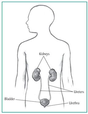
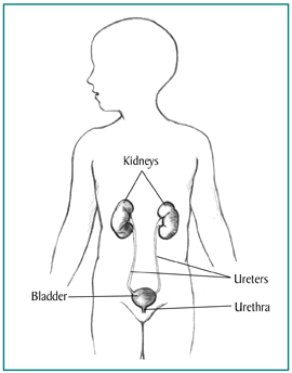
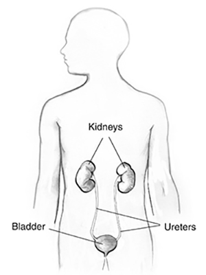
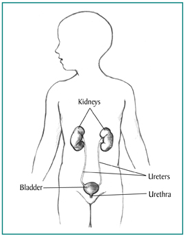
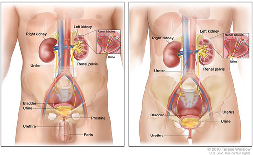
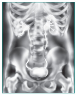
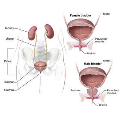
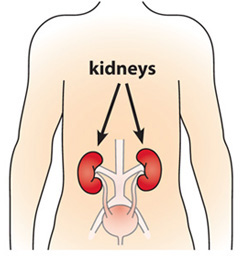
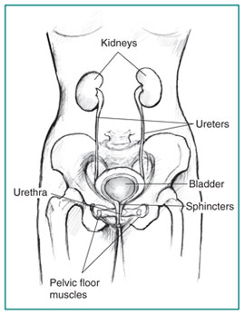
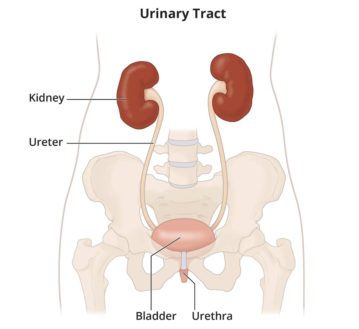
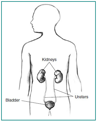
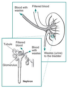
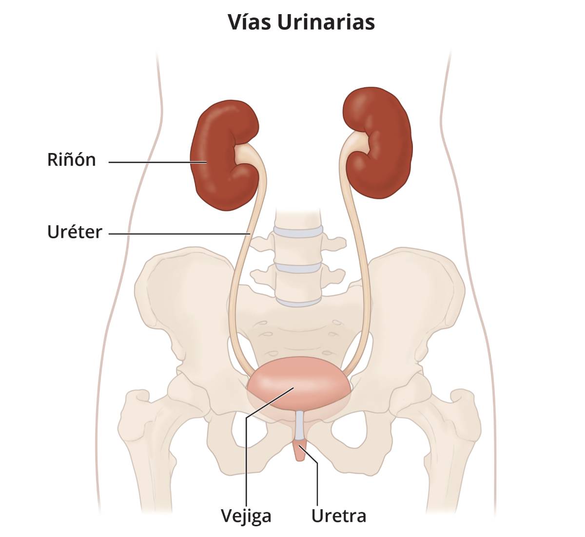
The kidneys are two bean-shaped organs, each about the size of a fist. They are located just below the rib cage, one on each side of the spine. Every day, the two kidneys filter about 120 to 150 quarts of blood to produce about 1 to 2 quarts of urine, composed of wastes and extra fluid. The urine flows from the kidneys to the bladder through tubes called ureters. The bladder stores urine. When the bladder empties, urine flows out of the body through a tube called the urethra, located at the bottom of the bladder. In men, the urethra is long, while in women it is short.

Drawing of the urinary tract inside the outline of the upper half of a human body. The kidneys, ureters, bladder, and urethra are labeled.
The kidneys are two bean-shaped organs, each about the size of a fist.

What types of amyloidosis affect the kidneys?

Primary amyloidosis and dialysis-related amyloidosis are the types of amyloidosis that can affect the kidneys.

Primary Amyloidosis of the Kidneys

The kidneys are the organs most commonly affected by primary amyloidosis. Amyloid deposits damage the kidneys and make it harder for them to filter wastes and break down proteins. When the kidneys become too damaged, they may no longer be able to function well enough to maintain health, resulting in kidney failure. Kidney failure can lead to problems such as high blood pressure, bone disease, and anemia—a condition in which the body has fewer red blood cells than normal.

Dialysis-related Amyloidosis

People who suffer from kidney failure and have been on long-term dialysis may develop dialysis-related amyloidosis. This type of amyloidosis occurs when a certain protein, called beta-2 microglobulin, builds up in the blood because dialysis does not remove it completely. The two types of dialysis are

  • hemodialysis. Hemodialysis uses a special filter called a dialyzer to remove wastes and extra fluid from the blood.
  • peritoneal dialysis. Peritoneal dialysis uses the lining of the abdominal cavity—the space in the body that holds organs such as the stomach, intestines, and liver—to filter the blood.

Dialysis-related amyloidosis is a complication of kidney failure because neither hemodialysis nor peritoneal dialysis effectively filters beta-2 microglobulin from the blood. As a result, elevated amounts of beta-2 microglobulin remain in the blood. Dialysis-related amyloidosis is relatively common in people with kidney failure, especially adults older than 60 years of age, who have been on dialysis for more than 5 years.1

More information is provided in the NIDDK health topics:

What are the signs and symptoms of primary amyloidosis of the kidneys?

The most common sign of primary amyloidosis of the kidneys is nephrotic syndrome––a collection of signs that indicate kidney damage. The signs of nephrotic syndrome include

  • albuminuria—an increased amount of albumin, a protein, in the urine. A person with nephrotic syndrome excretes more than half a teaspoon of albumin per day.
  • hyperlipidemia—a condition in which a person’s blood has more-than-normal amounts of fats and cholesterol.
  • edema—swelling, typically in a person’s legs, feet, or ankles and less often in the hands or face.
  • hypoalbuminemia—a condition in which a person’s blood has less-than-normal amounts of albumin.

More information is provided in the NIDDK health topic, Nephrotic Syndrome in Adults.

Other signs and symptoms of primary amyloidosis may include

  • fatigue, or feeling tired
  • shortness of breath
  • low blood pressure
  • numbness, tingling, or a burning sensation in the hands or feet
  • weight loss

What are the symptoms of dialysis-related amyloidosis?

The symptoms of dialysis-related amyloidosis may include

  • pain, stiffness, and fluid in the joints.
  • abnormal, fluid-containing sacs, called cysts, in some bones.
  • carpal tunnel syndrome, caused by unusual buildup of amyloid proteins in the wrists. The symptoms of carpal tunnel syndrome include numbness or tingling, sometimes associated with muscle weakness, in the fingers and hands.

Dialysis-related amyloidosis most often affects bones, joints, and the tissues that connect muscle to bone, called tendons. The disease may also affect the digestive tract and organs such as the heart and lungs. Bone cysts caused by dialysis-related amyloidosis can lead to bone fractures. Dialysis-related amyloidosis can also cause tears in tendons and ligaments. Ligaments are tissues that connect bones to other bones.

How is primary amyloidosis of the kidneys diagnosed?

A health care provider diagnoses primary amyloidosis of the kidneys with

  • a medical and family history
  • a physical exam
  • urinalysis
  • blood tests
  • a kidney biopsy

Medical and Family History

Taking a medical and family history may help a health care provider diagnose amyloidosis of the kidneys. He or she will ask the patient to provide a medical and family history.

Physical Exam

A physical exam may help diagnose primary amyloidosis of the kidneys. During a physical exam, a health care provider usually

  • examines a patient’s body to check for swelling
  • uses a stethoscope to listen to the lungs
  • taps on specific areas of the patient’s body

Urinalysis

A health care provider may use urinalysis—the testing of a urine sample—to check for albumin and amyloid proteins in urine. The patient provides a urine sample in a special container at a health care provider’s office or a commercial facility. A nurse or technician can test the sample in the same location or send it to a lab for analysis. More-than-normal amounts of albumin in urine may indicate kidney damage due to primary amyloidosis. Amyloid proteins in urine may indicate amyloidosis.

Blood Tests

The health care provider may use blood tests to see how well the kidneys are working and to check for amyloid proteins and hyperlipidemia. A blood test involves drawing a patient’s blood at a health care provider’s office or a commercial facility and sending the sample to a lab for analysis. Blood tests for kidney function measure the waste products in the blood that healthy kidneys normally filter out. Hyperlipidemia may indicate nephrotic syndrome. Amyloid proteins in blood may indicate amyloidosis.

Kidney Biopsy

Only a biopsy can show the amyloid protein deposits in the kidneys. A health care provider may recommend a kidney biopsy if other tests show kidney damage. A kidney biopsy is a procedure that involves taking a piece of kidney tissue for examination with a microscope. A health care provider performs a kidney biopsy in a hospital with light sedation and local anesthetic. The health care provider uses imaging techniques such as ultrasound or a computerized tomography (CT) scan to guide the biopsy needle into the kidney and take the tissue sample. A pathologist—a doctor who specializes in diagnosing diseases––examines the tissue in a lab for amyloid proteins and kidney damage.

The biopsy results can help the health care provider determine the best course of treatment. More information is provided in the NIDDK health topic, Kidney Biopsy.

How is dialysis-related amyloidosis diagnosed?

A health care provider diagnoses dialysis-related amyloidosis with

  • urinalysis
  • blood tests
  • imaging tests

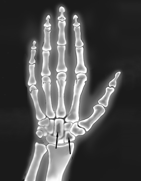
A health care provider can use urinalysis and blood tests to detect the amount of amyloid proteins in urine and blood. Imaging tests, such as x-rays and CT scans, can provide pictures of bone cysts and amyloid deposits in bones, joints, tendons, and ligaments. An x-ray technician performs imaging tests in a health care provider’s office, an outpatient center, or a hospital. A radiologist—a doctor who specializes in medical imaging—interprets the images. A patient does not require anesthesia.

X-ray image of the hand and wrist, with arrows pointing to darkened areas in two wrist bones, indicating amyloid deposits.
X-ray image showing amyloid deposits in the wrist

How is primary amyloidosis of the kidneys treated?

A health care provider treats primary amyloidosis of the kidneys with the following:

  • medication therapy, including chemotherapy
  • a stem cell transplant
  • treating other conditions

Medication therapy. The goal of medication therapy, including chemotherapy, is to reduce amyloid protein levels in the blood. Many health care providers recommend combination medication therapy such as

  • melphalan (Alkeran), a type of chemotherapy
  • dexamethasone (Decadron), an anti-inflammatory steroid medication

These medications can stop the growth of the cells that make amyloid proteins. These medications may cause hair loss and serious side effects, such as nausea, vomiting, and fatigue.

Stem cell transplant. A stem cell transplant is a procedure that replaces a patient’s damaged stem cells with healthy ones. Stem cells are found in the bone marrow and develop into three types of blood cells the body needs. To prepare for a stem cell transplant, the patient receives high doses of chemotherapy. The actual transplant is like a blood transfusion. The transplanted stem cells travel to the bone marrow to make healthy new blood cells. The chemotherapy a patient receives to prepare for the transplant can have serious side effects, so it is important to talk with the health care provider about the risks of this procedure.

Read more in What Is a Blood and Marrow Stem Cell Transplant? at www.nhlbi.nih.gov/health/health-topics/topics/bmsct.

Treating other conditions. Primary amyloidosis has no cure, so treating some of the side effects and other conditions seen with the disease is essential. Other conditions may include

  • anemia—treatment may include medications
  • depression—treatment may include talking with a mental health counselor and taking medications
  • fatigue—treatment may include changes in diet and activity level
  • kidney disease—treatment may include medications to help maintain kidney function or slow the progression of kidney disease

A patient and his or her family should talk with the health care provider about resources for support and treatment options.

More information about kidney disease is provided in the NIDDK health topic, Kidney Disease.

How is dialysis-related amyloidosis treated?

A health care provider treats dialysis-related amyloidosis with

  • medication therapy
  • newer, more effective hemodialysis filters
  • surgery
  • a kidney transplant

The goal of medication therapy and the use of newer, more effective hemodialysis filters is to reduce amyloid protein levels in the blood. Medication therapy can help reduce symptoms such as pain and inflammation. A health care provider may treat a person with dialysis-related amyloidosis who has bone, joint, and tendon problems, such as bone cysts and carpal tunnel syndrome, using surgery.

Dialysis-related amyloidosis has no cure; however, a successful kidney transplant may stop the disease from progressing.

More information is provided in the NIDDK health topic, Treatment Methods for Kidney Failure: Transplantation.

Eating, Diet, and Nutrition

Researchers have not found that eating, diet, and nutrition play a role in causing or preventing primary amyloidosis of the kidneys or dialysis-related amyloidosis. People with nephrotic syndrome may make dietary changes such as

  • limiting dietary sodium, often from salt, to help reduce edema and lower blood pressure
  • decreasing liquid intake to help reduce edema and lower blood pressure
  • eating a diet low in saturated fat and cholesterol to help control more-than-normal amounts of fats and cholesterol in the blood

Health care providers may recommend that people with kidney disease eat moderate or reduced amounts of protein. Proteins break down into waste products that the kidneys filter from the blood. Eating more protein than the body needs may burden the kidneys and cause kidney function to decline faster. However, protein intake that is too low may lead to malnutrition, a condition that occurs when the body does not get enough nutrients.

People with kidney disease on a restricted protein diet should receive blood tests that can show low nutrient levels. People with primary amyloidosis of the kidneys or dialysis-related amyloidosis should talk with a health care provider about dietary restrictions to best manage their individual needs.

Points to Remember

  • Amyloidosis is a rare disease that occurs when amyloid proteins are deposited in tissues and organs.
  • Primary amyloidosis and dialysis-related amyloidosis are the types of amyloidosis that can affect the kidneys.
  • The most common sign of primary amyloidosis of the kidneys is nephrotic syndrome.
  • The signs of nephrotic syndrome include
    • albuminuria—an elevated amount of albumin in the urine. A person with nephrotic syndrome excretes more than half a teaspoon of albumin per day.
    • hyperlipidemia—a condition in which a person’s blood has more-than-normal amounts of fats and cholesterol.
    • edema—swelling, typically in a person’s legs, feet, or ankles and less often in the hands or face.
    • hypoalbuminemia—a condition in which a person’s blood has less-than-normal amounts of albumin.
  • Other signs and symptoms of primary amyloidosis may include
    • fatigue, or feeling tired
    • shortness of breath
    • low blood pressure
    • numbness, tingling, or a burning sensation in the hands or feet
    • weight loss
  • The symptoms of dialysis-related amyloidosis may include
    • pain, stiffness, and fluid in the joints.
    • abnormal, fluid-containing sacs, called cysts, in some bones.
    • carpal tunnel syndrome, caused by unusual buildup of amyloid proteins in the wrists. The symptoms of carpal tunnel syndrome include numbness or tingling, sometimes associated with muscle weakness, in the fingers and hands.
  • A health care provider diagnoses primary amyloidosis of the kidneys with
    • a medical and family history
    • a physical exam
    • urinalysis
    • blood tests
    • a kidney biopsy
  • A health care provider diagnoses dialysis-related amyloidosis with
    • urinalysis
    • blood tests
    • imaging tests
  • A health care provider treats primary amyloidosis of the kidneys with the following:
    • medication therapy, including chemotherapy
    • a stem cell transplant
    • treating other conditions
  • A health care provider treats dialysis-related amyloidosis with
    • medication therapy
    • newer, more effective hemodialysis filters
    • surgery
    • a kidney transplant

References

Clinical Trials

The National Institute of Diabetes and Digestive and Kidney Diseases (NIDDK) and other components of the National Institutes of Health (NIH) conduct and support research into many diseases and conditions.

What are clinical trials, and are they right for you?

Clinical trials are part of clinical research and at the heart of all medical advances. Clinical trials look at new ways to prevent, detect, or treat disease. Researchers also use clinical trials to look at other aspects of care, such as improving the quality of life for people with chronic illnesses. Find out if clinical trials are right for you.

What clinical trials are open?

Clinical trials that are currently open and are recruiting can be viewed at www.ClinicalTrials.gov.

About the Kidney Failure Series

You and your doctor will work together to choose a treatment that's best for you. The publications of the NIDDK Kidney Failure Series can help you learn about the specific issues you will face.

Booklets

Fact Sheets

Learning as much as you can about your treatment will help make you an important member of your health care team.

This content is provided as a service of the National Institute of Diabetes and Digestive and Kidney Diseases (NIDDK), part of the National Institutes of Health. The NIDDK translates and disseminates research findings through its clearinghouses and education programs to increase knowledge and understanding about health and disease among patients, health professionals, and the public. Content produced by the NIDDK is carefully reviewed by NIDDK scientists and other experts.

The NIDDK would like to thank:
Glenn Chertow, M.D., University of California at San Francisco; William J. Stone, M.D., Vanderbilt University; Morie A. Gertz, M.D., Mayo Clinic

Anatomic Problems of the Lower GI Tract

What are anatomic problems of the lower gastrointestinal (GI) tract?

Anatomic problems of the lower GI tract are structural defects. Anatomic problems that develop before birth are known as congenital abnormalities. Other anatomic problems may occur any time after birth—from infancy into adulthood.

The GI tract is a series of hollow organs joined in a long, twisting tube from the mouth to the anus. The movement of muscles in the GI tract, along with the release of hormones and enzymes, allows for the digestion of food. Organs that make up the GI tract are the mouth, esophagus, stomach, small intestine, large intestine—which includes the appendix, cecum, colon, and rectum—and anus. The intestines are sometimes called the bowel. The last part of the GI tract—called the lower GI tract—consists of the large intestine and anus.

The large intestine is about 5 feet long in adults and absorbs water and any remaining nutrients from partially digested food passed from the small intestine. The large intestine then changes waste from liquid to a solid matter called stool. Stool passes from the colon to the rectum. The rectum is 6 to 8 inches long in adults and is located between the last part of the colon—called the sigmoid colon—and the anus. The rectum stores stool prior to a bowel movement. During a bowel movement, the muscles of the rectal wall contract to move stool from the rectum to the anus, a 1-inch-long opening through which stool leaves the body.

Drawing of the lower gastrointestinal tract inside the outline of a man’s torso. Inset of the lower gastrointestinal tract with the cecum, ascending colon, transverse colon, descending colon, sigmoid colon, and rectum labeled.
The lower GI tract

Anatomic problems of the lower GI tract may involve parts of organs being in the wrong place, shaped abnormally, or incorrectly connected to other organs. Anatomic problems that affect the large intestine or anus include

  • malrotation
  • volvulus
  • intussusception
  • fistula
  • imperforate anus
  • colonic atresia

How does the lower GI tract develop?

About 4 weeks into gestation—the 9-month period from conception to birth—the intestines of the developing baby, or fetus, consist of a thin, straight tube that connects the stomach and the rectum. Over the next 2 weeks, the rapidly developing intestines outgrow the baby’s abdomen and move into the umbilical cord, which connects the baby to the mother. During gestational weeks 10 to 12, the baby’s abdomen has grown large enough to hold the intestines, which return to the abdomen, rotating counterclockwise to their final position. The intestines are held in place by tissue called mesentery.

What is malrotation?

Malrotation is when the intestines do not rotate completely or correctly during gestation. Malrotation can cause serious medical problems in some infants and children, while others may never develop problems. Surgeons estimate that problems with malrotation occur in a small percentage of cases and are usually diagnosed in the first month of life. Boys are more likely than girls to be diagnosed with malrotation during infancy, but problems identified later in childhood are equally likely in boys and girls. Malrotation rarely occurs in adults.

Malrotation can prevent the cecum—the beginning of the large intestine—from moving to its normal position in the lower right area of the abdomen. If this happens, bands of mesentery can block the small intestine, creating an intestinal obstruction—also called bowel obstruction—a life-threatening event and a medical emergency. Malrotation may also leave the mesentery only narrowly attached to the back of the abdomen. This incomplete attachment may result in the intestine twisting—a serious condition called volvulus—see "What is volvulus?"

Symptoms of Malrotation

Infants who have serious problems resulting from malrotation experience pain that can be severe, and they often vomit bile—a greenish-yellow fluid. Other symptoms may include

  • abdominal tenderness, swelling, or bloating
  • bloody or dark-red stools
  • constipation—a condition in which a child has fewer than two bowel movements a week
  • dehydration, or abnormal loss of body fluids—decreased tears and little or no urine or dark-yellow urine may be observed
  • signs of shock—paleness, sweating, confusion, and rapid pulse
  • weight loss

Older children with problems from malrotation may have the above symptoms as well as nausea, abdominal pain, diarrhea, or an abnormal growth rate, as compared with their peers.

Infants or children with any of the above symptoms should be evaluated immediately by a health care provider.

Diagnosis and Treatment of Malrotation

Doctors use x rays of the abdomen and imaging studies to diagnose intestinal problems related to malrotation.

  • Computerized tomography (CT) scan. CT scans use a combination of x rays and computer technology to create three-dimensional (3-D) images. A CT scan may include the injection of a special dye, called contrast medium. CT scans require the child to lie on a table that slides into a tunnel-shaped device where the x rays are taken. CT scans can help diagnose malrotation. Infants and children may be given a sedative to help them fall asleep for the test.
  • Upper GI series. An upper GI series may be done to look at the small intestine. No eating or drinking is allowed for 8 hours before the procedure, if possible. During the procedure, a child is given barium—a chalky liquid—to drink. An infant will lie on a table and is given barium through a tiny tube placed in the nose that runs into the stomach. Infants and children may be given a sedative to help them fall asleep for the test. Barium coats the small intestine, making signs of malrotation show up more clearly on x rays.

    The child may experience bloating and nausea for a short time after the test. For several days afterward, barium liquid in the GI tract causes stools to be white or light colored. A health care provider will provide specific instructions about eating and drinking after the test.
  • Lower GI series. A lower GI series may be done to look at the large intestine. A health care provider may provide written bowel prep instructions to follow at home before the test. The child may be given a clear liquid diet for 1 to 3 days before the procedure. A laxative or enema may be used before the test. A laxative is medication that loosens stool and increases bowel movements. An enema involves flushing water, laxative, or sometimes a mild soap solution into the anus using a special plastic bottle.

    Infants and children may be given a sedative to help them fall asleep for the test. For the test, the child will lie on a table while the doctor inserts a flexible tube into the child’s anus. The large intestine is filled with barium, making signs of malrotation show up more clearly on x rays.

    For several days afterward, barium liquid in the GI tract causes stools to be white or light colored. Enemas and repeated bowel movements may cause anal soreness. A health care provider will provide specific instructions about eating and drinking after the test.

The above tests are all performed at a hospital or outpatient center by an x-ray technician, and the images are interpreted by a radiologist—a doctor who specializes in medical imaging.

Surgery is almost always required to correct problems resulting from malrotation. A surgeon performs the procedure in a hospital and the child is given general anesthesia. With early diagnosis and treatment, surgery is usually successful and may involve

  • repositioning the large and small intestines
  • dividing the bands of mesentery blocking the small intestine
  • removing the appendix, a 4-inch pouch attached to the cecum
  • untwisting the large intestine if volvulus has occurred

What is volvulus?

Volvulus occurs when the intestine twists around itself and the mesentery that supports it, creating an obstruction. The area of intestine above the obstruction continues to function and fills with food, fluid, and gas. The mesentery may become so tightly twisted that blood flow to the affected part of the intestine is cut off. This situation can lead to death of the blood-starved tissue and tearing of the intestinal wall—a life-threatening event and a medical emergency.

Volvulus can be caused by malrotation or by other medical conditions such as

  • an enlarged colon
  • Hirschsprung disease, a disease of the large intestine that causes severe constipation or intestinal obstruction
  • abdominal adhesions, or bands of scar tissue that form as part of the healing process following abdominal injury, infection, or surgery

Sigmoid volvulus—twisting of the sigmoid colon—accounts for the majority of cases, with cecal volvulus—twisting of the cecum and ascending colon—occurring less frequently.

Sigmoid Volvulus

Anatomic problems that increase a person’s risk of developing sigmoid volvulus include

  • an elongated or movable sigmoid colon that is not attached to the left wall of the abdomen
  • a narrow mesentery connection at the base of the sigmoid colon
  • malrotation that presents with problems in infancy

Sigmoid volvulus that occurs after infancy is more commonly seen in people who

  • are male
  • are older than age 60
  • live in a nursing or psychiatric facility
  • have a history of mental health conditions

Symptoms of Sigmoid Volvulus

Sigmoid volvulus symptoms can be severe and occur suddenly. Symptoms may include

  • abdominal cramping
  • bloody stools
  • constipation
  • nausea
  • signs of shock
  • vomiting

People with any of these symptoms should be evaluated immediately by a health care provider.

Other symptoms may develop more slowly but worsen with time, such as constipation, inability to pass gas, and abdominal swelling. People with these symptoms should also contact a health care provider.

Diagnosis and Treatment of Sigmoid Volvulus

Prompt diagnosis and appropriate treatment of sigmoid volvulus generally lead to a successful outcome. Doctors use x rays, upper or lower GI series, CT scans, and flexible sigmoidoscopy—another common diagnostic test—to help diagnose sigmoid volvulus.

  • Flexible sigmoidoscopy. This test is used to look inside the rectum and lower colon. Sigmoidoscopy is performed at a hospital, outpatient center, or doctor’s office by a gastroenterologist—a doctor who specializes in digestive diseases—or a radiologist. Infants and children may be given a sedative to help them fall asleep for the test. For the test, the person will lie on a table while the doctor inserts a flexible tube into the anus. A small camera on the tube sends a video image of the intestinal lining to a computer screen. The doctor can see sigmoid volvulus.

    Children and adults can usually go back to their normal diet after the test, though cramping or bloating may occur during the first hour after the test.

If volvulus is found, the doctor may use the sigmoidoscope to untwist the colon. However, if the colon is twisted tightly or if the blood flow has been cut off, immediate surgery will be needed. Surgery involves restoring the blood supply, if possible, to the affected part of the sigmoid colon. Sometimes the affected part of the colon must be removed and the healthy ends reattached, a procedure called an intestinal resection. Resection prevents volvulus from recurring; untwisting the volvulus with the sigmoidoscope may not prevent recurrence.

Cecal Volvulus

Cecal volvulus is twisting of the cecum and ascending colon. Normally, the cecum and ascending colon are fixed to the abdominal wall. If improperly attached, they can move and become twisted.

Symptoms of Cecal Volvulus

More commonly seen in people ages 30 to 60, cecal volvulus may be caused by abdominal adhesions, severe coughing, or pregnancy. People with cecal volvulus often have intermittent chronic symptoms—those that come and go over a longer period of time—including

  • abdominal cramping or swelling
  • nausea
  • vomiting

People with any of the above symptoms should be evaluated immediately by a health care provider.

Other symptoms may develop more slowly but worsen with time, such as constipation, inability to pass gas, and abdominal swelling. People with these symptoms should also contact a health care provider.

Diagnosis and Treatment of Cecal Volvulus

Doctors use x rays, upper or lower GI series, and CT scans to diagnose cecal volvulus. Imaging shows whether the cecum is out of place and inflated with trapped air. Imaging may also show that the appendix, which is attached to the cecum, is filled with air. To treat cecal volvulus, surgeons use a procedure called cecopexy to reposition the cecum and attach it to the abdominal wall. If the cecum is seriously damaged by volvulus, the surgeon will perform intestinal resection surgery. Cecopexy and intestinal resection surgery have high rates of success and usually prevent the recurrence of cecal volvulus.

What is intussusception?

Intussusception is a condition in which one section of either the large or small intestine folds into itself, much like a collapsible telescope. The condition can cause obstruction and cut off blood flow to the affected part of the intestine—a life-threatening event and a medical emergency.

Malrotation increases the risk that an infant or young child will develop intussusception. Sometimes, a viral infection or growth in the small or large intestine—such as a polyp or tumor—can trigger intussusception, but most childhood cases have no known cause.

Intussusception, though uncommon, is the leading cause of bowel obstruction in young children in the United States. About 1,400 cases occur in the United States each year, nearly all of them in children younger than 1 year old. Intussusception is 1.5 times as likely to affect boys as girls.1

Symptoms of Intussusception

Infants or children with intussusception may have symptoms including

  • abdominal pain, which may be intermittent
  • bloody stools
  • diarrhea
  • fever
  • lack of energy
  • signs of dehydration
  • stool mixed with mucus—a clear liquid made by the intestines that coats and protects tissues
  • swelling or a lump in the abdomen
  • vomiting with or without bile

Infants or children with any of the above symptoms should be evaluated immediately by a health care provider.

Diagnosis and Treatment of Intussusception

A range of tests may be required to diagnose intussusception. X rays of the abdomen may show an intestinal obstruction. Upper and lower GI series can locate the intussusception and show telescoping of the intestine. CT scans can also help diagnose the condition. If intussusception is not diagnosed promptly, it can cause serious damage to the affected part of the intestine.

In some cases, intussusception may be temporary and correct itself and, if no underlying problem is found, treatment is not required. When intussusception does not resolve on its own, doctors can usually correct the problem with a lower GI series, using air or barium to gently push the telescoped part of intestine into its proper position. However, intestinal resection surgery is sometimes necessary to successfully treat the condition.

What is a fistula?

A fistula is an abnormal passage, or tunnel, between two organs—called an internal fistula—or between an organ and the outside of the body—called an external fistula. In the lower GI tract, both internal and external fistulas can occur. Fistulas can develop during gestation or at any age after birth. Fistulas that develop during gestation are more common in boys than girls.

Fistulas may occur as a result of

  • complications following surgery—the most common cause
  • childbirth—a fistula can develop between a mother’s vagina and rectum
  • Crohn’s disease, a chronic inflammatory bowel disease that can affect any part of the GI tract
  • diverticulitis, an inflammation or infection of small pouches called diverticula that are created by bulging, weak spots on the colon
  • infection
  • trauma

Symptoms of Fistulas

Some people with a fistula in the lower GI tract have no symptoms; others may experience

  • abdominal pain that begins in one spot and spreads throughout the abdomen
  • dehydration
  • diarrhea
  • fatigue, muscle cramps, or slow growth due to malabsorption—a condition that occurs when the small intestine cannot absorb nutrients from food
  • fever, with or without chills

People with any of these symptoms should be evaluated immediately by a health care provider.

Diagnosis and Treatment of Fistulas

External fistulas can be found during a physical examination. Internal fistulas can be seen during an upper or lower GI series, CT scan, or colonoscopy.

  • Colonoscopy. Colonoscopy is used to look inside the rectum and entire colon for signs of fistulas. Colonoscopy is performed at a hospital or outpatient center by a gastroenterologist or a radiologist. The health care provider will provide written bowel prep instructions to follow at home before the test. The person may be asked to follow a clear liquid diet for 1 to 3 days before the test. Laxatives and enemas may be used before the test.

    For the test, the person will lie on a table while the doctor inserts a flexible tube into the anus. A small camera on the tube sends a video image of the intestinal lining to a computer screen. The doctor can see signs of fistulas. In most cases, a light sedative, and possibly pain medication, helps keep the person relaxed.

    Cramping or bloating may occur during the first hour after the test. Driving is not permitted for 24 hours after the test to allow the sedative time to wear off. Before the appointment, people should make plans for a ride home. Full recovery is expected by the next day.

Internal and external fistulas may close on their own, although this process could take weeks or months. The doctor may prescribe antibiotics to prevent or treat infection resulting from leakage of intestinal contents. Some people may need to stop eating and receive nourishment intravenously to ensure proper healing.

If a fistula does not close on its own, a surgeon may perform intestinal resection surgery.

What is imperforate anus?

Imperforate anus occurs during gestation and involves abnormal development of the rectum and anus. This condition results in a blocked or missing anus, which allows little or no stool to pass from the rectum. Imperforate anus is uncommon and occurs slightly more often in boys than in girls. Types of imperforate anus include

  • an anus that is narrow or blocked by a thin membrane—this condition is also called anal atresia
  • an anus that is missing or incorrectly placed
  • a rectum that is not connected to the anus
  • a rectum that is connected to the urinary tract or genitals by a fistula

Although most girls with imperforate anus have a less severe form of the condition, such as anal atresia, some are born with a more severe form of imperforate anus called cloaca—a common opening for the rectum, bladder, and vagina.

Symptoms of Imperforate Anus

Imperforate anus is observed when a newborn is first examined after birth. In addition to visible indications such as an incorrectly placed anus, imperforate anus may be associated with symptoms that include abdominal swelling and the absence of bowel movements.

Diagnosis and Treatment of Imperforate Anus

The severity of imperforate anus depends on where the blockage is situated—low, intermediate, or high—in relation to the set of muscles that support the rectum and other organs within the pelvic region. X rays of the abdomen and CT scans can help determine the severity of the condition. The doctor may perform other tests to look for abnormalities in the urinary tract.

Correcting imperforate anus almost always requires surgery, and the type of procedure depends on the location and severity of the defect. For example, a low imperforate anus may only require gently widening the anus. Sometimes anoplasty—a surgery to rebuild or move the anus—is needed within the first days after birth. Intermediate or high imperforate anus may require multiple surgeries done in stages. Girls with cloaca may require multiple extensive and complicated surgeries.

The outcome of surgery is measured by the child’s ability to eventually control bowel movements. Most children treated for imperforate anus develop voluntary bowel movements at the usual age of toilet training. However, some children may not achieve good bowel control after surgery because the anal muscles do not develop properly. Factors affecting the outcome of surgery include

  • location of the defect—treatment of low imperforate anus has a more successful long-term outcome than intermediate or high imperforate anus
  • the child’s sex—girls tend to have low imperforate anus, which can be corrected more easily and has more successful long-term results
  • age at the time of surgery—the younger the child when surgery is done, the more successful the outcome

What is colonic atresia?

Colonic atresia is an extremely rare congenital anomaly that occurs when a section of the colon closes before birth. Symptoms appear in infants soon after birth and include vomiting, abdominal swelling, and the absence of bowel movements. Intestinal resection surgery is performed immediately after diagnosis.

Eating, Diet, and Nutrition

Eating, diet, and nutrition have not been shown to play a role in causing or preventing anatomic problems of the lower GI tract.

Points to Remember

  • Anatomic problems of the lower gastrointestinal (GI) tract are structural defects.
  • Anatomic problems that develop before birth are known as congenital abnormalities. Other anatomic problems may occur any time after birth—from infancy into adulthood.
  • The GI tract is a series of hollow organs joined in a long, twisting tube from the mouth to the anus. The movement of muscles in the GI tract, along with the release of hormones and enzymes, allows for the digestion of food.
  • The last part of the GI tract—called the lower GI tract—consists of the large intestine and anus.
  • Anatomic problems of the lower GI tract may involve parts of organs being in the wrong place, shaped abnormally, or incorrectly connected to other organs.
  • Anatomic problems that affect the large intestine or anus include malrotation, volvulus, intussusception, fistula, imperforate anus, and colonic atresia.
  • Surgery is often needed to correct anatomic problems of the lower GI tract.
  • Some anatomic problems of the lower GI tract can be corrected without surgery and others resolve without treatment or never cause problems.

References

Clinical Trials

The National Institute of Diabetes and Digestive and Kidney Diseases (NIDDK) and other components of the National Institutes of Health (NIH) conduct and support research into many diseases and conditions.

What are clinical trials, and are they right for you?

Clinical trials are part of clinical research and at the heart of all medical advances. Clinical trials look at new ways to prevent, detect, or treat disease. Researchers also use clinical trials to look at other aspects of care, such as improving the quality of life for people with chronic illnesses. Find out if clinical trials are right for you.

What clinical trials are open?

Clinical trials that are currently open and are recruiting can be viewed at www.ClinicalTrials.gov.

This content is provided as a service of the National Institute of Diabetes and Digestive and Kidney Diseases (NIDDK), part of the National Institutes of Health. The NIDDK translates and disseminates research findings through its clearinghouses and education programs to increase knowledge and understanding about health and disease among patients, health professionals, and the public. Content produced by the NIDDK is carefully reviewed by NIDDK scientists and other experts.

The NIDDK would like to thank:
Robert Beart, M.D., University of Southern California; James W. Fleshman, M.D., Washington University and Barnes-Jewish Hospital; Kevan Jacobson, M.B.B.Ch, B.C.’s (British Columbia, Canada’s) Children’s Hospital; Joseph Levy, M.D., Children’s Hospital of New York-Presbyterian; John H. Pemberton, M.D., Mayo Clinic

Anemia

What is anemia?

Anemia is a condition in which the body has fewer red blood cells than normal. Red blood cells carry oxygen to tissues and organs throughout the body and enable them to use energy from food. With anemia, red blood cells carry less oxygen to tissues and organs—particularly the heart and brain—and those tissues and organs may not function as well as they should.

How is anemia related to chronic kidney disease?

Anemia commonly occurs in people with chronic kidney disease (CKD)—the permanent, partial loss of kidney function. Anemia might begin to develop in the early stages of CKD, when someone has 20 to 50 percent of normal kidney function. Anemia tends to worsen as CKD progresses. Most people who have total loss of kidney function, or kidney failure, have anemia.1 A person has kidney failure when he or she needs a kidney transplant or dialysis in order to live. The two forms of dialysis include hemodialysis and peritoneal dialysis. Hemodialysis uses a machine to circulate a person’s blood through a filter outside the body. Peritoneal dialysis uses the lining of the abdomen to filter blood inside the body.

What are the kidneys and what do they do?

The kidneys are two bean-shaped organs, each about the size of a fist. They are located just below the rib cage, one on each side of the spine. Every day, the kidneys filter about 120 to 150 quarts of blood to produce about 1 to 2 quarts of urine.

Healthy kidneys produce a hormone called erythropoietin (EPO). A hormone is a chemical produced by the body and released into the blood to help trigger or regulate particular body functions. EPO prompts the bone marrow to make red blood cells, which then carry oxygen throughout the body.

What causes anemia in chronic kidney disease?

When kidneys are diseased or damaged, they do not make enough EPO. As a result, the bone marrow makes fewer red blood cells, causing anemia. When blood has fewer red blood cells, it deprives the body of the oxygen it needs.

Other common causes of anemia in people with kidney disease include blood loss from hemodialysis and low levels of the following nutrients found in food:

  • iron
  • vitamin B12
  • folic acid

These nutrients are necessary for red blood cells to make hemoglobin, the main oxygen-carrying protein in the red blood cells.

If treatments for kidney-related anemia do not help, the health care provider will look for other causes of anemia, including

  • other problems with bone marrow
  • inflammatory problems—such as arthritis, lupus, or inflammatory bowel disease—in which the body’s immune system attacks the body’s own cells and organs
  • chronic infections such as diabetic ulcers
  • malnutrition
Top: process of normal red blood cell production with a healthy kidney. Bottom: process of reduced red blood cell production with a damaged kidney.
Healthy kidneys produce a hormone called EPO. EPO prompts the bone marrow to make red blood cells, which then carry oxygen throughout the body. When the kidneys are diseased or damaged, they do not make enough EPO. As a result, the bone marrow makes fewer red blood cells, causing anemia.

What are the signs and symptoms of anemia in someone with chronic kidney disease?

The signs and symptoms of anemia in someone with CKD may include

  • weakness
  • fatigue, or feeling tired
  • headaches
  • problems with concentration
  • paleness
  • dizziness
  • difficulty breathing or shortness of breath
  • chest pain

Anyone having difficulty breathing or with shortness of breath should seek immediate medical care. Anyone who has chest pain should call 911.

What are the complications of anemia in someone with chronic kidney disease?

Heart problems are a complication of anemia and may include

  • an irregular heartbeat or an unusually fast heartbeat, especially when exercising.
  • the harmful enlargement of muscles in the heart.
  • heart failure, which does not mean the heart suddenly stops working. Instead, heart failure is a long-lasting condition in which the heart can’t pump enough blood to meet the body’s needs.

How is anemia in chronic kidney disease diagnosed?

A health care provider diagnoses anemia based on

  • a medical history
  • a physical exam
  • blood tests

Medical History

Taking a medical history is one of the first things a health care provider may do to diagnose anemia. He or she will usually ask about the patient’s symptoms.

Physical Exam

A physical exam may help diagnose anemia. During a physical exam, a health care provider usually examines a patient’s body, including checking for changes in skin color.

Blood Tests

To diagnose anemia, a health care provider may order a complete blood count, which measures the type and number of blood cells in the body. A blood test involves drawing a patient’s blood at a health care provider’s office or a commercial facility. A health care provider will carefully monitor the amount of hemoglobin in the patient’s blood, one of the measurements in a complete blood count.

The Kidney Disease: Improving Global Outcomes Anemia Work Group recommends that health care providers diagnose anemia in males older than age 15 when their hemoglobin falls below 13 grams per deciliter (g/dL) and in females older than 15 when it falls below 12 g/dL.2 If someone has lost at least half of normal kidney function and has low hemoglobin, the cause of anemia may be decreased EPO production.

Two other blood tests help measure iron levels:

  • The ferritin level helps assess the amount of iron stored in the body. A ferritin score below 200 nanograms (ng) per milliliter may mean a person has iron deficiency that requires treatment.2
  • The transferrin saturation score indicates how much iron is available to make red blood cells. A transferrin saturation score below 30 percent can also mean low iron levels that require treatment.2

In addition to blood tests, the health care provider may order other tests, such as tests for blood loss in stool, to look for other causes of anemia.

How is anemia in chronic kidney disease treated?

Depending on the cause, a health care provider treats anemia with one or more of the following treatments:

Iron

The first step in treating anemia is raising low iron levels. Iron pills may help improve iron and hemoglobin levels. However, for patients on hemodialysis, many studies show pills do not work as well as iron given intravenously.2

Erythropoietin

If blood tests indicate kidney disease as the most likely cause of anemia, treatment can include injections of a genetically engineered form of EPO. A health care provider, often a nurse, injects the patient with EPO subcutaneously, or under the skin, as needed. Some patients learn how to inject the EPO themselves. Patients on hemodialysis may receive EPO intravenously during hemodialysis.

Studies have shown the use of EPO increases the chance of cardiovascular events, such as heart attack and stroke, in people with CKD. The health care provider will carefully review the medical history of the patient and determine if EPO is the best treatment for the patient’s anemia. Experts recommend using the lowest dose of EPO that will reduce the need for red blood cell transfusions. Additionally, health care providers should consider the use of EPO only when a patient’s hemoglobin level is below 10 g/dL. Health care providers should not use EPO to maintain a patient’s hemoglobin level above 11.5 g/dL.2 Patients who receive EPO should have regular blood tests to monitor their hemoglobin so the health care provider can adjust the EPO dose when the level is too high or too low.2 Health care providers should discuss the benefits and risks of EPO with their patients.

Many people with kidney disease need iron supplements and EPO to raise their red blood cell count to a level that will reduce the need for red blood cell transfusions. In some people, iron supplements and EPO will improve the symptoms of anemia.

Red Blood Cell Transfusions

If a patient’s hemoglobin falls too low, a health care provider may prescribe a red blood cell transfusion. Transfusing red blood cells into the patient’s vein raises the percentage of the patient’s blood that consists of red blood cells, increasing the amount of oxygen available to the body.

Vitamin B12 and Folic Acid Supplements

A health care provider may suggest vitamin B12 and folic acid supplements for some people with CKD and anemia. Using vitamin supplements can treat low levels of vitamin B12 or folic acid and help treat anemia. To help ensure coordinated and safe care, people should discuss their use of complementary and alternative medical practices, including their use of dietary supplements, with their health care provider.

Read more about vitamin B12 and folic acid on the MedlinePlus website at www.nlm.nih.gov/medlineplus. Read more about complementary and alternative medicine at nccih.nih.gov/.

Eating, Diet, and Nutrition

A health care provider may advise people with kidney disease who have anemia caused by iron, vitamin B12, or folic acid deficiencies to include sources of these nutrients in their diets. Some of these foods are high in sodium or phosphorus, which people with CKD should limit in their diet. Before making any dietary changes, people with CKD should talk with their health care provider or with a dietitian who specializes in helping people with kidney disease. A dietitian can help a person plan healthy meals.

Read more about nutrition for people with CKD on the National Kidney Disease Education Program page.

The following chart illustrates some good dietary sources of iron, vitamin B12, and folic acid.

Food Serving Size Iron Vitamin B12 Folic Acid
Recommended Daily Value    18 mg 6 mcg 400 mcg
100 percent fortified breakfast cereal ¾ cup (1 oz) 18 mg 6 mcg 394 mcg
beans, baked 1 cup (8 oz) 8 mg 0 mcg 37 mcg
beef, ground 3 oz 2 mg 2 mcg 8 mcg
beef liver 3 oz 5 mg 67 mcg 211 mcg
clams, fried 4 oz 3 mg 1 mcg 66 mcg
spinach, boiled 1 cup (3 oz) 2 mg 0 mcg 115 mcg
spinach, fresh 1 cup (1 oz) 1 mg 0 mcg 58 mcg
trout 3 oz 0 mg 5 mcg 16 mcg
tuna, canned 3 oz 1 mg 1 mcg 2 mcg

Points to Remember

  • Anemia is a condition in which the body has fewer red blood cells than normal. Red blood cells carry oxygen to tissues and organs throughout the body and enable them to use energy from food.
  • Anemia commonly occurs in people with chronic kidney disease (CKD)—the permanent, partial loss of kidney function. Most people who have total loss of kidney function, or kidney failure, have anemia.
  • When kidneys are diseased or damaged, they do not make enough erythropoietin (EPO). As a result, the bone marrow makes fewer red blood cells, causing anemia.
  • Other common causes of anemia in people with kidney disease include blood loss from hemodialysis and low levels of the following nutrients found in food:
    • iron
    • vitamin B12
    • folic acid
  • The first step in treating anemia is raising low iron levels.
  • If blood tests indicate kidney disease as the most likely cause of anemia, treatment can include injections of a genetically engineered form of EPO.
  • Many people with kidney disease need iron supplements and EPO to raise their red blood cell count to a level that will reduce the need for red blood cell transfusions.
  • A health care provider may suggest vitamin B12 and folic acid supplements for some people with CKD and anemia.
  • A health care provider may advise people with kidney disease who have anemia caused by iron, vitamin B12, or folic acid deficiencies to include sources of these nutrients in their diets.

References

Clinical Trials

The National Institute of Diabetes and Digestive and Kidney Diseases (NIDDK) and other components of the National Institutes of Health (NIH) conduct and support research into many diseases and conditions.

What are clinical trials, and are they right for you?

Clinical trials are part of clinical research and at the heart of all medical advances. Clinical trials look at new ways to prevent, detect, or treat disease. Researchers also use clinical trials to look at other aspects of care, such as improving the quality of life for people with chronic illnesses. Find out if clinical trials are right for you.

What clinical trials are open?

Clinical trials that are currently open and are recruiting can be viewed at www.ClinicalTrials.gov.

About the Kidney Failure Series

You and your doctor will work together to choose a treatment that's best for you. The publications of the NIDDK Kidney Failure Series can help you learn about the specific issues you will face.

Booklets

Fact Sheets

This content is provided as a service of the National Institute of Diabetes and Digestive and Kidney Diseases (NIDDK), part of the National Institutes of Health. The NIDDK translates and disseminates research findings through its clearinghouses and education programs to increase knowledge and understanding about health and disease among patients, health professionals, and the public. Content produced by the NIDDK is carefully reviewed by NIDDK scientists and other experts.

The NIDDK would like to thank:
John C. Stivelman, M.D., Emory University School of Medicine; Kerri Cavanaugh, M.D., M.H.S., Vanderbilt University

Appendicitis

Definition & Facts

What is appendicitis?

Appendicitis is inflammation of your appendix.

How common is appendicitis?

In the United States, appendicitis is the most common cause of acute abdominal pain requiring surgery. Over 5% of the population develops appendicitis at some point.1

Who is more likely to develop appendicitis?

Appendicitis most commonly occurs in the teens and twenties but may occur at any age.1

What are the complications of appendicitis?

If appendicitis is not treated, it may lead to complications. The complications of a ruptured appendix are

  • peritonitis, which can be a dangerous condition. Peritonitis happens if your appendix bursts and infection spreads in your abdomen. If you have peritonitis, you may be very ill and have
    • fever
    • nausea
    • severe tenderness in your abdomen
    • vomiting
  • an abscess of the appendix called an appendiceal abscess.

References


Symptoms & Causes

What are the symptoms of appendicitis?

The most common symptom of appendicitis is pain in your abdomen.

If you have appendicitis, you’ll most often have pain in your abdomen that

  • begins near your belly button and then moves lower and to your right
  • gets worse in a matter of hours
  • gets worse when you move around, take deep breaths, cough, or sneeze
  • is severe and often described as different from any pain you’ve felt before
  • occurs suddenly and may even wake you up if you’re sleeping
  • occurs before other symptoms

Other symptoms of appendicitis may include

  • loss of appetite
  • nausea
  • vomiting
  • constipation or diarrhea
  • an inability to pass gas
  • a low-grade fever
  • swelling in your abdomen
  • the feeling that having a bowel movement will relieve discomfort

Symptoms can be different for each person and can seem like the following conditions that also cause pain in the abdomen:

What causes appendicitis?

Appendicitis can have more than one cause, and in many cases the cause is not clear. Possible causes include:

  • Blockage of the opening inside the appendix
  • enlarged tissue in the wall of your appendix, caused by infection in the gastrointestinal (GI) tract or elsewhere in your body
  • inflammatory bowel disease
  • stool, parasites, or growths that can clog your appendiceal lumen
  • trauma to your abdomen

When should I seek a doctor's help?

Appendicitis is a medical emergency that requires immediate care. See a health care professional or go to the emergency room right away if you think you or a child has appendicitis. A doctor can help treat the appendicitis and reduce symptoms and the chance of complications.


Diagnosis

How do doctors diagnose appendicitis?

Most often, health care professionals suspect the diagnosis of appendicitis based on your symptoms, your medical history, and a physical exam. A doctor can confirm the diagnosis with an ultrasound, x-ray, or MRI exam.

Medical history

A health care professional will ask specific questions about your symptoms and health history to help rule out other health problems. The health care professional will want to know

  • when your abdominal pain began
  • the exact location and severity of your pain
  • when your other symptoms appeared
  • your other medical conditions, previous illnesses, and surgical procedures
  • whether you use medicines, alcohol, or illegal drugs

Physical exam

Health care professionals need specific details about the pain in your abdomen to diagnose appendicitis correctly. A health care professional will assess your pain by touching or applying pressure to specific areas of your abdomen.

The following responses to touch or pressure may indicate that you have appendicitis:

  • Rovsing's sign
  • Psoas sign
  • Obturator sign
  • Guarding
  • Rebound tenderness
  • Digital rectal exam
  • Pelvic exam

Lab tests

Doctors use lab tests to help confirm the diagnosis of appendicitis or find other causes of abdominal pain.

Blood tests. A health care professional draws your blood for a blood test at a doctor’s office or a commercial facility. The health care professional sends the blood sample to a lab for testing. Blood tests can show a high white blood cell count, a sign of infection. Blood tests also may show dehydration or fluid and electrolyte imbalances.

Urinalysis. Urinalysis is testing of a urine sample. You will provide a urine sample in a special container in a doctor’s office, a commercial facility, or a hospital. Health care professionals can test the urine in the same location or send it to a lab for testing. Doctors use urinalysis to rule out a urinary tract infection or a kidney stone.

Pregnancy test. For women, health care professionals also may order blood or urine samples to check for pregnancy.

Imaging tests

Doctors use imaging tests to confirm the diagnosis of appendicitis or find other causes of pain in the abdomen.

Abdominal ultrasound. In an ultrasound, a health care professional uses a device, called a transducer, to bounce safe, painless sound waves off of your organs to create an image of their structure. He or she can move the transducer to different angles to examine different organs.

In an abdominal ultrasound, a health care professional applies a gel to your abdomen and moves a hand-held transducer over your skin. A health care professional performs this procedure in a doctor’s office, an outpatient center, or a hospital, and you don’t need anesthesia.

A radiologist reviews the images, which can show signs of

  • a blockage in your appendiceal lumen
  • a burst appendix
  • inflammation
  • other sources of abdominal pain

Health care professionals use an ultrasound as the first imaging test for possible appendicitis in infants, children, young adults, and pregnant women.

Magnetic resonance imaging (MRI). MRI machines use radio waves and magnets to produce detailed pictures of your body’s internal organs and soft tissues without using x-rays.

A health care professional performs the procedure in an outpatient center or a hospital. A radiologist reviews the images. Patients don’t need anesthesia, although a health care professional may give light sedation, taken by mouth, to children and people with a fear of small spaces. A health care professional may inject a special dye, called contrast medium, into your body.

In most cases, you’ll lie on a table that slides into a tunnel-shaped device. The tunnel may be open ended or closed at one end.

An MRI can show signs of

  • a blockage in your appendiceal lumen
  • a burst appendix
  • inflammation
  • other sources of abdominal pain

When diagnosing appendicitis and other sources of abdominal pain, doctors can use an MRI as a safe, reliable alternative to a computerized tomography (CT) scan.2

CT scan. CT scans use x-rays and computer technology to create images.

A health care professional may give you a solution to drink and an injection of contrast medium. You’ll lie on a table that slides into a tunnel-shaped device that takes the x-rays. X-ray technicians perform CT scans in an outpatient center or a hospital. Radiologists review the images.

Patients don’t need anesthesia, although health care professionals may give children a sedative to help them fall asleep for the test.

A CT scan of the abdomen can show signs of inflammation, such as

  • an enlarged or a burst appendix
  • an appendiceal abscess
  • a blockage in your appendiceal lumen

Women of childbearing age should have a pregnancy test before having a CT scan. The radiation from CT scans can be harmful to a developing fetus.

References


Treatment

How do doctors treat appendicitis?

Doctors typically treat appendicitis with surgery to remove the appendix. Surgeons perform the surgery in a hospital with general anesthesia. Your doctor will recommend surgery if you have continuous abdominal pain and fever, or signs of a burst appendix and infection. Prompt surgery decreases the chance that your appendix will burst.

Health care professionals call the surgery to remove the appendix an appendectomy. A surgeon performs the surgery using one of the following methods:

  • Laparoscopic surgery. During laparoscopic surgery, surgeons use several smaller incisions and special surgical tools that they feed through the incisions to remove your appendix. Laparoscopic surgery leads to fewer complications, such as hospital-related infections, and has a shorter recovery time.
  • Laparotomy. Surgeons use laparotomy to remove the appendix through a single incision in the lower right area of your abdomen.

After surgery, most patients completely recover from appendicitis and don’t need to make changes to their diet, exercise, or lifestyle. Surgeons recommend that you limit physical activity for the first 10 to 14 days after a laparotomy and for the first 3 to 5 days after laparoscopic surgery.

What if the surgeon finds a normal appendix?

In some cases, a surgeon finds a normal appendix during surgery. In this case, many surgeons will remove it to eliminate the future possibility of appendicitis. Sometimes surgeons find a different problem, which they may correct during surgery.

Can doctors treat appendicitis without surgery?

Some cases of mild appendicitis may be cured with antibiotics alone. All patients suspected of having appendicitis are treated with antibiotics before surgery, and some patients may improve completely before surgery is performed.

How do doctors treat complications of a burst appendix?

Treating the complications of a burst appendix will depend on the type of complication. In most cases of peritonitis, a surgeon will remove your appendix immediately with surgery. The surgeon will use laparotomy to clean the inside of your abdomen to prevent infection and then remove your appendix. Without prompt treatment, peritonitis can cause death.

A surgeon may drain the pus from an appendiceal abscess during surgery or, more commonly, before surgery. To drain an abscess, the surgeon places a tube in the abscess through the abdominal wall. You leave the drainage tube in place for about 2 weeks while you take antibiotics to treat infection. When the infection and inflammation are under control, about 6 to 8 weeks later, surgeons operate to remove what remains of the burst appendix.


Eating, Diet, & Nutrition

How can your diet help prevent or relieve appendicitis?

Researchers have not found that eating, diet, and nutrition cause or prevent appendicitis.?


Clinical Trials

The National Institute of Diabetes and Digestive and Kidney Diseases (NIDDK) and other components of the National Institutes of Health (NIH) conduct and support basic and clinical research into many digestive disorders.

What are clinical trials and are they right for you?

Clinical trials are part of clinical research and at the heart of all medical advances. Clinical trials look at new ways to prevent, detect, or treat disease. Researchers also use clinical trials to look at other aspects of care, such as improving the quality of life for people with chronic illnesses. Find out if clinical trials are right for you.

What clinical trials are open?

Clinical trials that are currently open and are recruiting can be viewed at www.ClinicalTrials.gov.?


This content is provided as a service of the National Institute of Diabetes and Digestive and Kidney Diseases (NIDDK), part of the National Institutes of Health. The NIDDK translates and disseminates research findings through its clearinghouses and education programs to increase knowledge and understanding about health and disease among patients, health professionals, and the public. Content produced by the NIDDK is carefully reviewed by NIDDK scientists and other experts.

Syndicated Content Details:
Source URL: https://www.niddk.nih.gov/Syndication/appendicitis
Source Agency: National Institute of Diabetes and Digestive and Kidney Diseases (NIDDK)
Captured Date: 2015-12-05 06:52:00.0

Ayuda financiera para el tratamiento de la insuficiencia renal

¿Cuán costoso es el tratamiento de la insuficiencia renal?

El tratamiento de la insuficiencia renal—la hemodiálisis, la diálisis peritoneal y el trasplante de riñón—es costoso y la mayoría de la gente necesita ayuda financiera. El costo promedio de Medicare por persona en el 2011 era el siguiente:1

  • casi 88 millones de dólares para la hemodiálisis, un tratamiento para la insuficiencia renal que filtra la sangre fuera del cuerpo
  • más de 71 millones de dólares para la diálisis peritoneal, un tratamiento para la insuficiencia renal que utiliza el revestimiento de la cavidad abdominal del paciente como filtro
  • casi 33 millones de dólares para un trasplante, una cirugía para colocar un riñón sano de alguien que acaba de morir o de un donante vivo, por lo general un familiar, en el cuerpo del paciente

El Gobierno Federal y otras fuentes proveen ayuda financiera. En 1972, el Congreso de los EE. UU. aprobó una ley que permita que la mayoría de la gente con insuficiencia renal pueda recibir la cobertura de Medicare.

Los proveedores de atención médica con frecuencia le cobran costos adicionales a los planes de salud privados. Los estados y otras fuentes también pueden ayudar. Una persona puede vivir por años con la insuficiencia renal, por lo tanto es importante obtener ayuda para pagar por el tratamiento.

¿Qué es Medicare?

Medicare es un programa de seguro federal que paga los costos de atención médica de la gente que cumple con los requisitos y

  • tiene 65 años o más
  • tiene menos de 65 años, pero tiene una determinada discapacidad
  • tiene la ERT—no importa la edad

¿Cómo se puede obtener los servicios de Medicare para la insuficiencia renal?

Para obtener los servicios de Medicare para la insuficiencia renal, una persona debe cumplir con dos condiciones:

  1. Recibir la diálisis o tener un trasplante de riñón por causa de la ERT
  2. Haber pagado impuestos suficientes—o ser cónyuge o hijo de alguien que lo ha hecho—a través de
    • Seguro Social
    • Junta de Jubilación de los Empleados Ferroviarios (Railroad Retirement Board en inglés)
    • Empleo del gobierno

¿Qué planes de salud ofrece Medicare?

Existen tres tipos principales de planes de salud de Medicare:

  1. Medicare original tiene tres partes:
    • La Parte A (hospital) cubre la atención de pacientes hospitalizados, el trasplante de riñón, vivir en centros especializados de enfermería y los cuidados para pacientes terminales. La Parte A no tiene primas para aquellos que han pagado suficientes impuestos de Medicare. Una prima es la suma que debe pagar periódicamente el paciente—por mes o por trimestre—para Medicare, un otro plan de salud o un plan de cobertura de medicamentos. La Parte A sí tiene un deducible, una suma que debe pagar el paciente por recibir atención médica o recetas, antes de que pague por estas cosas el plan de salud. El paciente debe pagar una suma diaria por las estadías en el hospital que superen los 60 días.
    • La Parte B (pacientes ambulatorios) cubre la mayoría de los tratamientos y suministros de la diálisis, los costos de los proveedores de atención médica y las medicinas contra el rechazo para trasplantes. La Parte B tiene una prima mensual que se basa en el ingreso de la persona. Los precios cambian cada año. Después de que paga el paciente cada año el deducible, la Parte B paga como un pagador primario el 80 por ciento por la mayoría de los servicios cubiertos. El personal de facturación del proveedor de los servicios—un hospital o una clínica—puede calcular cuánto adeudará el paciente.
    • La Parte D (medicaciones) tiene una prima y cubre algunas medicinas. Las compañías privadas de seguros ofrecen diferentes planes de la Parte D, los cuales son aprobados por Medicare. Los costos y la cobertura varían según el plan. Una persona que tiene pocos activos y gana menos del 150 por ciento del nivel federal de pobreza puede calificar para recibir ayuda adicional para pagar las primas y los costos de las medicinas de la Parte D. Puede encontrar las reglas generales para el año actual en www.aspe.hhs.gov (en inglés) o por llamar al Seguro Social al 1–800–772–1213. Puede encontrar información y solicitudes de los planes para la Parte D en https://es.medicare.gov. También puede solicitar los servicios de la Parte D de una compañía de seguros que vende un plan así.
  2. Los planes de Medicare Advantage, que también se llaman la Parte C, los venden las compañías de seguros. Medicare debe aprobar los planes de Medicare Advantage. Cada plan de Medicare Advantage debe cubrir los servicios de la Parte A y la Parte B, y además puede cubrir otros servicios. Los planes de Medicare Advantage pueden tener la cobertura de medicamentos de la Parte D. Si no, una persona puede comprar por separado el plan de la Parte D. Todos los planes de Medicare Advantage no son iguales. Una persona con la insuficiencia renal que piensa elegir un plan de Medicare Advantage debe preguntar sobre las reglas del plan. Las reglas pueden especificar cuáles proveedores de atención médica u hospitales que puede utilizar el paciente. El plan puede requerir un remitido del proveedor de atención primaria para que el paciente pueda tener cita con un especialista. Es posible que el plan no cubra los gastos médicos que se adquiere durante un viaje. La suma de gastos extra que debe pagar el paciente cada año varía según el plan. Las personas que tienen un plan de Medicare Advantage no pueden tener un plan de Medigap para ayudar a pagar los gastos extra. Consulte la sección sobre Medigap.

    Existen cuatro tipos de planes de Medicare Advantage:
    • organizaciones del mantenimiento de la salud (HMO por sus siglas en inglés)
    • organizaciones de proveedores preferidos (PPO por sus siglas en inglés)
    • costo privado para planes de servicio
    • planes de necesidades especiales para determinados grupos

    Aquellos que ya reciben el tratamiento con diálisis no se pueden inscribir en la mayoría de los planes de Medicare Advantage. Sin embargo, una persona que ya tenía un plan de Medicare Advantage antes de que fallaron los riñones puede conservar el plan. En algunas regiones, se diseñan planes de necesidades especiales para aquellos que reciben el tratamiento de diálisis. Puede llamar al 1–800–MEDICARE (1–800–633–4227) para aprender si existen planes de necesidades especiales específicos a la región para los pacientes que reciben el tratamiento con diálisis.
  3. Otros planes de salud de Medicare son para determinados grupos, tales como las personas en estado delicado que viven en la comunidad y las personas con múltiples enfermedades crónicas, e incluyen la cobertura hospitalaria y médica. Algunos también pagan las medicinas recetadas. Algunos son financiados y administrados parcialmente por el estado—se llaman Medicaid. Los planes incluyen los siguientes:
    • Los planes de costos de Medicare (Medicare Cost Plans) son HMO, similares a los planes ofrecidos como Medicare Advantage, pero los proveedores fuera de la red reciben pagos como si el titular de la póliza tuviera Medicare Original.
    • El Programa de cobertura total de la salud para personas mayores (PACE por sus siglas en inglés) combina los servicios de atención médica, los servicios sociales y los de atención a largo plazo para las personas en estado delicado que viven y reciben atención médica dentro de la comunidad.
    • Los proyectos de innovación de Medicare son proyectos especiales que evalúan las mejoras en la cobertura, el pago y la calidad de atención de Medicare.

Se puede encontrar más información sobre los planes de costos de Medicare y los programas de Demostración o Piloto en el sitio web estatal de Medicaid en www.medicaid.gov (en inglés) o por llamar al 1–800–MEDICARE (1–800–633–4227). Las oficinas estatales de Medicaid pueden proporcionar más información sobre PACE. Consulte la sección sobre Medicaid.

¿Dónde se puede inscribir para Medicare?

Puede aplicar para los servicios de Medicare en la página web www.ssa.gov o en una oficina local del Seguro Social. El número gratuito del Seguro Social es 1–800–772–1213, TTY 1–800–325–0778. Puede llamar para coordinar una cita con alguien en una oficina local y aplicar para los servicios.

¿Cuándo comienzan los servicios de Medicare para las personas con ERT?

Una persona con ERT puede aplicar para Medicare al comienzo de la diálisis o al momento del trasplante de riñón. La fecha de inicio de los servicios de Medicare depende del tipo de tratamiento:

  • Diálisis en casa, incluso la diálisis peritoneal. Los servicios de Medicare pueden comenzar el primer mes de diálisis solamente si el paciente se capacita para la diálisis en casa.
  • Trasplante de riñón. Los servicios de Medicare pueden comenzar el mismo mes en que ocurre el trasplante. En algunos casos, los servicios de Medicare podrían comenzar 2 meses antes del trasplante si el paciente está hospitalizado y se demora el trasplante. Por ejemplo, si un paciente está hospitalizado para un trasplante en marzo y se demora el trasplante hasta mayo, el pago todavía comienza en marzo. Un asesor financiero de trasplante puede proporcionar más información.
  • Diálisis en el centro. Los servicios de Medicare no comenzarán hasta el cuarto mes de la diálisis en el centro. Por ejemplo, si una persona comienza la diálisis en una clínica en julio y no se capacita para la diálisis en casa, Medicare no comenzará a pagar hasta el 1 de octubre.
Julio Agosto Septiembre Octubre
Primer mes de diálisis Segundo mes de diálisis Tercer mes de diálisis Cuarto mes de diálisis. Comienza la cobertura de Medicare.

¿Cuándo terminan los servicios de Medicare para las personas con ERT?

Los servicios de Medicare continúan siempre y cuando que el paciente reciba diálisis y pague una prima, incluso para los pacientes que tienen trabajo. Los servicios de Medicare terminarán por las dos razones descritas a continuación:

  1. La insuficiencia renal es la única razón por qué tenía Medicare y el paciente obtenga un trasplante de riñón. Si el riñón nuevo funciona y ya no necesita diálisis, los servicios de Medicare terminan 3 años después de la cirugía de trasplante.
  2. La salud de los riñones mejora y ya no necesite diálisis. Los servicios de Medicare terminarán 12 meses después de que la persona deja la diálisis, a menos que vuelva a la diálisis o reciba un trasplante dentro de esos 12 meses. Los servicios de Medicare no terminarán si la persona cumple con los requisitos de edad o discapacidad.

¿Cómo paga Medicare si una persona tiene otro seguro médico?

Las reglas sobre cuál seguro paga primero dependen del tipo de seguro médico que tiene el paciente.

Tipo de plan de seguro Quién paga primero
Plan individual—no es proporcionado por un empleador Medicare siempre paga primero. Un plan individual siempre paga segundo.
Plan grupal del empleador o sindicato Respecto a la insuficiencia renal, el plan grupal del empleador o sindicato paga primero durante los 30 meses* después de que la persona cumpla con los requisitos de Medicare debido a la insuficiencia renal. El plazo de 30 meses comienza sin importar si la persona se inscriba en Medicare o no. Sin embargo, la persona eventualmente tendrá que inscribirse en Medicare. Después de los 30 meses, Medicare paga primero. El plan grupal del empleador o sindicato puede pagar la totalidad o una parte del remanente.

*Este período de tiempo se denomina el “período de coordinación del pagador secundario de Medicare”.

Tener los servicios de Medicare Parte B y también otro plan de seguro médico puede limitar la cantidad de los gastos extra que paga el paciente por la atención médica. En algunos casos, Medicare puede limitar la cantidad de dinero que puede cobrar un proveedor de atención médica por los servicios. Si una persona no tiene los servicios de la Parte B, el proveedor de atención médica o la clínica de diálisis puede facturar una tarifa más alta al plan médico individual o grupal y también al paciente. El paciente tendría que pagar lo que el plan no paga.

En algunas situaciones, una persona que tiene otro seguro puede ahorrar dinero si no se inscribe en la Parte B hasta que finalice el período de coordinación de 30 meses. La persona no tendría que pagar las primas de la Parte B durante ese período.

¿Debería esperar para comenzar a pagar la Parte B un paciente que tiene otro seguro?

  • SÍ, solamente si: El plan de la persona paga el 100 por ciento de todos los costos de atención médica durante ese período.
    • Si espera para inscribirse en ambas partes, la Parte A y la B, la inscripción en ambas partes puede realizarse en cualquier momento.
    • Si obtiene la Parte A—en espera para comenzar con la Parte B—solamente puede inscribirse una vez por año desde el 1 de enero hasta el 31 de marzo, y la Parte B no comenzará hasta el 1 de julio. Debe inscribirse en la Parte B a tiempo para evitar un vacío en la cobertura. Por ejemplo, si el período de coordinación de 30 meses finaliza el 30 de abril, el plan grupal del empleador o sindicato puede dejar de pagar primero en el 1 de mayo. Si se inscribió en la Parte B antes del 31 de marzo, la Parte B no comenzará a pagar primero hasta el 1 de julio. La persona tendrá grandes facturas durante mayo y junio.
  • NO, solamente si: El paciente tiene que pagar deducibles, copagos o coseguros anuales, los cuales son costos que no cubre el plan de seguros. Medicare puede pagar esos cargos o no. Sin embargo, tener la cobertura de Medicare limita lo que puede cobrar una clínica de diálisis. La prima para la Parte B por lo general cuesta menos que pagar los deducibles, los copagos o el coseguro.

¿Dónde puedo obtener más información sobre la cobertura de Medicare para el tratamiento de la insuficiencia renal?

Estos folletos de Medicare le ofrecen más información sobre la cobertura de Medicare para el tratamiento de la insuficiencia renal:

  • La Cobertura de Medicare de los Servicios de Diálisis Renal y Trasplante de Riñón
  • Medicare para los niños que padecen de Enfermedad Renal Terminal

¿Cuándo empieza a pagar el plan grupal del empleador o sindicato?

Un plan grupal del empleador o sindicato puede ayudar a pagar los costos del tratamiento y de las medicinas recetadas bajo las siguientes circunstancias:

  • Antes de que Medicare comience a pagar la diálisis, el plan grupal del empleador o sindicato puede cubrir la mayoría o la totalidad de los costos.
  • Durante el período de coordinación de 30 meses, el plan grupal del empleador o sindicato paga la mayor parte de los costos primero y, luego, Medicare paga su parte de los costos restantes.
  • Después de que finalice el período de coordinación de 30 meses, Medicare paga la mayor parte primero y, luego, el plan grupal del empleador o sindicato paga su parte de los costos restantes.

Las personas con la insuficiencia renal deben leer su póliza de seguro médico con cuidado para asegurarse de que cubra el tratamiento de la insuficiencia renal. Si tienen preguntas sobre los beneficios, deben consultar al agente de seguros o al asesor de beneficios del empleador.

¿Qué otros programas federales pueden ayudar?

Los siguientes programas federales pueden ayudar a cubrir el costo del tratamiento de la insuficiencia renal:

  • Seguro de discapacidad del Seguro Social (SSDI por sus siglas en inglés). El SSDI es un plan federal de seguros que paga una suma mensual a las personas que no pueden trabajar. Las personas ganan créditos laborales de SSDI cuando pagan los impuestos del Seguro Social. Deben tener créditos suficientes según la edad para calificar. Por lo tanto, si una enfermedad o lesión le impide a trabajar por al menos un año, los pagos del SSDI pueden ser una opción. Un cuadro en www.ssa.gov muestra cuántos créditos laborales que necesita.
  • Seguridad de ingreso suplementario (SSI por sus siglas en inglés). La SSI es un programa federal de red de seguridad que paga una suma mensual a los niños y adultos discapacitados quienes ganan poco y tienen pocos activos. Una persona que recibe la SSI puede obtener cupones de alimentos y también los servicios de Medicaid.

Se puede encontrar más información sobre el SSDI y la SSI, y sobre cómo aplicar, en www.ssa.gov o por llamar al 1–800–772–1213, TTY 1–800–325–0778.

¿Qué programas estatales pueden ayudar?

Los programas estatales como Medicaid, Medigap y los Programas de Ahorros de Medicare (Medicare Savings Programs) están financiados en parte por el Gobierno Federal. Sin embargo, los estados proporcionan los servicios y deciden quienes reciben la ayuda.

Medicaid. Medicaid es un programa estatal para aquellos con bajos ingresos y pocos activos. Cada estado administra su propio programa. El Gobierno Federal exige que los programas de Medicaid cubran un conjunto específico de servicios; sin embargo, los estados pueden elegir cubrir otros servicios además de los exigidos. Una persona puede tener Medicaid sólo o Medicare y Medicaid. Si una persona tiene los dos tipos de cobertura, Medicare paga primero y Medicaid paga segundo. Medicaid puede pagar por servicios por los que Medicare no paga. Una persona puede aplicar para los servicios de Medicaid en la oficina del Departamento de Servicios Sociales de la ciudad o del condado. Un trabajador social puede explicar el programa de Medicaid del estado y ayudar a una persona a aplicar. Se puede encontrar más información sobre Medicaid en www.medicaid.gov (en inglés).

Medigap, también conocido como suplemento de Medicare. Un plan de Medigap puede ayudar a pagar lo que no paga el Medicare Original por los servicios cubiertos. Las compañías de seguros venden cobertura Medigap. Las personas que tienen un plan de Medicare Advantage no pueden tener un plan de Medigap también.

Para las personas que tienen 65 años o más, la ley federal establece que, durante los primeros 6 meses que tiene los servicios de la Parte B, las empresas no pueden negarles una aplicación ni limitar los pagos para cualquier servicio que cubre Medicare Original, según la salud de la persona. Algunos estados hacen que las compañías de seguros vendan al menos un plan de cobertura de Medigap a aquellos que tienen menos de 65 años y también tienen la cobertura de Medicare. Las oficinas de seguros estatales pueden explicar los planes en su estado. Para buscar las oficinas locales, haga clic en el mapa en www.naic.org (en inglés).

Programas de ahorro de Medicare. Algunos estados pueden pagar las primas, los deducibles y el coseguro de Medicare si la persona tiene bajos ingresos y pocos activos. El Departamento de Servicios Sociales de la ciudad o del condado puede determinar si una persona cumple con los requisitos.

Programas de asistencia estatales específicos al riñón. Algunos estados utilizan fondos estatales para ayudar a pagar costos específicos relacionados con el riñón. Un trabajador social en una clínica de diálisis o de trasplante, o un trabajador social de un Departamento de Servicios Sociales local puede ayudarle a determinar si un estado tiene un programa renal.

Programa Estatal de Asistencia de Seguro de Salud (State Health Insurance Assistance Program, SHIP por sus siglas en inglés). Los SHIP obtienen dinero del Gobierno Federal para proveer consejos gratis sobre el seguro médico a aquellos que tienen Medicare. Alguien que necesita cobertura adicional del seguro médica debe hablar con un consejero de SHIP (en inglés) o un trabajador social.

¿Qué programas pueden ayudar a poblaciones especiales?

Muchos programas pueden ayudar a poblaciones específicas, como los veteranos de los EE. UU.; socios del servicio militar, los retirados y sus familias; los indios estadounidenses y los nativos de Alaska; los niños; las personas con discapacidades; las personas con insuficiencia renal que buscan trabajo; los receptores de trasplante de riñón y los donantes vivos de órganos.

Veteranos de los EE. UU.

Para los veteranos de los EE. UU., el Departamento de Asuntos de los Veteranos (VA por sus siglas en inglés) puede ayudar a pagar algunos costos de la salud. Los veteranos pueden aprender si califican para estos beneficios y aplicar en la página web www.va.gov (en inglés). Un veterano puede aprender más en un hospital local de VA o por llamar al 1–877–222–8387. Algunos hospitales de VA proporcionan la diálisis. Otros tienen contratos con clínicas de diálisis para proveer a los veteranos un mejor acceso al tratamiento. El VA también tiene su propia red de centros de trasplante.

Socios del servicio militar, los retirados y sus familias

Una persona puede obtener TRICARE—el programa de atención médica que sirve mundialmente a los socios uniformados del servicio, los retirados y sus familias—si la persona es

  • un socio activo del servicio
  • un militar retirado
  • un familiar de un socio activo del servicio o de un militar retirado
  • un socio de la Guardia Nacional/Reservas en servicio activo por 30 días
  • un familiar de un socio de la Guardia Nacional/Reservas en servicio activo por 30 días

TRICARE por Vida es un plan específico de TRICARE que ofrece cobertura secundaria para las personas que tienen los servicios de la Parte A y la Parte B de Medicare.

Puede encontrar información sobre TRICARE y los números de teléfono de los cuatro regiones de TRICARE en www.tricare.mil (en inglés).

Indios estadounidenses y nativos de Alaska

El Servicio de Salud Indígena (Indian Health Service) puede ayudar a los socios de las tribus de indios estadounidenses y nativos de Alaska reconocidas a nivel federal. Hay disponible más información en el sitio web del Servicio de Salud Indígena en www.IHS.gov (en inglés). Un indio americano o nativo de Alaska también puede cumplir con los requisitos para recibir ayuda de programas públicos, privados y estatales.

Niños

El Programa de Seguro Médico para Niños (CHIP por sus siglas en inglés) provee servicios de Medicaid de bajo costo o gratis a los niños cuyos padres ganan demasiado para recibir Medicaid, pero no lo suficiente para pagar por un plan de salud. El CHIP también puede proporcionar asistencia a los padres. El CHIP es un programa federal y estatal. Puede encontrar más información en http://espanol.insurekidsnow.gov o por llamar al 1–877–543–7669.

Personas con discapacidades

Puede encontrar ayuda estatal y local para las personas con discapacidades en www.dol.gov (en inglés). En este sitio web, puede aprender más sobre los derechos civiles, la vida en comunidad, la educación, la planificación para emergencias, el trabajo, la salud, la vivienda, la tecnología y el transporte. Un trabajador social puede referirse a recursos federales, estatales y locales.

Personas con insuficiencia renal que buscan trabajo

Las leyes tales como la Ley para Estadounidenses con Discapacidades (Americans with Disabilities Act, ADA por sus siglas en inglés) protegen a las personas con determinadas discapacidades, incluso la insuficiencia renal. Una persona con insuficiencia renal puede encontrar información sobre la capacitación laboral y la búsqueda de empleo a través de

  • programas estatales de rehabilitación vocacional
  • redes de empleo privadas que tienen contratos con el Seguro Social para ayudar a las personas con discapacidades a encontrar trabajo

Se puede encontrar más información sobre los incentivos laborales del Seguro Social, el programa “Boleto para Trabajar” y quiénes pueden ayudarle, incluso programas de rehabilitación vocacional y redes de empleo, en https://yourtickettowork.com (en inglés).

Receptores de trasplantes de riñón

La Red Unida para la Compartición de Órganos (UNOS por sus siglas en inglés) tiene un sitio web que se llama Transplant Living en www.trasplantesyvida.org. Lea más sobre la UNOS en Treatment Methods for Kidney Failure: Transplantation.

Donantes vivos de órganos

El National Living Donor Assistance Program puede ayudar a aquellos que desean donar órganos, pero no pueden pagar por hacerlo. Los donantes deben informar a los centros de trasplante que desean solicitar ayuda para pagar los costos de transporte y manutención. Puede encontrar más información sobre este programa en www.livingdonorassistance.org (en inglés).

El Gobierno Federal, algunos gobiernos estatales y compañías privadas proveen a los donantes licencia médica. Algunos estados dan deducciones de impuestos o créditos fiscales a los donantes vivos.

¿Qué organizaciones privadas pueden ayudar?

Las organizaciones privadas incluyen las organizaciones de beneficencia y las fundaciones. Algunas existen específicamente para ayudar a las personas con enfermedades renales y la insuficiencia renal, tales como

  • American Kidney Fund. El American Kidney Fund provee pequeños subsidios, según la necesidad, a los pacientes estadounidenses que reciben el tratamiento de diálisis o un trasplante. El American Kidney Fund provee subsidios para ayudar a pagar las primas de los planes de salud médicas. Un trabajador social puede ayudarle a aplicar. El American Kidney Fund depende de las donaciones, por eso puede haber momentos en que los fondos estén escasos. Puede encontrar más información en www.kidneyfund.org.
  • National Kidney Foundation. Las oficinas locales del National Kidney Foundation pueden ayudar con la nutrición, el transporte, las medicinas y las becas. Un trabajador social puede ayudarle a aplicar. El National Kidney Foundation también depende de las donaciones, entonces puede haber momentos en que los fondos estén escasos. Puede encontrar más información en www.kidney.org o por llamar al 1–800–622–9010.

¿Cómo puede pagar menos por las medicinas?

Medicare recomienda las siguientes maneras de pagar menos por las medicinas:

  • Consulte con un proveedor de atención médica o un farmacéutico sobre las medicinas genéricas y de bajo costo.
  • Pida ayuda a organizaciones de beneficencia.
  • Averigüe sobre Medicaid y otros programas estatales de medicinas.
  • Aplique para la “ayuda extra” de la Parte D del Seguro Social.
  • Averigüe sobre los programas de asistencia al paciente. Necesitará completar un formulario para solicitarlos. Los formularios preguntan sobre los ingresos y los planes de salud. Un trabajador social puede ayudarle a completar los formularios si sea necesario. Los siguientes programas de asistencia al paciente pueden ayudar a las personas que no pueden pagar por las medicinas:
    • Programas estatales de asistencia farmacéutica son disponibles en 22 estados y en un territorio. Hay más información disponible en http://es.medicare.gov.
    • El sitio web de la Alianza para la Asistencia con los Medicamentos Recetados en www.pparx.org enumera más de 475 programas que ayudan a pagar por las medicinas. Muchos de estos programas son proporcionados por los laboratorios farmacéuticos que producen los medicamentos.
    • NeedyMeds es un grupo sin fines de lucro que ayuda a los pacientes a encontrar programas que ayudan a pagar por los medicamentos. El sitio web www.needymeds.org permite que el usuario busque en una lista de programas por usar como palabra clave el nombre de la medicina o el productor. Algunos de los formularios para aplicar se encuentran en línea.
    • RxAssist tiene un sitio web en www.rxassist.org (en inglés) que provee información sobre los programas de laboratorios farmacéuticos, los programas estatales, las tarjetas de descuento para medicamentos y ayuda en copagos, entre otras cosas.

Puntos para recordar

  • El tratamiento de la insuficiencia renal—la hemodiálisis, la diálisis peritoneal y el trasplante de riñón—es costoso y la mayoría de la gente necesita ayuda financiera.
  • En 1972, el Congreso de los EE. UU. aprobó una ley que permita que la mayoría de la gente con insuficiencia renal pueda recibir la cobertura de Medicare.
  • Un trabajador social es una persona capacitada para ayudar a las personas a resolver los problemas de la vida cotidiana, especialmente las personas con discapacidades o con bajos ingresos. Un trabajador social puede ayudar con asuntos financieros y laborales. Las clínicas de diálisis y los centros de trasplante deben tener un trabajador social para ayudar a los pacientes.
  • Medicare es un programa de seguro federal que paga los costos de atención médica de la gente que cumple con los requisitos.
  • Tener los servicios de Medicare Parte B y también otro plan de seguro médico puede limitar la cantidad de gastos extra que paga el paciente por la atención médica.
  • Las personas con la insuficiencia renal deben leer su póliza de seguro médico con cuidado para asegurarse de que cubra el tratamiento de la insuficiencia renal. Si tienen preguntas sobre los beneficios, deben consultar al agente de seguros o al asesor de beneficios del empleador.
  • Los programas estatales, como Medicaid, Medigap y los Programas de Ahorros de Medicare (Medicare Savings Programs), están financiados en parte por el Gobierno Federal. Sin embargo, los estados proporcionan los servicios y deciden quienes reciben la ayuda.
  • Los Programas Estatales de Asistencia de Seguro de Salud (SHIP) obtienen dinero del Gobierno Federal para proveer consejos gratis sobre el seguro médico a aquellos que tienen Medicare.
  • Muchos programas pueden ayudar a poblaciones específicas, como los veteranos de los EE. UU., los indios estadounidenses, los niños y las personas con discapacidades.
  • Las leyes tales como la Ley para Estadounidenses con Discapacidades (ADA) protegen a las personas con determinadas discapacidades, incluso la insuficiencia renal.
  • Una persona con insuficiencia renal puede encontrar información sobre la capacitación laboral y la búsqueda de empleo.
  • La Red Unida para la Compartición de Órganos (UNOS) tiene un sitio web que se llama Transplant Living en www.trasplantesyvida.org.
  • Las organizaciones privadas incluyen las organizaciones de beneficencia y las fundaciones. Algunas existen específicamente para ayudar a las personas con enfermedades renales y la insuficiencia renal.
  • Los programas de asistencia al paciente pueden ayudar a las personas que no pueden pagar por las medicinas.

Referencias

Ensayos Clínicos

El Instituto Nacional de Diabetes y Enfermedades Digestivas y Renales (NIDDK por sus siglas en inglés) y otras organizaciones del Instituto Nacional de Salud (NIH) conducen y ayudan en la investigación de muchas enfermedades y condiciones médicas.

Que son ensayos clínicos, y serian una buena opción para usted?

Los ensayos clínicos son parte de la investigación clínica y a la raíz de todos avances médicos. Los ensayos clínicos buscan maneras nuevas de prevenir, detectar o tratar enfermedades. Los investigadores también usan los ensayos clínicos para estudiar otros aspectos de la atención clínica, como la manera de mejorar la calidad de vida de las personas con enfermedades crónicas. Averigüe si los ensayos clínicos son adecuados para usted.

Que ensayos clínicos están disponibles?

Para más información sobre los ensayos clínicos que están disponibles y están reclutando visite: www.ClinicalTrials.gov.

Acerca de la serie sobre insuficiencia renal

Usted y su médico trabajarán juntos para elegir el tratamiento que sea mejor para usted. La serie de publicaciones sobre insuficiencia renal puede ayudarle a enterarse de los problemas específicos a los que se enfrentará. Estas publicaciones son del National Institute of Diabetes and Digestive and Kidney Diseases, NIDDK por sus siglas, que en español se llama Instituto Nacional de la Diabetes y las Enfermedades Digestivas y Renales.

Nota: Las publicaciones con títulos en español están disponibles en este idioma. Las demás publicaciones se encuentran disponibles sólo en inglés en este momento.

Libritos

Hojas de información

Aprender todo lo que pueda sobre su tratamiento le ayudará a convertirse en un miembro importante de su equipo de profesionales de la salud.

This content is provided as a service of the National Institute of Diabetes and Digestive and Kidney Diseases (NIDDK), part of the National Institutes of Health. The NIDDK translates and disseminates research findings through its clearinghouses and education programs to increase knowledge and understanding about health and disease among patients, health professionals, and the public. Content produced by the NIDDK is carefully reviewed by NIDDK scientists and other experts.

El NIDDK quiere agradecer a:
Thomas Dudley, Maria Ciccanti, Eva Fung, Emerson Carvalho y Kelly Anderson, Centros para Servicios de Medicare y Medicaid (Centers for Medicare & Medicaid Services); Leslie Pionke, M.S.W., Centro de Trasplante Inova (Inova Transplant Center); Cindy Cornejo, M.S.W., Clínica de Diálisis DaVita (DaVita Dialysis Clinic); Beth Witten, M.S.W., A.C.S.W., L.S.C.S.W., Instituto de Educación Médica (Medical Education Institute); Dori Schatell, M.S., Instituto de Educación Médica (Medical Education Institute); Amy E. Witten, B.A., Centro para la Educación de la Enfermedad Renal Crónica del Programa Renal Missouri

Bariatric Surgery

Definition & Facts

What is obesity?

Obesity is defined as having a body mass index (BMI) of 30 or more. BMI is a measure of your weight in relation to your height. Class 1 obesity means a BMI of 30 to 35, Class 2 obesity is a BMI of 35 to 40, and Class 3 obesity is a BMI of 40 or more. Classes 2 and 3, also known as severe obesity, are often hard to treat with diet and exercise alone.

Calculate your BMI to learn your BMI category.

What is bariatric surgery?

Bariatric surgery is an operation that helps you lose weight by making changes to your digestive system. Some types of bariatric surgeries make your stomach smaller, allowing you to eat and drink less at one time and making you feel full sooner. Other bariatric surgeries also change your small intestine—the part of your body that absorbs calories and nutrients from foods and beverages.

Bariatric surgery may be an option if you have severe obesity and have not been able to lose weight or keep from gaining back any weight you lost using other methods such as lifestyle treatment or medications. Bariatric surgery also may be an option if you have serious health problems, such as type 2 diabetes or sleep apnea, related to obesity. Bariatric surgery can improve many of the medical conditions linked to obesity, especially type 2 diabetes.

Does bariatric surgery always work?

Studies show that many people who have bariatric surgery lose about 15 to 30 percent of their starting weight on average, depending on the type of surgery they have. However, no method, including surgery, is sure to produce and maintain weight loss. Some people who have bariatric surgery may not lose as much as they hoped. Over time, some people regain a portion of the weight they lost. The amount of weight people regain may vary. Factors that affect weight regain may include a person’s level of obesity and the type of surgery he or she had.

Bariatric surgery does not replace healthy habits, but may make it easier for you to consume fewer calories and be more physically active. Choosing healthy foods and beverages before and after the surgery may help you lose more weight and keep it off long term. Regular physical activity after surgery also helps keep the weight off. To improve your health, you must commit to a lifetime of healthy lifestyle habits and following the advice of your health care providers.

Three ladies exercising
Maintaining healthy lifestyle habits may help
you succeed after bariatric surgery.

How much does bariatric surgery cost?

On average, bariatric surgery costs between $15,000 and $25,000, depending on what type of surgery you have and whether you have surgery-related problems. Costs may be higher or lower based on where you live. The amount your medical insurance will pay varies by state and insurance provider.

Medicare and some Medicaid programs cover three common types of bariatric surgery—gastric bypass, gastric band, and gastric sleeve surgery—if you meet certain criteria and have a doctor’s recommendation. Some insurance plans may require you to use approved surgeons and facilities. Some insurers also require you to show that you were unable to lose weight by completing a nonsurgical weight-loss program or that you meet other requirements.

Your health insurance company or your regional Medicare or Medicaid office will have more information about bariatric surgery coverage, options, and requirements.


Types of Bariatric Surgery

The type of surgery that may be best to help a person lose weight depends on a number of factors. You should discuss with your doctor what kind of surgery might be best for you or your teen.

What is the difference between open and laparoscopic surgery?

In open bariatric surgery, surgeons make a single, large cut in the abdomen. More often, surgeons now use laparoscopic surgery, in which they make several small cuts and insert thin surgical tools through the cuts. Surgeons also insert a small scope attached to a camera that projects images onto a video monitor. Laparoscopic surgery has fewer risks than open surgery and may cause less pain and scarring than open surgery. Laparoscopic surgery also may lead to a faster recovery.

Open surgery may be a better option for certain people. If you have a high level of obesity, have had stomach surgery before, or have other complex medical problems, you may need open surgery.

What are the surgical options?

In the United States, surgeons use three types of operations most often:

  • laparoscopic adjustable gastric band
  • gastric sleeve surgery, also called sleeve gastrectomy
  • gastric bypass

Surgeons use a fourth operation, biliopancreatic diversion with duodenal switch, less often.

Laparoscopic Adjustable Gastric Band

In this type of surgery, the surgeon places a ring with an inner inflatable band around the top of your stomach to create a small pouch. This makes you feel full after eating a small amount of food. The band has a circular balloon inside that is filled with salt solution. The surgeon can adjust the size of the opening from the pouch to the rest of your stomach by injecting or removing the solution through a small device called a port placed under your skin.

After surgery, you will need several follow-up visits to adjust the size of the band opening. If the band causes problems or is not helping you lose enough weight, the surgeon may remove it.

The U.S. Food and Drug Administration (FDA) has approved use of the gastric band for people with a BMI of 30 or more who also have at least one health problem linked to obesity, such as heart disease or diabetes.

Gastric Sleeve

In gastric sleeve surgery, also called vertical sleeve gastrectomy, a surgeon removes most of your stomach, leaving only a banana-shaped section that is closed with staples. Like gastric band surgery, this surgery reduces the amount of food that can fit in your stomach, making you feel full sooner. Taking out part of your stomach may also affect gut hormones or other factors such as gut bacteria that may affect appetite and metabolism. This type of surgery cannot be reversed because some of the stomach is permanently removed.

Gastric Bypass

Gastric bypass surgery, also called Roux-en-Y gastric bypass, has two parts. First, the surgeon staples your stomach, creating a small pouch in the upper section. The staples make your stomach much smaller, so you eat less and feel full sooner.

Next, the surgeon cuts your small intestine and attaches the lower part of it directly to the small stomach pouch. Food then bypasses most of the stomach and the upper part of your small intestine so your body absorbs fewer calories. The surgeon connects the bypassed section farther down to the lower part of the small intestine. This bypassed section is still attached to the main part of your stomach, so digestive juices can move from your stomach and the first part of your small intestine into the lower part of your small intestine. The bypass also changes gut hormones, gut bacteria, and other factors that may affect appetite and metabolism. Gastric bypass is difficult to reverse, although a surgeon may do it if medically necessary.

Duodenal Switch

This surgery, also called biliopancreatic diversion with duodenal switch, is more complex than the others. The duodenal switch involves two separate surgeries. The first is similar to gastric sleeve surgery. The second surgery redirects food to bypass most of your small intestine. The surgeon also reattaches the bypassed section to the last part of the small intestine, allowing digestive juices to mix with food.

This type of surgery allows you to lose more weight than the other three. However, this surgery is also the most likely to cause surgery-related problems and a shortage of vitamins, minerals, and protein in your body. For these reasons, surgeons do not perform this surgery as often.

Most Common Weight-loss Surgeries

Gastric Band

Illustration of Gastric Band

What it is

Surgeon places an inflatable band around top part of stomach, creating a small pouch with an adjustable opening.

Pros

  • Can be adjusted and reversed.
  • Short hospital stay and low risk of surgery-related problems.
  • No changes to intestines.
  • Lowest chance of vitamin shortage.

Cons

  • Less weight loss than other types of bariatric surgery.
  • Frequent follow-up visits to adjust band; some people may not adapt to band.
  • Possible future surgery to remove or replace a part or all of the band system.

Gastric Sleeve

Illustration of Gastric Sleeve

What it is

Surgeon removes about 80 percent of stomach, creating a long, banana-shaped pouch.

Pros

  • Greater weight loss than gastric band.
  • No changes to intestines.
  • No objects placed in body.
  • Short hospital stay.

Cons

  • Cannot be reversed.
  • Chance of vitamin shortage.
  • Higher chance of surgery-related problems than gastric band.
  • Chance of acid reflux.

Gastric Bypass

Illustration of Gastric Bypass

What it is

Surgeon staples top part of stomach, creating a small pouch and attaching it to middle part of small intestine.

Pros

  • Greater weight loss than gastric band.
  • No objects placed in body.

Cons

  • Difficult to reverse.
  • Higher chance of vitamin shortage than gastric band or gastric sleeve.
  • Higher chance of surgery-related problems than gastric band.
  • May increase risk of alcohol use disorder.

Adapted from: Bariatric surgery procedures. American Society for Metabolic and Bariatric Surgery (ASMBS) website. asmbs.org/patients/bariatric-surgery-procedures. Accessed May 24, 2016.

Image credit: Walter Pories, M.D., FACS.

What should I expect before surgery?

Before surgery, you will meet with several health care providers, such as a dietitian, a psychiatrist or psychologist, an internist, and a bariatric surgeon.

  • The doctor will ask about your medical history, do a thorough physical exam, and order blood tests. If you are a smoker, he or she will likely ask you to stop smoking at least 6 weeks before your surgery.
  • The surgeon will tell you more about the surgery, including how to prepare for it and what type of follow-up you will need.
  • The dietitian will explain what and how much you will be able to eat and drink after surgery and help you to prepare for how your life will change after surgery.
  • The psychiatrist or psychologist may do an assessment to see if bariatric surgery is an option for you.

These health care providers also will advise you to become more active and adopt a healthy eating plan before and after surgery. In some cases, losing weight and bringing your blood sugar levels closer to normal before surgery may lower your chances of having surgery-related problems.

Some bariatric surgery programs have groups you can attend before and after surgery that can help answer questions about the surgery and offer support.

What should I expect after surgery?

After surgery, you will need to rest and recover. Although the type of follow-up varies by type of surgery, you will need to take supplements that your doctor prescribes to make sure you are getting enough vitamins and minerals.

Walking and moving around the house may help you recover more quickly. Start slowly and follow your doctor’s advice about the type of physical activity you can do safely. As you feel more comfortable, add more physical activity.

After surgery, most people move from a liquid diet to a soft diet such as cottage cheese, yogurt, or soup, and then to solid foods over several weeks. Your doctor, nurse, or dietitian will tell you which foods and beverages you may have and which ones you should avoid. You will need to eat small meals and chew your food well.

How much weight can I expect to lose?

The amount of weight people lose after bariatric surgery depends on the individual and on the type of surgery he or she had. A study following people for 3 years after surgery found that those who had gastric band surgery lost an average of about 45 pounds. People who had gastric bypass lost an average of 90 pounds.1 Most people regained some weight over time, but weight regain was usually small compared to their initial weight loss.

Researchers know less about the long-term results of gastric sleeve surgery, but the amount of weight loss seems to be similar to or slightly less than gastric bypass.

Your weight loss could be different. Remember, reaching your goal depends not just on the surgery but also on sticking with healthy lifestyle habits throughout your life.

Weight-loss Devices

The FDA has approved several new weight-loss devices that do not permanently change your stomach or small intestine. These devices cause less weight loss than bariatric surgery, and some are only temporary. The devices may have risks, so talk with your doctor if you’re thinking about any of these options. Researchers haven’t studied any of them over a long period of time and don’t know the long-term risks and benefits.

  • The electrical stimulation system uses a device implanted in your abdomen, by way of laparoscopic surgery, that blocks nerve activity between your stomach and brain. The device works on the vagus nerve, which helps signal the brain that the stomach feels full or empty.
  • The gastric balloon system consists of one or two balloons placed in your stomach through a tube inserted through your mouth. Your doctor or nurse will give you a sedative before the procedure. Once the balloons are in your stomach, doctors inflate them with salt water so they take up space in your stomach and help you feel fuller. You will need to have the balloons removed after 6 months or a year.
  • A new device uses a pump to drain part of the food in your stomach after a meal. The device includes a tube that goes from the inside of your stomach to a port on the outside of your abdomen. The port is a small valve that fits over the opening in your abdomen. About 20 to 30 minutes after eating, you attach tubing from the port to the pump and open the valve. The pump drains your stomach contents through a tube into the toilet, so that your body doesn’t absorb about 30 percent of calories you ate. You can have the device removed at any time.

1 Inge TH, Courcoulas AP, Jenkins TM, et al. Weight loss and health status 3 years after bariatric surgery in adolescents. The New England Journal of Medicine. 2016;374(2):113–123.


Bariatric Surgery Benefits

What are the benefits of bariatric surgery?

Bariatric surgery can help you lose weight and improve many health problems related to obesity. These health problems include

You may be better able to move around and be physically active after surgery. You might also notice your mood improve and feel like your quality of life is better.

Man playing soccer with his young son.
You may be better able to move around and be physically active after surgery.

Bariatric Surgery Side Effects

What are the side effects of bariatric surgery?

Side effects may include

  • bleeding
  • infection
  • leaking from the site where the sections of the stomach or small intestine, or both, are stapled or sewn together
  • diarrhea
  • blood clots in the legs that can move to the lungs and heart

Rarely, surgery-related problems can lead to death.

Other side effects may occur later. Your body may not absorb nutrients well, especially if you don’t take your prescribed vitamins and minerals. Not getting enough nutrients can cause health problems, such as anemia and osteoporosis. Gallstones can occur after rapid weight loss. Some doctors prescribe medicine for about 6 months after surgery to help prevent gallstones. Gastric bands can erode into the stomach wall and need to be removed.

Vitamins and nutrients in pill format sitting in a person’s hand.
Taking your prescribed vitamins and minerals will help you get enough nutrients.

Other problems that could occur later include strictures and hernias. Strictures—narrowing of the new stomach or connection between the stomach and small intestine—make it hard to eat solid food and can cause nausea, vomiting, and trouble swallowing. Doctors treat strictures with special instruments to expand the narrowing. Two kinds of hernias may occur after bariatric surgery—at the incision site or in the abdomen. Doctors repair hernias with surgery.

Some research suggests that bariatric surgery, especially gastric bypass, may change the way your body absorbs and breaks down alcohol, and may lead to more alcohol-related problems after surgery.


Potential Candidates for Bariatric Surgery

Who is a good adult candidate for bariatric surgery?

Bariatric surgery may be an option for adults who have

  • a body mass index (BMI) of 40 or more, OR
  • a BMI of 35 or more with a serious health problem linked to obesity, such as type 2 diabetes, heart disease, or sleep apnea
  • a BMI of 30 or more with a serious health problem linked to obesity, for the gastric band only

Having surgery to lose weight is a serious decision. If you are thinking about having bariatric surgery, you should know what’s involved. Your answers to the following questions may help you decide if surgery is an option for you:

  • Have you been unable to lose weight or keep it off using nonsurgical methods such as lifestyle changes or drug treatment?
  • Do you understand what the operation involves and its risks and benefits?
  • Do you understand how your eating and physical activity patterns will need to change after you have surgery?
  • Can you commit to following lifelong healthy eating and physical activity habits, medical follow-up, and the need to take extra vitamins and minerals?

Who is a good teen candidate for bariatric surgery?

Doctors sometimes use bariatric surgery to treat teens with severe obesity who also have obesity-related health problems. Bariatric surgery often improves health problems that could grow worse in adulthood if the teen remains obese.

Surgery may be an option for teens who have gone through puberty and reached their adult height, and have

  • a BMI of 35 or more with serious obesity-related health problems, such as type 2 diabetes or severe sleep apnea, OR
  • a BMI of 40 or more with less severe health problems, such as high blood pressure or high cholesterol2
A photo of a woman smiling
Bariatric surgery may be an option for teens who have a BMI of 35 or more.

Studies suggest that bariatric surgery is fairly safe for teens and can improve health problems such as type 2 diabetes for at least 3 years after surgery. Teens who took part in a study that followed them for 3 years after surgery lost an average of 90 pounds and kept most of the weight off.3 They also reported improved quality of life related to their weight. Researchers continue to study the long-term effects, which currently are unknown.

Like adults, teens who are thinking about weight-loss surgery should be prepared for the lifestyle changes they will need to make after the surgery. A surgical center that focuses on the unique needs of youth may help the teen patient prepare for and adjust to these changes. Parents and caregivers also should be prepared and ready to support their child.

References


Clinical Trials

The National Institute of Diabetes and Digestive and Kidney Diseases (NIDDK) and other components of the National Institutes of Health (NIH) conduct and support research into many diseases and conditions.

What are clinical trials and are they right for you?

Clinical trials are part of clinical research and at the heart of all medical advances. Clinical trials look at new ways to prevent, detect, or treat disease in people of all ages. Researchers also use clinical trials to look at other aspects of care, such as improving the quality of life for people with chronic illnesses. Find out more about clinical trials.

What clinical trials are open?

Clinical trials that are currently open and are recruiting can be viewed at www.ClinicalTrials.gov.

What is the Longitudinal Assessment of Bariatric Surgery?

From 2003 to 2016, the NIDDK partnered with researchers to create the Longitudinal Assessment of Bariatric Surgery, or LABS. The researchers examined the safety and effectiveness of weight-loss surgery in adults. A separate group of researchers are doing similar research with teens, called Teen-LABS.

LABS found that weight-loss surgery performed by experienced surgeons is relatively safe, leads to large weight loss, and improves many weight-related health problems. Three years after surgery, about half of adult gastric bypass patients had lost at least 90 pounds, or almost one-third of their starting weight. Half of gastric band patients had lost at least 44 pounds, or 16 percent of their starting weight.4

Teens who had gastric bypass or gastric sleeve surgery had similar results. Three years after surgery, teens lost an average of 27 percent of their starting weight, with improved overall health and quality of life.5

References


This content is provided as a service of the National Institute of Diabetes and Digestive and Kidney Diseases (NIDDK), part of the National Institutes of Health. The NIDDK translates and disseminates research findings through its clearinghouses and education programs to increase knowledge and understanding about health and disease among patients, health professionals, and the public. Content produced by the NIDDK is carefully reviewed by NIDDK scientists and other experts.

Barrett's Esophagus

Definition & Facts

What is Barrett's Esophagus?

Barrett’s esophagus is a condition in which tissue that is similar to the lining of your intestine replaces the tissue lining your esophagus. Doctors call this process intestinal metaplasia.

Are people with Barrett’s esophagus more likely to develop cancer?

People with Barrett’s esophagus are more likely to develop a rare type of cancer called esophageal adenocarcinoma.

The risk of esophageal adenocarcinoma in people with Barrett’s esophagus is about 0.5 percent per year.1 Typically, before this cancer develops, precancerous cells appear in the Barrett’s tissue. Doctors call this condition dysplasia and classify the dysplasia as low grade or high grade.

You may have Barrett’s esophagus for many years before cancer develops. Visit the National Cancer Institute to learn more about esophageal adenocarcinoma.

How common is Barrett’s esophagus?

Experts are not sure how common Barrett’s esophagus is. Researchers estimate that it affects 1.6 to 6.8 percent of people.2

Who is more likely to develop Barrett’s esophagus?

Men develop Barrett’s esophagus twice as often as women, and Caucasian men develop this condition more often than men of other races.1 The average age at diagnosis is 55.3 Barrett’s esophagus is uncommon in children.3

References


Symptoms & Causes

What are the symptoms of Barrett’s esophagus?

While Barrett’s esophagus itself doesn’t cause symptoms, many people with Barrett’s esophagus have gastroesophageal reflux disease (GERD), which does cause symptoms.

What causes Barrett’s esophagus?

Experts don’t know the exact cause of Barrett’s esophagus. However, some factors can increase or decrease your chance of developing Barrett’s esophagus.

What factors increase a person’s chances of developing Barrett’s esophagus?

Having GERD increases your chances of developing Barrett’s esophagus. GERD is a more serious, chronic form of gastroesophageal reflux, a condition in which stomach contents flow back up into your esophagus. Refluxed stomach acid that touches the lining of your esophagus can cause heartburn and damage the cells in your esophagus.

Between 10 and 15 percent of people with GERD develop Barrett’s esophagus.4

Obesity—specifically high levels of belly fat—and smoking also increase your chances of developing Barrett’s esophagus. Some studies suggest that your genetics, or inherited genes, may play a role in whether or not you develop Barrett’s esophagus.

What factors decrease a person’s chances of developing Barrett’s esophagus?

Having a Helicobacter pylori (H. pylori) infection may decrease your chances of developing Barrett’s esophagus. Doctors are not sure how H. pylori protects against Barrett’s esophagus. While the bacteria damage your stomach and the tissue in your duodenum, some researchers believe the bacteria make your stomach contents less damaging to your esophagus if you have GERD.

Researchers have found that other factors may decrease the chance of developing Barrett’s esophagus, including

  • frequent use of aspirin or other nonsteroidal anti-inflammatory drugs
  • a diet high in fruits, vegetables, and certain vitamins

References


Diagnosis

How do doctors diagnose Barrett’s esophagus?

Doctors diagnose Barrett’s esophagus with an upper gastrointestinal (GI) endoscopy and a biopsy. Doctors may diagnose Barrett’s esophagus while performing tests to find the cause of a patient’s gastroesophageal reflux disease (GERD) symptoms.

Medical history

Your doctor will ask you to provide your medical history. Your doctor may recommend testing if you have multiple factors that increase your chances of developing Barrett’s esophagus.

Upper GI endoscopy and biopsy

In an upper GI endoscopy, a gastroenterologist, surgeon, or other trained health care provider uses an endoscope to see inside your upper GI tract, most often while you receive light sedation. The doctor carefully feeds the endoscope down your esophagus and into your stomach and duodenum. The procedure may show changes in the lining of your esophagus.

The doctor performs a biopsy with the endoscope by taking a small piece of tissue from the lining of your esophagus. You won’t feel the biopsy. A pathologist examines the tissue in a lab to determine whether Barrett’s esophagus cells are present. A pathologist who has expertise in diagnosing Barrett’s esophagus may need to confirm the results.

Barrett’s esophagus can be difficult to diagnose because this condition does not affect all the tissue in your esophagus. The doctor takes biopsy samples from at least eight different areas of the lining of your esophagus.

Normal esophagus
Barrett's esophagus

Who should be screened for Barrett’s esophagus?

Your doctor may recommend screening for Barrett’s esophagus if you are a man with chronic—lasting more than 5 years—and/or frequent—happening weekly or more—symptoms of GERD and two or more risk factors for Barrett’s esophagus. These risk factors include

  • being age 50 and older
  • being Caucasian
  • having high levels of belly fat
  • being a smoker or having smoked in the past
  • having a family history of Barrett’s esophagus or esophageal adenocarcinoma

Treatment

How do doctors treat Barrett’s esophagus?

Your doctor will talk about the best treatment options for you based on your overall health, whether you have dysplasia, and its severity. Treatment options include medicines for GERD, endoscopic ablative therapies, endoscopic mucosal resection, and surgery.

Periodic surveillance endoscopy

Your doctor may use upper gastrointestinal endoscopy with a biopsy periodically to watch for signs of cancer development. Doctors call this approach surveillance.

Experts aren’t sure how often doctors should perform surveillance endoscopies. Talk with your doctor about what level of surveillance is best for you. Your doctor may recommend endoscopies more frequently if you have high-grade dysplasia rather than low-grade or no dysplasia. Read whether people with Barrett’s esophagus are more likely to develop cancer.

Medicines

If you have Barrett’s esophagus and gastroesophageal reflux disease (GERD), your doctor will treat you with acid-suppressing medicines called proton pump inhibitors (PPIs). These medicines can prevent further damage to your esophagus and, in some cases, heal existing damage.

PPIs include

All of these medicines are available by prescription. Omeprazole and lansoprazole are also available in over-the-counter strength.

Your doctor may consider anti-reflux surgery if you have GERD symptoms and don’t respond to medicines. However, research has not shown that medicines or surgery for GERD and Barrett’s esophagus lower your chances of developing dysplasia or esophageal adenocarcinoma.

Endoscopic ablative therapies

Endoscopic ablative therapies use different techniques to destroy the dysplasia in your esophagus. After the therapies, your body should begin making normal esophageal cells.

A doctor, usually a gastroenterologist or surgeon, performs these procedures at certain hospitals and outpatient centers. You will receive local anesthesia and a sedative. The most common procedures are the following:

  • Photodynamic therapy. Photodynamic therapy uses a light-activated chemical called porfimer (Photofrin), an endoscope, and a laser to kill precancerous cells in your esophagus. A doctor injects porfimer into a vein in your arm, and you return 24 to 72 hours later to complete the procedure.
  • Radiofrequency ablation. Radiofrequency ablation uses radio waves to kill precancerous and cancerous cells in the Barrett’s tissue. An electrode mounted on a balloon or an endoscope creates heat to destroy the Barrett’s tissue and precancerous and cancerous cells.

Endoscopic mucosal resection

In endoscopic mucosal resection, your doctor lifts the Barrett’s tissue, injects a solution underneath or applies suction to the tissue, and then cuts the tissue off. The doctor then removes the tissue with an endoscope. Gastroenterologists perform this procedure at certain hospitals and outpatient centers. You will receive local anesthesia to numb your throat and a sedative to help you relax and stay comfortable.

Before performing an endoscopic mucosal resection for cancer, your doctor will do an endoscopic ultrasound.

Complications can include bleeding or tearing of your esophagus. Doctors sometimes combine endoscopic mucosal resection with photodynamic therapy.

Surgery

Surgery called esophagectomy is an alternative to endoscopic therapies. Many doctors prefer endoscopic therapies because these procedures have fewer complications.

Esophagectomy is the surgical removal of the affected sections of your esophagus. After removing sections of your esophagus, a surgeon rebuilds your esophagus from part of your stomach or large intestine. The surgery is performed at a hospital. You’ll receive general anesthesia, and you’ll stay in the hospital for 7 to 14 days after the surgery to recover.

Surgery may not be an option if you have other medical problems. Your doctor may consider the less-invasive endoscopic treatments or continued frequent surveillance instead.


Eating, Diet, & Nutrition

How can your diet help prevent Barrett’s esophagus?

Researchers have not found that diet and nutrition play an important role in causing or preventing Barrett’s esophagus.?

If you have gastroesophageal reflux (GER) or gastroesophageal reflux disease (GERD), you can prevent or relieve your symptoms by changing your diet. Dietary changes that can help reduce your symptoms include

  • decreasing fatty foods
  • eating small, frequent meals instead of three large meals

Avoid eating or drinking the fol?lowing items that may make GER or GERD worse:

  • ?chocolate
  • coffee
  • peppermint
  • greasy or spicy foods?
  • tomatoes and tomato products
  • alcoholic drinks

Clinical Trials

The National Institute of Diabetes and Digestive and Kidney Diseases (NIDDK) and other components of the National Institutes of Health (NIH) conduct and support basic and clinical research into many digestive disorders.

What are clinical trials and are they right for you?

Clinical trials are part of clinical research and at the heart of all medical advances. Clinical trials look at new ways to prevent, detect, or treat disease. Researchers also use clinical trials to look at other aspects of care, such as improving the quality of life for people with chronic illnesses. Find out if clinical trials are right for you.

What clinical trials are open?

Clinical trials that are currently open and are recruiting can be viewed at www.ClinicalTrials.gov.?


This content is provided as a service of the National Institute of Diabetes and Digestive and Kidney Diseases (NIDDK), part of the National Institutes of Health. The NIDDK translates and disseminates research findings through its clearinghouses and education programs to increase knowledge and understanding about health and disease among patients, health professionals, and the public. Content produced by the NIDDK is carefully reviewed by NIDDK scientists and other experts.

Binge Eating Disorder

Definition & Facts

What is binge eating disorder?

Binge eating is when you eat a large amount of food in a short amount of time and feel that you can’t control what or how much you are eating. If you binge eat regularly—at least once a week for 3 months, you may have binge eating disorder.

If you have binge eating disorder, you may be very upset by your binge eating. You also may feel ashamed and try to hide your problem. Even your close friends and family members may not know you binge eat.

Woman eating cookies
If you have binge eating disorder, you may be very upset by your binge eating.

How is binge eating disorder different from bulimia nervosa?

Unlike people with binge eating disorder, people who have bulimia nervosa try to prevent weight gain after binge eating by vomiting, using laxatives or diuretics, fasting, or exercising too much.

How common is binge eating disorder?

Binge eating disorder is the most common eating disorder in the United States. About 3.5 percent of adult women and 2 percent of adult men have binge eating disorder. For men, binge eating disorder is most common in midlife, between the ages of 45 to 59.1

For women, binge eating disorder most commonly starts in early adulthood, between the ages of 18 and 29. About 1.6 percent of teenagers are affected.2 A much larger number of adults and children have episodes of binge eating or loss-of-control eating, but the episodes do not occur frequently enough to meet the criteria for binge eating disorder.

Binge eating disorder affects African Americans as often as whites. More research is needed on how often binge eating disorder affects people in other racial and ethnic groups.

Who is more likely to develop binge eating disorder?

Binge eating disorder can occur in people of average body weight but is more common in people with obesity, particularly severe obesity. However, it is important to note that most people with obesity do not have binge eating disorder.

Painful childhood experiences—such as family problems and critical comments about your shape, weight, or eating—also are associated with developing binge eating disorder. Binge eating disorder also runs in families, and there may be a genetic component as well.

What other health problems can you have with binge eating disorder?

Binge eating disorder may lead to weight gain and health problems related to obesity. Overweight and obesity are associated with many health problems, including type 2 diabetes, heart disease, and certain types of cancer. People with binge eating disorder may also have mental health problems such as depression or anxiety. Some people with binge eating disorder also have problems with their digestive system, or joint and muscle pain.

References


Symptoms & Causes

What are the symptoms of binge eating disorder?

If you have binge eating disorder, you may

  • eat a large amount of food in a short amount of time; for example, within 2 hours3
  • feel you lack control over your eating; for example, you cannot stop eating or control what or how much you are eating
Woman eating chocolate
If you have binge eating disorder, you may eat a large amount of food in a short amount of time.

You also may

  • eat more quickly than usual during binge episodes
  • eat until you feel uncomfortably full
  • eat large amounts of food even when you are not hungry
  • eat alone because you are embarrassed about the amount of food you eat
  • feel disgusted, depressed, or guilty after overeating4

If you think that you or someone close to you may have binge eating disorder, share your concerns with a health care provider. He or she can connect you to helpful sources of care.

What causes binge eating disorder?

No one knows for sure what causes binge eating disorder. Like other eating disorders, binge eating disorder may result from a mix of factors related to your genes, your thoughts and feelings, and social issues. Binge eating disorder has been linked to depression and anxiety

For some people, dieting in unhealthy ways—such as skipping meals, not eating enough food, or avoiding certain kinds of food—may contribute to binge eating. 

References


Diagnosis & Treatment

How do doctors diagnose binge eating disorder?

Most of us overeat from time to time, and some of us often feel we have eaten more than we should have. Eating a lot of food does not necessarily mean you have binge eating disorder.

Woman patient with doctor.
To determine if you have binge eating disorder, you may want to talk with a specialist in eating disorders.

To determine if you have binge eating disorder, you may want to talk with a specialist in eating disorders, such as a psychiatrist, psychologist, or other mental health professional. He or she will talk with you about your symptoms and eating patterns. If a health care provider determines you have binge eating disorder, he or she can work with you to find the best treatment options. 

How do doctors treat binge eating disorder?

Talk to your doctor if you think you have binge eating disorder. Ask him or her to refer you to a mental health professioinal in your area. A specialist, such as a psychiatrist, psychologist or other mental health professional, may be able to help you choose the best treatment for you.

Treatment may include therapy to help you change your eating habits, as well as thoughts and feelings that may lead to binge eating and other psychological symptoms. Types of therapy that have been shown to help people with binge eating disorder are called psychotherapies and include cognitive behavioral therapy, interpersonal psychotherapy, and dialectical behavior therapy. Your psychiatrist or other health care provider may also prescribe medication to help you with your binge eating, or to treat other medical or mental health problems.

Should you try to lose weight if you have binge eating disorder?

Losing weight may help prevent or reduce some of the health problems related to carrying excess weight. Binge eating may make it hard to lose weight and keep it off. If you have binge eating disorder and are overweight, a weight-loss program that also offers treatment for eating disorders may help you lose weight. However, some people with binge eating disorder do just as well in a behavioral treatment program designed only for weight loss as people who do not binge eat. Talk with your health care professional to help you decide whether you should try to manage your binge eating before entering a weight management program.

Patient standing on weight scale.
Binge eating may make it hard to lose weight and keep it off.

Clinical Trials

The National Institute of Diabetes and Digestive and Kidney Diseases (NIDDK) and other components of the National Institutes of Health (NIH) conduct and support basic and clinical research into many digestive disorders.

What are clinical trials and are they right for you?

Clinical trials are part of clinical research and at the heart of all medical advances. Clinical trials look at new ways to prevent, detect, or treat disease. Researchers also use clinical trials to look at other aspects of care, such as improving the quality of life for people with chronic illnesses. Find out if clinical trials are right for you.

What clinical trials are open?

Clinical trials that are currently open and are recruiting can be viewed at www.ClinicalTrials.gov.


This content is provided as a service of the National Institute of Diabetes and Digestive and Kidney Diseases (NIDDK), part of the National Institutes of Health. The NIDDK translates and disseminates research findings through its clearinghouses and education programs to increase knowledge and understanding about health and disease among patients, health professionals, and the public. Content produced by the NIDDK is carefully reviewed by NIDDK scientists and other experts.

Bladder Control Problems & Bedwetting in Children

Definition & Facts

What are bladder control problems in children?

Children may have a bladder control problem—also called urinary incontinence (UI)—if they leak urine by accident and are past the age of toilet training. A child may not stay dry during the day, called daytime wetting; or through the night, called bedwetting.

Children normally gain control over their bladders somewhere between ages 2 and 4—each in their own time. Occasional wetting is common even in 4- to 6-year-old children.

By age 4, when most children stay dry during the day, daytime wetting can be very upsetting and embarrassing. By ages 5 or 6, children might have a bedwetting problem if the bed is wet once or twice a week over a few months.

Most bladder control problems disappear naturally as children grow older. When needed, a health care professional can check for conditions that may lead to wetting.

Loss of urine is almost never due to laziness, a strong will, emotional problems, or poor toilet training. Parents and caregivers should always approach this problem with understanding and patience.

Learn more about the urinary tract and how it works.

Young children lined up in a school hallway.
Children who leak urine after most classmates stay dry may have a bladder control problem. Emotional support, new habits, or treatments may help.

Do bladder control problems have another name?

Bladder control problems are also called urinary incontinence or enuresis.

  • Primary enuresis is wetting in a child who has never regularly stayed dry.
  • Secondary enuresis is wetting that begins after at least 6 months of staying dry.

What are the types of bladder control problems in children?

Children usually have one of two main bladder control problems:

  • daytime wetting, also called diurnal enuresis
  • bedwetting, also called nocturnal enuresis

Some children may have trouble controlling their bladders both day and night.

Daytime wetting

For infants and toddlers, wetting is a normal part of development. Children gradually learn to control their bladders as they grow older. Problems that can occur during this process and lead to daytime wetting include

  • Holding urine too long. Your child’s bladder can overfill and leak urine.
  • Overactive bladder. Your child’s bladder squeezes without warning, causing frequent runs for the toilet and wet clothes.
  • Underactive bladder. Your child uses the toilet only a few times a day, with little urge to do so. Children may have a weak or interrupted stream of urine.
  • Disordered urination. Your child’s bladder muscles and nerves do not work together smoothly. Certain muscles cut off urine flow too soon. Urine left in the bladder may leak.

Bedwetting

Children who wet the bed fall into two groups: those who have never been dry at night, and children who started wetting the bed again after staying dry for 6 months.

How common are bladder control problems in children?

Bladder control problems are common in children. About 1 in 10 children has trouble with daytime wetting at age 5.1 Nighttime wetting is more common than daytime wetting.

Age2 Bedwetting Numbers
Age 5 About 1 in 6 children
Age 6 About 1 in 8 children
Age 7 1 in 10 children
Age 15 1-2 in 100 children
A child in pajamas lies near a wet spot on the bed.
Bedwetting often stops without formal treatment as children grow older.

Who is more likely to have bladder control problems?

Daytime wetting is more common in girls than boys.

Bedwetting is more common in boys—and in all children whose parents wet the bed when they were young. Your child’s chances of wetting the bed are about 1 in 3 when one parent was affected as a child. If both parents were affected, the chances that your child will wet the bed are 7 in 10.2

Most children with bladder control problems are physically and emotionally normal. Certain health conditions can make a child more likely to experience wetting, including

  • a bladder or kidney infection (urinary tract infection)
  • constipation—fewer than two bowel movements a week, or bowel movements in which stool is painful or hard to pass
  • nerve problems, such those seen with spina bifida, a birth defect
  • vesicouretal reflux (VUR), backward flow of urine from the bladder to the kidneys
  • diabetes, a condition in which blood glucose, also called blood sugar, is too high
  • problems with the structure of the urinary tract, such as a blockage or a narrowed urethra
  • obstructive sleep apnea (OSA), a condition in which breathing is interrupted during sleep, often because of inflamed or enlarged tonsils
  • ADHD, or attention deficit hyperactivity disorder

What are the complications of bladder control problems?

Children can manage or outgrow most bladder control problems with no lasting health effects. However, accidental wetting can cause emotional distress and poor self-esteem for a child as well as frustration for families.

Bladder control problems can sometimes lead to bladder or kidney infections (UTIs). Bedwetting that is never treated during childhood can last into the teen years and adulthood, causing emotional distress.

References


Symptoms & Causes

What are the signs and symptoms of bladder control problems in children?

Losing urine by accident is the main sign of a bladder control problem. Your child may often have wet or stained underwear—or a wet bed.

Young girl squatting to avoid leaking urine.
Squatting, leg crossing, and heel sitting can be signs of an overactive bladder.

Daytime Wetting

Signs that your child may have a condition that causes daytime wetting include

  • the urgent need to urinate, often with urine leaks
  • urinating 8 or more times a day, called frequency
  • infrequent urination—emptying the bladder only 2 to 3 times a day, rather the usual 4 to 7 times a day
  • incomplete urination—not fully emptying the bladder during bathroom visits
  • squatting, squirming, leg crossing, or heel sitting to avoid leaking urine

Bedwetting

Nighttime wetting is normal for many children—and is often not considered a health problem at all—especially when it runs in the family.

At ages 5 and older, signs that your child may have a nighttime bladder control problem—whether due to slow physical development, an illness, or any cause—can include

  • never being dry at night
  • wetting the bed 2 to 3 times a week over 3 months or more
  • wetting the bed again after 6 months of dry nights
Doctor listens to a boy in her office.
Your child’s doctor can suggest when treatments may help control bedwetting.

When should my child see a doctor about bladder control problems?

If you or your child are worried about accidental wetting, talk with a health care professional. He or she can check for medical problems and offer treatment, or reassure you that your child is developing normally.

Take your child to a health care professional if there are signs of a medical problem, including

  • symptoms of bladder infection such as
    • pain or burning when urinating
    • cloudy, dark, bloody, or foul-smelling urine
    • urinating more often than usual
    • strong urges to urinate, but passing only a small amount of urine
    • pain in the lower belly area or back
    • crying while urinating
    • fever
    • restlessness
  • your child dribbles urine or has a weak urine stream, which can be signs of a birth defect in the urinary tract
  • your child was dry, but started wetting again

Although each child is unique, providers often use a child’s age to decide when to look for a bladder control problem. In general,

  • by age 4, most children are dry during the day
  • by ages 5 or 6, most children are dry at night

Seek care right away

If your child has symptoms of a bladder or kidney infection, or has a fever without a clear cause, see a health care professional within 24 hours. Quick treatment is important to prevent a urinary tract infection from causing more serious health problems.

What causes bladder control problems in children?

Bathroom habits, such as holding urine too long, and slow physical development cause many of the bladder control problems seen in children. Less often, a medical condition can cause wetting. Learn which children are more likely to have bladder control problems.

What causes daytime wetting in children?

Daytime wetting in children is commonly caused by holding urine too long, constipation, or bladder systems that don’t work together smoothly. Health problems can sometimes cause daytime wetting, too, such as bladder or kidney infections (UTIs), structural problems in the urinary tract, or nerve problems.

When children hold their urine too long, it can trigger problems in how the bladder works or make existing problems worse. These bladder problems include:

Overactive bladder or urge incontinence

Bladder muscles squeeze at the wrong time, without warning, causing a loss of urine. Your child may have strong, sudden urges to urinate. She may urinate frequently—8 or more times a day.

Underactive bladder

Children only empty the bladder a few times a day, with little urge to urinate. Bladder contractions can be weak, and your child may strain when urinating, have a weak stream, or stop-and-go urine flow.

Disordered urination

Muscles and nerves of the bladder may not work together smoothly. As the bladder empties, sphincter or pelvic floor muscles may cut off urine flow too soon, before the bladder empties all the way. Urine left in the bladder may leak.

What causes bedwetting in children?

Nighttime wetting is often related to slow physical development, a family history of bedwetting, or making too much urine at night. In many cases, there is more than one cause. Children almost never wet the bed on purpose—and most children who wet the bed are physically and emotionally normal.

Sometimes a health condition can lead to bedwetting, such as diabetes or constipation.

Boy sleeping under colorful blanket.
Bedwetting often runs in families, where it is usually a normal growth pattern, not an illness.

Slow physical development

Between ages 5 and 10, slow physical development can cause your child to wet the bed. Your child may have a small bladder, deep sleep cycles, or a nervous system that’s still growing and developing. The nervous system handles the body’s alarms—sending signals about a full or emptying bladder—and the need to wake up.

Family history

Bedwetting often runs in families. Researchers have found genes that are linked to bedwetting. Genes are parts of the master code that children inherit from each parent for hair color and many other features and traits.

Making too much urine

Your child’s kidneys may make too much urine overnight, leading to an overfull bladder. If your child doesn’t wake up in time, a wet bed is likely. Often this excess urine at night is due to low levels of a natural substance called antidiuretic hormone (ADH). ADH tells the kidneys to release less water at night.

Sleep disorders

Sleepwalking and obstructive sleep apnea (OSA) can lead to bedwetting. With OSA, children breathe poorly and get less oxygen, which triggers the kidneys to make extra urine at night. Bedwetting can be a sign that your child has OSA. Other symptoms include snoring, mouth breathing, ear and sinus infections, a dry mouth in the morning, and daytime sleepiness.

Stress

Stress can sometimes lead to bedwetting, and worry about daytime or nighttime wetting can make the problem worse. Stresses that may affect your child include a new baby in the family, sleeping alone, moving or starting a new school, abuse, or a family crisis.


Diagnosis

How do doctors diagnose bladder control problems in children?

To diagnose a bladder control problem, doctors use a child’s

  • medical history
  • physical exam
  • lab tests
  • imaging tests, if needed

In addition, doctors will ask questions about

  • symptoms
  • when and how often the wetting happens
  • dry periods
  • family history of bedwetting

Bladder and liquids diary

Before an office visit, it’s helpful to use a bladder diary (PDF, 487 KB) to keep track your child’s bathroom habits and how much liquid your child drinks. Write down when your child uses the toilet, the amount of urine passed, and when your child leaks urine. Record the timing and amount of liquid your child drinks, too, including whether your child drinks fluids before bedtime.

Because constipation can cause wetting or make it worse, your child’s doctor may ask you to record how often your child passes stool and whether it’s hard or soft.

What tests do doctors use to diagnose bladder control problems in children?

Lab tests

Health care professionals often test a urine sample, which is called urinalysis, to help diagnose bladder control problems in children. The lab may also perform a urine culture, if requested. White blood cells and bacteria in the urine can be signs of a urinary tract infection.

Small, empty plastic jars, with lids, that are used for urine samples.
Your child may need to collect a urine sample in a container. Lab tests can help diagnose the cause of bladder leaks.

Other tests

In a few cases, health care professionals may order imaging tests or tests of how the urinary tract works. These tests can show a birth defect or a blockage in the urinary tract that may lead to wetting. Special tests can find nerve or spine problems. Testing can also help show a small bladder, weak muscles, or muscles that don’t work together well.

Ultrasound. An ultrasound uses sound waves to look at structures inside the body without exposing your child to radiation. During this painless test, your child lies on a padded table. A technician gently moves a wand called a transducer over your child’s belly and back. No anesthesia is needed.

Voiding cystourethrogram (VCUG). A voiding cystourethrogram uses x-rays of the bladder and urethra to show how urine flows. A technician uses a catheter to fill your child’s bladder with a special dye. The technician then takes x-rays before, during and after your child urinates. A VCUG uses only a small amount of radiation. Anesthesia is not needed, but the doctor may offer your child a calming medicine, called a sedative.

MRI. Magnetic resonance imaging (MRI) uses magnets and radio waves to make pictures of the urinary tract and spine. During this test, your child lies on a table inside a tunnel-like machine. MRI scans do not expose your child to radiation. No anesthesia is needed, but the doctor may offer your child a calming medicine or suggest watching a children’s program during the test.

Urodynamic testing. Urodynamic testing is a group of tests that look at how well the bladder, sphincters, and urethra are storing and releasing urine. These studies are not used often, but they may be helpful when simple bladder management methods are not as successful as expected.

Learn more about imaging tests of the urinary tract.


Treatment

How can my child’s doctor and I treat a bladder control problem?

When a health condition causes the wetting—such as diabetes or a birth defect in the urinary tract—doctors will treat the health problem, and the wetting is likely to stop.

Other common treatments for wetting include bladder training, moisture alarms, medicines, and home care. Teamwork is important among you, your child, and your child’s doctor. You should reward your child for following a program, rather than for staying dry—because a child often cannot control wetting.

If your child wets both day and night, the doctor is likely to treat daytime wetting first. Children usually stay dry during the day before they gain bladder control at night.

Daytime wetting

Treatments for daytime wetting depend on what’s causing the wetting, and will often start with changes in bladder and bowel habits. Your child’s doctor will treat any constipation, so that hard stools don’t press against the bladder and lead to wetting.

Bladder training

Bladder training helps your child get to the bathroom sooner and may help reset bladder systems that don’t work together smoothly. Programs can include

  • urinating on schedule every 2 to 3 hours, called timed voiding.
  • urinating twice during one visit, called double voiding. This method may help the bladder empty completely in children who have an underactive or “lazy” bladder or vesicoureteral reflux (VUR)
  • relaxing the pelvic floor muscles so children can empty the bladder fully. A few sessions of biofeedback can retrain muscles that don’t work together in the right order.

In extremely rare cases, doctors may suggest using a thin, flexible tube, called a catheter, to empty the bladder. Occasional use of a catheter may help develop better bladder control in children with a weak, underactive bladder.

Medicine

Your child’s doctor may suggest medicine to limit daytime wetting or prevent a urinary tract infection (UTI).

Oxybutynin (Ditropan) is often the first choice of medicine to calm an overactive bladder until a child matures and outgrows the problem naturally.

If your child often has bladder infections, the doctor may prescribe an antibiotic, which is a medicine that kills the bacteria that cause infections. Your child’s doctor may suggest taking a low-dose antibiotic for several months to prevent repeated bladder infections.

Home care and support

Changes in your child’s routines and behavior may greatly improve daytime wetting, even without other treatments. Encourage your child to

  • use the bathroom whenever the urge occurs.
  • drink more liquid, mainly water, if the doctor suggests doing so. Drinking more liquid produces more urine and more trips to the bathroom.
  • take extra time in the bathroom to relax and empty the bladder completely.
  • avoid drinks with caffeine or bubbles, citrus juices, and sports drinks. These drinks may irritate the bladder or produce extra urine.

Children need plenty of support from parents and caregivers to overcome daytime wetting, not blame or punishment. Calming your child’s stresses may help—stresses about a new baby or new school, for example. A counselor or psychologist can help treat anxiety.

Bedwetting

If your child’s provider suggests treatment, it’s likely to start with ways to motivate your child and change his or her behavior. The next steps include moisture alarms or medicine.

For a bedwetting treatment program to work, both the parent and child must be motivated. Treatment doesn’t always completely stop bedwetting—and there are likely to be some setbacks. However, treatment can greatly reduce how often your child wets the bed.

Motivational therapy

For motivational therapy, you and your child agree on ways to manage bedwetting and rewards for following the program. Keep a record of your child’s tasks and progress, such as a calendar with stickers. You can give rewards to your child for remembering to use the bathroom before bed, helping to change and clean wet bedding, and having a dry night.

Motivational therapy helps children gain a sense of control over bedwetting. Many children learn to stay dry with this approach, and many others have fewer wet nights. Taking back rewards, shaming, penalties, and punishments don’t work; your child is not wetting the bed on purpose. If there’s no change in your child’s wetting after 3 to 6 months, talk with a health care professional about other treatments.

Health care professional places a sticker on the shirt of a little girl who is sitting on an examining table.
Tracking good bathroom habits may help children develop fewer wet days or nights over time. Rewards are given for effort, because a child can’t always control wetting.

Moisture alarms

Moisture alarms detect the first drops of urine in a child’s underwear and sound an alarm to wake the child. A sensor clips to your child’s clothes or bedding. At first you may need to wake your child, get him or her to the bathroom, and clean up wet clothes and bedding. Eventually, your child learns to wake up when his or her bladder is full and get to the bathroom in time.

Moisture alarms work well for many children and can end bedwetting for good. Families need to use the alarm regularly for 3 to 4 months as the child learns to sense his or her signals and control the bladder. Signs of progress usually appear in the first few weeks—smaller wet spots, fewer alarms each night, and your child waking on his or her own.

Medicine

Your child’s doctor may suggest medicine when other treatments haven’t worked well.

Desmopressin (DDAVP) is often the first choice of medicine for bedwetting. This medicine slows the amount of urine your child’s body makes overnight, so the bladder doesn’t overfill and leak. Desmopressin can work well, but bedwetting often returns when a child stops taking the medicine. You can use desmopressin for sleepovers, camp, and other short periods of time. You can also keep a child on desmopressin safely for long periods of time.

Home care

Changes in your child’s routines may improve bedwetting, when used alone or with other treatments. Encourage your child to

  • drink most of his or her liquids during the morning and early afternoon.
  • urinate regularly during the day—every 2 to 3 hours—and just before bed, which is a total of about 4 to 7 times a day.
  • urinate twice before bedtime (about a half hour apart) to fully empty the bladder and allow room for new urine made overnight.
  • avoid drinks with caffeine or bubbles, citrus juices, and sports drinks. These drinks may irritate the bladder or produce extra urine.
View from a hallway shows a girl washing her hands in a bathroom next to her bedroom.
Children who wet the bed should use the bathroom just before bedtime.

How can I help my child cope with bladder control problems?

Your patience, understanding, and encouragement are vital to help your child cope with a bladder control problem. If you think a health problem may be causing your child’s wetting, make an appointment with your child’s health care provider.

Clothing, bedding, and wearable products

For children with daytime wetting, clothes that come on and off easily may help prevent accidents. A wristwatch alarm set to vibrate can privately remind your child to visit the toilet, without help from a teacher or parent.

For children who wet the bed, the following practices can make life easier and may boost your child’s confidence:

  • Leave out dry pajamas and towels so your child can clean up easily.
  • Layer waterproof pads and fitted sheets on the bed. Your child can quickly pull off wet bedding and put it in a hamper. Fewer signs of wetting may help your child feel less embarrassed.
  • Have your child help with the clean-up and laundry the next day. However, don’t make it a punishment.
  • Be sure your child showers or bathes every day to wash away the smell of urine.
  • Plan to stop using diapers, training pants, or disposable training pants, except when sleeping away from home. These items may discourage your child from getting out of bed to use the toilet.

Don’t make a habit of waking your child during the night to use the bathroom. Researchers don’t think it helps children overcome bedwetting.3

Father and son work together to put dirty laundry into a washing machine.
Easy clean-up routines may give children a sense of control while they outgrow bedwetting.

Emotional support

Let your child know that bedwetting is very common and most children outgrow it. If your child is age 4 or older, ask him or her for ideas on how to stop or manage the wetting. Involving your child in finding solutions may provide a sense of control.

Calming your child’s stresses may help—stresses about a new baby or new school, for example. A counselor or psychologist can help treat anxiety.

References


Prevention

How can I help my child prevent bladder control problems?

Often, you can’t prevent a bladder control problem, especially bedwetting, which is a common pattern of normal child development. However, good habits may help your child have more dry days and nights, including

  • avoid or treat constipation.
  • urinate every 2 to 3 hours during the day—4 to 7 times total in a day.
  • drink the right amount of liquid, with most liquids consumed between morning and about 5 p.m. Ask your child’s health care provider how much liquid is healthy, based on age, weather, and activities.
  • avoid drinks with caffeine or bubbles, citrus juices, and sports drinks. These drinks may irritate the bladder or produce extra urine.

Clinical Trials

The National Institute of Diabetes and Digestive and Kidney Diseases (NIDDK) and other components of the National Institutes of Health (NIH) conduct and support basic and clinical research into many diseases and conditions.

What are clinical trials and what role do children play in research?

Clinical trials are research studies involving people of all ages. Clinical trials look at safe and effective new ways to prevent, detect, or treat disease. Researchers also use clinical trials to look at other aspects of care, such as improving quality of life. Research involving children helps scientists

  • identify care that is best for a child
  • find the best dose of medicines
  • find treatments for conditions that only affect children
  • treat conditions that behave differently in children
  • understand how treatment affects a growing child’s body

Find out more about clinical trials and children.

What clinical trials are open?

Clinical trials that are currently open and are recruiting can be viewed at www.ClinicalTrials.gov.


This content is provided as a service of the National Institute of Diabetes and Digestive and Kidney Diseases (NIDDK), part of the National Institutes of Health. The NIDDK translates and disseminates research findings through its clearinghouses and education programs to increase knowledge and understanding about health and disease among patients, health professionals, and the public. Content produced by the NIDDK is carefully reviewed by NIDDK scientists and other experts.

Bladder Infection (Urinary Tract Infection) in Adults

Definition & Facts

What is a bladder infection?

A bladder infection is an illness caused by bacteria. Bladder infections are the most common type of urinary tract infection (UTI).1 A UTI can develop in any part of your urinary tract, including your urethra, bladder, ureters, or kidneys.

Your body has ways to defend against infection in the urinary tract. For example, urine normally flows from your kidneys, through the ureters to your bladder. Bacteria that enter your urinary tract are flushed out when you urinate. This one-way flow of urine helps to keep bacteria from infecting your urinary tract. Learn more about your urinary tract and how it works.

Sometimes your body’s defenses fail and the bacteria may cause a bladder infection. If you have bladder infection symptoms, see a health care professional.

Most of the time, getting treatment right away for an infection in your urethra or bladder can prevent a kidney infection. A kidney infection can develop from a UTI that moves upstream to one or both of your kidneys. Kidney infections are often very painful and can cause serious health problems, so it’s best to get early treatment for a UTI.

When a bladder infection or other UTI is diagnosed and treated properly, most people won't have complications.

Illustration of the urinary tract showing the kidneys, ureters, bladder, and urethra. Close-up of male bladder shows the prostate gland surrounding the urethra.
Most UTIs occur in the bladder. In a few cases, an infection can spread to one or both kidneys.

Is there another name for a bladder infection?

Bladder infections are also called cystitis. Sometimes people use the more general term, urinary tract infection (UTI), to mean a bladder infection, although UTIs can occur in other parts of the urinary system. UTIs that occur in the urethra only are called urethritis. A kidney infection is called pyelonephritis.

How common are bladder infections?

Bladder infections are common, especially among women. Research suggests that at least 40 to 60 percent of women develop a UTI during their lifetime, and most of these infections are bladder infections. One in 4 women is likely to have a repeat infection.1

Who is more likely to develop a bladder infection?

People of any age or sex can develop bladder infections, but women are at higher risk than men. Some people are more prone to getting these infections than others, especially those who have certain medical conditions or lifestyle factors.

You are more likely to develop a bladder infection if you

  • are sexually active
  • are a woman who has gone through menopause
  • are a woman who uses certain types of birth control, such as diaphragms or spermicide
  • have trouble emptying your bladder completely, like people with a spinal cord injury or nerve damage around the bladder
  • have a problem in your urinary tract that blocks, or obstructs, the normal flow of urine, such as a kidney stone or enlarged prostate
  • have an abnormality of the urinary tract, such as vesicoureteral reflux (VUR)
  • have diabetes or problems with your body’s immune, or natural defense, system
  • recently used a urinary catheter
  • had a UTI in the past

Women are more likely to develop a bladder infection than men, mainly due to differences in anatomy:

  • Women have a shorter urethra than men, which means bacteria have a shorter distance to travel to reach and infect a woman’s bladder.
  • In women, the opening to the urethra is closer to the rectum, where the bacteria that cause bladder infections live.
A young woman talks with a health care professional who is wearing a stethoscope.
People of any age or sex can develop a bladder infection. However, women are much more likely to develop this type of infection than men.

What are the complications of bladder infections?

If infections in the lower urinary tract, such as bladder infections, are not treated, they can lead to kidney infections. If you have a kidney infection, a health care professional will provide treatment to relieve your symptoms and help prevent complications.

Health care professionals routinely test pregnant women for bacteria in the urine because a bladder infection during pregnancy is more likely to become a kidney infection.

Complications from bladder infections are rare when you work with your health care provider to find the best treatment and complete it. If your infection is treated with antibiotics, it’s important to follow directions carefully and finish all the medicine, even after you start to feel better. If you stop taking antibiotics too soon, you may get another infection that is harder to treat.

References


Symptoms & Causes

What are the symptoms of a bladder infection?

Symptoms of a bladder infection may include

  • a burning feeling when you urinate
  • frequent or intense urges to urinate, even when you have little urine to pass

Seek care right away

If you have symptoms of a bladder infection, see a health care professional right away, especially if you have severe pain in your back near your ribs or in your lower abdomen, along with vomiting and nausea, fever, or other symptoms that may indicate a kidney infection.

Kidney infections are often very painful and can cause serious health problems, so it’s best to get early treatment.

What causes a bladder infection?

Most of the time a bladder infection is caused by bacteria that are normally found in your bowel. The bladder has several systems to prevent infection. For example, urination most often flushes out bacteria before it reaches the bladder. Sometimes your body can’t fight the bacteria and the bacteria cause an infection. Read the reasons you may be at risk for UTIs.


Diagnosis

How do health care professionals diagnose a bladder infection?

Health care professionals use your medical history, a physical exam, and tests to diagnose a bladder infection.

A health care professional will ask if you have a history of health conditions that make you more likely to develop any type of UTI. During a physical exam, the health care professional will ask you about your symptoms.

A woman sits on an examining table and talks to a health care professional.
Health care professionals use your medical history, a physical exam, and tests to diagnose a bladder infection.

Which tests do health care professionals use to diagnose a bladder infection?

Health care professionals typically test a sample of your urine to diagnose a bladder infection. In rare cases, a health care professional may also order another test to look at your urinary tract.

Lab tests

Urinalysis. You will collect a urine sample in a special container at a doctor’s office or at a lab. A health care professional will test the sample for bacteria and white blood cells, which the body produces to fight infection. Bacteria also can be found in the urine of healthy people, so a bladder infection is diagnosed based both on your symptoms and lab tests.

Urine culture. In some cases, a health care professional may culture your urine to find out what type of bacteria is causing the infection. Urine culture is not required in every case, but is important in certain circumstances, such as having repeated UTIs or certain medical conditions. The results of a urine culture take about 2 days to return and will help your health care professional determine the best treatment for you.

Imaging and other tests

If you have repeated bladder infections or have a complicated infection, a doctor may order imaging tests to look at your urinary tract. A complicated UTI is an infection linked to certain other conditions, such as a kidney stone, or a structural problem in your urinary tract. Read more about imaging tests for your urinary tract.

Doctors may use cystoscopy to look inside the urethra and bladder. Doctors use a cystoscope, a tube-like instrument, during cystoscopy to look for swelling, redness, and other signs of infection in addition to structural problems that may be causing the infection.

Doctors may also use urodynamic testing, which is any procedure that shows how well your bladder, sphincters, and urethra are storing and releasing urine.


Treatment

How do health care professionals treat a bladder infection?

If you have a bladder infection caused by bacteria, a health care professional is likely to prescribe antibiotics. If the diagnosis is not certain, based on your symptoms or lab test results, you may not need antibiotics. Instead, your health care professional will work to find the cause and the best treatment for your symptoms.

Medicines

Which antibiotic you take is based on the type of bacteria causing your infection and any allergies you may have to antibiotics.

The length of treatment depends on

  • how severe the infection is
  • whether your symptoms and infection go away
  • whether you have repeated infections
  • whether you have problems with your urinary tract

Men may need to take antibiotics longer because bacteria can move into the prostate gland, which surrounds the urethra. Bacteria can hide deep inside prostate tissue.

Follow your health care professional’s instructions carefully and completely when taking antibiotics. Although you may feel relief from your symptoms, make sure to take the entire antibiotic treatment.

If needed, a health care professional may prescribe other medicines to relieve any pain or discomfort from your bladder infection.

At-home treatments

Drink a lot of liquids and urinate often to speed healing. Water is best. Talk with a health care professional if you can’t drink a lot of liquids due to other health problems, such as urinary incontinence, urinary frequency, or heart or kidney failure.

A heating pad on your back or abdomen may help you manage pain from a kidney or bladder infection.

Research

Researchers are studying ways to treat or prevent bladder infections without taking antibiotics. The bacteria that cause these infections can become stronger and harder to fight when a person takes antibiotics repeatedly. Alternate approaches include probiotics, vaginal estrogen, and "watchful waiting." Talk to your health care professional about any treatment for a bladder infection before you start it, including home remedies and supplements. Some supplements can have side effects or react poorly with other medications you take.

Man drinks from a large glass of water as he works on a computer.
Drink lots of liquids and urinate often to speed healing. Water is best.

How can I prevent a bladder infection?

Changing some of your daily habits and lifestyle choices may help you prevent repeated bladder infections.

Drink enough liquids

Most people should try drinking six to eight, 8-ounce glasses of liquid a day. Talk with a health care professional if you can’t drink this amount due to other health problems, such as urinary incontinence, urinary frequency, or heart or kidney failure.

Be aware of your bathroom habits

Urinate often and when you first feel like you need to go. Bacteria can grow when urine stays in the bladder too long and can cause an infection. Urinate shortly after having sex to flush away bacteria that might have entered your urethra during sex.

After urinating or having a bowel movement, always wipe from front to back. This step is most important after a bowel movement to keep from getting bacteria into your urethra.

Wear loose-fitting clothing

Consider wearing cotton underwear and loose-fitting clothes so air can keep the area around the urethra dry.

Consider switching birth control methods if you have repeat bladder infections

If you have trouble with repeat bladder infections, talk with a health care professional about your birth control. Consider switching to a new form of birth control if you use diaphragms, unlubricated condoms, or spermicide, all of which can increase your chances of developing a bladder infection. Consider using lubricated condoms without spermicide or using a nonspermicidal lubricant.


Eating, Diet, & Nutrition

Can my eating, diet, and nutrition help prevent bladder infections?

Experts don’t think eating, diet, and nutrition play a role in preventing or treating bladder infections. Although some research shows that cranberry juice, extract, or pills may help prevent these infections, not enough evidence shows this. Research shows that cranberry products are not effective in treating a bladder infection if you already have one.2

Can drinking liquid help prevent or relieve bladder infections?

Yes. Drink six to eight, 8-ounce glasses of liquid a day. Talk with a health care professional if you can’t drink this amount due to other health problems, such as urinary incontinence, urinary frequency, or kidney failure. The amount of liquid you need to drink depends on the weather and your activity level. If you live, work, or exercise in hot weather, you may need more liquid to replace the fluid you lose through sweat.

References


Clinical Trials

The National Institute of Diabetes and Digestive and Kidney Diseases (NIDDK) and other components of the National Institutes of Health (NIH) conduct and support research into many diseases and conditions.

What are clinical trials, and are they right for you?

Clinical trials are part of clinical research and at the heart of all medical advances. Clinical trials look at new ways to prevent, detect, or treat disease. Researchers also use clinical trials to look at other aspects of care, such as improving the quality of life for people with chronic illnesses. Find out if clinical trials are right for you.

What clinical trials are open?

Clinical trials that are currently open and are recruiting can be viewed at www.ClinicalTrials.gov.


This content is provided as a service of the National Institute of Diabetes and Digestive and Kidney Diseases (NIDDK), part of the National Institutes of Health. The NIDDK translates and disseminates research findings through its clearinghouses and education programs to increase knowledge and understanding about health and disease among patients, health professionals, and the public. Content produced by the NIDDK is carefully reviewed by NIDDK scientists and other experts.

Body Weight Planner

The Body Weight Planner allows users to make personalized calorie and physical activity plans to reach a goal weight within a specific time period and to maintain it afterwards.

Use SuperTracker to get a personalized meal plan based on your calorie results from the Body Weight Planner. SuperTracker is a free food, physical activity, and weight tracking tool from ChooseMyPlate.gov.

Disclaimer: This information is for use in adults defined as individuals 18 years of age or older and not by younger people, or pregnant or breastfeeding women. This information is not intended to provide medical advice. A health care provider who has examined you and knows your medical history is the best person to diagnose and treat your health problem. If you have specific health questions, please consult your health care provider.

This content is provided as a service of the National Institute of Diabetes and Digestive and Kidney Diseases (NIDDK), part of the National Institutes of Health. The NIDDK translates and disseminates research findings through its clearinghouses and education programs to increase knowledge and understanding about health and disease among patients, health professionals, and the public. Content produced by the NIDDK is carefully reviewed by NIDDK scientists and other experts.

Bowel Control Problems (Fecal Incontinence)

Definition & Facts

What is fecal incontinence?

Fecal incontinence, also called accidental bowel leakage, is the accidental passing of bowel movements—including solid stools, liquid stools, or mucus—from your anus.

The most common type of fecal incontinence is called urge incontinence. When you have urge incontinence, you feel a strong urge to have a bowel movement but cannot stop it before reaching a toilet. If you have urge incontinence, your pelvic floor muscles may be too weak to hold back a bowel movement due to muscle injury or nerve damage.

Another type of fecal incontinence is called passive incontinence. When you have passive incontinence, leakage occurs without you knowing it. If you have passive incontinence, your body may not be able to sense when your rectum is full.

Fecal incontinence can be upsetting and embarrassing. Some people may feel ashamed and try to hide the problem. You may be afraid or embarrassed to talk about fecal incontinence with your doctor. However, talking openly and honestly with your doctor is important in diagnosing and treating your fecal incontinence.

An upset woman with her head down and hand on her forehead.
Fecal incontinence can be upsetting and embarrassing. You may feel ashamed and try to hide the problem.

Does fecal incontinence have other names?

Fecal incontinence is also called

  • accidental bowel leakage
  • bowel incontinence
  • encopresis—a term used mostly for fecal incontinence in children

How common is fecal incontinence?

Medical experts consider fecal incontinence a common problem, affecting about 1 in 3 people who see a primary health care provider.1

  • Fecal incontinence is more common in older adults.2
  • Among adults who are not in hospitals or nursing homes, between 7 and 15 out of 100 have fecal incontinence.1
  • Among adults who are in hospitals, between 18 and 33 out of 100 have fecal incontinence.3
  • Among adults who are in nursing homes, between 50 and 70 out of 100 have fecal incontinence.2

Fecal incontinence occurs in about 2 out of 100 children.4

Who is more likely to have fecal incontinence?

You may be more likely to have fecal incontinence if you

  • are older than age 65
  • are not physically active
  • have certain chronic diseases, medical conditions, or health problems
  • have had your gallbladder removed
  • are a current smoker

Children who were born with certain birth defects of the spinal cord, anus, or rectum are more likely to have fecal incontinence. Children who are constipated are also more likely to have fecal incontinence.

What other health problems do people with fecal incontinence have?

If you have fecal incontinence, you may also have other health problems, including

What problems may fecal incontinence cause?

The problems that fecal incontinence may cause include

  • discomfort or irritation of the skin around the anus
  • emotional and social distress, such as fear, embarrassment, social isolation, loss of self-esteem, anger, or depression
  • quality-of-life issues, such as not being able to exercise, work, attend school, or go to social gatherings

References


Symptoms & Causes

What are the symptoms of fecal incontinence?

The symptoms of fecal incontinence depend on the type.

  • If you have urge fecal incontinence, you will know when you need to pass stool but not be able to control passing stool before reaching a toilet.
  • If you have passive fecal incontinence, you will pass stool or mucus from your anus without knowing it.

Some medical experts include streaks or stains of stool or mucus on your underwear—called soiling—as a symptom of fecal incontinence.

When should I see a doctor for fecal incontinence?

You should see a doctor if your fecal incontinence is frequent or severe. Although some people are able to manage mild or infrequent fecal incontinence on their own, you should see a doctor if your fecal incontinence is affecting your quality of life or causing emotional or social distress.

What causes fecal incontinence in adults?

Fecal incontinence has many causes, including digestive tract disorders and chronic diseases. Some causes of fecal incontinence, such as childbirth by vaginal delivery, happen only in women.

Diarrhea

Loose, watery stools from diarrhea fill your rectum quickly and are harder to hold in than solid stools. Diarrhea is the most common risk factor for fecal incontinence for people not staying in hospitals, nursing homes, or other similar institutions. Diarrhea may be caused by digestive tract problems such as

Constipation

Constipation can lead to large, hard stools that are difficult to pass. The hard stools stretch and, over time, weaken the muscles in your rectum. The weakened muscles let watery stools that build up behind the hard stool leak out.

Muscle injury or weakness

If the muscles in your anus, pelvic floor, or rectum are injured or weakened, they may not be able to keep your anus closed, letting stool leak out. These muscles can be injured or weakened by

Nerve damage

If the nerves that control your anus, pelvic floor, and rectum are damaged, the muscles can’t work the way they should. Damage to the nerves that tell you when there is stool in your rectum makes it hard to know when you need to look for a toilet. Nerves can be damaged by

  • a long-term habit of straining to pass stool
  • brain injury
  • spinal cord injury

Neurologic diseases

Neurologic diseases that affect the nerves of the anus, pelvic floor, or rectum can cause fecal incontinence. These diseases include

Loss of stretch in the rectum

If your rectum is scarred or inflamed, it becomes stiff and can’t stretch as much to hold stool. Your rectum can get full quickly, and stool can leak out. Rectal surgery, radiation therapy in the pelvic area, and inflammatory bowel disease can cause scarring and inflammation in your rectum.

Hemorrhoids

Hemorrhoids can keep the muscles around your anus from closing completely, which lets small amounts of stool or mucus to leak out.

Rectal prolapse

Rectal prolapse—a condition that causes your rectum to drop down through your anus—can also keep the muscles around your anus from closing completely, which lets small amounts of stool or mucus leak out.

Physical inactivity

If you are not physically active, especially if you spend many hours a day sitting or lying down, you may be holding a lot of stool in your rectum. Liquid stool can then leak around the more solid stool. Frail, older adults are most likely to develop constipation-related fecal incontinence for this reason.

Childbirth by vaginal delivery

Childbirth sometimes causes injuries to the anal sphincters, which can cause fecal incontinence. The chances are greater if

  • your baby was large
  • forceps were used to help deliver your baby
  • you had a vacuum-assisted delivery
  • the doctor made a cut, called an episiotomy, in your vaginal area to prevent the baby’s head from tearing your vagina during birth

Rectocele

Rectocele is a condition that causes your rectum to bulge out through your vagina. Rectocele can happen when the thin layer of muscles separating your rectum from your vagina becomes weak. Stool may stay in your rectum because the rectocele makes it harder to push stool out.

What causes fecal incontinence in children?

For children older than age 4, the most common cause of fecal incontinence is constipation with a large amount of stool in the rectum. When this happens, a child may not be able to sense when a new stool is coming into the rectum. The child may not know that he or she needs to have a bowel movement. A large amount of stool in the rectum can cause the internal anal sphincters to become chronically relaxed, which lets soft stool seep around hard stool in the rectum and leak out.

Birth defects of the anus, rectum, or colon, such as Hirschsprung disease, can cause fecal incontinence in children. These birth defects may weaken pelvic floor muscles or damage nerves in the anus or rectum. Injuries to the nerves in the anus and rectum can also cause fecal incontinence, as can spinal cord injuries and birth defects of the spinal cord.


Diagnosis

How do doctors diagnose fecal incontinence?

Doctors use your medical history, a physical exam, and medical tests to diagnose fecal incontinence and its causes.

Medical history

In addition to reviewing your general medical history, your doctor may ask the following questions:

  • When did your fecal incontinence start?
  • Did your fecal incontinence start after
    • the birth of your child?
    • a motor vehicle accident?
    • a fall?
    • the start of another illness?
  • How often does your fecal incontinence happen?
  • How much stool passes?
  • Do you pass liquid or solid stool?
  • Do you have a strong urge to have a bowel movement before your fecal incontinence happens?
  • Do you know when you need to have a bowel movement before it happens?
  • Does your fecal incontinence happen without you knowing?
  • Do you leak liquid stool or mucus?
  • Do you have fecal incontinence when you have diarrhea or constipation?
  • Is your fecal incontinence worse after eating?
  • Do certain foods seem to make your fecal incontinence worse?
  • How does fecal incontinence affect your daily life?

Your doctor may ask you to keep a stool diary to help answer these questions. A stool diary is a chart for recording details of your daily bowel movements. Your doctor may give you a stool diary form that he or she has created. Or, you can create your own stool diary form or record your bowel movement details in a notebook.

You may feel embarrassed or shy about answering your doctor’s questions. However, your doctor will not be shocked or surprised. The more details and examples you can give about your problem, the better your doctor will be able to help you. You can play an active role in your diagnosis by talking openly and honestly with your doctor.

A patient answering a doctor’s questions.
You may feel embarrassed or shy about answering your doctor’s questions, but your doctor will not be shocked or surprised by what you say.

Physical exam

Your doctor will perform a physical exam, including a

  • digital rectal exam
  • pelvic exam—an exam to check if internal female reproductive organs are normal by feeling their shape and size

What medical tests do doctors use to diagnose fecal incontinence?

Lab tests

Your doctor may use one or more of the following lab tests to look for signs of certain diseases and conditions that may be causing your fecal incontinence.

Bowel function tests

Your doctor may perform one or more of the following tests to see how well the muscles and nerves in your anus, pelvic floor, and rectum are working:

  • anorectal manometry—a test that checks how sensitive your rectum is, how well it works, and how well the anal sphincters work
  • defecography—an x-ray of the area around the anus and rectum to see how well you can hold and release stool
  • electromyography—a test that checks how well the muscles and nerves of your anus and pelvic floor are working

Endoscopy

Your doctor may perform an endoscopy to look inside your anus, rectum, and colon for signs of inflammation and digestive tract problems that may be causing your fecal incontinence. Endoscopies for fecal incontinence include

Imaging tests

To look for problems in the anus, pelvic floor, or rectum that may be causing your fecal incontinence, your doctor may perform an imaging test such as


Treatment

The first step in treating your fecal incontinence is to see a doctor. Your doctor will talk to you about the causes of fecal incontinence and how they can be treated. Simple treatments—such as diet changes, medicines, bowel training, and exercises to strengthen your pelvic floor muscles—can improve symptoms by about 60 percent.5 These treatments can stop fecal incontinence in 1 out of 5 people.5

Your doctor can recommend ways you can help manage and treat your fecal incontinence. Your doctor can also recommend ways to relieve anal discomfort and cope with your fecal incontinence.

You can play an active role in your treatment by talking openly and honestly with your doctor about your symptoms and how well your treatments are working.

How can I manage and treat my fecal incontinence?

You can help manage and treat your fecal incontinence in the following ways.

Wearing absorbent pads

Wearing absorbent pads inside your underwear is the most frequently used treatment for fecal incontinence. For milder forms of fecal incontinence—few bowel leakage accidents, small volumes of stool, or staining of underwear—wearing absorbent pads may make a big difference in your quality of life. Wearing absorbent pads can be combined with other treatments.

Diet changes

Changing what you eat can help prevent or relieve your fecal incontinence. If diarrhea is the problem, your doctor will recommend avoiding foods and drinks that make your diarrhea worse. To find out which foods and drinks make your fecal incontinence better or worse, your doctor may recommend keeping a food diary to track

  • what you eat each day
  • how much of certain foods you eat
  • when you eat
  • what symptoms you have
  • what types of bowel movements you have, such as diarrhea or constipation
  • when your fecal incontinence happens
  • which foods or drinks make your fecal incontinence better or worse

Take your food diary to your doctor to talk about the foods and drinks that affect your fecal incontinence.

If constipation or hemorrhoids are causing your fecal incontinence, your doctor may recommend eating more fiber and drinking more liquids. Talk with your doctor or a dietitian about how much fiber and liquids are right for you.

Over-the-counter medicines

Depending on the cause, over-the-counter medicines can help reduce or relieve your fecal incontinence. If diarrhea is causing your fecal incontinence, your doctor may recommend medicines such as loperamide (Imodium) and bismuth subsalicylate (Pepto-Bismol, Kaopectate). If constipation is causing your fecal incontinence, your doctor may recommend laxatives, stool softeners, or fiber supplements such as psyllium (Metamucil) or methylcellulose (Citrucel).

Pink liquid medicine being poured from a bottle into a spoon.
Depending on the cause, over-the-counter medicines can help reduce or relieve your fecal incontinence.

Bowel training

Your doctor may recommend that you train yourself to have bowel movements at certain times of the day, such as after meals. Developing regular bowel movements may take weeks to months to improve fecal incontinence.

Pelvic floor muscle exercises

Pelvic floor muscle exercises, also called Kegel exercises, can improve fecal incontinence symptoms. Tightening and relaxing your pelvic floor muscles many times a day can strengthen the muscles in your anus, pelvic floor, and rectum. Your doctor can help make sure you’re doing the exercises the right way.

How do doctors treat fecal incontinence?

How doctors treat fecal incontinence depends on the cause. Your doctor may recommend one or more of the following treatments:

Biofeedback therapy

Biofeedback therapy uses devices to help you learn how to do exercises to strengthen your pelvic floor muscles. This therapy can also help you learn how to

Biofeedback therapy can be more effective than learning pelvic floor exercises on your own. Ask your doctor about getting a biofeedback machine or device.

Sacral nerve stimulation

The sacral nerves control the anal sphincters, colon, and rectum. Doctors use sacral nerve stimulation—a type of electrical stimulation—when the nerves are not working properly. For this treatment, your doctor places thin wires under your skin near the sacral nerves, just above the tailbone. A battery-operated device sends mild electrical pulses through the wires to the sacral nerves.

Electrical stimulation of the sacral nerves helps them work properly. The electrical pulses do not hurt. You can turn the electrical stimulation on or off at any time.

Prescription medicines

If over-the-counter medicines to treat your fecal incontinence aren’t helping your symptoms, your doctor may prescribe prescription medicines that are stronger. These medicines may treat the causes of fecal incontinence, such as irritable bowel syndrome, Crohn’s disease, and ulcerative colitis.

Vaginal balloons

For women with fecal incontinence, your doctor may prescribe a device that inflates a balloon inside your vagina. The balloon puts pressure on the wall of your rectum through the vaginal wall. Pressure on the wall of your rectum keeps stool from passing. After your doctor makes sure the device fits right, you can add or remove air from the device as needed to control the passing of stool.

Nonabsorbable bulking agents

Nonabsorbable bulking agents are substances injected into the wall of your anus to bulk up the tissue around the anus. The bulkier tissues make the opening of your anus narrower so the sphincters are able to close better.

Surgery

Surgery may be an option for fecal incontinence that fails to improve with other treatments, or for fecal incontinence caused by injuries to the pelvic floor muscles or anal sphincters.

Sphincteroplasty. Sphincteroplasty—the most common fecal incontinence surgery—reconnects the separated ends of an anal sphincter torn by childbirth or another injury.

Artificial anal sphincter. This surgery involves placing a cuff around your anus and implanting a small pump under the skin so that you can inflate or deflate the cuff. Inflating the cuff controls the passage of stool. This surgery is not a common treatment because it may cause side effects.

Colostomy. Colostomy is a surgery in which the colon is brought through an opening in the abdominal wall, and stools are collected in a bag on the outside of the abdomen. Doctors may recommend this surgery as a last resort for the treatment of fecal incontinence. However, this surgery is rarely used to treat fecal incontinence because of the colostomy’s effect on quality of life.

Other surgeries. Doctors may perform other surgeries to treat the causes of fecal incontinence, such as

What should I do about anal discomfort?

Fecal incontinence can cause anal discomfort such as irritation, pain, or itching. You can help relieve anal discomfort by

  • washing the anal area after a bowel movement
  • changing soiled underwear as soon as possible
  • keeping the anal area dry
  • using a moisture-barrier cream in the area around your anus
  • using nonmedicated powders
  • using wicking pads or disposable underwear
  • wearing clothes and underwear that let air pass through easily

Talk with your doctor or a health care professional about which moisture-barrier creams and nonmedicated powders are right for you.

White powder being sprinkled from a bottle onto a hand.
You can help relieve anal discomfort by using nonmedicated powders.

How do I cope with my fecal incontinence?

Doing the following can help you cope with your fecal incontinence:

  • using the toilet before leaving home
  • carrying a bag with cleanup supplies and a change of clothes when leaving the house
  • finding public restrooms before one is needed
  • wearing absorbent pads inside your underwear
  • wearing disposable underwear
  • using fecal deodorants—over-the-counter pills that reduce the smell of stool and gas
  • taking over-the-counter medicines to help prevent diarrhea before eating in restaurants or at social gatherings

As part of coping with your fecal incontinence, remember that fecal incontinence

  • isn't something to be ashamed of—it's simply a medical problem
  • can often be treated—a wide range of successful treatments are available
  • isn't always a normal part of aging
  • won't usually go away on its own—most people need treatment

What should I do if my child has fecal incontinence?

If your child has fecal incontinence and is older than age 4 and toilet trained, you should see a doctor to find out the cause. How the doctor treats your child’s incontinence depends on the cause.

References


Eating, Diet, & Nutrition

How can my diet help prevent or relieve fecal incontinence?

Depending on the cause, changing what you eat and drink can help prevent or relieve your fecal incontinence.

What should I eat if I have fecal incontinence?

You should eat a healthy, well-balanced diet. Your doctor or a dietitian can recommend a healthy eating plan that is right for you.

If your fecal incontinence is caused by constipation or hemorrhoids, eating more fiber and drinking more liquids can improve your symptoms. Talk with your doctor or a dietitian about how much fiber and liquids are right for you.

Healthy foods representing a well-balanced diet.
Eating a healthy, well-balanced diet can help you prevent or relieve your fecal incontinence.

What should I avoid eating if I have fecal incontinence?

If your fecal incontinence is caused by diarrhea, you should avoid foods that make your symptoms worse, such as

  • alcoholic beverages
  • drinks and foods containing caffeine
  • dairy products such as milk, cheese, and ice cream
  • fatty and greasy foods
  • drinks and foods containing fructose
  • fruits such as apples, peaches, and pears
  • spicy foods
  • products, including candy and gum, with sweeteners ending in “–ol,” such as sorbitol, mannitol, xylitol, and maltitol

Keeping a food diary

Your doctor or dietitian may recommend keeping a food diary, which can help you find out which foods and drinks make your symptoms better or worse. After a few days, the diary may show a link between certain foods and drinks and your fecal incontinence. Changing the foods and drinks linked to your fecal incontinence may improve your symptoms.


Clinical Trials

The National Institute of Diabetes and Digestive and Kidney Diseases (NIDDK) and other components of the National Institutes of Health (NIH) conduct and support research into many diseases and conditions.

What are clinical trials, and are they right for you?

Clinical trials are part of clinical research and at the heart of all medical advances. Clinical trials look at new ways to prevent, detect, or treat disease. Researchers also use clinical trials to look at other aspects of care, such as improving the quality of life for people with chronic illnesses. Find out if clinical trials are right for you.

What clinical trials are open?

Clinical trials that are currently open and are recruiting can be viewed at www.ClinicalTrials.gov.


This content is provided as a service of the National Institute of Diabetes and Digestive and Kidney Diseases (NIDDK), part of the National Institutes of Health. The NIDDK translates and disseminates research findings through its clearinghouses and education programs to increase knowledge and understanding about health and disease among patients, health professionals, and the public. Content produced by the NIDDK is carefully reviewed by NIDDK scientists and other experts.

Carbohydrate Counting & Diabetes

What is carbohydrate counting?

Carbohydrate counting, also called carb counting, is a meal planning tool for people with type 1 or type 2 diabetes. Carbohydrate counting involves keeping track of the amount of carbohydrate in the foods you eat each day.

Carbohydrates are one of the main nutrients found in food and drinks. Protein and fat are the other main nutrients. Carbohydrates include sugars, starches, and fiber. Carbohydrate counting can help you control your blood glucose, also called blood sugar, levels because carbohydrates affect your blood glucose more than other nutrients.

Healthy carbohydrates, such as whole grains, fruits, and vegetables, are an important part of a healthy eating plan because they can provide both energy and nutrients, such as vitamins and minerals, and fiber. Fiber can help you prevent constipation, lower your cholesterol levels, and control your weight.

Unhealthy carbohydrates are often food and drinks with added sugars. Although unhealthy carbohydrates can also provide energy, they have little to no nutrients. More information about which carbohydrates provide nutrients for good health and which carbohydrates do not is provided in the NIDDK health topic, Diabetes Diet and Eating.

The amount of carbohydrate in foods is measured in grams. To count grams of carbohydrate in foods you eat, you’ll need to

  • know which foods contain carbohydrates
  • learn to estimate the number of grams of carbohydrate in the foods you eat
  • add up the number of grams of carbohydrate from each food you eat to get your total for the day

Your doctor can refer you to a dietitian or diabetes educator who can help you develop a healthy eating plan based on carbohydrate counting.

Which foods contain carbohydrates?

Foods that contain carbohydrates include

  • grains, such as bread, noodles, pasta, crackers, cereals, and rice
  • fruits, such as apples, bananas, berries, mangoes, melons, and oranges
  • dairy products, such as milk and yogurt
  • legumes, including dried beans, lentils, and peas
  • snack foods and sweets, such as cakes, cookies, candy, and other desserts
  • juices, soft drinks, fruit drinks, sports drinks, and energy drinks that contain sugars
  • vegetables, especially “starchy” vegetables such as potatoes, corn, and peas

Potatoes, peas, and corn are called starchy vegetables because they are high in starch. These vegetables have more carbohydrates per serving than nonstarchy vegetables.

Examples of nonstarchy vegetables are asparagus, broccoli, carrots, celery, green beans, lettuce and other salad greens, peppers, spinach, tomatoes, and zucchini.

Foods that do not contain carbohydrates include meat, fish, and poultry; most types of cheese; nuts; and oils and other fats.

Drawing of foods that contain carbohydrates, including cereal, pasta, bread, fruits, pinto beans, milk, and a potato.
Foods that contain carbohydrates include grains,fruits, dairy products, vegetables, and legumes.

What happens when I eat foods containing carbohydrates?

When you eat foods containing carbohydrates, your digestive system breaks down the sugars and starches into glucose. Glucose is one of the simplest forms of sugar. Glucose then enters your bloodstream from your digestive tract and raises your blood glucose levels. The hormone insulin, which comes from the pancreas or from insulin shots, helps cells throughout your body absorb glucose and use it for energy. Once glucose moves out of the blood into cells, your blood glucose levels go back down.

How can carbohydrate counting help me?

Carbohydrate counting can help keep your blood glucose levels close to normal. Keeping your blood glucose levels as close to normal as possible may help you

  • stay healthy longer
  • prevent or delay diabetes problems such as kidney disease, blindness, nerve damage, and blood vessel disease that can lead to heart attacks, strokes, and amputations—surgery to remove a body part
  • feel better and more energetic

You may also need to take diabetes medicines or have insulin shots to control your blood glucose levels. Discuss your blood glucose targets with your doctor. Targets are numbers you aim for. To meet your targets, you will need to balance your carbohydrate intake with physical activity and diabetes medicines or insulin shots.

Drawing of a woman walking.

How much carbohydrate do I need each day?

The daily amount of carbohydrate, protein, and fat for people with diabetes has not been defined—what is best for one person may not be best for another. Everyone needs to get enough carbohydrate to meet the body’s needs for energy, vitamins and minerals, and fiber.

Experts suggest that carbohydrate intake for most people should be between 45 and 65 percent of total calories. People on low-calorie diets and people who are physically inactive may want to aim for the lower end of that range.

One gram of carbohydrate provides about 4 calories, so you’ll have to divide the number of calories you want to get from carbohydrates by 4 to get the number of grams. For example, if you want to eat 1,800 total calories per day and get 45 percent of your calories from carbohydrates, you would aim for about 200 grams of carbohydrate daily. You would calculate that amount as follows:

  • .45 x 1,800 calories = 810 calories
  • 810 ÷ 4 = 202.5 grams of carbohydrate

You’ll need to spread out your carbohydrate intake throughout the day. A dietitian or diabetes educator can help you learn what foods to eat, how much to eat, and when to eat based on your weight, activity level, medicines, and blood glucose targets.

Drawing of a female registered dietitian consulting with a male patient.

How can I find out how much carbohydrate is in the foods I eat?

You will need to learn to estimate the amount of carbohydrate in foods you typically eat. For example, the following amounts of carbohydrate-rich foods each contain about 15 grams of carbohydrate:

  • one slice of bread
  • one 6-inch tortilla
  • 1/3 cup of pasta
  • 1/3 cup of rice
  • 1/2 cup of canned or fresh fruit or fruit juice or one small piece of fresh fruit, such as a small apple or orange
  • 1/2 cup of pinto beans
  • 1/2 cup of starchy vegetables such as mashed potatoes, cooked corn, peas, or lima beans
  • 3/4 cup of dry cereal or 1/2 cup cooked cereal
  • 1 tablespoon of jelly

Some foods are so low in carbohydrates that you may not have to count them unless you eat large amounts. For example, most nonstarchy vegetables are low in carbohydrates. A 1/2-cup serving of cooked nonstarchy vegetables or a cup of raw vegetables has only about 5 grams of carbohydrate.

As you become familiar with which foods contain carbohydrates and how many grams of carbohydrate are in food you eat, carbohydrate counting will be easier.

Nutrition Labels

You can find out how many grams of carbohydrate are in the foods you eat by checking the nutrition labels on food packages. Following is an example of a nutrition label:

Sample nutrition label for macaroni and cheese showing a serving size of 1 cup and total carbohydrate amount of 31 grams.
Nutrition labels tell you the total grams of carbohydrate per serving, along with other nutrition information.

Nutrition labels tell you

  • the food’s serving size––such as one slice or 1/2 cup
  • the total grams of carbohydrate per serving
  • other nutrition information, including calories and the amount of protein and fat per serving

If you have two servings instead of one, such as one cup of pinto beans instead of 1/2 cup, you multiply the number of grams of carbohydrate in one serving—for example, 15—by two to get the total number of grams of carbohydrate—30.

15 x 2 = 30

Cooking at Home

To find out the amount of carbohydrate in homemade foods, you’ll need to estimate and add up the grams of carbohydrate from the ingredients. You can use books or websites that list the typical carbohydrate content of homemade items to estimate the amount of carbohydrate in a serving.

You can also weigh foods with a scale or measure amounts with measuring cups or spoons to estimate the amount of carbohydrate. For example, if a nutrition label shows that 1 1/2 cups of cereal contain 45 grams of carbohydrate, then 1/2 cup will have 15 grams of carbohydrate and 1 cup will have 30 grams of carbohydrate.

Eating Out

Some restaurants provide nutrition information that lists grams of carbohydrate. You can also use carbohydrate counting food lists to estimate the amount of carbohydrate in restaurant meals. 

Can I eat sweets and other foods and drinks with added sugars?

Yes, you can eat sweets and other foods and drinks with added sugars. However, you should limit your intake of these high-carbohydrate foods and drinks because they are often high in calories and low in vitamins, minerals, and fiber. Fiber-rich whole grains, fruits, vegetables, and beans are wiser choices.

Instead of eating sweets every day, try eating them in small amounts once in a while so you don’t fill up on foods that are low in nutrition. Ask your dietitian or diabetes educator about including sweets in your eating plan.

How can I tell whether carbohydrate counting is working for me?

Checking your blood glucose levels can help you tell whether carbohydrate counting is working for you. You can check your blood glucose levels using a glucose meter.

You should also have an A1C blood test at least twice a year. The A1C test reflects the average amount of glucose in your blood during the past 3 months.

If your blood glucose levels are too high, you may need to make changes in your eating plan or other lifestyle changes. For example, you may need to make wiser food choices, be more physically active, or make changes to your diabetes medicines. Talk with your doctor about what changes you need to make to control your blood glucose levels.

If you use an insulin pump or take more than one daily insulin shot, ask your doctor how to adjust your insulin when you eat something that isn’t in your usual eating plan.

Drawing of hands  holding a blood glucose meter and writing results in a record book.

Can I use carbohydrate counting if I am pregnant?

You can use carbohydrate counting to help control your blood glucose levels when you are pregnant. Meeting your blood glucose targets during pregnancy is important for your and your baby’s health. High blood glucose during pregnancy can harm the baby and increase the baby’s chances of having type 2 diabetes later in life.

Women diagnosed with gestational diabetes—a type of diabetes that develops only during pregnancy—can also use carbohydrate counting to help control their blood glucose levels.

Talk with your doctor about using carbohydrate counting to help meet your blood glucose targets during your pregnancy.

Drawing of a pregnant woman standing at her open refrigerator. She is holding grapes and looking inside the refrigerator at other healthy food.

More information about diabetes during pregnancy is provided in the NIDDK health topics:

Where can I find more help with carbohydrate counting?

The Internet has carbohydrate counting tools that let you enter a type of food and find out what nutrients the food contains, including carbohydrates. Try visiting these sites:

Points to Remember

  • Carbohydrate counting is a meal planning tool for people with type 1 or type 2 diabetes. Carbohydrate counting involves keeping track of the amount of carbohydrate in the foods you eat each day.
  • Carbohydrates are one of the main nutrients found in food and drinks. Carbohydrates include sugars, starches, and fiber.
  • Carbohydrate counting can help you control your blood glucose, also called blood sugar, levels because carbohydrates affect your blood glucose more than other nutrients.
  • Carbohydrates are an important part of a healthy eating plan because they provide energy. Most foods containing carbohydrates also contain important vitamins and minerals. Many foods that contain carbohydrates are good sources of fiber, which can help you prevent constipation, lower your cholesterol levels, and control your weight.
  • To count carbohydrates, you’ll need to know which foods contain carbohydrates and learn to estimate the number of grams of carbohydrate in the foods you eat. Then you’ll add up the number of grams of carbohydrate from each food you eat to get your total for the day.
  • Foods that contain carbohydrates include grains, fruits, dairy products, vegetables, legumes, snack foods and sweets, and drinks that contain sugar. When you eat carbohydrates, your digestive system breaks down the sugars and starches into glucose.
  • To meet your blood glucose targets, you will need to balance your carbohydrate intake with physical activity and diabetes medicines or insulin shots.
  • Nutrition labels on food packages tell you the food’s serving size, the total grams of carbohydrate per serving, and other nutrition information.
  • Foods and drinks with added sugars are high-carbohydrate foods that are often high in calories and low in vitamins, minerals, and fiber. Fiber-rich whole grains, fruits, vegetables, and beans are wiser choices.

Clinical Trials

The National Institute of Diabetes and Digestive and Kidney Diseases (NIDDK) and other components of the National Institutes of Health (NIH) conduct and support research into many diseases and conditions.

What are clinical trials, and are they right for you?
Clinical trials are part of clinical research and at the heart of all medical advances. Clinical trials look at new ways to prevent, detect, or treat disease. Researchers also use clinical trials to look at other aspects of care, such as improving the quality of life for people with chronic illnesses. Find out if clinical trials are right for you.

What clinical trials are open?
Clinical trials that are currently open and are recruiting can be viewed at www.ClinicalTrials.gov.

This content is provided as a service of the National Institute of Diabetes and Digestive and Kidney Diseases (NIDDK), part of the National Institutes of Health. The NIDDK translates and disseminates research findings through its clearinghouses and education programs to increase knowledge and understanding about health and disease among patients, health professionals, and the public. Content produced by the NIDDK is carefully reviewed by NIDDK scientists and other experts.

The NIDDK would like to thank:
Marion J. Franz, M.S., R.D., L.D., C.D.E., Nutrition Concepts by Franz, Inc.; Hope Warshaw, M.M.Sc., R.D., C.D.E., Hope Warshaw Associates, LLC

Fauquier Wellness Center and Inova Diabetes Center Support Group for facilitating field-testing of this publication.

Caring for a Child with Kidney Disease

How can chronic kidney disease (CKD) affect children and their families?

The lives of children with serious and long-lasting conditions such as CKD are affected in many ways. CKD is any condition that causes reduced kidney function over an extended period of time. Children with CKD may have a negative self-image and may have relationship problems with family members due to the stress of living with a chronic disease. The condition can lead to behavior problems and make participating in school and extracurricular activities more difficult. CKD can cause learning problems because the buildup of wastes in the body can slow down nerve and brain function. Children with CKD may have trouble concentrating and may develop language and motor skills more slowly than their peers. The most severe problems occur when CKD is present starting early in infancy.

CKD that leads to kidney failure—described as end-stage kidney disease or ESRD when treated with a kidney transplant or blood-filtering treatments called dialysis—can increase these challenges. Fortunately, a kidney transplant can reverse or improve most of these problems. Dialysis can also improve or correct these problems. Most children with CKD who receive appropriate treatment can attend school, graduate from high school, and go on to college or vocational school. However, families of children with CKD or kidney failure need to recognize that these children may need additional guidance and understanding.

More information about kidney disease in children and its treatment is provided in the NIDDK health topics, Overview of Kidney Disease in Children and Treatment Methods for Kidney Failure in Children.

How can parents and other adults help children with chronic kidney disease in daily life?

Parents and other adults can help children with CKD fit in at school, deal with low self-esteem, make friends, be physically active, and follow their treatment regimen. As children with CKD approach adulthood, they may need help with preparing to enter the workforce.

Attending School

School attendance is vital in helping children with CKD lead the best life possible. Many people are unaware of how CKD affects children. School administrators, teachers, and classmates should receive education and information about the effects of CKD.

Children with kidney failure may miss school each week because of dialysis and medical appointments. These absences can compound the learning problems many children with CKD face. Parents or guardians should make every effort to schedule treatments outside of school hours.

The Individuals with Disabilities Education Act Public Law 94-142 states that every child is entitled to an appropriate education and directs school districts to provide special services to ensure children with disabilities can participate in regular classrooms to the fullest extent possible. Children with CKD may need

  • tutoring
  • vocational rehabilitation
  • special accommodations

To receive special education services, children must be evaluated to see if they have a disability and must receive an Individualized Education Program—a written document that details the services each child will receive. Parents or guardians of children with CKD should talk with the school counselor about having their child evaluated.

Dealing with Low Self-esteem

Having a chronic illness can make a child feel depressed and powerless. Learning and growth problems may also contribute to low self-esteem. One way to help children feel empowered is to give them as much control and responsibility over their own care as possible:

  • Children can learn more about their medications, including doses.
  • Children on dialysis should be encouraged to take an active part in their treatments.
  • Parents or guardians should allow children to participate in treatment decision making.

Urinary incontinence—the loss of bladder control, which results in the accidental loss of urine—is common in children with CKD. Urinary incontinence can be embarrassing for children and can make it harder to build relationships with their peers. However, several strategies and treatment options are available to help prevent urinary incontinence.

More information is provided in the NIDDK health topic, Urinary Incontinence in Children.

Making Friends

Children with CKD may have trouble fitting in with children their own age because of their small stature or delayed mental development. For children who have had a transplant, the side effects—such as a full face, weight gain, acne, or facial hair—of some of the medications they take may make it harder to make friends and may also lower self-esteem. Participating in regular classroom and extracurricular activities may help children improve their social skills. Summer camps and recreational programs for children with special needs can be a good place to make new friends.

Participating in Physical Activities and Sports

Children with CKD should be encouraged to participate in physical activities, including exercise and sports. In general, exercise has physical and psychological benefits. Parents or guardians may feel protective of children with CKD; however, they should not try to limit activities unless instructed to by a health care provider. Some children may even need to be encouraged to get outside and play. Parents or guardians should talk with their child’s health care provider about the right activity level and appropriate sports for their child.

Children who have had a kidney transplant can compete in sports. Every year the Transplant Games, sponsored by the National Kidney Foundation, allow people with transplants to participate in friendly competition and show the world that transplantation provides a new lease on life and health.

Following Treatment Regimens

Children with CKD may need to take multiple medications, eat a specific diet, and follow their health care provider’s orders to help control their disease. Many children have a hard time following the treatment regimen. Health care providers use the term nonadherence to describe the failure or refusal to take prescribed medications or follow a health care provider’s directions. Adherence can be improved with a combination of health education, motivational techniques, and behavioral skill methods. Strategies need to be tailored to each child and the child’s family. The health care provider should

  • teach the child about the condition and treatment regimen
  • talk with the child to learn about the child’s wishes, beliefs, and feelings to find ways to improve motivation
  • suggest methods for remembering to take medications, such as a calendar, a pillbox, or text message reminders

Preparing to Enter the Work Force

Young adults who have been treated for CKD during childhood have to overcome a number of barriers to prepare for a career and find a job. Completing the training required for employment takes more effort, and some people with CKD report that their small stature creates problems fitting into adult environments. Employers may have trouble understanding kidney problems and the accommodations that need to be made for people with CKD. The employment section of the U.S. Department of Labor’s Disability website offers a variety of resources for employers and job seekers about career planning, workplace accommodations, work incentive programs, and other related issues.

Who can help families deal with issues related to chronic kidney disease in children?

Many skilled professionals are available to ensure that children with CKD get the best possible care. The family may want to talk with a social worker, mental health professional, financial counselor, and dietitian. If a child reaches kidney failure, the medical staff at the dialysis center or transplantation clinic can provide help.

Social Worker

A social worker can help families locate services such as transportation and family counseling. The social worker can provide information about

  • finding support groups in the community
  • helping a child with a chronic illness rejoin school activities
  • reducing the stress of caring for a child with a chronic illness

The social worker can also help families submit applications for Medicare and Medicaid. Medicare is a program that helps people older than 65 and people with disabilities, including people of any age with kidney failure, pay for medical care. Medicaid is a health care program for certain low-income individuals and families who fit into an eligibility group that is recognized by federal and state law.

Mental Health Professional

A mental health professional, such as a psychologist, can help children with CKD find ways to deal with the emotional turmoil caused by having a chronic illness. Some child psychologists also specialize in helping children with disabilities and medical problems rejoin school activities. They may also be able to suggest techniques that reinforce adherence with taking medication and following the health care provider’s instructions.

Family members may also find that counseling helps them handle the conflicts and stresses they face. Many couples report increased tension in their marriage when their child has a serious illness such as CKD. Siblings may resent the amount of attention given to their sibling with CKD and feel guilty about having bad thoughts about their sibling.

Financial Counselor

A financial counselor can help families meet the financial obligations that chronic illness creates. Medical bills can strain family finances; in some cases, a parent or guardian may need to stop working to care for a child full time.

More information is provided in the NIDDK health topic, Financial Help for Treatment of Kidney Failure.

Dietitian

Proper nutrition is extremely important for children with CKD. Every dialysis clinic has a dietitian to help people understand how the food they eat affects their health. The dietitian

  • helps develop meal plans to fit a child’s restricted diet
  • provides information about possible nutritional deficiencies caused by kidney disease
  • recommends special dietary supplements or formulas to improve the child’s nutrition
  • provides recipes and recommends cookbooks appropriate for people with kidney disease

Following the restrictions of a kidney disease diet might be hard at first; however, making tasty and satisfying meals is possible with just a little creativity.

Eating, Diet, and Nutrition

For children with CKD, learning about nutrition is vital because their diet can affect how well their kidneys work. Parents or guardians should always consult with their child’s health care team before making any dietary changes. Staying healthy with CKD requires paying close attention to the following elements of a diet:

  • Protein. Children with CKD should eat enough protein for growth while limiting high protein intake. Too much protein can put an extra burden on the kidneys and cause kidney function to decline faster. Protein needs increase when a child is on dialysis because the dialysis process removes protein from the child’s blood. The health care team recommends the amount of protein needed for the child. Foods with protein include
    • eggs
    • milk
    • cheese
    • chicken
    • fish
    • red meats
    • beans
    • yogurt
    • cottage cheese
  • Sodium. The amount of sodium children need depends on the stage of their kidney disease, their age, and sometimes other factors. The health care team may recommend limiting or adding sodium and salt to the diet. Foods high in sodium include
    • canned foods
    • some frozen foods
    • most processed foods
    • some snack foods, such as chips and crackers
  • Potassium. Potassium levels need to stay in the normal range for children with CKD, because too little or too much potassium can cause heart and muscle problems. Children may need to stay away from some fruits and vegetables or reduce the number of servings and portion sizes to make sure they do not take in too much potassium. The health care team recommends the amount of potassium a child needs. Low-potassium fruits and vegetables include
    • apples
    • cranberries
    • strawberries
    • blueberries
    • raspberries
    • pineapple
    • cabbage
    • boiled cauliflower
    • mustard greens
    • uncooked broccoli
    High-potassium fruits and vegetables include
    • oranges
    • melons
    • apricots
    • bananas
    • potatoes
    • tomatoes
    • sweet potatoes
    • cooked spinach
    • cooked broccoli
  • Phosphorus. Children with CKD need to control the level of phosphorus in their blood because too much phosphorus pulls calcium from the bones, making them weaker and more likely to break. Too much phosphorus also can cause itchy skin and red eyes. As CKD progresses, a child may need to take a phosphate binder with meals to lower the concentration of phosphorus in the blood. Phosphorus is found in high-protein foods. Foods with low levels of phosphorus include
    • liquid nondairy creamer
    • green beans
    • popcorn
    • unprocessed meats from a butcher
    • lemon-lime soda
    • root beer
    • powdered iced tea and lemonade mixes
    • rice and corn cereals
    • egg whites
    • sorbet
  • Fluids. Early in CKD, a child’s damaged kidneys may produce either too much or too little urine, which can lead to swelling or dehydration. As CKD progresses, children may need to limit fluid intake. The health care provider will tell the child and parents or guardians the goal for fluid intake.

More information is provided in the NIDDK health topics, Nutrition for Chronic Kidney Disease in Children and Kidney Failure: Eat Right to Feel Right on Hemodialysis.

Points to Remember

  • Children with chronic kidney disease (CKD) may have a negative self-image and may have relationship problems with family members due to the stress of living with a chronic disease. The condition can lead to behavior problems and make participating in school and extracurricular activities more difficult.
  • CKD can cause learning problems because the buildup of wastes in the body can slow down nerve and brain function. Children with CKD may have trouble concentrating and may develop language and motor skills more slowly than their peers.
  • Parents and other adults can help children with CKD fit in at school, deal with low self-esteem, make friends, be physically active, and follow their treatment regimen. As children with CKD approach adulthood, they may need help with preparing to enter the workforce.
  • School attendance is vital in helping children with CKD lead the best life possible.
  • One way to help children feel empowered is to give them as much control and responsibility over their own care as possible:
    • Children can learn more about their medications, including doses.
    • Children on dialysis should be encouraged to take an active part in their treatments.
    • Parents or guardians should allow children to participate in treatment decision making.
  • Participating in regular classroom and extracurricular activities may help children improve their social skills. Summer camps and recreational programs for children with special needs can be a good place to make new friends.
  • Parents or guardians may feel protective of children with CKD; however, they should not try to limit activities unless instructed to by a health care provider.
  • Children with CKD may need to take multiple medications, eat a specific diet, and follow their health care provider’s orders to help control their disease. Many children have a hard time following the treatment regimen. Health care providers use the term nonadherence to describe the failure or refusal to take prescribed medications or follow a health care provider’s directions.
  • Adherence can be improved with a combination of health education, motivational techniques, and behavioral skill methods.
  • Many skilled professionals are available to ensure that children with CKD get the best possible care. The family may want to talk with a social worker, mental health professional, financial counselor, and dietitian. If a child reaches kidney failure, the medical staff at the dialysis center or transplantation clinic can provide help.
  • For children with CKD, learning about nutrition is vital because their diet can affect how well their kidneys work. Parents or guardians should always consult with their child’s health care team before making any dietary changes.

Resources

American Society of Transplantation

Facts about Kidney Transplantation: Pediatric Patient Education Brochure (PDF, 312 KB)

National Kidney Foundation

Children with Chronic Kidney Disease: Tips for Parents

Family Focus newsletter

Employers’ Guide

Nemours KidsHealth Website

When Your Child Has a Chronic Kidney Disease

What’s the Deal with Dialysis?

Nephkids

Cyber-support group

United Network for Organ Sharing

Organ Transplants: What Every Kid Needs to Know (PDF, 1.67 MB)

U.S. Department of Health and Human Services, Centers for Medicare & Medicaid Services

Medicare Coverage of Kidney Dialysis & Kidney Transplant Services (PDF, 743 KB)

U.S. Social Security Administration

Benefits for Children with Disabilities (PDF, 413 KB)

Clinical Trials

The National Institute of Diabetes and Digestive and Kidney Diseases (NIDDK) and other components of the National Institutes of Health (NIH) conduct and support research into many diseases and conditions.

What are clinical trials, and are they right for you?

Clinical trials are part of clinical research and at the heart of all medical advances. Clinical trials look at new ways to prevent, detect, or treat disease. Researchers also use clinical trials to look at other aspects of care, such as improving the quality of life for people with chronic illnesses. Find out if clinical trials are right for you.

What clinical trials are open?

Clinical trials that are currently open and are recruiting can be viewed at www.ClinicalTrials.gov.

This content is provided as a service of the National Institute of Diabetes and Digestive and Kidney Diseases (NIDDK), part of the National Institutes of Health. The NIDDK translates and disseminates research findings through its clearinghouses and education programs to increase knowledge and understanding about health and disease among patients, health professionals, and the public. Content produced by the NIDDK is carefully reviewed by NIDDK scientists and other experts.

The NIDDK would like to thank:
Barbara Fivush, M.D., and Kathy Jabs, M.D., of the American Society of Pediatric Nephrology (ASPN); Steve Alexander, M.D.; John Brandt, M.D.; Manju Chandra, M.D.; Ira Davis, M.D.; Joseph Flynn, M.D.; Ann Guillott, M.D.; Deborah Kees-Folts, M.D.; Tej Mattoo, M.D.; Alicia Neu, M.D.; William Primack, M.D.; and Steve Wassner, M.D., all on ASPN’s Clinical Affairs Committee; Frederick Kaskel, M.D., ASPN; Sharon Andreoli, M.D., ASPN

Causas de enfermedad de los riñones

La diabetes y la presión arterial alta son las causas más comunes de la enfermedad de los riñones. Su médico buscará en su historia médica y es posible que desee realizar pruebas para indagar por qué tiene la enfermedad de los riñones. La causa de su enfermedad de los riñones puede afectar el tipo de tratamiento que recibe.

Diabetes

Demasiada glucosa, también llamada azúcar en su sangre, daña los filtros de sus riñones. Con el tiempo, sus riñones están tan dañados que ya no hacen un buen trabajo filtrando los desechos y el exceso de líquido de su sangre.

A menudo, el primer signo de la enfermedad de los riñones por diabetes es la presencia de proteínas en la orina. Cuando los filtros se dañan, una proteína llamada albúmina, la cual es necesaria para mantenerse saludable, sale de su sangre a la orina. Un riñón sano no deja pasar la albúmina de la sangre a la orina.

La nefropatía diabética es el término médico para la enfermedad de los riñones producida por la diabetes.

Presión arterial alta

La presión arterial alta puede dañar los vasos sanguíneos en los riñones de modo que no funcionen tan bien. Si los vasos sanguíneos en los riñones se dañan, es posible que sus riñones no trabajen tan bien para eliminar los desechos y el exceso de líquido de su cuerpo. El exceso de líquido en los vasos sanguíneos puede aumentar la presión arterial aún más, creando un ciclo peligroso.

Más información se puede encontrar en el artículo de salud, La presión arterial alta y la enfermedad de los riñones del NIDDK.

Foto de un anciano que se revisa su presión arterial en casa.
La presión arterial alta puede dañar los vasos sanguíneos en sus riñones.

Otras causas de la enfermedad de los riñones

Otras causas de la enfermedad de los riñones incluyen:

  • un trastorno genético que produce que varios quistes crezcan en los riñones, enfermedad poliquística renal.
  • una infección.
  • un fármaco que es tóxico para los riñones.
  • una enfermedad que afecta a todo el cuerpo, tal como la diabetes o el lupus. La nefritis lúpica es el término médico para la enfermedad de los riñones producida por el lupus.
  • glomerulonefritis por IgA.
  • trastornos en los que el sistema inmunológico ataca a sus propias células y órganos, tales como el síndrome de Goodpasture.
  • toxicidad por metales pesados, tales como la toxicidad por plomo.
  • enfermedades genéticas raras, tales como el síndrome de Alport.
  • síndrome hemolítico urémico en niños.
  • púrpura de Schoenlein Henoch.
  • estenosis de la arteria renal.

This content is provided as a service of the National Institute of Diabetes and Digestive and Kidney Diseases (NIDDK), part of the National Institutes of Health. The NIDDK translates and disseminates research findings through its clearinghouses and education programs to increase knowledge and understanding about health and disease among patients, health professionals, and the public. Content produced by the NIDDK is carefully reviewed by NIDDK scientists and other experts.

Causes of CKD

Diabetes and high blood pressure are the most common causes of chronic kidney disease (CKD). Your health care provider will look at your health history and may do tests to find out why you have kidney disease. The cause of your kidney disease may affect the type of treatment you receive.

Diabetes

Too much glucose, also called sugar, in your blood damages your kidneys’ filters. Over time, your kidneys can become so damaged that they no longer do a good job filtering wastes and extra fluid from your blood.

Often, the first sign of kidney disease from diabetes is protein in your urine. When the filters are damaged, a protein called albumin, which you need to stay healthy, passes out of your blood and into your urine. A healthy kidney doesn’t let albumin pass from the blood into the urine.

Diabetic kidney disease is the medical term for kidney disease caused by diabetes.

High blood pressure

High blood pressure can damage blood vessels in the kidneys so they don’t work as well. If the blood vessels in your kidneys are damaged, your kidneys may not work as well to remove wastes and extra fluid from your body. Extra fluid in the blood vessels may then raise blood pressure even more, creating a dangerous cycle.

More information is provided in the NIDDK health topic, High Blood Pressure and Kidney Disease.

A photo of an older man checking his blood pressure at home.
High blood pressure can damage blood vessels in your kidneys.

Other causes of kidney disease

Other causes of kidney disease include

This content is provided as a service of the National Institute of Diabetes and Digestive and Kidney Diseases (NIDDK), part of the National Institutes of Health. The NIDDK translates and disseminates research findings through its clearinghouses and education programs to increase knowledge and understanding about health and disease among patients, health professionals, and the public. Content produced by the NIDDK is carefully reviewed by NIDDK scientists and other experts.

Celebrate the Beauty of Youth

You lead a busy life. Being young is exciting, but it can also be a bit hectic. So many things to take care of! Getting the little ones ready in the morning and tucked in bed at night, juggling work tasks, keeping in touch with your parents, and spending quality time with your partner may leave you with little time for yourself. This tip sheet, part of the Sisters Together Series, will give you ideas on how to stay active, healthy, and strong during this exciting phase of your life.

Why should I move more and eat better?

Being physically active and making smart food choices is good for your health. But moving more and eating better have lots of other benefits as well. They can help you do the following:

  • Feel good about yourself and have more energy.
  • Look good in the latest fashions.
  • Prevent weight gain and related health problems like heart disease and diabetes.
  • Reduce stress, boredom, or the blues.
  • Tone your body (without losing your curves).

How can I move more?

Physical activity can be fun! Do things you enjoy, like

  • dancing
  • fast walking
  • group fitness classes, such as dance or aerobics
  • running

If you can, be physically active with a friend or a group. That way, you can cheer each other on, have a good time while being active, and feel safer when you are outdoors. Find a local school track or park where you can walk or run with your friends, or join a recreation center so you can work out or take a fun fitness class together. Think you don't have time for physical activity? The good news is that you can still benefit from being active for short periods of time throughout the day—even 10 minutes at a time. When fitting in physical activity, remember that any activity is better than none. So try to move more by making these small changes to your daily routine:

  • Get off the bus or subway one stop early and walk the rest of the way (be sure the area is safe).
  • Go for a walk during breaks or at lunchtime while at work, if your schedule permits.
  • Play with your kids—dance, jump rope, play hide-and-seek or tag.
  • Put physical activity on your to-do list for the day. For example, plan on exercising right after work, before you can get distracted by dinner or going out

What if I don't want to mess up my hair?

If you avoid physical activity because you don't want to ruin your hairstyle, try

  • a natural hairstyle, short haircut, braids, twists, or locs
  • a scarf to wrap around hair while you exercise, then blow-dry your hair to remove moisture and remove the wrap
  • a style that can be wrapped or pulled back 

Tip:

Day-to-day activities can cause salt build-up in your hair. To remove salt, shampoo with a mild, pH-balanced product at least once a week. For more tips on keeping natural, relaxed, or braided hairstyles looking good during and after exercise, see Hair Care Tips for Sisters on the Move. See the Additional Links section for a website link.

How can I eat better?

Eating healthy can be hard when you don't have time to cook or a fast food place appears around every corner. Here are some simple things you can do to eat better:

  • Start every day with breakfast. Try oatmeal topped with berries and a few walnuts, or whole-wheat toast with a teaspoon of peanut butter or fruit spread.
  • Fill half of your plate with fruits and vegetables.
  • Choose whole grains like 100 percent whole-wheat bread, brown rice, or oatmeal instead of refined bread, pasta, and rice.
  • Choose low-fat dairy products or substitutes like soy milk with added vitamin D and calcium.
  • Pack a healthy lunch for work. If you love sandwiches, use a variety of whole-grain breads, pitas, and wraps. Choose lean fillings like lean meats, low-fat cheese, sliced eggs, or tuna fish with assorted greens, onions, sliced cucumbers, and/or tomatoes.
  • Reduce sodium (salt), which can increase your blood pressure. Aim for 1,500 mg a day (about 2/3 teaspoon, including what you eat in processed foods).

Tip:

Make sure you are getting enough folate, a B vitamin that helps the body make healthy new cells and prevents birth defects in babies. Dried beans and peas, fortified cereals, fruits (like citrus fruits and juices), and leafy green vegetables (like spinach and turnip greens) are all good sources. Taking a multivitamin that has folic acid (a form of folate) may also be helpful. Most women should get 400 micrograms a day. If you are pregnant, aim for 600 micrograms a day.

Many people think that bigger is better. We are so used to value-sized servings that it is easy to eat more than our bodies need. Eating smaller portions will help you cut down on calories and fat (and might save you money, too).If you want to eat a favorite food or treat once in a while, eat small portions. Here are sensible serving sizes:

  • cheese pizza—two small slices or one large slice
  • french fries—one small serving (equal to a child's order)
  • ice cream—ask for the kiddie cone

Tip:

Sometimes you may eat without paying attention to how much you are eating or whether you are really hungry. You may do this because you are distracted in front of the TV or because you are bored, nervous, or sad. Be aware of when, where, and why you eat, and try to eat balanced meals throughout the day.

Can I still have my favorite foods and drinks as part of a healthy eating plan?

You can enjoy your favorite foods and drinks in healthy ways when you're hanging out with your friends and kids, whether at home or out and about. Try these tips:

  • Order vegetable toppings like mushrooms, peppers, and spinach instead of salty, high-fat meats like pepperoni or sausage when you want to eat pizza.
  • Remember that, while alcohol may have benefits in moderate amounts, it is also a source of calories and may contain sugars as well. Limit your alcoholic beverages to one drink a day.

Have a recipe makeover potluck!

Invite some friends over and have them bring their favorite dishes "made over" for eating healthy. Each person can explore changing a favorite recipe by using

  • low-fat cheese or milk instead of whole-milk dairy products
  • oil instead of butter or shortening (or using a reduced amount of fat)
  • whole grains instead of refined grains

You may also brighten casseroles and pastas with color and enrich them with vitamins and fiber by adding your favorite vegetables like carrots or red peppers. Have a chat to discuss whose recipe turned out the best. You may all create brand new, healthy tastes that you love!

Celebrate Youth!

Enjoy these action-packed years! Love, laugh, spend time with your kids, family members, and friends. Support each other in staying healthy, active, and strong!

Clinical Trials

The National Institute of Diabetes and Digestive and Kidney Diseases (NIDDK) and other components of the National Institutes of Health (NIH) conduct and support research into many diseases and conditions.

What are clinical trials, and are they right for you?

Clinical trials are part of clinical research and at the heart of all medical advances. Clinical trials look at new ways to prevent, detect, or treat disease. Researchers also use clinical trials to look at other aspects of care, such as improving the quality of life for people with chronic illnesses. Find out if clinical trials are right for you.

What clinical trials are open?

Clinical trials that are currently open and are recruiting can be viewed at www.ClinicalTrials.gov.

The Sisters Together Series includes the following publications:

This content is provided as a service of the National Institute of Diabetes and Digestive and Kidney Diseases (NIDDK), part of the National Institutes of Health. The NIDDK translates and disseminates research findings through its clearinghouses and education programs to increase knowledge and understanding about health and disease among patients, health professionals, and the public. Content produced by the NIDDK is carefully reviewed by NIDDK scientists and other experts.

The NIDDK would like to thank:
Mark Johnson, M.S.S.W., Lexington-Fayette County Health Department for reviewing this tip sheet.

Celiac Disease

Definition & Facts

What is celiac disease?

Celiac disease is a digestive disorder that damages the small intestine. The disease is triggered by eating foods containing gluten. Gluten is a protein found naturally in wheat, barley, and rye, and is common in foods such as bread, pasta, cookies, and cakes. Many pre-packaged foods, lip balms and lipsticks, hair and skin products, toothpastes, vitamin and nutrient supplements, and, rarely, medicines, contain gluten.

Celiac disease can be very serious. The disease can cause long-lasting digestive problems and keep your body from getting all the nutrients it needs. Celiac disease can also affect the body outside the intestine.

Celiac disease is different from gluten sensitivity or wheat intolerance. If you have gluten sensitivity, you may have symptoms similar to those of celiac disease, such as abdominal pain and tiredness. Unlike celiac disease, gluten sensitivity does not damage the small intestine.

A picture of a wheat field.
Celiac disease is triggered by eating foods containing gluten.

Celiac disease is also different from a wheat allergy. In both cases, your body’s immune system reacts to wheat. However, some symptoms in wheat allergies, such as having itchy eyes or a hard time breathing, are different from celiac disease. Wheat allergies also do not cause long-term damage to the small intestine.1

How common is celiac disease?

As many as one in 141 Americans has celiac disease, although most don’t know it.2

Who is more likely to develop celiac disease?

Although celiac disease affects children and adults in all parts of the world, the disease is more common in Caucasians and more often diagnosed in females. You are more likely to develop celiac disease if someone in your family has the disease. Celiac disease also is more common among people with certain other diseases, such as Down syndrome, Turner syndrome, and type 1 diabetes.

What other health problems do people with celiac disease have?

If you have celiac disease, you also may be at risk for

What are the complications of celiac disease?

Long-term complications of celiac disease include

  • malnutrition, a condition in which you don’t get enough vitamins, minerals, and other nutrients you need to be healthy
  • accelerated osteoporosis or bone softening, known as osteomalacia
  • nervous system problems
  • problems related to reproduction

Rare complications can include

  • intestinal cancer
  • liver diseases
  • lymphoma, a cancer of part of the immune system called the lymph system that includes the gut

In rare cases, you may continue to have trouble absorbing nutrients even though you have been following a strict gluten-free diet. If you have this condition, called refractory celiac disease, your intestines are severely damaged and can’t heal. You may need to receive nutrients through an IV.

References


Symptoms & Causes

What are the symptoms of celiac disease?

Most people with celiac disease have one or more symptoms. However, some people with the disease may not have symptoms or feel sick. Sometimes health issues such as surgery, a pregnancy, childbirth, bacterial gastroenteritis, a viral infection, or severe mental stress can trigger celiac disease symptoms.

If you have celiac disease, you may have digestive problems or other symptoms. Digestive symptoms are more common in children and can include

  • bloating, or a feeling of fullness or swelling in the abdomen
  • chronic diarrhea
  • constipation
  • gas
  • nausea
  • pale, foul-smelling, or fatty stools that float
  • stomach pain
  • vomiting

For children with celiac disease, being unable to absorb nutrients when they are so important to normal growth and development can lead to

  • damage to the permanent teeth’s enamel
  • delayed puberty
  • failure to thrive in infants
  • mood changes or feeling annoyed or impatient
  • slowed growth and short height
  • weight loss
A person standing on a scale.
Celiac disease in children may lead to weight loss.

Adults are less likely to have digestive symptoms and, instead, may have one or more of the following:

  • anemia
  • a red, smooth, shiny tongue
  • bone or joint pain
  • depression or anxiety
  • dermatitis herpetiformis
  • headaches
  • infertility or repeated miscarriage
  • missed menstrual periods
  • mouth problems such a canker sores or dry mouth
  • seizures
  • tingling numbness in the hands and feet
  • tiredness
  • weak and brittle bones

Adults who have digestive symptoms with celiac disease may have

  • abdominal pain and bloating
  • intestinal blockages
  • tiredness that lasts for long periods of time
  • ulcers, or sores on the stomach or lining of the intestine

Celiac disease also can produce a reaction in which your immune system, or your body’s natural defense system, attacks healthy cells in your body. This reaction can spread outside your digestive tract to other areas of your body, including your

  • bones
  • joints
  • nervous system
  • skin
  • spleen

Depending on how old you are when a doctor diagnoses your celiac disease, some symptoms, such as short height and tooth defects, will not improve.

Dermatitis herpetiformis

Dermatitis herpetiformis is an itchy, blistering skin rash that usually appears on the elbows, knees, buttocks, back, or scalp. The rash affects about 10 percent of people with celiac disease. The rash can affect people of all ages but is most likely to appear for the first time between the ages of 30 and 40. Men who have the rash also may have oral or, rarely, genital sores. Some people with celiac disease may have the rash and no other symptoms.

Why are celiac disease symptoms so varied?

Symptoms of celiac disease vary from person to person. Your symptoms may depend on

  • how long you were breastfed as an infant; some studies have shown that the longer you were breastfed, the later celiac disease symptoms appear
  • how much gluten you eat
  • how old you were when you started eating gluten
  • the amount of damage to your small intestine
  • your age—symptoms can vary between young children and adults

People with celiac disease who have no symptoms can still develop complications from the disease over time if they do not get treatment.

What causes celiac disease?

Research suggests that celiac disease only happens to individuals who have particular genes. These genes are common and are carried by about one-third of the population. Individuals also have to be eating food that contains gluten to get celiac disease. Researchers do not know exactly what triggers celiac disease in people at risk who eat gluten over a long period of time. Sometimes the disease runs in families. About 10 to 20 percent of close relatives of people with celiac disease also are affected.3

Your chances of developing celiac disease increase when you have changes in your genes, or variants. Certain gene variants and other factors, such as things in your environment, can lead to celiac disease.

References


Diagnosis

How do doctors diagnose celiac disease?

Celiac disease can be hard to diagnose because some of the symptoms are like symptoms of other diseases, such as irritable bowel syndrome (IBS) and lactose intolerance. Your doctor may diagnose celiac disease with a medical and family history, physical exam, and tests. Tests may include blood tests, genetic tests, and biopsy.

Medical and family history

Your doctor will ask you for information about your family’s health—specifically, if anyone in your family has a history of celiac disease.

A doctor speaking with a patient.
Your doctor will ask you for information about your family's health.

Physical exam

During a physical exam, a doctor most often

  • checks your body for a rash or malnutrition, a condition that arises when you don’t get enough vitamins, minerals, and other nutrients you need to be healthy
  • listens to sounds in your abdomen using a stethoscope
  • taps on your abdomen to check for pain and fullness or swelling

Dental exam

For some people, a dental visit can be the first step toward discovering celiac disease. Dental enamel defects, such as white, yellow, or brown spots on the teeth, are a pretty common problem in people with celiac disease, especially children. These defects can help dentists and other health care professionals identify celiac disease.

What tests do doctors use to diagnose celiac disease?

Blood tests

A health care professional may take a blood sample from you and send the sample to a lab to test for antibodies common in celiac disease. If blood test results are negative and your doctor still suspects celiac disease, he or she may order more blood tests.

Genetic tests

If a biopsy and other blood tests do not clearly confirm celiac disease, your doctor may order genetic blood tests to check for certain gene changes, or variants.4 You are very unlikely to have celiac disease if these gene variants are not present. Having these variants alone is not enough to diagnose celiac disease because they also are common in people without the disease. In fact, most people with these genes will never get celiac disease.

Intestinal biopsy

If blood tests suggest you have celiac disease, your doctor will perform a biopsy to be sure. During a biopsy, the doctor takes a small piece of tissue from your small intestine during a procedure called an upper GI endoscopy.

Skin biopsy

If a doctor suspects you have dermatitis herpetiformis, he or she will perform a skin biopsy. For a skin biopsy, the doctor removes tiny pieces of skin tissue to examine with a microscope.

A doctor examines the skin tissue and checks the tissue for antibodies common in celiac disease. If the skin tissue has the antibodies, a doctor will perform blood tests to confirm celiac disease. If the skin biopsy and blood tests both suggest celiac disease, you may not need an intestinal biopsy.

Do doctors screen for celiac disease?

Screening is testing for diseases when you have no symptoms. Doctors in the United States do not routinely screen people for celiac disease. However, blood relatives of people with celiac disease and those with type 1 diabetes should talk with their doctor about their chances of getting the disease.

Many researchers recommend routine screening of all family members, such as parents and siblings, for celiac disease.5 However, routine genetic screening for celiac disease is not usually helpful when diagnosing the disease.

References


Treatment

How do doctors treat celiac disease?

A gluten-free diet

Doctors treat celiac disease with a gluten-free diet. Gluten is a protein found naturally in wheat, barley, and rye that triggers a reaction if you have celiac disease. Symptoms greatly improve for most people with celiac disease who stick to a gluten-free diet. In recent years, grocery stores and restaurants have added many more gluten-free foods and products, making it easier to stay gluten free.

Your doctor may refer you to a dietitian who specializes in treating people with celiac disease. The dietitian will teach you how to avoid gluten while following a healthy diet. He or she will help you

  • check food and product labels for gluten
  • design everyday meal plans
  • make healthy choices about the types of foods to eat
A person holding a grocery basket filled with vegetables.
A dietitian can help you make healthy food choices.

For most people, following a gluten-free diet will heal damage in the small intestine and prevent more damage. You may see symptoms improve within days to weeks of starting the diet. The small intestine usually heals in 3 to 6 months in children. Complete healing can take several years in adults. Once the intestine heals, the villi, which were damaged by the disease, regrow and will absorb nutrients from food into the bloodstream normally.

Gluten-free diet and dermatitis herpetiformis

If you have dermatitis herpetiformis—an itchy, blistering skin rash—skin symptoms generally respond to a gluten-free diet. However, skin symptoms may return if you add gluten back into your diet. Medicines such as dapsone, taken by mouth, can control the skin symptoms. People who take dapsone need to have regular blood tests to check for side effects from the medicine.

Dapsone does not treat intestinal symptoms or damage, which is why you should stay on a gluten-free diet if you have the rash. Even when you follow a gluten-free diet, the rash may take months or even years to fully heal—and often comes back over the years.

Avoiding medicines and nonfood products that may contain gluten

In addition to prescribing a gluten-free diet, your doctor will want you to avoid all hidden sources of gluten.  If you have celiac disease, ask a pharmacist about ingredients in

  • herbal and nutritional supplements
  • prescription and over-the-counter medicines
  • vitamin and mineral supplements

You also could take in or transfer from your hands to your mouth other products that contain gluten without knowing it. Products that may contain gluten include

  • children’s modeling dough, such as Play-Doh
  • cosmetics
  • lipstick, lip gloss, and lip balm
  • skin and hair products
  • toothpaste and mouthwash
  • communion wafers
A picture of several bottles of lotion.
Products that may contain gluten include skin and hair products.

Medications are rare sources of gluten. Even if gluten is present in a medicine, it is likely to be in such small quantities that it would not cause any symptoms.

Reading product labels can sometimes help you avoid gluten. Some product makers label their products as being gluten-free. If a product label doesn’t list the product’s ingredients, ask the maker of the product for an ingredients list.

What if changing to a gluten-free diet isn't working?

If you don’t improve after starting a gluten-free diet, you may still be eating or using small amounts of gluten. You probably will start responding to the gluten-free diet once you find and cut out all hidden sources of gluten. Hidden sources of gluten include additives made with wheat, such as

  • modified food starch
  • malt flavoring
  • preservatives
  • stabilizers

If you still have symptoms even after changing your diet, you may have other conditions or disorders that are more common with celiac disease, such as irritable bowel syndrome (IBS), lactose intolerance, microscopic colitis, dysfunction of the pancreas, and small intestinal bacterial overgrowth.


Eating, Diet, & Nutrition

What should I avoid eating if I have celiac disease?

Avoiding foods with gluten, a protein found naturally in wheat, rye, and barley, is critical in treating celiac disease. Removing gluten from your diet will improve symptoms, heal damage to your small intestine, and prevent further damage over time. While you may need to avoid certain foods, the good news is that many healthy, gluten-free foods and products are available.

A bowl of pasta with a side of bread.
Avoiding foods with gluten is critical in treating celiac disease.

You should avoid all products that contain gluten, such as most cereal, grains, and pasta, and many processed foods. Be sure to always read food ingredient lists carefully to make sure the food you want to eat doesn’t have gluten. In addition, discuss gluten-free food choices with a dietitian or health care professional who specializes in celiac disease.

What should I eat if I have celiac disease?

Foods such as meat, fish, fruits, vegetables, rice, and potatoes without additives or seasonings do not contain gluten and are part of a well-balanced diet. You can eat gluten-free types of bread, pasta, and other foods that are now easier to find in stores, restaurants, and at special food companies. You also can eat potato, rice, soy, amaranth, quinoa, buckwheat, or bean flour instead of wheat flour.

In the past, doctors and dietitians advised against eating oats if you have celiac disease. Evidence suggests that most people with the disease can safely eat moderate amounts of oats, as long as they did not come in contact with wheat gluten during processing. You should talk with your health care team about whether to include oats in your diet.

When shopping and eating out, remember to

  • read food labels—especially on canned, frozen, and processed foods—for ingredients that contain gluten
  • identify foods labelled “gluten-free;” by law, these foods must contain less than 20 parts per million, well below the threshold to cause problems in the great majority of patients with celiac disease
  • ask restaurant servers and chefs about how they prepare the food and what is in it
  • find out whether a gluten-free menu is available
  • ask a dinner or party host about gluten-free options before attending a social gathering

Foods labeled gluten-free tend to cost more than the same foods that have gluten. You may find that naturally gluten-free foods are less expensive. With practice, looking for gluten can become second nature.

If you have just been diagnosed with celiac disease, you and your family members may find support groups helpful as you adjust to a new approach to eating.

Is a gluten-free diet safe if I don't have celiac disease?

In recent years, more people without celiac disease have adopted a gluten-free diet, believing that avoiding gluten is healthier or could help them lose weight. No current data suggests that the general public should maintain a gluten-free diet for weight loss or better health. 6, 7

A gluten-free diet isn’t always a healthy diet. For instance, a gluten-free diet may not provide enough of the nutrients, vitamins, and minerals the body needs, such as fiber, iron, and calcium. Some gluten-free products can be high in calories and sugar.

If you think you might have celiac disease, don’t start avoiding gluten without first speaking with your doctor. If your doctor diagnoses you with celiac disease, he or she will put you on a gluten-free diet.

Gluten-free food labeling requirements

The U.S. Food and Drug Administration (FDA) published a rule defining what “gluten-free” means on food labels. The "gluten-free" for food labeling rule requires that any food with the terms “gluten-free,” “no gluten,” "free of gluten,” and “without gluten” on the label must meet all of the definition’s requirements.

While the FDA rule does not apply to foods regulated by the U.S. Department of Agriculture, including meat and egg products, it is often still observed.

References


Clinical Trials

The National Institute of Diabetes and Digestive and Kidney Diseases (NIDDK) and other components of the National Institutes of Health (NIH) conduct and support basic and clinical research into many digestive disorders.

What are clinical trials and are they right for you?

Clinical trials are part of clinical research and at the heart of all medical advances. Clinical trials look at new ways to prevent, detect, or treat disease. Researchers also use clinical trials to look at other aspects of care, such as improving the quality of life for people with chronic illnesses. Find out if clinical trials are right for you.

What clinical trials are open?

Clinical trials that are currently open and are recruiting can be viewed at www.ClinicalTrials.gov.?


This content is provided as a service of the National Institute of Diabetes and Digestive and Kidney Diseases (NIDDK), part of the National Institutes of Health. The NIDDK translates and disseminates research findings through its clearinghouses and education programs to increase knowledge and understanding about health and disease among patients, health professionals, and the public. Content produced by the NIDDK is carefully reviewed by NIDDK scientists and other experts.

Celiac Disease Testing (for Health Care Professionals)

Serologic tests for celiac disease provide an effective first step in identifying candidates for intestinal biopsy.

If serologic or genetic tests indicate the possibility of celiac disease, a biopsy should be done promptly and before initiating any dietary changes.  Genetic tests that confirm the presence or absence of specific genes associated with celiac disease may be beneficial in some cases.

Serologic Tests

Serologic tests look for three antibodies common in celiac disease:

  • anti-tissue transglutaminase (tTG) antibodies
  • endomysial antibodies (EMA)
  • deamidated gliadin peptide (DGP) antibodies

The most sensitive antibody tests are of the immunoglobulin A (IgA) class; however, immunoglobulin G (IgG) tests may be used in people with IgA deficiency.  Panels are often used because no one serologic test is ideal.  However, the tests included in a celiac panel vary by lab, and one or more may be unwarranted.  Some reference labs—labs used for specialized tests—have developed cascades of tests in an attempt to minimize the use of less accurate tests whose automatic inclusion in a panel would add little or no sensitivity and/or detract from specificity.  For accurate diagnostic test results, patients must be on a gluten-containing diet.

tTG

Image of blood samples taken from an arm.

The tTG-IgA test is an enzyme-linked immunosorbent assay (ELISA) test.  The tTG-IgA test is the preferred screening method and has a sensitivity of 93 percent, yielding few false negative results.  The tTG test also has a specificity of more than 98 percent.1

The performance of the tTG-IgA test may depend on the degree of intestinal damage, making the test less sensitive among people with milder celiac disease.  In addition to screening, the tTG test may be used to assess initiation and maintenance of a gluten-free diet.

Point-of-care tTG tests have been developed commercially; however, because of lower sensitivity and specificity, assay results may differ from those in the lab.

The tTG-IgG test is only useful in those subjects who have IgA deficiency, which is 1/400 of the general population or 2 to 3 percent of people with celiac disease.2

EMA

The test for EMA-IgA is highly specific for celiac disease, with 99 percent accuracy.1 The reason the test has a variable sensitivity of 70 to 100 percent may be due in part to the high technical difficulty in performing this test. EMA are measured by indirect immunofluorescent assay, a more expensive and time-consuming process than ELISA testing. In addition, the EMA test is qualitative, making the results more subjective than those for tTG. EMA is often used as an adjunctive test to the routine tTG-IgA test when EMA make celiac disease more certain.3

A jejunal biopsy may help diagnose patients who are EMA or tTG negative and suspected of having celiac disease.

DGP

A new generation of tests that use DGP antibodies has sensitivity and specificity that is substantially better than the older gliadin tests. However, based on a meta-analysis of 11 studies, insufficient evidence exists to support the use of DGP over tTG or EMA tests. The tTG test is less expensive than the DGP test and offers better diagnostic performance.4

IgA Deficiency

If tTG-IgA or EMA-IgA is negative and celiac disease is still suspected, total IgA should be measured to identify selective IgA deficiency. In cases of IgA deficiency, tTG-IgG or DGP-IgG should be measured. DGP-IgG may be sensitive for celiac disease, and it is preferable to tTG-IgG if used in a cascade. DGP-IgG has reasonable sensitivity for celiac disease in IgA-sufficient as well as IgA-deficient patients.

Genetic Screening Tests

Most people with celiac disease have gene pairs that encode for at least one of the human leukocyte antigen (HLA) gene variants, or alleles, designated HLA-DQ2—found in 95 percent of people with the disease—and HLA-DQ8. However, these alleles are found in about 30 to 35 percent of Caucasians, and most people with the variants do not develop celiac disease.1 Negative findings for HLA-DQ2 and HLA-DQ8 make current or future celiac disease very unlikely in patients for whom other tests, including biopsy, do not provide a clear diagnostic result. An increased risk of developing celiac disease has recently been described in individuals who carry a new HLA-G I allele in addition to HLA-DQ2.5

References

Clinical Trials

The National Institute of Diabetes and Digestive and Kidney Diseases (NIDDK) and other components of the National Institutes of Health (NIH) conduct and support research into many diseases and conditions.

What are clinical trials, and are they right for you?

Clinical trials are part of clinical research and at the heart of all medical advances. Clinical trials look at new ways to prevent, detect, or treat disease. Researchers also use clinical trials to look at other aspects of care, such as improving the quality of life for people with chronic illnesses. Find out if clinical trials are right for you.

What clinical trials are open?

Clinical trials that are currently open and are recruiting can be viewed at www.ClinicalTrials.gov.

This content is provided as a service of the National Institute of Diabetes and Digestive and Kidney Diseases (NIDDK), part of the National Institutes of Health. The NIDDK translates and disseminates research findings through its clearinghouses and education programs to increase knowledge and understanding about health and disease among patients, health professionals, and the public. Content produced by the NIDDK is carefully reviewed by NIDDK scientists and other experts.

The NIDDK would like to thank:
Joseph A. Murray, M.D., Mayo Clinic

Changing Your Habits for Better Health

Are you thinking about being more active? Have you been trying to cut back on less healthy foods? Are you starting to eat better and move more but having a hard time sticking with these changes?

Old habits die hard. Changing your habits is a process that involves several stages. Sometimes it takes a while before changes become new habits. And, you may face roadblocks along the way.

Adopting new, healthier habits may protect you from serious health problems like obesity and diabetes. New habits, like healthy eating and regular physical activity, may also help you manage your weight and have more energy. After a while, if you stick with these changes, they may become part of your daily routine.

Smiling man and woman riding bikes.
New habits may help you look better and have more energy.

The information below outlines four stages you may go through when changing your health habits or behavior. You will also find tips to help you improve your eating, physical activity habits, and overall health. The four stages of changing a health behavior are

  • contemplation
  • preparation
  • action
  • maintenance

What stage of change are you in?

Contemplation: “I’m thinking about it.”

In this first stage, you are thinking about change and becoming motivated to get started.

You might be in this stage if you

  • have been considering change but are not quite ready to start
  • believe that your health, energy level, or overall well-being will improve if you develop new habits
  • are not sure how you will overcome the roadblocks that may keep you from starting to change

Preparation: “I have made up my mind to take action.”

In this next stage, you are making plans and thinking of specific ideas that will work for you.

You might be in this stage if you

  • have decided that you are going to change and are ready to take action
  • have set some specific goals that you would like to meet
  • are getting ready to put your plan into action

Action: “I have started to make changes.”

In this third stage, you are acting on your plan and making the changes you set out to achieve.

You might be in this stage if you

  • have been making eating, physical activity, and other behavior changes in the last 6 months or so
  • are adjusting to how it feels to eat healthier, be more active, and make other changes such as getting more sleep or reducing screen time
  • have been trying to overcome things that sometimes block your success

Maintenance: “I have a new routine.”

In this final stage, you have become used to your changes and have kept them up for more than 6 months.

You might be in this stage if

  • your changes have become a normal part of your routine
  • you have found creative ways to stick with your routine
  • you have had slip-ups and setbacks but have been able to get past them and make progress

Did you find your stage of change? Read on for ideas about what you can do next.

Contemplation: Are you thinking of making changes?

Making the leap from thinking about change to taking action can be hard and may take time. Asking yourself about the pros (benefits) and cons (things that get in the way) of changing your habits may be helpful. How would life be better if you made some changes?

Think about how the benefits of healthy eating or regular physical activity might relate to your overall health. For example, suppose your blood glucose, also called blood sugar, is a bit high and you have a parent, brother, or sister who has type 2 diabetes. This means you also may develop type 2 diabetes. You may find that it is easier to be physically active and eat healthy knowing that it may help control blood glucose and protect you from a serious disease.

Woman with her hand on her chin thinking about making changes in her habits.
Making the leap from thinking about change to taking action can be hard and may take a while.

You may learn more about the benefits of changing your eating and physical activity habits from a health care professional. This knowledge may help you take action.

Look at the lists of pros and cons below. Find the items you believe are true for you. Think about factors that are important to you.

Healthy Eating

Pros Cons
  • have more energy
  • improve my health
  • lower my risk for health problems
  • maintain a healthy weight
  • feel proud of myself
  • set an example for friends and family
  • _______________________
  • _______________________
  • may spend more money and time on food
  • may need to cook more often at home
  • may need to eat less of foods I love
  • may need to buy different foods
  • may need to convince my family that we all have to eat healthier foods
  • _______________________
  • _______________________

Physical Activity

Pros Cons
  • improve my health
  • reduce my risk for serious health problems
  • feel better about myself
  • become stronger
  • have fun
  • take time to care for myself
  • meet new people and spend time with them
  • have more energy
  • maintain a healthy weight
  • become a role model for others
  • _______________________
  • _______________________
  • takes too much time and energy
  • it is too hot or cold outside
  • feel self-conscious
  • am nervous about my health
  • could hurt myself
  • am not good at being active
  • do not know what to do
  • have no one to be active with
  • am not young or fit enough
  • keeps me from family and friends
  • _______________________
  • _______________________

Preparation: Have you made up your mind?

If you are in the preparation stage, you are about to take action. To get started, look at your list of pros and cons. How can you make a plan and act on it?

The chart below lists common roadblocks you may face and possible solutions to overcome roadblocks as you begin to change your habits. Think about these things as you make your plan.

Roadblock Solution
I don’t have time. Make your new healthy habit a priority. Fit in physical activity whenever and wherever you can. Try taking the stairs or getting off the bus a stop early if it is safe to do so. Set aside one grocery shopping day a week, and make healthy meals that you can freeze and eat later when you don’t have time to cook.
Healthy habits cost too much. You can walk around the mall, a school track, or a local park for free. Eat healthy on a budget by buying in bulk and when items are on sale, and by choosing frozen or canned fruits and vegetables.
I can’t make this change alone. Recruit others to be active with you, which will help you stay motivated and safe. Consider signing up for a fun fitness class like salsa dancing. Get your family or coworkers on the healthy eating bandwagon. Plan healthy meals together with your family, or start a healthy potluck once a week at work.
I don’t like physical activity. Forget the old notion that being physically active means lifting weights in a gym. You can be active in many ways, including dancing, walking, or gardening. Make your own list of options that appeal to you. Explore options you never thought about, and stick with what you enjoy.
I don’t like healthy foods. Try making your old favorite recipes in healthier new ways. For example, you can trim fat from meats and reduce the amount of butter, sugar, and salt you cook with. Use low-fat cheeses or milk rather than whole-milk foods. Add a cup or two of broccoli, carrots, or spinach to casseroles or pasta.

Once you have made up your mind to change your habits, make a plan and set goals for taking action. Here are some ideas for making your plan:

  • learn more about healthy eating and food portions
  • learn more about being physically active
  • make lists of
    • healthy foods that you like or may need to eat more of—or more often
    • foods you love that you may need to eat less often
    • things you could do to be more physically active
    • fun activities you like and could do more often, such as dancing

After making your plan, start setting goals for putting your plan into action. Start with small changes. For example, “I’m going to walk for 10 minutes, three times a week.” What is the one step you can take right away?

Action: Have you started to make changes?

You are making real changes to your lifestyle, which is fantastic! To stick with your new habits

  • review your plan
  • look at the goals you set and how well you are meeting them
  • overcome roadblocks by planning ahead for setbacks
  • reward yourself for your hard work

Track your progress

  • Tracking your progress helps you spot your strengths, find areas where you can improve, and stay on course. Record not only what you did, but how you felt while doing it—your feelings can play a role in making your new habits stick.
  • Recording your progress may help you stay focused and catch setbacks in meeting your goals. Remember that a setback does not mean you have failed. All of us experience setbacks. The key is to get back on track as soon as you can.
  • You can track your progress with online tools such as the SuperTracker and the NIH Body Weight Planner. The SuperTracker is a free, online physical activity-, food-, and weight-tracking tool. The NIH Body Weight Planner, part of the SuperTracker, lets you make eating and physical activity plans to reach your personal goals within a specific time period.

Overcome roadblocks

  • Remind yourself why you want to be healthier. Perhaps you want the energy to play with your nieces and nephews or to be able to carry your own grocery bags. Recall your reasons for making changes when slip-ups occur. Decide to take the first step to get back on track.
  • Problem-solve to “outsmart” roadblocks. For example, plan to walk indoors, such as at a mall, on days when bad weather keeps you from walking outside.
  • Ask a friend or family member for help when you need it, and always try to plan ahead. For example, if you know that you will not have time to be physically active after work, go walking with a coworker at lunch or start your day with an exercise video.

Reward yourself

  • After reaching a goal or milestone, allow for a nonfood reward such as new workout gear or a new workout device. Also consider posting a message on social media to share your success with friends and family.
  • Choose rewards carefully. Although you should be proud of your progress, keep in mind that a high-calorie treat or a day off from your activity routine are not the best rewards to keep you healthy.
  • Pat yourself on the back. When negative thoughts creep in, remind yourself how much good you are doing for your health by moving more and eating healthier.

Maintenance: Have you created a new routine?

Make your future a healthy one. Remember that eating healthy, getting regular physical activity, and other healthy habits are lifelong behaviors, not one-time events. Always keep an eye on your efforts and seek ways to deal with the planned and unplanned changes in life.

Man and woman shopping for produce.
Eating healthy and being physically active are lifelong behaviors, not one-time events.

Now that healthy eating and regular physical activity are part of your routine, keep things interesting, avoid slip-ups, and find ways to cope with what life throws at you.

Add variety and stay motivated

  • Mix up your routine with new physical activities and goals, physical activity buddies, foods, recipes, and rewards.

Deal with unexpected setbacks

  • Plan ahead to avoid setbacks. For example, find other ways to be active in case of bad weather, injury, or other issues that arise. Think of ways to eat healthy when traveling or dining out, like packing healthy snacks while on the road or sharing an entrée with a friend in a restaurant.
  • If you do have a setback, don’t give up. Setbacks happen to everyone. Regroup and focus on meeting your goals again as soon as you can.

Challenge yourself!

  • Revisit your goals and think of ways to expand them. For example, if you are comfortable walking 5 days a week, consider adding strength training twice a week. If you have limited your saturated fat intake by eating less fried foods, try cutting back on added sugars, too. Small changes can lead to healthy habits worth keeping.

Clinical Trials

The National Institute of Diabetes and Digestive and Kidney Diseases (NIDDK) and other components of the National Institutes of Health (NIH) conduct and support research into many diseases and conditions.

What are clinical trials, and are they right for you?

Clinical trials are part of clinical research and at the heart of all medical advances. Clinical trials look at new ways to prevent, detect, or treat disease. Researchers also use clinical trials to look at other aspects of care, such as improving the quality of life for people with chronic illnesses. Find out if clinical trials are right for you.

What clinical trials are open?

Clinical trials that are currently open and are recruiting can be viewed at www.ClinicalTrials.gov.

This content is provided as a service of the National Institute of Diabetes and Digestive and Kidney Diseases (NIDDK), part of the National Institutes of Health. The NIDDK translates and disseminates research findings through its clearinghouses and education programs to increase knowledge and understanding about health and disease among patients, health professionals, and the public. Content produced by the NIDDK is carefully reviewed by NIDDK scientists and other experts.

The NIDDK would like to thank:
Dr. Carla Miller, Associate Professor, Ohio State University

Childhood Nephrotic Syndrome

What is childhood nephrotic syndrome?

Childhood nephrotic syndrome is not a disease in itself; rather, it is a group of symptoms that

  • indicate kidney damage—particularly damage to the glomeruli, the tiny units within the kidney where blood is filtered
  • result in the release of too much protein from the body into the urine

When the kidneys are damaged, the protein albumin, normally found in the blood, will leak into the urine. Proteins are large, complex molecules that perform a number of important functions in the body.

The two types of childhood nephrotic syndrome are

  • primary—the most common type of childhood nephrotic syndrome, which begins in the kidneys and affects only the kidneys
  • secondary—the syndrome is caused by other diseases

A health care provider may refer a child with nephrotic syndrome to a nephrologist—a doctor who specializes in treating kidney disease. A child should see a pediatric nephrologist, who has special training to take care of kidney problems in children, if possible. However, in many parts of the country, pediatric nephrologists are in short supply, so the child may need to travel. If traveling is not possible, some nephrologists who treat adults can also treat children.

What are the kidneys and what do they do?

The kidneys are two bean-shaped organs, each about the size of a fist. They are located just below the rib cage, one on each side of the spine. Every day, the kidneys filter about 120 to 150 quarts of blood to produce about 1 to 2 quarts of urine, composed of wastes and extra fluid. Children produce less urine than adults and the amount produced depends on their age. The urine flows from the kidneys to the bladder through tubes called ureters. The bladder stores urine. When the bladder empties, urine flows out of the body through a tube called the urethra, located at the bottom of the bladder.

Urinary tract inside the outline of the upper half of a human body.
The urine flows from the kidneys to the bladder through tubes called ureters.

Kidneys work at the microscopic level. The kidney is not one large filter. Each kidney is made up of about a million filtering units called nephrons. Each nephron filters a small amount of blood. The nephron includes a filter, called the glomerulus, and a tubule. The nephrons work through a two-step process. The glomerulus lets fluid and waste products pass through it; however, it prevents blood cells and large molecules, mostly proteins, from passing. The filtered fluid then passes through the tubule, which sends needed minerals back to the bloodstream and removes wastes.

Drawing of the kidney. Blood with wastes enters the kidney, filtered blood exits, and wastes go to the bladder. Inset shows a nephron with glomerulus and tubule.
Each kidney is made up of about a million filtering units called nephrons. Each nephron filters a small amount of blood. The nephron includes a filter, called the glomerulus, and a tubule.

What causes childhood nephrotic syndrome?

While idiopathic, or unknown, diseases are the most common cause of primary childhood nephrotic syndrome, researchers have linked certain diseases and some specific genetic changes that damage the kidneys with primary childhood nephrotic syndrome.

The cause of secondary childhood nephrotic syndrome is an underlying disease or infection. Called a primary illness, it’s this underlying disease or infection that causes changes in the kidney function that can result in secondary childhood nephrotic syndrome.

Congenital diseases—diseases that are present at birth—can also cause childhood nephrotic syndrome.

Primary Childhood Nephrotic Syndrome

The following diseases are different types of idiopathic childhood nephrotic syndrome:

  • Minimal change disease involves damage to the glomeruli that can be seen only with an electron microscope. This type of microscope shows tiny details better than any other microscope. Scientists do not know the exact cause of minimal change disease.
    Minimal change disease is the most common cause of idiopathic childhood nephrotic syndrome.1
  • Focal segmental glomerulosclerosis is scarring in scattered regions of the kidney:
    • “Focal” means that only some of the glomeruli become scarred.
    • “Segmental” means damage affects only part of an individual glomerulus.
  • Membranoproliferative glomerulonephritis is a group of disorders involving deposits of antibodies that build up in the glomeruli, causing thickening and damage. Antibodies are proteins made by the immune system to protect the body from foreign substances such as bacteria or viruses.

Secondary Childhood Nephrotic Syndrome

Some common diseases that can cause secondary childhood nephrotic syndrome include

  • diabetes, a condition that occurs when the body cannot use glucose—a type of sugar—normally
  • Henoch-Schönlein purpura, a disease that causes small blood vessels in the body to become inflamed and leak
  • hepatitis, inflammation of the liver caused by a virus
  • human immunodeficiency virus (HIV), a virus that alters the immune system
  • lupus, an autoimmune disease that occurs when the body attacks its own immune system
  • malaria, a disease of the blood that is spread by mosquitos
  • streptococcal infection, an infection that results when the bacteria that causes strep throat or a skin infection is left untreated

Other causes of secondary childhood nephrotic syndrome can include certain medications, such as aspirin, ibuprofen, or other nonsteroidal anti-inflammatory drugs, and exposure to chemicals, such as mercury and lithium.

Congenital Diseases and Childhood Nephrotic Syndrome

Congenital nephrotic syndrome is rare and affects infants in the first 3 months of life.2 This type of nephrotic syndrome, sometimes called infantile nephrotic syndrome, can be caused by

  • inherited genetic defects, which are problems passed from parent to child through genes
  • infections at the time of birth

More information about underlying diseases or infections that cause changes in kidney function is provided in the NIDDK health topic, Glomerular Diseases.

Which children are more likely to develop childhood nephrotic syndrome?

In cases of primary childhood nephrotic syndrome for which the cause is idiopathic, researchers are unable to pinpoint which children are more likely to develop the syndrome. However, as researchers continue to study the link between genetics and childhood nephrotic syndrome, it may be possible to predict the syndrome for some children.

Children are more likely to develop secondary childhood nephrotic syndrome if they

  • have diseases that can damage their kidneys
  • take certain medications
  • develop certain types of infections

What are the signs and symptoms of childhood nephrotic syndrome?

The signs and symptoms of childhood nephrotic syndrome may include

  • edema—swelling, most often in the legs, feet, or ankles and less often in the hands or face
  • albuminuria—when a child’s urine has high levels of albumin
  • hypoalbuminemia—when a child’s blood has low levels of albumin
  • hyperlipidemia—when a child’s blood cholesterol and fat levels are higher than normal

In addition, some children with nephrotic syndrome may have

  • blood in their urine
  • symptoms of infection, such as fever, lethargy, irritability, or abdominal pain
  • loss of appetite
  • diarrhea
  • high blood pressure

What are the complications of childhood nephrotic syndrome?

The complications of childhood nephrotic syndrome may include

  • infection. When the kidneys are damaged, a child is more likely to develop infections because the body loses proteins that normally protect against infection. Health care providers will prescribe medications to treat infections. Children with childhood nephrotic syndrome should receive the pneumococcal vaccine and yearly flu shots to prevent those infections. Children should also receive age-appropriate vaccinations, although a health care provider may delay certain live vaccines while a child is taking certain medications.
  • blood clots. Blood clots can block the flow of blood and oxygen through a blood vessel anywhere in the body. A child is more likely to develop clots when he or she loses proteins through the urine. The health care provider will treat blood clots with blood-thinning medications.
  • high blood cholesterol. When albumin leaks into the urine, the albumin levels in the blood drop. The liver makes more albumin to make up for the low levels in the blood. At the same time, the liver makes more cholesterol. Sometimes children may need treatment with medications to lower blood cholesterol levels.

How is childhood nephrotic syndrome diagnosed?

A health care provider diagnoses childhood nephrotic syndrome with

  • a medical and family history
  • a physical exam
  • urine tests
  • a blood test
  • ultrasound of the kidney
  • kidney biopsy

Medical and Family History

Taking a medical and family history is one of the first things a health care provider may do to help diagnose childhood nephrotic syndrome.

Physical Exam

A physical exam may help diagnose childhood nephrotic syndrome. During a physical exam, a health care provider most often

  • examines a child’s body
  • taps on specific areas of the child’s body

Urine Tests

A health care provider may order the following urine tests to help determine if a child has kidney damage from childhood nephrotic syndrome.

Dipstick test for albumin. A dipstick test performed on a urine sample can detect the presence of albumin in the urine, which could mean kidney damage. The child or a caretaker collects a urine sample in a special container. For the test, a nurse or technician places a strip of chemically treated paper, called a dipstick, into the child’s urine sample. Patches on the dipstick change color when albumin is present in urine.

Urine albumin-to-creatinine ratio. A health care provider uses this measurement to estimate the amount of albumin passed into the urine over a 24-hour period. The child provides a urine sample during an appointment with the health care provider. Creatinine is a waste product filtered in the kidneys and passed in the urine. A high urine albumin-to-creatinine ratio indicates that the kidneys are leaking large amounts of albumin into the urine.

Blood Test

A blood test involves drawing blood at a health care provider’s office or a commercial facility and sending the sample to a lab for analysis. The lab tests the sample to estimate how much blood the kidneys filter each minute, called the estimated glomerular filtration rate, or eGFR. The test results help the health care provider determine the amount of kidney damage. Health care providers may also order other blood tests to help determine the underlying disease that may be causing childhood nephrotic syndrome.

Ultrasound of the Kidney

Ultrasound uses a device, called a transducer, that bounces safe, painless sound waves off organs to create an image of their structure. A specially trained technician performs the procedure in a health care provider’s office, an outpatient center, or a hospital. A radiologist—a doctor who specializes in medical imaging—interprets the images to see if the kidneys look normal; a child does not need anesthesia.

Kidney Biopsy

Biopsy is a procedure that involves taking a small piece of kidney tissue for examination with a microscope. A health care provider performs the biopsy in an outpatient center or a hospital. The health care provider will give the child light sedation and local anesthetic; however, in some cases, the child will require general anesthesia. A pathologist—a doctor who specializes in diagnosing diseases—examines the tissue in a lab. The test can help diagnose childhood nephrotic syndrome.

When the health care provider suspects a child has minimal change disease, he or she often starts treatment with medications without performing a biopsy. If the medication is effective, the child does not need a biopsy. In most cases, a health care provider does not perform a biopsy on children younger than age 12 unless he or she thinks that another disease is the cause.

How is childhood nephrotic syndrome treated?

Health care providers will decide how to treat childhood nephrotic syndrome based on the type:

  • primary childhood nephrotic syndrome: medications
  • secondary childhood nephrotic syndrome: treat the underlying illness or disease
  • congenital nephrotic syndrome: medications, surgery to remove one or both kidneys, and transplantation

Primary Childhood Nephrotic Syndrome

Health care providers treat idiopathic childhood nephrotic syndrome with several types of medications that control the immune system, remove extra fluid, and lower blood pressure.

  • Control the immune system. Corticosteroids are a group of medications that reduce the activity of the immune system, decrease the amount of albumin lost in the urine, and decrease swelling. Health care providers commonly use prednisone or a related corticosteroid to treat idiopathic childhood nephrotic syndrome. About 90 percent of children achieve remission with daily corticosteroids for 6 weeks and then a slightly smaller dose every other day for 6 weeks.2 Remission is a period when the child is symptom-free.

    Many children relapse after initial therapy, and health care providers treat them with a shorter course of corticosteroids until the disease goes into remission again. Children may have multiple relapses; however, they most often recover without long-term kidney damage.

    When a child has frequent relapses or does not respond to treatment, a health care provider may prescribe other medications that reduce the activity of the immune system. These medications prevent the body from making antibodies that can damage kidney tissues. They include
    • cyclophosphamide
    • mycophenolate (CellCept, Myfortic)
    • cyclosporine
    • tacrolimus (Hecoria, Prograf)
    A health care provider may use these other immune system medications with corticosteroids or in place of corticosteroids.
  • Remove extra fluid. A health care provider may prescribe a diuretic, a medication that helps the kidneys remove extra fluid from the blood. Removing the extra fluid can often help to lower blood pressure.
  • Lower blood pressure. Some children with childhood nephrotic syndrome develop high blood pressure and may need to take additional medications to lower their blood pressure. Two types of blood pressure-lowering medications, angiotensin-converting enzyme inhibitors and angiotensin receptor blockers, have the additional benefit of slowing the progression of kidney disease. Many children with nephrotic syndrome require two or more medications to control their blood pressure.

Secondary Childhood Nephrotic Syndrome

Health care providers treat secondary childhood nephrotic syndrome by treating the underlying cause of the primary illness. For example, a health care provider may treat children by

  • prescribing antibiotics for an infection
  • adjusting medications to treat lupus, HIV, or diabetes
  • changing or stopping medications that are known to cause secondary childhood nephrotic syndrome

While treating the underlying cause, the health care provider will also treat the child to improve or restore kidney function with the same medications used to treat primary childhood nephrotic syndrome.

Caretakers should make sure that children take all prescribed medications and follow the treatment plan recommended by their health care provider.

More information about specific treatments for secondary childhood nephrotic syndrome is provided in the NIDDK health topic, Glomerular Diseases.

Congenital Nephrotic Syndrome

Researchers have found that medications are not effective in treating congenital nephrotic syndrome, and that most children will need a kidney transplant by the time they are 2 or 3 years old. A kidney transplant is surgery to place a healthy kidney from someone who has just died or a living donor, most often a family member, into a person’s body to take over the job of the failing kidney. To keep the child healthy until the transplant, the health care provider may recommend the following:

  • albumin injections to make up for the albumin lost in urine
  • diuretics to help remove extra fluid that causes swelling
  • antibiotics to treat the first signs of infection
  • growth hormones to promote growth and help bones mature
  • removal of one or both kidneys to decrease the loss of albumin in the urine
  • dialysis to artificially filter wastes from the blood if the kidneys fail

More information is provided in the NIDDK health topic, Treatment Methods for Kidney Failure in Children.

How can childhood nephrotic syndrome be prevented?

Researchers have not found a way to prevent childhood nephrotic syndrome when the cause is idiopathic or congenital.

Eating, Diet, and Nutrition

Children who have nephrotic syndrome may need to make changes to their diet, such as

  • limiting the amount of sodium, often from salt, they take in each day
  • reducing the amount of liquids they drink each day
  • eating a diet low in saturated fat and cholesterol to help control elevated cholesterol levels

Parents or caretakers should talk with the child’s health care provider before making any changes to the child’s diet.

More information is provided in the NIDDK health topic, Nutrition for Chronic Kidney Disease in Children.

Points to Remember

  • Childhood nephrotic syndrome is not a disease in itself; rather, it is a group of symptoms that
    • indicate kidney damage—particularly damage to the glomeruli, the tiny units within the kidney where blood is filtered
    • result in the release of too much protein from the body into the urine
  • The two types of childhood nephrotic syndrome are
    • primary—the most common type of childhood nephrotic syndrome, which begins in the kidneys and affects only the kidneys
    • secondary—the syndrome is caused by other diseases
  • The signs and symptoms of childhood nephrotic syndrome may include
    • edema—swelling, most often in the legs, feet, or ankles and less often in the hands or face
    • albuminuria—when a child’s urine has high levels of albumin
    • hypoalbuminemia—when a child’s blood has low levels of albumin
    • hyperlipidemia—when a child’s blood cholesterol and fat levels are higher than normal
  • A health care provider may order urine tests to help determine if a child has kidney damage from childhood nephrotic syndrome.
  • Health care providers will decide how to treat childhood nephrotic syndrome based on the type:
    • primary childhood nephrotic syndrome: medications
    • secondary childhood nephrotic syndrome: treat the underlying illness or disease
    • congenital nephrotic syndrome: medications, surgery to remove one or both kidneys, or transplantation

Resources

National Kidney Foundation
Children with Chronic Kidney Disease: Tips for Parents
www.kidney.org

Family Focus newsletter
www.kidney.org

Employers’ Guide
www.kidney.org

Nemours KidsHealth Website
When Your Child Has a Chronic Kidney Disease
www.kidshealth.org

What’s the Deal With Dialysis?
www.kidshealth.org

Nephkids
Cyber-support group
www.cybernephrology.ualberta.ca

United Network for Organ Sharing
Organ Transplants: What Every Kid Needs to Know
www.unos.org (PDF, 1.67 MB)

U.S. Department of Health and Human Services, Centers for Medicare & Medicaid Services
Medicare Coverage of Kidney Dialysis & Kidney Transplant Services
www.medicare.gov (PDF, 1,080 KB)

U.S. Social Security Administration
Benefits For Children With Disabilities
www.ssa.gov (PDF, 413 KB)

References

Clinical Trials

The National Institute of Diabetes and Digestive and Kidney Diseases (NIDDK) and other components of the National Institutes of Health (NIH) conduct and support research into many diseases and conditions.

What are clinical trials, and are they right for you?

Clinical trials are part of clinical research and at the heart of all medical advances. Clinical trials look at new ways to prevent, detect, or treat disease. Researchers also use clinical trials to look at other aspects of care, such as improving the quality of life for people with chronic illnesses. Find out if clinical trials are right for you.

What clinical trials are open?

Clinical trials that are currently open and are recruiting can be viewed at www.ClinicalTrials.gov.

This content is provided as a service of the National Institute of Diabetes and Digestive and Kidney Diseases (NIDDK), part of the National Institutes of Health. The NIDDK translates and disseminates research findings through its clearinghouses and education programs to increase knowledge and understanding about health and disease among patients, health professionals, and the public. Content produced by the NIDDK is carefully reviewed by NIDDK scientists and other experts.

The NIDDK would like to thank:
Barbara Fivush, M.D., and Kathy Jabs, M.D., of the American Society of Pediatric Nephrology (ASPN); Tej Mattoo, M.D.; William Primack, M.D.; Joseph Flynn, M.D.; Ira Davis, M.D.; Ann Guillott, M.D.; Steve Alexander, M.D.; Deborah Kees-Folts, M.D.; Alicia Neu, M.D.; Steve Wassner, M.D.; John Brandt, M.D.; and Manju Chandra, M.D., all members of the ASPN’s Clinical Affairs Committee; Frederick Kaskel, M.D., Ph.D., and Sharon Andreoli, M.D., ASPN

Choose More than 50 Ways to Prevent Type 2 Diabetes

Reduce Portion Sizes

Portion size is the amount of food you eat, such as 1 cup of fruit or 6 ounces of meat. If you are trying to eat smaller portions, eat a half of a bagel instead of a whole bagel or have a 3-ounce hamburger instead of a 6-ounce hamburger. Three ounces is about the size of your fist or a deck of cards.

Put less on your plate, Nate.

Old woman eating salad

1. Drink a large glass of water 10 minutes before your meal so you feel less hungry.

2. Keep meat, chicken, turkey, and fish portions to about 3 ounces.

3. Share one dessert.

Eat a small meal, Lucille.

4. Use teaspoons, salad forks, or child-size forks, spoons, and knives to help you take smaller bites and eat less.

5. Make less food look like more by serving your meal on a salad or breakfast plate.

6. Eat slowly. It takes 20 minutes for your stomach to send a signal to your brain that you are full.

7. Listen to music while you eat instead of watching TV (people tend to eat more while watching TV).

Plate of food with proportions for each

How much should I eat?

Try filling your plate like this:

  • 1/4 protein
  • 1/4 grains
  • 1/2 vegetables and fruit
  • dairy (low-fat or skim milk)

Move More Each Day

Find ways to be more active each day. Try to be active for at least 30 minutes, 5 days a week. Walking is a great way to get started and you can do it almost anywhere at any time. Bike riding, swimming, and dancing are also good ways to move more.

If you are looking for a safe place to be active, contact your local parks department or health department to ask about walking maps, community centers, and nearby parks.

Dance it away, Faye.

8. Show your kids the dances you used to do when you were their age.

9. Turn up the music and jam while doing household chores.

10. Work out with a video that shows you how to get active.

Let's go, Flo.

11. Deliver a message in person to a co-worker instead of sending an e-mail.

12. Take the stairs to your office. Or take the stairs as far as you can, and then take the elevator the rest of the way.

13. Catch up with friends during a walk instead of by phone.

14. March in place while you watch TV.

15. Choose a place to walk that is safe, such as your local mall.

16. Get off of the bus one stop early and walk the rest of the way home or to work if it is safe.

Make Healthy Food Choices

Find ways to make healthy food choices. This can help you manage your weight and lower your chances of getting type 2 diabetes.

Choose to eat more vegetables, fruits, and whole grains. Cut back on high-fat foods like whole milk, cheeses, and fried foods. This will help you reduce the amount of fat and calories you take in each day.

Snack on a veggie, Reggie.

Man making a salad

17. Buy a mix of vegetables when you go food shopping.

18. Choose veggie toppings like spinach, broccoli, and peppers for your pizza.

19. Try eating foods from other countries. Many of these dishes have more vegetables, whole grains, and beans.

20. Buy frozen and low-salt (sodium) canned vegetables. They may cost less and keep longer than fresh ones.

21. Serve your favorite vegetable and a salad with low-fat macaroni and cheese.

Cook with care, Claire.

22. Stir fry, broil, or bake with non-stick spray or low-salt broth. Cook with less oil and butter.

23. Try not to snack while cooking or cleaning the kitchen.

24. Cook with smaller amounts of cured meats (smoked turkey and turkey bacon). They are high in salt.

Cook in style, Kyle.

25. Cook with a mix of spices instead of salt.

26. Try different recipes for baking or broiling meat, chicken, and fish.

27. Choose foods with little or no added sugar to reduce calories.

28. Choose brown rice instead of white rice.

Eat healthy on the go, Jo.

29. Have a big vegetable salad with low-calorie salad dressing when eating out. Share your main dish with a friend or have the other half wrapped to go.

30. Make healthy choices at fast food restaurants. Try grilled chicken (with skin removed) instead of a cheeseburger.

31. Skip the fries and chips and choose a salad.

32. Order a fruit salad instead of ice cream or cake.

Rethink your drink, Linc.

33. Find a water bottle you really like (from a church or club event, favorite sports team, etc.) and drink water from it every day.

34. Peel and eat an orange instead of drinking orange juice.

35. If you drink whole milk, try changing to 2% milk. It has less fat than whole milk. Once you get used to 2% milk, try 1% or fat-free (skim) milk. This will help you reduce the amount of fat and calories you take in each day.

36. Drink water instead of juice and regular soda.

Eat smart, Bart.

37. Make at least half of your grains whole grains, such as whole grain breads and cereals, brown rice, and quinoa.

38. Use whole grain bread for toast and sandwiches.

39. Keep a healthy snack with you, such as fresh fruit, a handful of nuts, and whole grain crackers.

40. Slow down at snack time. Eating a bag of low-fat popcorn takes longer than eating a candy bar.

41. Share a bowl of fruit with family and friends.

42. Eat a healthy snack or meal before shopping for food. Do not shop on an empty stomach.

43. Shop at your local farmers market for fresh, local food.

Keep track, Jack.

44. Make a list of food you need to buy before you go to the store.

45. Keep a written record of what you eat for a week. It can help you see when you tend to overeat or eat foods high in fat or calories.

Read the label, Mabel.

46. Compare food labels on packages.

47. Choose foods lower in saturated fats, trans fats, cholesterol (ko-LESS-tuh-ruhl), calories, salt, and added sugars.

Take Care of Your Mind, Body, and Soul

Candles and oils

You can exhale, Gail.

48. Take time to change the way you eat and get active. Try one new food or activity a week.

49. Find ways to relax. Try deep breathing, taking a walk, or listening to your favorite music.

50. Pamper yourself. Read a book, take a long bath, or meditate.

51. Think before you eat. Try not to eat when you are bored, upset, or unhappy.

Be Creative

Honor your health as your most precious gift. There are many more ways to prevent or delay type 2 diabetes by making healthy food choices and moving more. Discover your own and share them with your family, friends, and neighbors.

Make up your own, Tyrone or Simone.

52.

53.

54.

Track Your Progress

Visit National Diabetes Education Program or call 1-888-693-6337 / TTY: 1-866-569-1162 to get your free GAME PLAN to Prevent Type 2 Diabetes booklet. It has charts to help you track the foods you eat and how much you move each day.

Things to Remember:

  • Talk to your doctor about your risk for getting type 2 diabetes and what you can do to lower your chances.
  • Take steps to prevent diabetes by making healthy food choices, staying at a healthy weight, and moving more every day.
  • Find ways to stay calm during your day. Being active and reading a good book can help you lower stress.
  • Keep track of the many ways you are moving more and eating healthy by writing them down.

This content is provided as a service of the National Institute of Diabetes and Digestive and Kidney Diseases (NIDDK), part of the National Institutes of Health. The NIDDK translates and disseminates research findings through its clearinghouses and education programs to increase knowledge and understanding about health and disease among patients, health professionals, and the public. Content produced by the NIDDK is carefully reviewed by NIDDK scientists and other experts.

Choosing a Safe and Successful Weight-loss Program

Do you think you need to lose weight? Have you been thinking about trying a weight-loss program?

You are not alone. More than 70 percent of U.S. adults are overweight or have obesity1 —and many of them try to lose the extra pounds through different kinds of weight-loss programs. A number of these programs are advertised in magazines and newspapers, as well as on the radio, TV, and internet. But are they safe? And will they work for you?

Here you’ll find tips on how to choose a program that may help you lose weight safely and keep it off over time. You’ll also learn how to talk with a health care professional about your weight.

Your health care professional may be able to help you make lifestyle changes to reach and maintain a healthy weight. However, if you’re having trouble making these lifestyle changes—or if these changes aren’t enough to help you reach and stay at a healthy weight—you may want to consider a weight-loss program or other types of treatment.

Where do I start?

Talking with a health care professional about your weight is an important first step. Sometimes, health care professionals may not address issues such as healthy eating, physical activity, and weight during general office visits. You may need to raise these issues yourself. If you feel uneasy talking about your weight, bring your questions with you and practice talking about your concerns before your office visit. Aim to work with your health care professional to improve your health.

Woman talking with a female health care professional
Talking with your health care professional about your weight is an important first step.

Prepare for your visit

Before your visit with a health care professional, think about the following questions:

  • How can I change my eating habits so I can be healthier and reach a healthy weight?
  • How much and what type of physical activity do I think I need to be healthier and reach a healthy weight?
  • Could I benefit from seeing a nutrition professional or weight-loss specialist, or joining a weight-loss program?

You can be better prepared for a visit with a health care professional if you

  • write down all of your questions ahead of time
  • record all of the medicines and dietary supplements you take, or bring them with you
  • write down the types of diets or programs you have tried in the past to lose weight
  • bring a pen and paper, smartphone, or other mobile device to read your questions and take notes

During your visit, a health care professional may

  • review any medical problems you have and medicines you take to see whether they may be affecting your weight or your ability to lose weight
  • ask you about your eating, drinking, and physical activity habits
  • determine your body mass index (BMI) to see whether you’re overweight or have obesity

People who are overweight have a BMI between 25.0 and 29.9. People with obesity have a BMI of 30.0 or higher, and those with extreme obesity have a BMI of 40.0 or higher. You can use this online tool or chart to see what your BMI is.

If a health care professional says you should lose weight, you may want to ask for a referral to a weight-loss program, dietitian, or weight-loss specialist. If you decide to choose a weight-loss program on your own, consider talking with the health care professional about the program before you sign up, especially if you have any health problems.

Ask questions if you don’t understand something your health care professional has said, or if you need more information.

Questions to ask a health care professional

You may want to ask a health care professional the following questions:

  • What is a healthy weight or BMI for me?
  • Will losing weight improve my general health, as well as specific health problems I have?
  • Could any of my medical conditions or medications be causing weight gain or making it harder for me to lose weight?
  • Are there any types or amounts of physical activity I should not do because of my health?
  • What dietary approaches do you recommend I try or avoid?

What should I look for in a weight-loss program?

To reach and stay at a healthy weight over the long term, you must focus on your overall health and lifestyle habits, not just on what you eat. Successful weight-loss programs should promote healthy behaviors that help you lose weight safely, that you can stick with every day, and that help you keep the weight off.

Safe and successful weight-loss programs should include

  • behavioral treatment, also called lifestyle counseling, that can teach you how to develop and stick with healthier eating and physical activity habits—for example, keeping food and activity records or journals
  • information about getting enough sleep, managing stress, and the benefits and drawbacks of weight-loss medicines
  • ongoing feedback, monitoring, and support throughout the program, either in person, by phone, online, or through a combination of these approaches
  • slow and steady weight-loss goals—usually 1 to 2 pounds per week (though weight loss may be faster at the start of a program)
  • a plan for keeping the weight off, including goal setting, self-checks such as keeping a food journal, and counseling support

The most successful weight-loss programs provide 14 sessions or more of behavioral treatment over at least 6 months—and are led by trained staff.2

Some commercial weight-loss programs have all of these components for a safe and successful weight-loss program. Check for these features in any program you are thinking about trying.

Some weight-loss programs use very low-calorie diets to promote quick weight loss—3 or more pounds a week for several weeks—in people with a lot of excess weight. You should be monitored closely by a health care professional if you are on a very low-calorie diet that provides 800 calories a day or less.

Although these diets may help some people lose a lot of weight quickly—for example, 15 pounds in a month—they may not help people keep the weight off long term. These diets also may have related health risks, the most common being gallstones.3

For people who are overweight or have obesity, experts recommend a beginning weight-loss goal of 5 to 10 percent of your starting weight within 6 months.2 If you weigh 200 pounds, that would amount to a loss of 10 pounds, which is 5 percent of starting weight, to 20 pounds, which is 10 percent of starting weight, in 6 months.

Changing your lifestyle isn’t easy, but adopting healthy habits that you don’t give up after a few weeks or months may help you maintain your weight loss. Read how to change your habits for better health.

What if the program is offered online?

Many weight-loss programs are now being offered partly or completely online and through apps for mobile devices. Researchers are studying how well these programs work on their own or together with in-person programs, especially long term. However, experts suggest that these weight-loss programs should provide the following:

  • organized, weekly lessons, offered online or by podcast, and tailored to your personal goals
  • support from a qualified staff person to meet your goals
  • a plan to track your progress on changing your lifestyle habits, such as healthy eating and physical activity, using tools such as cellphones, activity counters, and online journals
  • regular feedback on your goals, progress, and results provided by a counselor through email, phone, or text messages
  • the option of social support from a group through bulletin boards, chat rooms, or online meetings

Whether a program is online or in person, you should get as much background as you can before you decide to join.

Woman holding a laptop and sitting on the floor with outstretched legs
Many weight-loss programs are now being offered online and through apps for mobile devices.

What questions should I ask about a weight-loss program?

Weight-loss program staff should be able to answer questions about the program’s features, safety, costs, and results. Find out if the program you’re interested in is based on current research about what works for reaching and maintaining a healthy weight.

A first and very important question to ask of commercial weight-loss programs is, “Has your company published any reports in peer-reviewed, scientific journals about the safety and effectiveness of your program?”

If the response is “yes,” ask for a copy of the report or how you could get it. If the answer is “no,” the program is harder to evaluate and may not be as favorable a choice as programs that have published such information. If you have questions about the findings, discuss the report with your health care professional.

Here are some other questions you may want to ask:

What does the program include?

Eating

  • Am I expected to follow a specific meal plan?
  • Am I encouraged to write down what I eat each day?
  • Do I have to buy special meals or supplements? If so, what are the daily or weekly costs?
  • Does the program offer healthy meal-plan suggestions that I could stick with?
  • If the program requires special foods, can I make changes based on my likes, dislikes, and any food allergies I may have?

Physical Activity

  • Does the program include a physical activity plan?
  • Does the program offer ways to help me be more physically active and stay motivated?

Counseling

  • Does the program offer one-on-one or group counseling to help me develop and stick with my healthier habits?
  • Does the program include a trained coach or counselor to help me overcome roadblocks and stay on track?

Weight Maintenance

  • Does the program include a plan to help me keep off the weight I’ve lost?
  • What does that program include? Will there be ongoing counseling support?

Other Features

  • How long is the actual weight-loss program?
  • How long is the weight-loss maintenance program?
  • Does the program require that I take any kind of medicine?
  • Can I speak with a doctor or certified health professional if I need to?
  • Can I change the program to meet my lifestyle, work schedule, and cultural needs?
  • Will the program help me cope with such issues as stress or social eating, getting enough sleep, changes in work schedules, lack of motivation, and injury or illness?
  • Is the program in person? Is there an online part to the program?
Group of men and women in an exercise class
Find out if the program offers ways to help you be more physically active.

What kind of education or training do staff members have?

These questions are especially important if you are considering a medically supervised program that encourages quick weight loss (3 or more pounds a week for several weeks):

  • Does a doctor or other certified health professional run or oversee the program?
  • Does the program include specialists in nutrition, physical activity, behavior change, and weight loss?
  • What type of certifications, education, experience, and training do staff members have? How long, on average, have most of the staff been working with the program?

Does the program or product carry any risks?

  • Could the program cause health problems or be harmful to me in any way?
  • Is there ongoing input and follow-up to ensure my safety while I’m in the program?
  • Will the program’s doctor or staff work with my health care professional if needed—for example, to address how the program may affect an ongoing medical issue?

How much does the program cost?

  • What is the total cost of the program, from beginning to end?
  • Are there costs that are not included in that total, such as membership fees or fees for
    • weekly visits
    • food, meal replacements, supplements, or other products
    • medical tests
    • counseling sessions
    • follow-up to maintain the weight I’ve lost

What results do people in the program typically achieve?

  • How much weight does the average person lose?
  • How long does the average person keep the weight off?
  • Do you have written information on these and other program results?
  • Are the results of the program published in a peer-reviewed scientific journal?

What if I need more help losing weight?

If a weight-loss program is not enough to help you reach a healthy weight, ask your health care professional about other types of weight-loss treatments. Prescription medicines to treat overweight and obesity, combined with healthy lifestyle changes, may help some people reach a healthy weight. For some people who have extreme obesity, bariatric surgery may be an option.

References

Clinical Trials

The National Institute of Diabetes and Digestive and Kidney Diseases (NIDDK) and other components of the National Institutes of Health (NIH) conduct and support research into many diseases and conditions.

What are clinical trials, and are they right for you?

Clinical trials are part of clinical research and at the heart of all medical advances. Clinical trials look at new ways to prevent, detect, or treat disease. Researchers also use clinical trials to look at other aspects of care, such as improving the quality of life for people with chronic illnesses. Find out if clinical trials are right for you.

What clinical trials are open?

Clinical trials that are currently open and are recruiting can be viewed at www.ClinicalTrials.gov.

This content is provided as a service of the National Institute of Diabetes and Digestive and Kidney Diseases (NIDDK), part of the National Institutes of Health. The NIDDK translates and disseminates research findings through its clearinghouses and education programs to increase knowledge and understanding about health and disease among patients, health professionals, and the public. Content produced by the NIDDK is carefully reviewed by NIDDK scientists and other experts.

The NIDDK would like to thank:
Thomas A. Wadden, Ph.D., Albert J. Stunkard Professor in Psychiatry; Director Emeritus, Center for Weight and Eating Disorders, Department of Psychiatry, Perelman School of Medicine, University of Pennsylvania

Chronic Diarrhea in Children

Definition & Facts

What is chronic diarrhea in children?

Chronic diarrhea is passing loose, watery stools three or more times a day for at least 4 weeks. Children with chronic diarrhea may have loose, watery stools continually, or their diarrhea may come and go. Chronic diarrhea can affect children of any age.

A young child sitting on a potty.
Chronic diarrhea can affect children of any age.

Diarrhea that lasts only a short time is called acute diarrhea. Acute diarrhea, a common problem in children, usually lasts a few days and goes away on its own.

What are the complications of chronic diarrhea in children?

Malabsorption

Certain diseases and conditions that cause chronic diarrhea may cause malabsorption—a condition in which a child’s body can’t absorb nutrients from foods, resulting in shortages of protein, calories, and vitamins. See a list of malabsorption symptoms.

A child who does not absorb enough nutrients from the food he or she eats may become malnourished. Malnourishment during the years when nutrition is important to a child’s normal growth and development can result in health problems.

Dehydration

Chronic diarrhea may lead to dehydration. With diarrhea, especially acute diarrhea, a child’s body loses more fluid and electrolytes in loose stools than solid stools. See a list of dehydration symptoms.


Symptoms & Causes

What are the symptoms of chronic diarrhea in children?

The main symptom of chronic diarrhea in children is passing loose, watery stools three or more times a day for at least 4 weeks.

Depending on the cause, children with chronic diarrhea may also have one or more of the following symptoms:

  • bloody stools
  • chills
  • fever
  • loss of control of bowel movements
  • nausea or vomiting
  • pain or cramping in the abdomen

Chronic diarrhea may cause malabsorption and may lead to dehydration.

A mother and father with sick child.
Chronic diarrhea may cause malabsorption and may lead to dehydration.

What are the symptoms of malabsorption and dehydration in children?

Malabsorption

Symptoms of malabsorption may include

  • bloating
  • changes in appetite
  • gas
  • loose, greasy, foul-smelling bowel movements
  • weight loss or poor weight gain

Dehydration

Symptoms of dehydration may include

  • thirst
  • urinating less than usual, or no wet diapers for 3 hours or more
  • lack of energy
  • dry mouth
  • no tears when crying
  • decreased skin turgor, meaning that when your child’s skin is pinched and released, the skin does not flatten back to normal right away
  • sunken eyes, cheeks, or soft spot in the skull

What causes chronic diarrhea in children?

Common diseases and disorders that cause chronic diarrhea in children include

Infections of the digestive tract

Infections from harmful viruses, bacteria, or parasites sometimes lead to chronic diarrhea. Children may become infected through contaminated water, beverages, or food; or through person-to-person contact. After an infection, some children have problems digesting carbohydrates such as lactose or proteins in foods such as milk, milk products, or soy. These problems can cause prolonged diarrhea—often for up to 6 weeks—after an infection. Also, some bacterial and parasitic infections that cause diarrhea do not go away quickly without treatment.

Read more about infections that cause diarrhea, such as foodborne illnesses and viral gastroenteritis.

Celiac disease

Celiac disease is a digestive disorder that damages the small intestine. The disease is triggered by eating foods containing gluten. Gluten is a protein found naturally in wheat, barley, and rye. Gluten is common in foods such as bread, pasta, cookies, and cakes. Celiac disease can cause chronic diarrhea in children of any age.

Functional GI disorders

In functional GI disorders, symptoms are caused by changes in how the digestive tract works. Children with a functional GI disorder have frequent symptoms, yet the digestive tract does not become damaged. Functional GI disorders are not diseases; they are groups of symptoms that occur together.

Two functional GI disorders that cause chronic diarrhea in children are toddler’s diarrhea and irritable bowel syndrome (IBS).

Toddler’s diarrhea
Toddler’s diarrhea—also called functional diarrhea, or chronic nonspecific diarrhea of childhood—is a common cause of chronic diarrhea in toddlers (ages 1 to 3), and preschool-age children (ages 3 to 5). Children with toddler’s diarrhea pass four or more watery or loose stools a day and do not have any other symptoms. They typically are growing well, gaining weight, and are healthy.

Toddler’s diarrhea develops between the ages of 6 months and 3 years, and it usually goes away on its own by the time children begin grade school. Researchers think that drinking too many sugar-sweetened beverages, especially those high in high-fructose corn syrup and sorbitol, may cause toddler’s diarrhea.

IBS
The most common symptoms of IBS are abdominal pain, discomfort, or cramping; along with diarrhea, constipation, or both. The pain or discomfort of IBS typically gets better with the passage of stool or gas. IBS does not cause symptoms such as weight loss, vomiting, or blood in the stool.

IBS is a common cause of chronic diarrhea in grade school-age children and adolescents. Doctors rarely diagnose IBS in younger children because younger children are not able to report symptoms of IBS such as abdominal pain or discomfort.

Food allergies and intolerances

Food allergies, lactose intolerance, fructose intolerance, and sucrose intolerance are common causes of chronic diarrhea.

Food allergies
Milk, milk products, and soy allergies are the most common food allergies that affect the digestive tract in children. Food allergies usually appear in the first year of life. Many children outgrow allergies to milk, milk-products, and soy by age 3. Allergies to other foods such as cereal grains, eggs, and seafood may also affect the digestive tract in children.

Lactose intolerance
Lactose intolerance is a common condition that may cause diarrhea after eating foods or drinking beverages that contain milk or milk products. Low levels of lactase—the enzyme that helps digest lactose—or lactase deficiency, and malabsorption of lactose cause lactose intolerance.

The most common type of lactase deficiency in children develops over time, beginning after about age 2, when a child’s body begins to produce less lactase. Children who have lactase deficiency may not experience symptoms of lactose intolerance until they become older teenagers or adults.

Infants—newborns to age 1—rarely have lactose intolerance at birth. However, premature infants may experience lactose intolerance for a short time after birth. People sometimes mistake milk allergy, which can cause diarrhea in infants, for lactose intolerance.

Fructose intolerance
Fructose intolerance is a condition that may cause diarrhea after eating foods or drinking beverages that contain fructose, a sugar found in fruits, fruit juices, and honey. Fructose is added to many foods and soft drinks as a sweetener called high-fructose corn syrup. Fructose malabsorption causes fructose intolerance. The amount of fructose that a child’s body can absorb varies. A child’s ability to absorb fructose increases with age. Some children may be able to tolerate more fructose as they get older.

Sucrose intolerance
Sucrose intolerance is a condition that may cause diarrhea after eating foods or drinking beverages that contain sucrose, also known as table sugar or white sugar. Sucrose malabsorption causes sucrose intolerance. Children who are sucrose intolerant lack the enzyme that helps digest sucrose. Most children with sucrose intolerance are better able to tolerate sucrose as they get older.

Inflammatory bowel disease (IBD)

The two main types of IBD are Crohn’s disease and ulcerative colitis. These disorders can affect children at any age. However, they commonly begin in the grade school years or in adolescence.

Small intestinal bacterial overgrowth (SIBO)

SIBO is an increase in the number of bacteria or a change in the type of bacteria in your small intestine. SIBO is often related to diseases that damage the digestive system such as Crohn’s disease.


Diagnosis

How do doctors find the cause of chronic diarrhea in children?

To find the cause of a child’s chronic diarrhea, doctors may use information from the child’s medical and family history, a physical exam, or tests.

Medical and family history

Your child’s doctor will ask you for information about your child’s symptoms, such as

  • how long your child has had diarrhea
  • how much stool your child passes
  • how often your child has diarrhea
  • how your child’s stool looks, such as color and consistency
  • whether your child has other symptoms along with diarrhea

The doctor will ask about the foods your child eats and beverages he or she drinks. The doctor may recommend keeping a diary of what your child eats and drinks and his or her bowel habits.

Your child’s doctor may also ask about family medical history. Some of the conditions that cause chronic diarrhea, such as Crohn’s disease and ulcerative colitis, run in families.

A doctor talking with a mother holding her child.
The doctor will ask about the foods your child eats and beverages he or she drinks.

Physical exam

During a physical exam, a doctor typically

What tests do doctors use to find the cause of chronic diarrhea in children?

Doctors may use the following tests to help find the cause of a child’s chronic diarrhea:

Stool test

Stool tests can show the presence of blood and signs of infection, food allergies, and digestive tract problems, such as malabsorption of certain sugars, proteins, or nutrients. A health care professional will give you a container for catching and storing a sample of your child’s stool, along with instructions on where to send or take the sample for testing. A doctor may also do a digital rectal exam to check for blood in your child’s stool.

Blood tests

A health care professional may take a sample of your child’s blood to test for signs of certain diseases or disorders that can cause chronic diarrhea, such as infections or celiac disease.

Hydrogen breath tests

This test measures the amount of hydrogen in a child’s breath. Normally, little hydrogen is found in your child’s breath. However, bacteria break down sugars—such as lactose, fructose, and sucrose—that are not digested by the small intestine and produce high levels of hydrogen. By measuring the amount of hydrogen in your child’s breath, a doctor can diagnose

For a lactose intolerance test, your child will drink a beverage that contains a known amount of lactose. For a fructose intolerance test, your child will drink a beverage that contains a known amount of fructose For a sucrose intolerance test, your child will drink a beverage that contains a known amount of sucrose. For a SIBO test, your child will drink a beverage that contains a known amount of sugar. Your child will then breathe into a balloon-type container that measures hydrogen. If the hydrogen level is high, your doctor will diagnose one of these disorders.

Fasting tests

To find out if a food allergy or intolerance is causing your child’s chronic diarrhea, the doctor may recommend that your child avoid foods with lactose, carbohydrates, wheat, or other ingredients to see if a change in diet reduces or stops the diarrhea.

Endoscopy

Your doctor may use endoscopy to look inside your child’s body to help find the cause of his or her chronic diarrhea. Endoscopic procedures include


Treatment

How do doctors treat chronic diarrhea in children?

How doctors treat chronic diarrhea in children depends on the cause. Doctors may be able to reduce or stop chronic diarrhea by treating the cause.

Infections of the digestive tract

Your child’s doctor may prescribe antibiotics to treat bacterial infections and medicines that target parasites to treat parasitic infections. If your child has long-lasting problems digesting certain carbohydrates or proteins after an infection, a doctor may recommend changes in the foods your child eats.

Your child’s doctor may prescribe antibiotics to treat your child’s small intestinal bacterial overgrowth (SIBO) and may recommend changes in what your child eats.

Functional GI disorders

Toddler’s diarrhea
For toddler’s diarrhea, treatment is usually not needed. Most children outgrow toddler’s diarrhea by the time they start school (around age 5). In many children, reducing sugar-sweetened beverages and increasing the amount of fiber and fat in the diet may improve symptoms of toddler’s diarrhea.

Irritable bowel syndrome
A doctor may treat your child’s irritable bowel syndrome with changes in what your child eats and medicines.

Food allergies and intolerances

Food allergies
If your child has a food allergy, his or her doctor will recommend avoiding foods that trigger the allergy. Keeping a diary of what your child eats and drinks and his or her bowel habits will help your child’s doctor find out what foods trigger the allergy.

Various foods that may cause food allergies.
If your child has a food allergy, his or her doctor will recommend avoiding foods that trigger the allergy.

Celiac disease
If your child has celiac disease, his or her doctor will recommend avoiding all foods and products that contain gluten.

Lactose intolerance
If your child is lactose intolerant, his or her doctor will recommend reducing or avoiding foods and beverages that contain milk or milk products. Most children with lactose intolerance can tolerate some amount of lactose in what they eat or drink. The amount of change needed in what a child eats or drinks depends on how much lactose a child can consume without symptoms. Your child’s doctor may recommend dietary supplements that contain lactase, the enzyme that helps digest lactose. Your child’s doctor may also recommend calcium supplements.

Fructose intolerance
If your child is fructose intolerant, his or her doctor will recommend reducing or avoiding foods and beverages that contain fructose.

Sucrose intolerance
If your child is sucrose intolerant, his or her doctor will recommend reducing or avoiding foods and beverages that contain sucrose.

Inflammatory bowel disease

A doctor may use medicines, surgery, and changes in what your child eats to treat inflammatory bowel diseases such as Crohn’s disease and ulcerative colitis.


Eating, Diet, & Nutrition

How can what a child eats help prevent or treat chronic diarrhea?

Eating, diet, and nutrition play a major role in treating chronic diarrhea in children. Depending on the cause, changing what your child eats can reduce or stop chronic diarrhea. Changing what your child eats may also help manage symptoms of some of the causes of chronic diarrhea. Talk with your child’s doctor before changing what he or she eats. Your child’s doctor or a dietitian can recommend a healthy eating plan that is right for your child.

A young boy sitting at a table eating.
Eating, diet, and nutrition play a major role in treating chronic diarrhea in children.

What should a child with chronic diarrhea eat?

A child with chronic diarrhea should eat foods that may improve symptoms and ensure good nutrition for normal growth and development.

Children whose chronic diarrhea was caused by an infection of the digestive tract should eat the foods they normally eat. If your child has long-lasting problems digesting certain carbohydrates or proteins after an infection, talk with your child’s doctor about what foods your child should eat.

Increasing the amount of fat and fiber your child eats may improve symptoms of toddler’s diarrhea.

What should a child with chronic diarrhea avoid eating?

What foods your child should avoid eating depends on the cause of chronic diarrhea. In general, your child should avoid foods that make symptoms worse.

To find out if certain foods trigger your child’s symptoms, keep a diary and track

  • what your child eats each day
  • what symptoms your child has
  • when the symptoms occur
  • what foods make the symptoms worse

Take your notes to your child’s doctor and talk about which foods seem to make your child’s symptoms worse. Your child may need to avoid these foods or eat less of them.

A woman making notes in a food diary.
To find out if certain foods trigger your child’s symptoms, keep a diary of what your child eats and what foods make the symptoms worse.

More information on what your child should eat or avoid eating if he or she has chronic diarrhea caused by certain digestive tract problems is available in these NIDDK health topics:


Clinical Trials

The National Institute of Diabetes and Digestive and Kidney Diseases (NIDDK) and other components of the National Institutes of Health (NIH) conduct and support basic and clinical research into many diseases and conditions.

What are clinical trials and what role do children play in research?

Clinical trials are research studies involving people of all ages. Clinical trials look at safe and effective new ways to prevent, detect, or treat disease. Researchers also use clinical trials to look at other aspects of care, such as improving quality of life. Research involving children helps scientists

  • identify care that is best for a child
  • find the best dose of medicines
  • find treatments for conditions that only affect children
  • treat conditions that behave differently in children
  • understand how treatment affects a growing child’s body

Find out more about clinical trials and children.

What clinical trials are open?

Clinical trials that are currently open and are recruiting can be viewed at www.ClinicalTrials.gov.??


This content is provided as a service of the National Institute of Diabetes and Digestive and Kidney Diseases (NIDDK), part of the National Institutes of Health. The NIDDK translates and disseminates research findings through its clearinghouses and education programs to increase knowledge and understanding about health and disease among patients, health professionals, and the public. Content produced by the NIDDK is carefully reviewed by NIDDK scientists and other experts.

Syndicated Content Details:
Source URL: https://www.niddk.nih.gov/Syndication/chronic-diarrhea-children
Source Agency: National Institute of Diabetes and Digestive and Kidney Diseases (NIDDK)
Captured Date: 2015-12-05 06:53:00.0

Tests & Diagnosis

How can I tell if I have kidney disease?

Early kidney disease usually doesn’t have any symptoms. Testing is the only way to know how well your kidneys are working. Get checked for kidney disease if you have

If you have diabetes, get checked every year. If you have high blood pressure, heart disease, or a family history of kidney failure, talk with your health care provider about how often you should get tested. The sooner you know you have kidney disease, the sooner you can get treatment to help protect your kidneys.

A photo of a nurse drawing blood from an older man at a doctor’s office.
Get tested to find out if you have kidney disease. Early treatment may help protect your kidneys.

What tests do doctors use to diagnose and monitor kidney disease?

To check for kidney disease, health care providers use

  • a blood test that checks how well your kidneys are filtering your blood, called GFR. GFR stands for glomerular filtration rate.
  • a urine test to check for albumin. Albumin is a protein that can pass into the urine when the kidneys are damaged.

If you have kidney disease, your health care provider will use the same two tests to help monitor your kidney disease and make sure your treatment plan is working.

Watch a video on how to check for kidney disease.

Blood test for GFR

Your health care provider will use a blood test to check your kidney function. The results of the test mean the following:

  • a GFR of 60 or more is in the normal range. Ask your health care provider when your GFR should be checked again.
  • a GFR of less than 60 may mean you have kidney disease. Talk with your health care provider about how to keep your kidney health at this level.
  • a GFR of 15 or less is called kidney failure. Most people below this level need dialysis or a kidney transplant. Talk with your health care provider about your treatment options.
A diagram of a GFR dial showing how a GFR that is 60 or higher is normal; a GFR below 60 may mean kidney disease; and a GFR of 15 or lower may mean kidney failure.
GFR results show whether your kidneys are filtering at a normal level.

You can’t raise your GFR, but you can try to keep it from going lower. Learn more about what you can do to keep your kidneys healthy.

Creatinine. Creatinine is a waste product from the normal breakdown of muscles in your body. Your kidneys remove creatinine from your blood. Providers use the amount of creatinine in your blood to estimate your GFR. As kidney disease gets worse, the level of creatinine goes up.

Urine Test for Albumin

If you are at risk for kidney disease, your provider may check your urine for albumin.

Albumin is a protein found in your blood. A healthy kidney doesn’t let albumin pass into the urine. A damaged kidney lets some albumin pass into the urine. The less albumin in your urine, the better. Having albumin in the urine is called albuminuria.

A diagram showing a healthy kidney with albumin only found in blood, and a damaged kidney that has albumin in both blood and urine.
A healthy kidney doesn’t let albumin pass into the urine. A damaged kidney lets some albumin pass into the urine.

A health care provider can check for albumin in your urine in two ways:

Dipstick test for albumin. A provider uses a urine sample to look for albumin in your urine. You collect the urine sample in a container in a health care provider’s office or lab. For the test, a provider places a strip of chemically treated paper, called a dipstick, into the urine. The dipstick changes color if albumin is present in the urine.

Urine albumin-to-creatinine ratio (UACR). This test measures and compares the amount of albumin with the amount of creatinine in your urine sample. Providers use your UACR to estimate how much albumin would pass into your urine over 24 hours. A urine albumin result of

  • 30 mg/g or less is normal
  • more than 30 mg/g may be a sign of kidney disease

If you have albumin in your urine, your provider may want you to repeat the urine test one or two more times to confirm the results. Talk with your provider about what your specific numbers mean for you.

If you have kidney disease, measuring the albumin in your urine helps your provider know which treatment is best for you. A urine albumin level that stays the same or goes down may mean that treatments are working.

How do I know if my kidney disease is getting worse?

You can keep track of your test results (PDF, 262 KB)  over time. You can tell that your treatments are working if your

  • GFR stays the same
  • urine albumin stays the same or goes down

Your health care provider will work with you to manage your kidney disease.

This content is provided as a service of the National Institute of Diabetes and Digestive and Kidney Diseases (NIDDK), part of the National Institutes of Health. The NIDDK translates and disseminates research findings through its clearinghouses and education programs to increase knowledge and understanding about health and disease among patients, health professionals, and the public. Content produced by the NIDDK is carefully reviewed by NIDDK scientists and other experts.

Colon Polyps

Definition & Facts

What are colon polyps?

Colon polyps are growths on the lining of your colon and rectum. You can have more than one colon polyp.

Are colon polyps cancerous?

Colon and rectal cancer—also called colorectal cancer—most often begins as polyps. Most polyps are not cancerous, but some may turn into cancer over time. Removing polyps can help prevent colorectal cancer. Colorectal cancer is the second leading cause of cancer death in the United States.1

How common are colon polyps?

Colon polyps are common in American adults. Anywhere between 15 and 40 percent of adults may have colon polyps. Colon polyps are more common in men and older adults.2

Who is more likely to develop colon polyps?

Although anyone can develop colon polyps, you may have a greater chance of developing them if you

An older and younger man sitting together on a couch and smiling.
You may have a greater chance of developing polyps if someone in your family has had them.

When should I start colon polyp screening?

Screening is testing for diseases when you have no symptoms. Finding and removing polyps can help prevent colorectal cancer. Your doctor will recommend screening for colorectal cancer starting at age 50 if you don’t have health problems or other factors that make you more likely to develop colorectal cancer.4

If you are at higher risk for colorectal cancer, your doctor may recommend screening at a younger age. You also may need to be tested more often.

If you are older than age 75, talk with your doctor about whether you should be screened. For more information, read the current colorectal cancer screening guidelines from the U.S. Preventive Services Task Force.

References


Symptoms & Causes

What are the symptoms of colon polyps?

Most people with colon polyps don’t have symptoms. You can’t tell that you have polyps because you feel well. When colon polyps do cause symptoms, you may

  • have bleeding from your rectum. You might notice blood on your underwear or on toilet paper after you’ve had a bowel movement.
  • have blood in your stool. Blood can make stool look black or can show up as red streaks in your stool.
  • feel tired because you have anemia and not enough iron in your body. Bleeding from colon polyps can lead to anemia and a lack of iron.

Many other health problems can also cause these symptoms. However, if you have bleeding from your rectum or blood in your stool, contact your doctor right away.

What causes colon polyps?

Experts aren’t sure what causes colon polyps. However, research suggests that certain factors, such as age and family history, can raise your chances of developing colon polyps. Learn who is more likely to develop colon polyps.


Diagnosis

How do doctors diagnose colon polyps?

Doctors can find colon polyps only by using certain tests or procedures, such as a colonoscopy or imaging study. Your doctor may first take a medical and family history and perform a physical exam to help decide which test or procedure is best for you.

For example, your doctor may ask if you have any symptoms. He or she may also ask if you have a family history of colon polyps or colorectal cancer. After taking a medical and family history, your doctor may perform a physical exam.

Health care professional speaking with a patient and sharing a document
Your doctor can find colon polyps only by using certain tests or procedures.

Tests and procedures

  • Flexible sigmoidoscopy. For a flexible sigmoidoscopy, a trained medical professional uses a sigmoidoscope—a flexible, narrow tube with a light and tiny camera on one end—to look inside your rectum and lower colon. Flexible sigmoidoscopy can show irritated or swollen tissue, ulcers, polyps, and cancer.
  • Colonoscopy. During a colonoscopy, a trained medical professional uses a long, flexible, narrow tube with a light and tiny camera on one end, called a colonoscope, to look inside your rectum and colon. Colonoscopy can show irritated and swollen tissue, ulcers, polyps, and cancer.
  • Virtual colonoscopy. Virtual colonoscopy uses x-rays and a computer to create images of your rectum and colon from outside the body. Virtual colonoscopy can show ulcers, polyps, and cancer. Doctors can’t remove polyps during virtual colonoscopy.
  • Lower gastrointestinal series. For a lower gastrointestinal (GI) series, a doctor uses x-rays and a chalky liquid called barium to view your large intestine. The barium will make your large intestine easier to see on an x-ray. A lower GI series is also called a barium enema.

Treatment

How do doctors treat colon polyps?

Doctors treat colon polyps by removing them.

In most cases, doctors use special tools during a colonoscopy or flexible sigmoidoscopy to remove colon polyps. After doctors remove the polyp, they send it for testing to check for cancer. A pathologist will review the test results and send a report to your doctor. Doctors can remove almost all polyps without surgery.

If you have colon polyps, your doctor will ask you to get tested regularly in the future because you have a higher chance of developing more polyps.

Seek Care Right Away

Call your doctor right away if you have any of the following symptoms after he or she removes a colon polyp:

  • severe pain in your abdomen
  • fever
  • bloody bowel movements that do not get better
  • bleeding from your anus that does not stop
  • dizziness
  • weakness

How can I prevent colon polyps?

Researchers don’t know a sure way to prevent colon polyps. However, you can take steps to lower your chances of developing colon polyps.

Eating, diet, and nutrition

Eating, diet, and nutrition changes—such as eating less red meat and more fruits and vegetables—may lower your chances of developing colon polyps.

Healthy lifestyle choices

You can make the following healthy lifestyle choices to help lower your chances of developing colon polyps:

Woman walking a dog outdoors
Being physically active and losing weight if you’re overweight may lower your chances of developing colon polyps.

Aspirin

Taking a low dose of aspirin every day for a long period of time may help prevent polyps from developing into colorectal cancer in some people.5 However, taking aspirin daily may cause side effects such as bleeding in your stomach or intestines. Talk with your doctor before you start taking aspirin daily.

References


Eating, Diet, & Nutrition

What type of eating plan is best to prevent colon polyps?

Research suggests that making the following changes may have health benefits and may lower your chances of developing colon polyps:

Shopper handing a bunch of broccoli to the checkout clerk at a grocery store
Eating more fruits, vegetables, and other foods with fiber may lower your chances of developing colon polyps.

Foods to limit

Research suggests that eating less of the following foods may have health benefits and may lower your chances of developing polyps:

  • fatty foods, such as fried foods
  • red meat, such as beef and pork
  • processed meat, such as bacon, sausage, hot dogs, and lunch meats

Clinical Trials

The National Institute of Diabetes and Digestive and Kidney Diseases (NIDDK) and other components of the National Institutes of Health (NIH) conduct and support basic and clinical research into many digestive disorders.

What are clinical trials and are they right for you?

Clinical trials are part of clinical research and at the heart of all medical advances. Clinical trials look at new ways to prevent, detect, or treat disease. Researchers also use clinical trials to look at other aspects of care, such as improving the quality of life for people with chronic illnesses. Find out if clinical trials are right for you.

What clinical trials are open?

Clinical trials that are currently open and are recruiting can be viewed at www.ClinicalTrials.gov.


This content is provided as a service of the National Institute of Diabetes and Digestive and Kidney Diseases (NIDDK), part of the National Institutes of Health. The NIDDK translates and disseminates research findings through its clearinghouses and education programs to increase knowledge and understanding about health and disease among patients, health professionals, and the public. Content produced by the NIDDK is carefully reviewed by NIDDK scientists and other experts.

Colonoscopy

What is colonoscopy?

Colonoscopy is a procedure in which a doctor uses a colonoscope or scope, to look inside your rectum and colon. Colonoscopy can show irritated and swollen tissue, ulcers, polyps, and cancer.

How is virtual colonoscopy different from colonoscopy?

Virtual colonoscopy and colonoscopy are different in several ways:

  • Virtual colonoscopy is an x-ray test, takes less time, and you don’t need anesthesia.
  • With virtual colonoscopy, your doctor doesn’t view the entire length of your colon.
  • Virtual colonoscopy may not find certain polyps as easily as a colonoscopy can.
  • Doctors can’t remove polyps or treat certain other problems during a virtual colonoscopy.
  • Your health insurance coverage may be different for the two procedures.

Why do doctors use colonoscopy?

A colonoscopy can help a doctor find the cause of symptoms, such as

  • bleeding from your anus
  • changes in your bowel activity, such as diarrhea
  • pain in your abdomen
  • unexplained weight loss

Doctors also use colonoscopy as a screening tool for colon polyps and cancer. Screening is testing for diseases when you have no symptoms. Screening may find diseases at an early stage, when a doctor has a better chance of curing the disease.

Screening for Colon and Rectal Cancer

Your doctor will recommend screening for colon and rectal cancer —also called colorectal cancer—starting at age 50 if you don’t have health problems or risk factors that make you more likely to develop colon cancer.1

You have risk factors for colorectal cancer if you2

If you are more likely to develop colorectal cancer, your doctor may recommend screening at a younger age, and more often.

If you are older than age 75, talk with your doctor about whether you should be screened. For more information, read the current colorectal cancer screening guidelines from the U.S. Preventive Services Task Force (USPSTF).

Government health insurance plans, such as Medicare, and private insurance plans sometimes change whether and how often they pay for cancer screening tests. Check with your insurance plan to find out how often your plan will cover a screening colonoscopy.

How do I prepare for a colonoscopy?

To prepare for a colonoscopy, you will need to talk with your doctor, change your diet for a few days, clean out your bowel, and arrange for a ride home after the procedure.

Talk with your doctor

You should talk with your doctor about any health problems you have and all prescribed and over-the-counter medicines, vitamins, and supplements you take, including

Change your diet and clean out your bowel

A health care professional will give you written bowel prep instructions to follow at home before the procedure so that little or no stool remains in your intestine. A complete bowel prep lets you pass stool that is clear and liquid. Stool inside your intestine can prevent your doctor from clearly seeing the lining.

You may need to follow a clear liquid diet for 1 to 3 days before the procedure. You should avoid red and purple-colored drinks or gelatin. The instructions will include details about when to start and stop the clear liquid diet. In most cases, you may drink or eat the following:

  • fat-free bouillon or broth
  • gelatin in flavors such as lemon, lime, or orange
  • plain coffee or tea, without cream or milk
  • sports drinks in flavors such as lemon, lime, or orange
  • strained fruit juice, such as apple or white grape—avoid orange juice
  • water

Different bowel preps may contain different combinations of laxatives—pills that you swallow or powders that you dissolve in water or clear liquids. Some people will need to drink a large amount, often a gallon, of liquid laxative over a scheduled amount of time—most often the night before and the morning of the procedure. Your doctor may also prescribe an enema.

The bowel prep will cause diarrhea, so you should stay close to a bathroom. You may find this part of the bowel prep hard; however, finishing the prep is very important. Call a health care professional if you have side effects that keep you from finishing the prep.

Your doctor will tell you how long before the procedure you should have nothing by mouth.

A woman drinking a clear liquid from a glass.
The instructions will include details about when to start and stop the clear liquid diet.

Arrange for a ride home

For safety reasons, you can’t drive for 24 hours after the procedure, as the sedatives or anesthesia need time to wear off. You will need to make plans for getting a ride home after the procedure.

How do doctors perform a colonoscopy?

A doctor performs a colonoscopy in a hospital or an outpatient center. A colonoscopy usually takes 30 to 60 minutes.

A health care professional will place an intravenous (IV) needle in a vein in your arm or hand to give you sedatives, anesthesia, or pain medicine, so you won’t be aware or feel pain during the procedure. The health care staff will check your vital signs and keep you as comfortable as possible.

For the procedure, you’ll lie on a table while the doctor inserts a colonoscope through your anus and into your rectum and colon. The scope inflates your large intestine with air for a better view. The camera sends a video image to a monitor, allowing the doctor to examine your large intestine.

The doctor may move you several times on the table to adjust the scope for better viewing. Once the scope reaches the opening to your small intestine, the doctor slowly removes the scope and examines the lining of your large intestine again.

Drawing of a torso and a colonoscope in the anus, rectum, and colon. An inset shows a health professional performing a colonoscopy and a patient on his side.
For the procedure, you will lie on a table while the doctor inserts a colonoscope through your anus and into your rectum and colon.

During the procedure, the doctor may remove polyps and will send them to a lab for testing. You will not feel the polyp removal. Colon polyps are common in adults and are harmless in most cases. However, most colon cancer begins as a polyp, so removing polyps early helps to prevent cancer.

If your doctor finds abnormal tissue, he or she may perform a biopsy. You won’t feel the biopsy.

What should I expect after a colonoscopy?

After a colonoscopy, you can expect the following:

  • The anesthesia takes time to wear off completely. You’ll stay at the hospital or outpatient center for 1 to 2 hours after the procedure.
  • You may feel cramping in your abdomen or bloating during the first hour after the procedure.
  • After the procedure, you—or a friend or family member—will receive instructions on how to care for yourself after the procedure. You should follow all instructions.
  • You’ll need your pre-arranged ride home, since you won’t be able to drive after the procedure.
  • You should expect a full recovery and return to your normal diet by the next day.

After the sedatives or anesthesia wear off, your doctor may share what was found during the procedure with you or, if you choose, with a friend or family member.

If the doctor removed polyps or performed a biopsy, you may have light bleeding from your anus. This bleeding is normal. A pathologist will examine the biopsy tissue, and results take a few days or longer to come back. A health care professional will call you or schedule an appointment to go over the results.

A doctor talking with a male patient who is seated on an examination table.
After the procedure, you—or a friend or family member—will receive instructions on how to care for yourself after the procedure. You should follow all instructions.

What are the risks of colonoscopy?

The risks of colonoscopy include

  • bleeding
  • perforation of the colon
  • a reaction to the sedative, including breathing or heart problems
  • severe pain in your abdomen
  • death, although this risk is rare

A study of screening colonoscopies found roughly 4 to 8 serious complications for every 10,000 procedures.3

Bleeding and perforation are the most common complications from colonoscopy. Most cases of bleeding occur in patients who have polyps removed. The doctor can treat bleeding that happens during the colonoscopy right away.

You may have delayed bleeding up to 2 weeks after the procedure. The doctor can diagnose and treat delayed bleeding with a repeat colonoscopy. The doctor may need to treat perforation with surgery.

Seek Care Right Away

If you have any of the following symptoms after a colonoscopy, seek medical care right away:

  • severe pain in your abdomen
  • fever
  • bloody bowel movements that do not get better
  • bleeding from the anus that does not stop
  • dizziness
  • weakness

References

This content is provided as a service of the National Institute of Diabetes and Digestive and Kidney Diseases (NIDDK), part of the National Institutes of Health. The NIDDK translates and disseminates research findings through its clearinghouses and education programs to increase knowledge and understanding about health and disease among patients, health professionals, and the public. Content produced by the NIDDK is carefully reviewed by NIDDK scientists and other experts.

Comer correctamente en la enfermedad de los riñones

Usted puede necesitar cambiar lo que come para controlar su enfermedad de los riñones. Trabaje con un dietista certificado para desarrollar un plan alimentación que incluya alimentos que disfrute comer al mismo tiempo que mantiene la salud de sus riñones.

Las medidas que se encuentran a continuación lo ayudarán a comer adecuadamente mientras controla su enfermedad de los riñones. Las primeras tres medidas (1-3) son importantes para todos las personas con enfermedad de los riñones. Las últimas dos medidas (4-5) son importantes para si la función de sus riñones disminuye.

Las primeras medidas para comer correctamente

Paso 1: escoja y prepare alimentos con menos sal y sodio

¿Por qué? Para ayudar a controlar su presión arterial. Su dieta debe contener menos de 2300 miligramos de sodio diarios.

  • Compre alimentos frescos. El sodio (una parte de la sal) se añade a muchos alimentos preparados o empacados que compra en el supermercado o en los restaurantes.
  • Cocine los alimentos desde cero en lugar de comer alimentos preparados, comidas "rápidas", alimentos congelados y enlatados que son altos en sodio. Cuando prepara su propia comida, usted controla lo que le pone.
  • Use especias, hierbas y condimentos libres de sodio en lugar de la sal.
  • Revise el sodio en la etiqueta de la Información Nutricional de los empaques de comida. Un valor diario de 20 por ciento o más significa que el alimento es alto en sodio.
  • Pruebe las versiones bajas en sodio de las comidas congeladas y otros alimentos fáciles de preparar.
  • Lave con agua los vegetales, frijoles, carnes y pescados enlatados antes de comerlos.

Busque las etiquetas en los alimentos que tengan palabras como libre de sodio o libre de sal; o bajo en sodio, sodio reducido, o sin sal o sodio; o ligeramente salado.

Ejemplo de una etiqueta de información nutricional que muestra un valor del porcentaje diario de 5 por ciento de sodio por porción.
Busque el sodio en la etiqueta de los alimentos. Una etiqueta que muestre un Porcentaje del valor diario de 5% o menos es bajo en sodio. También busque la cantidad de grasas saturadas y trans en la lista de la etiqueta.

Paso 2: coma la cantidad y el tipo correcto de proteínas

¿Por qué? Para ayudar a proteger sus riñones. Cuando su cuerpo usa proteínas, produce desechos. Sus riñones eliminan este desecho. Comer más proteínas de las que necesita puede hacer que sus riñones trabajen más.

  • Coma porciones pequeñas de alimentos proteicos.
  • Las proteínas se encuentran en los alimentos que provienen de plantas y animales. La mayoría de las personas comen ambos tipos de proteínas. Hable con su dietista sobre cómo escoger la combinación correcta de proteínas para usted.

Alimentos con proteína animal:

  • Pollo
  • Pescado
  • Carne
  • Huevos
  • Lácteos

Una porción cocida de pollo, pescado o carne es alrededor de 2 a 3 onzas o casi el tamaño de un mazo de cartas. Una porción de alimentos lácteos es ½ taza de leche o yogurt o una rebanada de queso.

Alimentos con proteína vegetal:

  • Frijoles
  • Nueces
  • Granos

Una porción cocida de frijoles es casi de ½ taza y una porción de nueces es ¼ de taza. Una porción de pan es una sola rebanada y una porción de arroz cocido o fideos cocidos es ½ taza.

Paso 3: escoja los alimentos que son saludables para su corazón

¿Por qué? Para ayudar a evitar que la grasa se acumule en sus vasos sanguíneos, corazón y riñones. Así como también para ayudar a evitar que la grasa se acumule en sus vasos sanguíneos, corazón y riñones.

  • Alimentos a la parrilla, asado, rostizado o salteados en lugar de fritos.
  • Cocine con aceite en aerosol antiadherente o una pequeña cantidad de aceite de oliva en lugar de mantequilla.
  • Elimine la grasa de la carne y remueva la piel de las aves antes de comerlas.
  • Trate de limitar las grasas saturadas y trans. Lea la etiqueta de los alimentos.

Alimentos saludables para el corazón:

  • Cortes de carne magra, tales como lomito o rueda
  • Aves sin piel
  • Pescado
  • Frijoles
  • Vegetales
  • Frutas
  • Leche, yogurt y queso semidescremados o descremados.
Foto de una pareja de ancianos haciendo una comida saludable para el corazón.
Escoja alimentos saludables para el corazón para ayudar a proteger sus vasos sanguíneos, corazón y riñones.

Limite el alcohol
Tome alcohol solo moderadamente: no más de una bebida diaria si es mujer y no más de dos si es hombre. Tomar mucho alcohol puede dañar el hígado, el corazón y el cerebro y producir problemas graves de salud. Pregunte a su médico cuánto alcohol puede tomar de manera segura.

Las siguientes medidas para comer correctamente

A medida que la función de sus riñones disminuye, puede necesitar comer alimentos con menos fósforo y potasio. Su médico ordenará exámenes de laboratorio para evaluar sus niveles de fósforo y potasio en su sangre y puede trabajar con su dietista para ajustar su plan alimenticio.

Paso 4: escoja alimentos y bebidas con menos fósforo

¿Por qué? Para ayudar a proteger sus huesos y vasos sanguíneos. Cuando se padece de enfermedad de los riñones, el fósforo se puede acumular en su sangre. Demasiado fósforo en su sangre extrae el calcio de sus huesos, lo que los hace más delgados, débiles y con mayor probabilidad de romperse. Los niveles elevados de fósforo en la sangre también producen picazón y dolor en los huesos y articulaciones.

  • Muchos alimentos empacados tienen fósforo. Busque el fósforo —o las letras “FOS” o “PHOS,” por sus siglas en inglés— en las etiquetas de los ingredientes.
  • Los embutidos y algunas carnes y aves frescas pueden tener fósforo. Pida al carnicero que lo ayude a escoger carnes frescas sin fósforo añadido.

Su médico puede hablarle sobre tomar un quelante de fosfato con las comidas para disminuir la cantidad de fósforo en la sangre. Un quelante de fosfato es un medicamento que actúa como una esponja que absorbe o se une al fósforo mientras está en el estómago. Gracias a esa unión, el fósforo no pasa a la sangre. En su lugar, su cuerpo lo elimina a través de las heces.

Paso 5: escoja alimentos con la cantidad correcta de potasio

¿Por qué? Para ayudar a que sus nervios y músculos trabajen correctamente. Cuando los niveles de potasio en la sangre están muy altos o muy bajos pueden aparecer problemas. Los riñones dañados permiten que el potasio se acumule en la sangre, lo cual puede ocasionar problemas cardíacos graves. Sus elecciones de alimentos y bebidas pueden ayudarlo a disminuir los niveles de potasio, de ser necesario.

  • Los sustitutos de la sal son altos en potasio. Lea la etiqueta de ingredientes. Revise con su médico el uso de sustitutos de la sal.
  • Drene las frutas y vegetales enlatados antes de comerlos.

Alimentos bajos en potasio

  • Manzanas, duraznos
  • Zanahorias, guisantes
  • Pan blanco y pasta
  • Arroz blanco
  • Leche de arroz (no enriquecida)
  • Arroz cocido y cereales de trigo, sémola
  • Jugo de manzana, uva o arándanos

Alimentos ricos en potasio

  • Naranjas, bananas y jugo de naranja
  • Papas, tomates
  • Arroz integral y salvaje
  • Cereales de salvado de trigo
  • Lácteos
  • Pan y pasta de trigo integral
  • Frijoles y nueces

Alimentos bajos en fósforo

  • Frutas frescas y vegetales
  • Pan, pasta, arroz
  • Leche de arroz (no enriquecida)
  • Cereales de maíz y arroz
  • Refrescos/gaseosas de color claro, tales como los de limón-lima o té helado casero.

Alimentos ricos en fósforo

  • Carne, aves, pescado
  • Cereales de salvado de trigo y avena
  • Lácteos
  • Frijoles, lentejas, nueces
  • Refrescos/gaseosas de color oscuro, coctel de frutas, algunos tés helados enlatados o embotellados que tienen fósforo añadido.

Algunas medicinas también pueden elevar el nivel de potasio. Su médico puede ajustar las medicinas que toma.

Vea e imprima los consejos para personas con la enfermedad de los riñones:

This content is provided as a service of the National Institute of Diabetes and Digestive and Kidney Diseases (NIDDK), part of the National Institutes of Health. The NIDDK translates and disseminates research findings through its clearinghouses and education programs to increase knowledge and understanding about health and disease among patients, health professionals, and the public. Content produced by the NIDDK is carefully reviewed by NIDDK scientists and other experts.

Constipation

Definition & Facts

What is Constipation?

Constipation is a condition in which you typically have:

  • fewer than three bowel movements a week
  • bowel movements with stools that are hard, dry, and small, making them painful or difficult to pass

Some people think they are constipated if they don’t have a bowel movement every day. However, people can have different bowel movement patterns. Some people may have three bowel movements a day. Other people may only have three bowel movements a week.

Constipation most often lasts for only a short time and is not dangerous. You can take steps to prevent or relieve constipation.

How common is constipation?

Constipation is one of the most common gastrointestinal (GI) problems, affecting about 42 million people in the United States.1

Who is more likely to become constipated?

Constipation is common among all ages and populations in the United States, yet certain people are more likely to become constipated, including

  • women, especially during pregnancy or after giving birth
  • older adults
  • non-Caucasians
  • people with lower incomes
  • people who just had surgery
  • people taking medicines to treat depression or to relieve pain from things such as a broken bone, a pulled tooth, or back pain
Young pregnant woman looking into the refrigerator

What are the complications of constipation?

Chronic, or long-lasting, constipation can lead to health problems such as hemorrhoids, anal fissures, rectal prolapse, or fecal impaction.

Hemorrhoids

Hemorrhoids are swollen and inflamed veins around your anus or in your lower rectum. You can develop hemorrhoids if you strain to have a bowel movement. If you have hemorrhoids, you may have bleeding in your rectum. You have bleeding in the rectum when you see bright red blood in your stool, on toilet paper, or in the toilet after a bowel movement.

Anal fissures

Anal fissures are small tears in your anus that may cause itching, pain, or bleeding.

Rectal prolapse

Rectal prolapse happens when your rectum slips so that it sticks out of your anus. Rectal prolapse can happen if you strain during bowel movements, among other reasons. Rectal prolapse may cause mucus to leak from your anus. Rectal prolapse is most common in older adults with a history of constipation, and is also more common in women than men, especially postmenopausal women.2

Fecal impaction

Fecal impaction happens when hard stool packs your intestine and rectum so tightly that the normal pushing action of your colon is not enough to push the stool out. Fecal impaction occurs most often in children and older adults.

References


Symptoms & Causes

What are the symptoms of constipation?

The most common symptoms of constipation are

  • fewer-than-normal bowel movements
  • stool that is difficult or painful to pass
  • pain or bloating in your abdomen

What causes constipation?

Constipation can happen for many reasons, and constipation may have more than one cause at a time. Among the most common causes of constipation are

  • slow movement of stool through the colon
  • delayed emptying of the colon from pelvic disorders, especially in women
  • a form of irritable bowel syndrome (IBS) that has symptoms of both IBS and constipation, also called IBS with constipation, or IBS-C.

Constipation may become worse because of the following factors:

Diets low in fiber

Fiber helps stool stay soft. Drink liquids to help fiber keep stool soft.

Older adults commonly have constipation because of limited dietary fiber, lack of physical activity, and medications.

Lack of physical activity

If you don’t exercise or move around regularly you may get constipated. For example, people may be less active because they

  • have other health problems
  • sit all day and don’t exercise regularly
  • have to stay in bed most of the time because of an illness or accident

Medicines

Some medicines that doctors prescribe to treat other health problems can cause constipation. Medicines that can cause constipation include

  • antacids—used to neutralize stomach acid—that contain aluminum and calcium
  • anticholinergics—used to treat muscle spasms in the intestines
  • anticonvulsants—used to decrease abnormal electrical activity in the brain to prevent seizures
  • antispasmodics—used to reduce muscle spasms in the intestines
  • calcium channel blockers—used to treat high blood pressure and heart disease
  • diuretics—used to help the kidneys remove fluid from the blood
  • iron supplements—used to build up higher iron levels in the blood
  • medicines used to treat Parkinson’s disease
  • narcotics—used to treat severe pain
  • some medicines used to treat depression

Life changes or daily routine changes

Constipation can happen when your life or daily routine changes. For example, your bowel movements can change

  • when you travel
  • if you become pregnant
  • as you get older

Ignoring the urge to have a bowel movement

If you ignore the urge to have a bowel movement, over time, you may stop feeling the need to have one. You may delay having a bowel movement because you do not want to use toilets outside of your home, do not have access to a toilet, or may feel you are too busy. This habit can lead to constipation.

Certain health problems

Some health problems can make stool move more slowly through your colon, rectum, or anus, causing constipation. These health problems include

  • disorders that affect your brain and spine, such as Parkinson’s disease
  • spinal cord or brain injuries
  • diabetes
  • hypothyroidism

Gastrointestinal (GI) tract problems

Problems in your GI tract that compress or narrow your colon and rectum can cause constipation. These problems include

Functional GI disorders

Functional GI disorders happen when your GI tract behaves in an abnormal way, yet without evidence of damage due to a disease. For example, IBS is a common functional GI disorder, and many people with IBS can have IBS with constipation.


Diagnosis

How do doctors diagnose constipation?

Doctors diagnose constipation by

  • taking a medical history
  • performing a physical exam
  • performing diagnostic tests, such as a blood test

Medical history

The medical history will include questions about your constipation, such as

  • how often you have a bowel movement
  • how long you’ve had symptoms
  • what your stools look like and whether you have blood in your stool
  • your eating habits
  • your level of physical activity
  • the medicines you take
A doctor taking the medical history of a patient at a desk.
To find out why you have constipation, your doctor will take your medical history.

Physical exam

The physical exam may include a digital rectal exam. During a digital rectal exam, your doctor will have you bend over a table or lie on your side while holding your knees close to your chest. After putting on a glove, the doctor slides a lubricated finger into your anus to check for tenderness, blockage, or blood, and will ask you to squeeze your anal muscles.

Diagnostic tests

The tests your doctor may order for constipation depend on

  • how long you’ve been constipated
  • how severe your constipation is
  • your age
  • whether you’ve had blood in your stool, recent changes in your bowel movement pattern, or weight loss

What tests do doctors use to diagnose constipation?

A doctor may use one or more of the following tests to diagnose constipation.

Blood test

A health care professional may take a blood sample from you to test for certain conditions that can cause constipation, such as anemia or hypothyroidism.

Flexible sigmoidoscopy or colonoscopy

Although the tests are similar, doctors use a colonoscopy to view your rectum and entire colon, while they use a flexible sigmoidoscopy to view just your rectum and lower colon. For either procedure, you’ll lie on a table while your doctor inserts a flexible tube into your anus. A small camera on the tube sends a video image of your bowel lining to a monitor. The procedures can show signs of problems in your lower gastrointestinal (GI) tract.

During these two tests, your doctor may also perform a biopsy. The doctor may take several small pieces of tissue from different areas of your bowel lining. A pathologist will look at the tissue using a microscope. You won’t feel the biopsy.

A patient lying on their side while a doctor performs a colonoscopy and a depiction of the lower GI tract examined during a colonoscopy (including the colon, rectum, colonoscope, and anus.
Colonoscopy is used to view the entire colon and rectum. © October 2014 Terese Winslow LLC, U.S. Govt. has certain rights

Colorectal transit studies

Colorectal transit studies are tests that show how well stool moves through your colon.

  • Radiopaque markers. For this test, you swallow capsules with small radioactive markers that doctors can see on an x-ray. Three to 7 days after you swallow the capsules, a health care professional will take several x-rays of your abdomen that track the movement of the markers through your colon. You eat a high-fiber diet to help stool and the markers move through your GI tract. The markers pass when you have a bowel movement.
  • Scintigraphy. For this test, you eat a meal that contains radioactive substances. The dose of radioactive substances is small, so scintigraphy isn’t likely to damage your cells. A health care professional will use computers and special cameras placed outside your body over your abdomen to create pictures of the radioactive substances as they move through your intestines.

Anorectal function tests

Anorectal function tests can show problems in your anus or rectum.

  • Anal manometry uses pressure sensors and a balloon that a health care professional inflates inside your rectum to check how sensitive your rectum is and how well it works. Anal manometry also checks the tightness of the muscles around your anus and how well they respond to nerve signals. For this test, a health care professional puts a thin tube that has pressure sensors and a balloon on its tip into your anus. Once the balloon reaches your rectum and the pressure sensors are in your anus, the health care professional slowly pulls the tube out to measure muscle tone and contractions. The test takes about 30 minutes.
  • Balloon expulsion tests consist of a health care professional filling a balloon with different amounts of water after he or she places it into your rectum. The health care professional will give you a stopwatch and instructions to go to the restroom and measure the amount of time it takes you to push the balloon out. If you can’t push out a balloon filled with less than 150 milliliters of water, or it takes longer than 1 minute to push the balloon out, you may have a problem pushing out stool.

Lower GI series

A lower GI series is an x-ray exam that doctors use to look at your large intestine. A health care professional will perform the procedure at a hospital or an outpatient center. A health care professional may give you written instructions about what to do at home before the procedure, called a bowel prep.

The health care professional may ask you to follow a clear liquid diet for 1 to 3 days before the procedure. You may also need to use a laxative or an enema before the procedure. Laxatives and enemas cause diarrhea, so you should stay close to a bathroom during the bowel prep.

For the procedure, you’ll lie on a table while your doctor inserts a flexible tube into your anus. Your doctor then fills your large intestine with barium, which makes signs of problems that may be causing constipation show up more clearly on x-rays.

Traces of barium in your large intestine can cause white or light-colored stools for several days. Enemas and repeated bowel movements may cause anal soreness. A health care professional will give you specific instructions about eating and drinking after the procedure.

Defecography

Defecography is a video x-ray of the area around your anus and rectum. The procedure may show problems with how you have a bowel movement.

During the test, a health care professional fills your rectum with a soft paste that shows up on x-rays and feels like stool. You sit on a toilet next to an x-ray machine. The health care professional will ask you to first pull in and squeeze muscles to keep stool in. Then, he or she will tell you to strain to have a bowel movement. The health care professional looks for problems as you push the paste out.

Magnetic resonance imaging (MRI)

MRI machines use radio waves and magnets to produce detailed pictures of your body’s internal organs and soft tissues without using x-rays.

Patients don’t need anesthesia, although a health care professional may give light sedation, taken by mouth, to children and people with a fear of small spaces. A health care professional may inject a special dye, called contrast medium, into your body. In most cases, you’ll lie on a table that slides into a tunnel-shaped device. The tunnel may be open ended or closed at one end. A radiologist reviews the images. A doctor can use the MRI images to diagnose health problems that may be causing your constipation.

Computerized tomography (CT) scan

CT scans use x-rays and computer technology to create images. A health care professional may give you a solution to drink and an injection of contrast medium. You’ll lie on a table that slides into a tunnel-shaped device that takes the x-rays. Radiologists review the images. A doctor can use the MRI images to diagnose health problems that may be causing your constipation.

Patients don’t need anesthesia, although health care professionals may give children a sedative to help them fall asleep for the test.

Women of childbearing age should have a pregnancy test before having a CT scan. The radiation from CT scans can be harmful to a developing fetus.


Treatment

How do doctors treat constipation?

Treatment for constipation depends on

  • what’s causing your constipation
  • how bad your constipation is
  • how long you’ve been constipated

Treatment for constipation may include the following:

Changes in eating, diet, and nutrition

Changes in your eating, diet, and nutrition can treat constipation. These changes include

  • drinking liquids throughout the day. A health care professional can recommend how much and what kind of liquids you should drink.
  • eating more fruits and vegetables.
  • eating more fiber.

Read about what you should eat to help prevent and relieve constipation and foods to avoid if you are constipated.

Exercise and lifestyle changes

Exercising every day may help prevent and relieve constipation.

You can also try to have a bowel movement at the same time each day. Picking a specific time of day may help you have a bowel movement regularly. For example, some people find that trying to have a bowel movement 15 to 45 minutes after breakfast helps them have a bowel movement. Eating helps your colon move stool. Make sure you give yourself enough time to have a bowel movement. You should also use the bathroom as soon as you feel the urge to have a bowel movement.

Two people walking for exercise.

Over-the-counter medicines

Your doctor may suggest using a laxative for a short time if you’re doing all the right things and are still constipated. Your doctor will tell you what type of laxative is best for you. Over-the-counter laxatives come in many forms, including liquid, tablet, capsule, powder, and granules.

If you’re taking an over-the-counter or prescription medicine or supplement that can cause constipation, your doctor may suggest you stop taking it or switch to a different one.

Bulk-forming agents. Bulk-forming agents absorb fluid in your intestines, making your stool bulkier. Bulkier stool helps trigger the bowel to contract and push stool out. Be sure to take bulk-forming agents with water or they can cause an obstruction or a blockage in your bowel. They can also cause bloating and pain in your abdomen. Brand names include

Osmotic agents. Osmotic agents help stool retain fluid. Stools with more fluid increase your number of bowel movements and soften stool. Older adults and people with heart or kidney failure should be careful when taking osmotic agents. They can cause dehydration or a mineral imbalance. Brand names include

Stool softeners. Stool softeners help mix fluid into stools to soften them. Doctors recommend stool softeners for people who should avoid straining while having a bowel movement. Doctors often recommend stool softeners after surgery or for women after childbirth. Brand names include

Lubricants. Lubricants work by coating the surface of stool, which helps the stool hold in fluid and pass more easily. Lubricants are simple, inexpensive laxatives. Doctors may recommend lubricants for people with anorectal blockage. Brand names include

  • Fleet
  • Zymenol

If these laxatives don’t work for you, your doctor may recommend other types of laxatives, including

Stimulants. Stimulant laxatives cause the intestines to contract, which moves stool. You should only use stimulants if your constipation is severe or other treatments have not worked. Brand names include

People should not use stimulant laxatives containing phenolphthalein. Phenolphthalein may increase your chances of cancer. Most laxatives sold in the United States do not contain phenolphthalein. Make sure to check the ingredients on the medicine’s package or bottle.

If you’ve been taking laxatives for a long time and can’t have a bowel movement without taking a laxative, talk with your doctor about how you can slowly stop using them. If you stop taking laxatives, over time, your colon should start moving stool normally.

Prescription medicines

If over-the-counter medicines do not relieve your symptoms, your doctor may prescribe one of the following medicines:

Chloride channel activator. If you have irritable bowel syndrome (IBS) with long-lasting or idiopathic—meaning the cause is not known—constipation, your doctor may prescribe lubiprostone (Amitiza). Lubiprostone is a chloride channel activator available with a prescription. Research has shown lubiprostone to be safe when used for 6 to 12 months. This type of medicine increases fluid in your GI tract, which helps to

  • reduce pain or discomfort in your abdomen
  • make your stool softer
  • reduce your need to strain when having a bowel movement
  • increase how often you have bowel movements

Guanylate cyclase-C agonist. If you have IBS with long-lasting or idiopathic constipation, your doctor may prescribe linaclotide (Linzess) to help make your bowel movements regular. Linaclotide is a guanylate cyclase-C agonist that eases pain in your abdomen and speeds up how often you have bowel movements.

Biofeedback

If you have problems with the muscles that control bowel movements, your doctor may recommend biofeedback to retrain your muscles. Biofeedback uses special sensors to measure bodily functions. A video monitor shows the measurements as line graphs, and sounds from the equipment tell you when you’re using the correct muscles. By watching the monitor and listening to the sounds, you learn how to change the muscle function. Practicing at home can improve muscle function. You may have to practice for 3 months before you get all the benefit from the training.

Surgery

You may need surgery to treat an anorectal blockage caused by rectal prolapse if other treatments don’t work. You may need surgery to remove your colon if your colon muscles don’t work correctly. Your doctor can tell you about the benefits and risks of surgery.

How do doctors treat complications of constipation?

Doctors can treat or tell you how to treat complications of constipation. Hemorrhoids, anal fissures, rectal prolapse, and fecal impaction all have different treatments.

Hemorrhoids

You can treat hemorrhoids at home by

  • making dietary changes to prevent constipation
  • taking warm tub baths
  • applying over-the-counter hemorrhoid cream to the area or using suppositories—a medicine you insert into your rectum—before bedtime

Talk with your doctor about hemorrhoids that do not respond to at-home treatments.

Anal fissures

You can treat anal fissures at home by

  • making changes in your diet to prevent constipation
  • applying over-the-counter hemorrhoid cream to numb the area or relax your muscles
  • using stool softeners
  • taking warm tub baths

Your doctor may recommend surgery to treat anal fissures that don’t heal with at-home treatments.

Rectal prolapse

Your doctor may be able to treat your rectal prolapse in his or her office by manually pushing the rectum back through your anus. If you have a severe or chronic—long-lasting—rectal prolapse, you may need surgery. The surgery will strengthen and tighten your anal sphincter muscle and repair the prolapsed lining. You can help prevent rectal prolapse caused by constipation by not straining during a bowel movement.

Fecal impaction

You can soften a fecal impaction with mineral oil that you take by mouth or through an enema. After softening the impaction, a health care professional may break up and remove part of the hardened stool by inserting one or two gloved, lubricated fingers into your anus.


Eating, Diet, & Nutrition

How can my diet help prevent and relieve constipation?

You can drink water and other fluids, such as fruit and vegetable juices and clear soups, to help the fiber in your diet work better. This change should make your stools more normal and regular. Ask your doctor about how much you should drink each day based on your health and activity level and where you live.

Depending on their age and sex, adults should get 22 to 34 grams of fiber a day.3 Older adults sometimes don’t get enough fiber in their diets, because they may lose interest in food. If you are older and have lost interest in food, talk with your doctor if

  • food doesn’t taste the same as it once did
  • you don’t feel hungry as often
  • you don’t want to cook
  • you have problems chewing or swallowing

Talk with your doctor to plan a diet with the right amount of fiber for you. Be sure to add fiber to your diet a little at a time so that your body gets used to the change.

Use this table as a tool to help replace less healthy foods with foods that have fiber.

Portions of food for constipation
Examples of Foods That Have Fiber 3
Beans, cereals, and breads
Fiber
½ cup of beans (navy, pinto, kidney, etc.), cooked 6.2–9.6 grams
½ cup of shredded wheat, ready-to-eat cereal 2.7-3.8 grams
? cup of 100% bran, ready-to-eat cereal 9.1 grams
1 small oat bran muffin 3.0 grams
1 whole-wheat English muffin  4.4 grams
Fruits
1 small apple, with skin 3.6 grams
1 medium pear, with skin 5.5 grams
½ cup of raspberries 4.0 grams
½ cup of stewed prunes 3.8 grams
Vegetables
½ cup of winter squash, cooked 2.9 grams
1 medium sweet potato, baked in skin 3.8 grams
½ cup of green peas, cooked 3.5-4.4 grams
1 small potato, baked, with skin 3.0 grams
½ cup of mixed vegetables, cooked 4.0 grams
½ cup of broccoli, cooked  2.6-2.8 grams
½ cup of greens (spinach, collards, turnip greens), cooked 2.5-3.5 grams

What should I avoid eating if I'm constipated?

If you’re constipated, try not to eat too many foods with little or no fiber, such as

  • cheese
  • chips
  • fast food
  • ice cream
  • meat
  • prepared foods, such as some frozen meals and snack foods
  • processed foods, such as hot dogs or some microwavable dinners

References


Clinical Trials

The National Institute of Diabetes and Digestive and Kidney Diseases (NIDDK) and other components of the National Institutes of Health (NIH) conduct and support basic and clinical research into many digestive disorders.

What are clinical trials and are they right for you?

Clinical trials are part of clinical research and at the heart of all medical advances. Clinical trials look at new ways to prevent, detect, or treat disease. Researchers also use clinical trials to look at other aspects of care, such as improving the quality of life for people with chronic illnesses. Find out if clinical trials are right for you.

What clinical trials are open?

Clinical trials that are currently open and are recruiting can be viewed at www.ClinicalTrials.gov.?


This content is provided as a service of the National Institute of Diabetes and Digestive and Kidney Diseases (NIDDK), part of the National Institutes of Health. The NIDDK translates and disseminates research findings through its clearinghouses and education programs to increase knowledge and understanding about health and disease among patients, health professionals, and the public. Content produced by the NIDDK is carefully reviewed by NIDDK scientists and other experts.

Syndicated Content Details:
Source URL: https://www.niddk.nih.gov/Syndication/constipation
Source Agency: National Institute of Diabetes and Digestive and Kidney Diseases (NIDDK)
Captured Date: 2015-12-05 06:53:00.0

Constipation in Children

Definition & Facts

What is constipation in children?

Constipation in children is a condition in which a child may have

  • fewer than two bowel movements a week
  • bowel movements with stools that are hard, dry, and small, making them painful or difficult to pass

In most cases, constipation in children lasts a short time and is not dangerous.

How common is constipation in children?

Almost 5 percent of visits to pediatricians are for constipation. About 25 percent of the children who visit gastroenterologists are constipated.1

What are the complications of constipation in children?

Constipation can lead to health problems such as fecal impaction, anal fissures, or rectal prolapse.

Fecal impaction

Fecal impaction happens when hard stool packs a child’s intestine and rectum so tightly that the normal pushing action of the colon is not enough to push the stool out.

Anal fissures

Anal fissures are small tears in the anus that may cause itching, pain, or bleeding.

Rectal prolapse

Rectal prolapse happens when a child’s rectum slips so that it sticks out from his or her anus. Rectal prolapse in children is not common in developed countries.2 Rectal prolapse can happen if a child strains during bowel movements, among other reasons. Rectal prolapse may cause mucus to leak from the child’s anus.

References


Symptoms & Causes

What are the symptoms of constipation in children?

If a child is constipated, he or she may have the following symptoms:

Posturing or changing positions

Posturing or changing positions can show that a child is trying to hold in stool or is constipated. When a child postures or changes position, he or she may

  • stand on tiptoes and then rock back on his or her heels
  • clench his or her buttocks muscles
  • do unusual, dancelike movements

Parents or caretakers often mistake these postures as ways to try and have a bowel movement.

Abdominal pain and bloating

A child may feel pain or bloating in his or her abdomen.

Stool in a child’s underwear

If a child delays having a bowel movement, he or she may develop a large amount of stool in the rectum—something health care professionals call a fecal impaction. Some of this stool may leak and soil a child’s underwear. Parents or caretakers often mistake this soiling as a sign of diarrhea.

Urinary incontinence

Stool in a child’s colon can press against his or her bladder. This pressure may cause daytime or nighttime wetting called urinary incontinence.

When should a child with constipation see a doctor?

A child should see a doctor if his or her symptoms of constipation last for more than 2 weeks. You should take a child to see a doctor right away if he or she has one or more of the following symptoms:

  • fever
  • vomiting
  • blood in his or her stool
  • a swollen abdomen
  • weight loss

What causes constipation in children?

Constipation happens when stool stays too long in a child’s colon. Causes of constipation in children may include the following:

Ignoring the urge to have a bowel movement

Children most often get constipated from holding in stool. When a child holds in stool, the colon absorbs too much fluid and his or her stool becomes hard, dry, and difficult to pass.

Children may hold in stool because they

  • are feeling stressed about potty training
  • are embarrassed to use a public bathroom
  • do not want to interrupt playtime
  • are worried about having a painful or an unpleasant bowel movement

Diets low in fiber

Another common cause of constipation in children is a diet with too little fiber. Fiber helps stool stay soft so that it moves smoothly through a child’s colon. Liquids such as water and juice help fiber work better.

Medicines

Some medicines that doctors prescribe to treat other health problems can cause constipation in children. Medicines that can cause constipation in children include

  • antacids—used to neutralize stomach acid—that contain aluminum and calcium
  • anticholinergics—used to treat muscle spasms in the intestines
  • narcotics—used to treat severe pain
  • some medicines used to treat depression

Certain health problems

Certain health problems can make stool move more slowly through a child’s colon, rectum, or anus, causing constipation:

  • Hirschsprung disease—a birth defect in which the large intestine lacks some nerve cells. The signals that tell your muscles to push stool along are missing, so stool stays in the large intestine and causes blockage.
  • obstructions that block part of the lower gastrointestinal (GI) tract
  • tumors or narrowing of the GI tract

Functional GI disorders

Functional GI disorders happen when something changes the way a child’s GI tract works, yet doesn’t cause damage. Functional constipation happens when the muscles in a child’s colon or anus move stool more slowly, and it often happens during one of three times:

  • when infants transition from breast milk to formula or when they start eating solid foods
  • when parents or caretakers are potty training toddlers, and toddlers are learning how to control bowel movements
  • when children start school and avoid using the bathroom at school for bowel movements

Irritable bowel syndrome (IBS) is also a functional GI disorder. Children with IBS can be constipated.


Diagnosis

How do doctors diagnose constipation in children?

To find out why a child is constipated, the child’s doctor will take a medical history and perform a physical exam, and may order tests.

Medical history

The medical history will include questions about the child’s constipation, such as

  • what are the child’s bowel movement patterns, including how often the child has bowel movements
  • when the first bowel movement after birth happened
  • what are the child’s eating habits, including when and what the child most often eats and drinks
  • what are the child’s social situations like, including
    • his or her day care attendance
    • his or her potty training
  • whether the child has any health problems
  • whether the child is taking medicine that can cause constipation
  • what is the family’s history of constipation

Doctors primarily use a child’s medical history to diagnose functional constipation. The child’s history and symptoms may be different depending on his or her age.

Physical exam

During a physical exam, a doctor will listen for bowel sounds and feel the child’s abdomen for

  • swelling
  • tenderness
  • masses, or lumps

The physical exam may include a rectal exam. After putting on a glove, a doctor will slide a lubricated finger into a child’s anus to check for tenderness, blockage, or blood.

Diagnostic tests

Since functional constipation is so common in children, doctors do not normally use diagnostic tests for children with constipation unless they do not respond to treatment or the doctor suspects a specific cause.

What tests do doctors use to diagnose constipation in children?

A doctor may use one or more of the following tests to diagnose constipation:

Blood test

A blood test might show an abnormality, such as anemia, indicating that a disease might be the cause of a child’s constipation.

X-ray

A doctor may order an x-ray of the child’s abdomen to look for problems causing the constipation. The child will lie on a table or stand during the x-ray. A health care professional positions the x-ray machine over the child’s abdomen. The child will hold his or her breath while the health care professional takes the x-ray so that the picture will not be blurry. The health care professional may ask the child to change position for more x-rays.


Treatment

How do doctors treat constipation in children?

Parents or caretakers can most often treat a child at home. However, if a child does not respond to treatment, call the child’s doctor. Treatment for constipation in children may include changes in eating, diet, and nutrition; behavioral changes; and enemas and laxatives:

Changes in eating, diet, and nutrition

Changes in a child’s eating, diet, and nutrition can treat constipation. These changes include

  • drinking liquids throughout the day. A health care professional can recommend how much and what kind of liquids a child should drink.
  • eating more fruits and vegetables.
  • eating more fiber.

Read what a child should eat to help prevent and relieve constipation and foods to avoid if a child is constipated.

Behavioral changes

Changing a child’s patterns and behaviors about having bowel movements can help treat constipation. You can help the child by

  • encouraging older children to use the toilet shortly after meals to build a routine
  • using a reward system when children use the bathroom regularly
  • taking a break from potty training until the constipation stops

Enemas and laxatives

Some children need to have an enema or take medicines to treat constipation. Most often, a doctor will first recommend using an enema. Cleansing a child’s bowel with an enema flushes water or a laxative into his or her anus using a special squirt bottle, which helps the child pass stool.

A doctor may prescribe a laxative for a child to take by mouth until his or her bowel movements are normal. Laxatives clean out the bowel and help a child have a bowel movement. Once a child has better eating and bowel habits, the doctor will recommend stopping the laxative. If you stop giving a child the laxative too soon then the child could become constipated again. You should not give a child laxatives unless told to do so by a doctor.

How do doctors treat complications of constipation in children?

Doctors can treat or recommend how to treat complications of constipation in children. Fecal impaction, anal fissures, and rectal prolapse all have different treatments.

Fecal impaction

Talk with the child’s doctor for how to treat fecal impaction in a child under 2 years old.

For a child who is 2 years old or older, you can soften his or her fecal impaction with mineral oil that he or she takes by mouth or through an enema. A health care professional may then recommend that you bring the child in to the doctor’s office. The health care professional can break up and remove part of the hardened stool by inserting one or two gloved, lubricated fingers into the child’s anus.

Anal fissures

You can treat a child’s anal fissures by

  • making changes in his or her diet to prevent constipation
  • applying over-the-counter anesthetic cream to numb the area or relax his or her muscles
  • using stool softeners
  • having him or her take warm tub baths to soothe the area

Rectal prolapse

A child’s doctor may be able to treat rectal prolapse during an office visit by manually pushing the rectum back through the child’s anus. Helping a child prevent constipation is the best way to prevent rectal prolapse.


Eating, Diet, & Nutrition

How can a child's diet help prevent or relieve constipation?

A child should drink water and other fluids, such as fruit and vegetable juices and clear soups, to help the fiber in his or her diet work better. This change should make the child’s stools more normal and regular. A doctor can help you plan a diet with the appropriate amount of fiber to help treat a child with constipation. A list of high-fiber foods appears below. Use this table as a tool to help replace less healthy foods with foods that have fiber.

Children ages 1 to 18, depending on their age and sex, should get 14 to 31 grams of fiber a day.3 Fiber guidelines are not available for infants less than 1 year old, who normally eat little to no solid food yet. Talk with the infant’s doctor about possibly breastfeeding the infant or what kind of foods he or she should eat.

Portions of food for constipation
Examples of Food That Have Fiber 3
?Beans, cereals, and breads ?Fiber
½ cup of beans (navy, pinto, kidney, etc.), cooked 6.2–9.6 grams
½ cup of shredded wheat, ready-to-eat cereal 2.7–3.8 grams
? cup of 100% bran, ready-to-eat cereal 9.1 grams
1 small oat bran muffin 3.0 grams
?1 whole-wheat English muffin 4.4 grams
??Fruits
1 small apple, with skin 3.6 grams?
1 medium pear, with skin 5.5 grams
½ cup of raspberries 4.0 grams
½ cup of stewed prunes 3.8 grams
??Vegetables
½ cup of winter squash, cooked 2.9 grams?
1 medium sweet potato, baked in skin 3.8 grams
½ cup of green peas, cooked 3.5–4.4 grams
1 small potato, baked, with skin 3.0 grams
½ cup of mixed vegetables, cooked 4.0 grams
½ cup of broccoli, cooked 2.6–2.8 grams
½ cup of greens (spinach, collards, turnip greens), cooked 2.5–3.5 grams

What should my child avoid eating if he or she is constipated?

If a child is constipated, try not to give him or her too many foods with little or no fiber, such as

  • ?cheese
  • chips
  • fast food
  • ice cream
  • meat
  • prepared foods, such as some frozen meals and snack foods, such as saltine or animal crackers, angel food cake, and vanilla wafers
  • processed foods, such as hot dogs or some microwavable dinners, such as pizza, Salisbury steak, and pot pie?

References


Clinical Trials

The National Institute of Diabetes and Digestive and Kidney Diseases (NIDDK) and other components of the National Institutes of Health (NIH) conduct and support basic and clinical research into many diseases and conditions.

What are clinical trials and what role do children play in research?

Clinical trials are research studies involving people of all ages. Clinical trials look at safe and effective new ways to prevent, detect, or treat disease. Researchers also use clinical trials to look at other aspects of care, such as improving quality of life. Research involving children helps scientists

  • identify care that is best for a child
  • find the best dose of medicines
  • find treatments for conditions that only affect children
  • treat conditions that behave differently in children
  • understand how treatment affects a growing child’s body

Find out more about clinical trials and children.

What clinical trials are open?

Clinical trials that are currently open and are recruiting can be viewed at www.ClinicalTrials.gov.??


This content is provided as a service of the National Institute of Diabetes and Digestive and Kidney Diseases (NIDDK), part of the National Institutes of Health. The NIDDK translates and disseminates research findings through its clearinghouses and education programs to increase knowledge and understanding about health and disease among patients, health professionals, and the public. Content produced by the NIDDK is carefully reviewed by NIDDK scientists and other experts.

Syndicated Content Details:
Source URL: https://www.niddk.nih.gov/Syndication/constipation-children
Source Agency: National Institute of Diabetes and Digestive and Kidney Diseases (NIDDK)
Captured Date: 2015-12-05 06:54:00.0

Control de la enfermedad de los riñones

Si tiene la enfermedad de los riñones, puede tomar medidas para proteger sus riñones de más daño.

Cuanto antes sepa que tiene la enfermedad de los riñones, mejor. Las medidas que puede tomar para proteger sus riñones de daño también pueden ayudar a evitar una enfermedad cardíaca y mejorar su salud general. Hacer estos cambios cuando no tiene síntomas puede ser difícil, pero vale la pena.

Controlar su presión arterial

El paso más importante que puede tomar para tratar la enfermedad de los riñones es controlar su presión arterial. La presión arterial alta puede dañar sus riñones. Puede proteger sus riñones manteniendo su presión arterial al nivel o menos del nivel establecido por su médico. Para la mayoría de las personas, la meta de presión arterial es menos de 140/90 mm Hg.

Trabaje con su médico para desarrollar un plan para alcanzar sus metas de presión arterial. Las medidas que puede tomar para alcanzar sus metas de presión arterial pueden incluir comer alimentos bajos en sodio y saludables para el corazón, dejar de fumar, ser físicamente activo, dormir suficiente y tomar sus medicinas como se le prescriben.

Foto que muestra un acercamiento de un médico evaluando la presión arterial de un paciente.
El paso más importante que puede tomar para tratar la falla de los riñones es controlar su presión arterial.

Lograr su meta de glucemia si sufre de diabetes

Para alcanzar su meta de glucemia, revise su nivel de glucosa en la sangre de manera regular. Use los resultados para guiar las decisiones sobre los alimentos, la actividad física y las medicinas. Pregunte a su médico cuán frecuente debe controlar su nivel de glucemia.

Su médico también evaluará su A1C. El A1C es un examen de sangre que mide su nivel de glucemia promedio en los últimos 3 meses. Este examen es distinto a los controles de glucemia que se hace regularmente. Entre más alto sea su cifra de A1C, mayores han sido sus niveles de glucemia en los últimos 3 meses. Manténgase cerca a sus cifras de glucemia diarias para ayudarse a alcanzar su meta de A1C.

La meta de A1C para muchas personas con diabetes es por debajo del 7 por ciento. Pregunte a su médico cuál debería ser su meta. Alcanzar sus niveles planteados lo ayudará a proteger sus riñones. Conozca más sobre cómo controlar la diabetes.

Trabajar con su equipo de cuidado de la salud para monitorear la salud de sus riñones

Los exámenes que los médicos usan para detectar la enfermedad de los riñones también se pueden usar para hacer un seguimiento de los cambios en la función y el daño de los riñones. La enfermedad de los riñones tiende a progresar con el tiempo. Cada vez que se le examine, pregunte a su médico cómo están los resultados en comparación con los últimos. Sus metas serán:

  • mantener su GFR igual.
  • mantener su albúmina en la orina igual o más baja.

Su médico también evaluará su presión arterial y, si tiene diabetes, su nivel de A1C, para asegurarse de que alcance sus metas de presión arterial y glucemia.

Lleve este documento con usted a su cita para ayudar a mantener un registro de los resultados de sus exámenes de los riñones (PDF, 252 KB) .

¿Cómo me puedo preparar para las consultas con mi médico?

Entre más planifique sus consultas, será más capaz de aprender sobre su salud y opciones de tratamiento.

Haga una lista de preguntas
Es normal tener muchas preguntas. Escriba sus preguntas como le vengan a la mente de modo que pueda recordar todo lo que desee preguntar cuando vaya a la consulta. Pudiera querer preguntar sobre qué exámenes se le están haciendo, qué significan los resultados de los exámenes o los cambios que necesita hacer en su dieta y medicinas.

Lleve a un amigo o familiar con usted para que le apoye
Un amigo de confianza o un miembro de la familia puede tomar notas, hacer preguntas que a usted no se ocurrieron, brindar apoyo y ayudar a recordar lo que el médico dijo durante la consulta. Hable con anticipación sobre lo que desea obtener de la consulta y el rol que le gustaría que tenga su amigo o familiar.

¿Quién forma parte de mi equipo de cuidado de la salud?

Los siguientes proveedores de cuidado de la salud pueden formar parte del equipo de cuidado de la salud involucrado en su tratamiento:

Foto de un médico escuchando a un paciente anciano y a su acompañante.

Médico de atención primaria. Su proveedor de atención primaria —médico, enfermera practicante o auxiliar médico— es la persona que lo atenderá en sus consultas médicas rutinarias. Su médico de atención primaria puede monitorear la salud de sus riñones y ayudarle a controlar su diabetes e presión arterial alta. Un médico de atención primaria también prescribe medicinas y lo puede remitir a especialistas.

Enfermera. Una enfermera puede ayudarle con su tratamiento y enseñarle sobre monitorear y tratar la enfermedad de los riñones, así como también controlar sus problemas de salud. Algunas enfermeras se especializan en la enfermedad de los riñones.

Dietista certificado. Un dietista certificado es un experto en alimentos y nutrición que ayuda a las personas a crear un plan de alimentación saludable cuando tienen problemas de salud como la enfermedad de los riñones. Los dietistas pueden ayudarle a crear un plan de alimentación basándose en cómo funcionan sus riñones. Los “dietistas renales” a menudo trabajan en centros de diálisis y están capacitados especialmente para trabajar con personas con la enfermedad de los riñones.

Educador de diabetes. Un educador de diabetes enseña a las personas con diabetes a cómo controlar su enfermedad y manejar los problemas relacionados con la diabetes.

Farmacéutico. Un farmacéutico le enseña sobre sus medicinas y le surte sus prescripciones. Una función importante del farmacéutico es revisar todas sus medicinas, incluyendo las medicinas sin receta y los complementos para evitar combinaciones riesgosas y efectos secundarios.

Trabajador social. Cuando esté próximo a necesitar diálisis, puede tener la oportunidad de visitar a un trabajador social. Un trabajador social de diálisis ayuda a las personas y a sus familias a enfrentar los cambios de la vida y los costos que se generan cuando se tiene la enfermedad de los riñones o la falla de los riñones. Un trabajador social de diálisis también puede ayudar a la gente con falla de los riñones a solicitar ayuda para cubrir los costos del tratamiento.

Nefrólogo. Un nefrólogo es un médico especialista en riñones. Su médico de atención primaria lo puede remitir a un nefrólogo si usted tiene un cuadro complicado de la enfermedad de los riñones, su enfermedad avanza con rapidez o se encuentra en una etapa avanzada.

Tome las medicinas como se le prescriben

Muchas personas con la enfermedad de los riñones toman medicinas prescritas para disminuir la presión arterial, controlar la glucemia y disminuir el colesterol.

Existen dos tipos de medicinas para la presión arterial, los inhibidores de la enzima convertidores de la angiotensina (IECA) y los antagonistas de los receptores de la angiotensina II (ARA-II) (ACEs y ARBs, por sus siglas en inglés). Estas medicinas pueden retrasar el progreso de la enfermedad de los riñones y demorar a que llegue a la etapa de falla de los riñones, incluso en personas que no no tienen presión arterial alta (hipertensión). Los nombres de estas medicinas terminan en -pril o en -sartan.

Muchas personas necesitan tomar dos o más medicinas para la presión arterial. También puede ameritar tomar un diurético, a veces llamado píldora de agua. El objetivo es alcanzar su meta de presión arterial. Estas medicinas funcionan mejor si limita la ingesta de sal.

Sepa que sus medicinas pueden cambiar a lo largo del tiempo

Su médico puede cambiar sus medicinas a medida que progresa su enfermedad de los riñones. Sus riñones no filtran tan bien como lo hacían en el pasado y esto puede producir acumulación riesgosa de medicinas en su sangre. Algunas medicinas también pueden ser nocivas para sus riñones. En consecuencia, su médico le puede decir que:

  • tome una medicina menos frecuentemente o tome una dosis menor.
  • deje de tomar una medicina o cámbiela a otra diferente.

Su farmacéutico y médico necesitan saber sobre todas las medicinas que toma, incluyendo las medicinas, vitaminas y complementos sin prescripción.

Foto de un médico hablando de medicinas con un paciente anciano.
Hable con su proveedor sobre todas las medicinas que toma, incluyendo las medicinas, vitaminas y suplementos sin prescripción.

Tenga cuidado con las medicinas sin prescripción que toma

Si toma medicinas sin prescripción o con prescripción para los dolores de cabeza, el dolor, la fiebre o los resfriados, usted pudiera estar tomando fármacos Antiinflamatorios No Esteroideos (AINE, o NSAIDs, por sus siglas en inglés). Los AINE incluyen con frecuencia los analgésicos y medicinas para el resfriado que pueden dañar sus riñones y producir lesión renal aguda, en especial en aquellas personas con enfermedad de los riñones, diabetes e presión arterial alta.

El ibuprofeno y el naproxeno son ejemplos de AINE comunes. Los AINE se venden bajo numerosos nombres comerciales, así que pregunte a su farmacéutico o médico si las medicinas que toma son seguras para usted.

También puede buscar un AINE en las etiquetas de información del medicamento como la que se muestra a continuación:

Ejemplo de etiqueta de información del fármaco de un antiinflamatorio no esteroideo (AINE) que muestra el ingrediente activo, ibuprofeno, y su propósito como analgésico.

Si ha estado tomando AINE de manera regular para el control del dolor crónico, pudiera querer preguntar a su médico sobre otras maneras de tratar el dolor, tales como la meditación u otras técnicas de relajación. Puede leer más sobre el manejo del dolor en el sitio web del Centro Nacional para la Salud Integrada y Complementaria de los Institutos Nacionales de Salud (National Institutes of Health, NIH).

Consejos para administrar sus medicinas

La próxima vez que tenga una prescripción o compre una medicina o suplemento sin prescripción, pregunte a su farmacéutico cómo el producto puede:

  • afectar a sus riñones.
  • interferir con otras medicinas que tome.

Adquiera sus medicinas en una sola farmacia o cadena de farmacias de manera que su farmacéutico pueda:

  • llevar un registro de sus medicinas y complementos.
  • revisar interacciones nocivas con otras medicinas.

Mantenga un registro de sus medicinas y complementos:

  • Mantenga una lista actualizada de sus medicinas y complementos en su cartera. Lleve la lista con usted o lleve las botellas de sus medicinas a todas sus consultas de cuidado de la salud.
Foto de un paciente que le muestra todas las botellas de sus medicinas a un médico.
Puede que desee traer todas las botellas de sus medicinas con usted a las consultas de cuidado de la salud.

Trabaje con un dietista para desarrollar un plan de alimentación

Lo que come y bebe lo puede ayudar a:

  • proteger sus riñones.
  • alcanzar sus metas de presión arterial y glucemia.
  • prevenir o retrasar problemas de salud ocasionados por la enfermedad de los riñones.

A medida que su enfermedad de los riñones progresa, puede requerir hacer más cambios en lo que come y lo que bebe.

Un dietista que tiene conocimientos sobre la enfermedad de los riñones puede trabajar con usted para crear un plan de alimentación que incluya alimentos saludables y que le guste comer. Cocinar y preparar su comida desde cero lo puede ayudar a comer más saludablemente.

La asesoría nutricional por parte de un dietista certificado que lo ayude a alcanzar sus metas de salud o médicas se llama terapia de nutrición médica (medical nutrition therapy, MNT, por sus siglas en inglés). Si tiene diabetes o enfermedad de los riñones y una referencia de su médico de atención primaria, su seguro médico puede cubrir la MNT. Si usted califica para Medicare, tiene cobertura para la MNT.

Su médico puede referirlo a un dietista. También puede conseguir un dietista certificado en línea a través de la Academia de Nutrición y Dietética. Trabaje de cerca con su dietista para aprender a comer adecuadamente para la enfermedad de los riñones.

Haga de la actividad física parte de su rutina

Haga actividad física por 30 minutos o más la mayoría de los días. La actividad física lo puede ayudar a reducir el estrés, controlar su peso y alcanzar sus metas de presión arterial y glucemia. Si no tiene actividad ahora, pregunte a su médico sobre el tipo y la cantidad de actividad física que está bien para usted.

Vea los recursos de actividad física y control de peso que lo ayudarán a mantenerse motivado.

Aspire tener un peso saludable

Tener sobrepeso hace que sus riñones trabajen más duro y los puede dañar. El Planificador de peso corporal del NIH es una herramienta en línea que lo ayudará a ajustar sus planes de alcanzar y mantenerse en un peso saludable. El Planificador de peso corporal es parte del SuperTracker, una herramienta gratuita de registro de alimentos, actividad física y peso de ChooseMyPlate.gov que lo ayudará a establecer una dieta saludable, controlar su peso y reducir el riesgo de enfermedades crónicas, incluyendo la enfermedad de los riñones.

Duerma suficiente

Aspire dormir de 7 a 8 horas cada noche. Dormir suficiente es importante para su salud física y mental general y lo puede ayudar a alcanzar sus metas de presión arterial y glucemia. Puede tomar medidas para mejorar sus hábitos de sueño.

Deje de fumar

Fumar cigarrillos puede empeorar daños en los riñones. Dejar de fumar puede ayudarlo a alcanzar sus metas de presión arterial, lo cual es bueno para sus riñones y puede disminuir las probabilidades de tener un infarto o un derrame cerebral. Para obtener consejos sobre cómo dejar de fumar, visite Smokefree.gov.

Consiga maneras saludables de manejar el estrés y la depresión

El estrés a largo plazo puede elevar su presión arterial y su glucemia y llevar a la depresión. Algunas de las medidas que debe tomar para controlar su enfermedad de los riñones son también maneras saludables de manejar el estrés. Por ejemplo: la actividad física y el sueño ayudan a reducir el estrés. Escuchar su música favorita, enfocarse en algo tranquilo y pacífico o meditar también lo puede ayudar.

La depresión es común entre personas con enfermedades crónicas o de largo plazo. La depresión puede hacer más difícil controlar su enfermedad de los riñones Pida ayuda si se siente triste. Busque ayuda de un profesional de la salud mental. Hablar con un grupo de apoyo, un miembro del clero, un amigo o un familiar que escuche sus sentimientos puede ayudar.

This content is provided as a service of the National Institute of Diabetes and Digestive and Kidney Diseases (NIDDK), part of the National Institutes of Health. The NIDDK translates and disseminates research findings through its clearinghouses and education programs to increase knowledge and understanding about health and disease among patients, health professionals, and the public. Content produced by the NIDDK is carefully reviewed by NIDDK scientists and other experts.

Crohn's Disease

Definition & Facts

What is Crohn’s disease?

Crohn’s disease is a chronic disease that causes inflammation and irritation in your digestive tract. Most commonly, Crohn’s affects your small intestine and the beginning of your large intestine. However, the disease can affect any part of your digestive tract, from your mouth to your anus. Learn more about your digestive system and how it works.

Crohn’s disease is an inflammatory bowel disease (IBD). Ulcerative colitis and microscopic colitis are other common types of IBD.

Crohn’s disease most often begins gradually and can become worse over time. You may have periods of remission that can last for weeks or years.

How common is Crohn’s disease?

Researchers estimate that more than half a million people in the United States have Crohn’s disease.1 Studies show that, over time, Crohn’s disease has become more common in the United States and other parts of the world.1,2 Experts do not know the reason for this increase.

Who is more likely to develop Crohn’s disease?

Crohn’s disease can develop in people of any age and is more likely to develop in people

  • between the ages of 20 and 292
  • who have a family member, most often a sibling or parent, with IBD
  • who smoke cigarettes

What are the complications of Crohn’s disease?

Complications of Crohn’s disease can include the following:

  • Intestinal obstruction. Crohn’s disease can thicken the wall of your intestines. Over time, the thickened areas of your intestines can narrow, which can block your intestines. A partial or complete intestinal obstruction, also called a bowel blockage, can block the movement of food or stool through your intestines.
  • Fistulas. In Crohn’s disease, inflammation can go through the wall of your intestines and create tunnels, or fistulas. Fistulas are abnormal passages between two organs, or between an organ and the outside of your body. Fistulas may become infected.
  • Abscesses. Inflammation that goes through the wall of your intestines can also lead to abscesses. Abscesses are painful, swollen, pus-filled pockets of infection.
  • Anal fissures. Anal fissures are small tears in your anus that may cause itching, pain, or bleeding.
  • Ulcers. Inflammation anywhere along your digestive tract can lead to ulcers or open sores in your mouth, intestines, anus, or perineum.
  • Malnutrition. Malnutrition develops when your body does not get the right amount of vitamins, minerals, and nutrients it needs to maintain healthy tissues and organ function.
  • Inflammation in other areas of your body. You may have inflammation in your joints, eyes, and skin.

What other health problems do people with Crohn’s disease have?

If you have Crohn’s disease in your large intestine, you may be more likely to develop colon cancer. If you receive ongoing treatment for Crohn’s disease and stay in remission, you may reduce your chances of developing colon cancer.3

Talk with your doctor about how often you should get screened for colon cancer. Screening is testing for diseases when you have no symptoms. Screening for colon cancer can include colonoscopy with biopsies. Although screening does not reduce your chances of developing colon cancer, it may help to find cancer at an early stage and improve the chance of curing the cancer.

References


Symptoms & Causes

What are the symptoms of Crohn’s Disease?

The most common symptoms of Crohn’s disease are

Other symptoms include

  • anemia
  • eye redness or pain
  • feeling tired
  • fever
  • joint pain or soreness
  • nausea or loss of appetite
  • skin changes that involve red, tender bumps under the skin

Your symptoms may vary depending on the location and severity of your inflammation.

Some research suggests that stress, including the stress of living with Crohn’s disease, can make symptoms worse. Also, some people may find that certain foods can trigger or worsen their symptoms.

What causes Crohn’s disease?

Doctors aren’t sure what causes Crohn’s disease. Experts think the following factors may play a role in causing Crohn’s disease.

Autoimmune reaction

One cause of Crohn’s disease may be an autoimmune reaction—when your immune system attacks healthy cells in your body. Experts think bacteria in your digestive tract can mistakenly trigger your immune system. This immune system response causes inflammation, leading to symptoms of Crohn’s disease.

Genes

Crohn’s disease sometimes runs in families. Research has shown that if you have a parent or sibling with Crohn’s disease, you may be more likely to develop the disease. Experts continue to study the link between genes and Crohn’s disease.

Other factors

Some studies suggest that other factors may increase your chance of developing Crohn’s disease:

  • Smoking may double your chance of developing Crohn’s disease.4
  • Nonsteroidal anti-inflammatory drugs (NSAIDs) such as aspirin or ibuprofen,5 antibiotics,6 and birth-control pills6 may slightly increase the chance of developing Crohn’s disease.
  • A high-fat diet may also slightly increase your chance of getting Crohn’s disease.7

Stress and eating certain foods do not cause Crohn’s disease.

References


Diagnosis

How do doctors diagnose Crohn’s disease?

Doctors typically use a combination of tests to diagnose Crohn’s disease. Your doctor will also ask you about your medical history—including medicines you are taking—and your family history and will perform a physical exam.

Physical exam

During a physical exam, a doctor most often

  • checks for bloating in your abdomen
  • listens to sounds within your abdomen using a stethoscope
  • taps on your abdomen to check for tenderness and pain and to see if your liver or spleen is abnormal or enlarged

Diagnostic tests

Your doctor may use the following tests to help diagnose Crohn’s disease:

  • lab tests
  • intestinal endoscopy
  • upper gastrointestinal (GI) series
  • computed tomography (CT) scan

Your doctor may also perform tests to rule out other diseases, such as ulcerative colitis, diverticular disease, or cancer, that cause symptoms similar to those of Crohn’s disease.

What tests do doctors use to diagnose Crohn’s disease?

Your doctor may perform the following tests to help diagnose Crohn’s disease.

Lab tests

Lab tests to help diagnose Crohn’s disease include:

Blood tests. A health care professional may take a blood sample from you and send the sample to a lab to test for changes in

  • red blood cells. If your red blood cells are fewer or smaller than normal, you may have anemia.
  • white blood cells. When your white blood cell count is higher than normal, you may have inflammation or infection somewhere in your body.
Image of a nurse taking blood
Blood test

Stool tests. A stool test is the analysis of a sample of stool. Your doctor will give you a container for catching and storing the stool. You will receive instructions on where to send or take the kit for analysis. Doctors use stool tests to rule out other causes of digestive diseases.

Intestinal endoscopy

Intestinal endoscopies are the most accurate methods for diagnosing Crohn’s disease and ruling out other possible conditions, such as ulcerative colitis, diverticular disease, or cancer. Intestinal endoscopies include the following:

Colonoscopy. Colonoscopy is a procedure in which a doctor uses a long, flexible, narrow tube with a light and tiny camera on one end, called a colonoscope or endoscope, to look inside your rectum and colon. The doctor may also examine your ileum to look for signs of Crohn’s disease.

A trained specialist performs a colonoscopy in a hospital or an outpatient center. A health care professional will give you written bowel prep instructions to follow at home before the procedure. You will receive sedatives, anesthesia, or pain medicine during the procedure.

During a colonoscopy, you’ll be asked to lie on a table while the doctor inserts a colonoscope into your anus and slowly guides it through your rectum and colon and into the lower part of your ileum. If your doctor suspects that you have Crohn’s disease, the colonoscopy will include biopsies of your ileum, colon, and rectum. You won’t feel the biopsies.

Upper GI endoscopy and enteroscopy. In an upper GI endoscopy, your doctor uses an endoscope to see inside your upper digestive tract, also called your upper GI tract.

A trained specialist performs the procedure at a hospital or an outpatient center. You should not eat or drink before the procedure. A health care professional will tell you how to prepare for an upper GI endoscopy. You most often receive a liquid anesthetic to numb your throat and a light sedative to help you stay relaxed and comfortable during the procedure.

During the procedure, the doctor carefully feeds the endoscope down your esophagus and into your stomach and duodenum.

During an enteroscopy, a doctor examines your small intestine with a special, longer endoscope using one of the following procedures:

  • push enteroscopy, which uses a long endoscope to examine the upper portion of your small intestine
  • single- or double-balloon enteroscopy, which uses small balloons to help move the endoscope into your small intestine
  • spiral enteroscopy, which uses a tube attached to an endoscope that acts as a corkscrew to move the instrument into your small intestine

Capsule endoscopy. In capsule endoscopy, you swallow a capsule containing a tiny camera that allows your doctor to see inside your digestive tract. You should not eat or drink before the procedure. A health care professional will tell you how to prepare for a capsule endoscopy. You don’t need anesthesia for this procedure.

The test begins in a doctor’s office, where you swallow the capsule. You can leave the doctor’s office during the test. As the capsule passes through your digestive tract, the camera will record and transmit images to a small receiver device that you wear. When the recording is done, your doctor downloads and reviews the images. The camera capsule leaves your body during a bowel movement, and you can safely flush it down the toilet.

Upper GI series

An upper GI series is a procedure in which a doctor uses x-rays, fluoroscopy, and a chalky liquid called barium to view your upper GI tract.

An x-ray technician and a radiologist perform this test at a hospital or an outpatient center. You should not eat or drink before the procedure. A health care professional will tell you how to prepare for an upper GI series. You don’t need anesthesia for this procedure.

For the procedure, you’ll be asked to stand or sit in front of an x-ray machine and drink barium. The barium will make your upper GI tract more visible on an x-ray. You will then lie on the x-ray table, and the radiologist will watch the barium move through your upper GI tract on the x-ray and fluoroscopy.

CT scan

A CT scan uses a combination of x-rays and computer technology to create images of your digestive tract.

For a CT scan, a health care professional may give you a solution to drink and an injection of a special dye, called contrast medium. Contrast medium makes the structures inside your body easier to see during the procedure. You’ll lie on a table that slides into a tunnel-shaped device that takes the x-rays. CT scans can diagnose both Crohn’s disease and the complications of the disease.


Treatment

How do doctors treat Crohn’s disease?

Doctors treat Crohn’s disease with medicines, bowel rest, and surgery.

No single treatment works for everyone with Crohn’s disease. The goals of treatment are to decrease the inflammation in your intestines, to prevent flare-ups of your symptoms, and to keep you in remission.

Medicines

Many people with Crohn’s disease need medicines. Which medicines your doctor prescribes will depend on your symptoms.

Image of a woman taking medications with a glass of water
Many people with Crohn’s disease need medicines. Which
medicines your doctor prescribes will depend on your symptoms.

Although no medicine cures Crohn’s disease, many can reduce symptoms.

Aminosalicylates. These medicines contain 5-aminosalicylic acid (5-ASA), which helps control inflammation. Doctors use aminosalicylates to treat people newly diagnosed with Crohn’s disease who have mild symptoms. Aminosalicylates include

Some of the common side effects of aminosalicylates include

Corticosteroids. Corticosteroids, also known as steroids, help reduce the activity of your immune system and decrease inflammation. Doctors prescribe corticosteroids for people with moderate to severe symptoms. Corticosteroids include

Side effects of corticosteroids include

In most cases, doctors do not prescribe corticosteroids for long-term use.

Immunomodulators. These medicines reduce immune system activity, resulting in less inflammation in your digestive tract. Immunomodulators can take several weeks to 3 months to start working. Immunomodulators include

Doctors prescribe these medicines to help you go into remission or help you if you do not respond to other treatments. You may have the following side effects:

  • a low white blood cell count, which can lead to a higher chance of infection
  • feeling tired
  • nausea and vomiting
  • pancreatitis

Doctors most often prescribe cyclosporine only if you have severe Crohn’s disease because of the medicine’s serious side effects. Talk with your doctor about the risks and benefits of cyclosporine.

Biologic therapies. These medicines target proteins made by the immune system. Neutralizing these proteins decreases inflammation in the intestines. Biologic therapies work to help you go into remission, especially if you do not respond to other medicines. Biologic therapies include

Doctors most often give patients infliximab every 6 to 8 weeks at a hospital or an outpatient center. Side effects may include a toxic reaction to the medicine and a higher chance of developing infections, particularly tuberculosis.

Other medicines. Other medicines doctors prescribe for symptoms or complications may include

  • acetaminophen for mild pain. You should avoid using ibuprofen, naproxen, and aspirin because these medicines can make your symptoms worse.
  • antibiotics to prevent or treat complications that involve infection, such as abscesses and fistulas.
  • loperamide to help slow or stop severe diarrhea. In most cases, people only take this medicine for short periods of time because it can increase the chance of developing megacolon.

Bowel rest

If your Crohn’s disease symptoms are severe, you may need to rest your bowel for a few days to several weeks. Bowel rest involves drinking only certain liquids or not eating or drinking anything. During bowel rest, your doctor may

  • ask you to drink a liquid that contains nutrients
  • give you a liquid that contains nutrients through a feeding tube inserted into your stomach or small intestine
  • give you intravenous (IV) nutrition through a special tube inserted into a vein in your arm

You may stay in the hospital, or you may be able to receive the treatment at home. In most cases, your intestines will heal during bowel rest.

Surgery

Even with medicines, many people will need surgery to treat their Crohn’s disease. One study found that nearly 60 percent of people had surgery within 20 years of having Crohn’s disease.8 Although surgery will not cure Crohn’s disease, it can treat complications and improve symptoms. Doctors most often recommend surgery to treat

  • fistulas
  • bleeding that is life threatening
  • intestinal obstructions
  • side effects from medicines when they threaten your health
  • symptoms when medicines do not improve your condition

A surgeon can perform different types of operations to treat Crohn’s disease.

For any surgery, you will receive general anesthesia. You will most likely stay in the hospital for 3 to 7 days following the surgery. Full recovery may take 4 to 6 weeks.

Small bowel resection. Small bowel resection is surgery to remove part of your small intestine. When you have an intestinal obstruction or severe Crohn’s disease in your small intestine, a surgeon may need to remove that section of your intestine. The two types of small bowel resection are

  • laparoscopic—when a surgeon makes several small, half-inch incisions in your abdomen. The surgeon inserts a laparoscope—a thin tube with a tiny light and video camera on the end—through the small incisions. The camera sends a magnified image from inside your body to a video monitor, giving the surgeon a close-up view of your small intestine. While watching the monitor, the surgeon inserts tools through the small incisions and removes the diseased or blocked section of small intestine. The surgeon will reconnect the ends of your intestine.
  • open surgery—when a surgeon makes one incision about 6 inches long in your abdomen. The surgeon will locate the diseased or blocked section of small intestine and remove or repair that section. The surgeon will reconnect the ends of your intestine.

Subtotal colectomy. A subtotal colectomy, also called a large bowel resection, is surgery to remove part of your large intestine. When you have an intestinal obstruction, a fistula, or severe Crohn’s disease in your large intestine, a surgeon may need to remove that section of intestine. A surgeon can perform a subtotal colectomy by

  • laparoscopic colectomy—when a surgeon makes several small, half-inch incisions in your abdomen. While watching the monitor, the surgeon removes the diseased or blocked section of your large intestine. The surgeon will reconnect the ends of your intestine.
  • open surgery—when a surgeon makes one incision about 6 to 8 inches long in your abdomen. The surgeon will locate the diseased or blocked section of large intestine and remove that section. The surgeon will reconnect the ends of your intestine.

Proctocolectomy and ileostomy. A proctocolectomy is surgery to remove your entire colon and rectum. An ileostomy is a stoma, or opening in your abdomen, that a surgeon creates from a part of your ileum. The surgeon brings the end of your ileum through an opening in your abdomen and attaches it to your skin, creating an opening outside your body. The stoma is about three-quarters of an inch to a little less than 2 inches wide and is most often located in the lower part of your abdomen, just below the beltline.

A removable external collection pouch, called an ostomy pouch or ostomy appliance, connects to the stoma and collects stool outside your body. Stool passes through the stoma instead of passing through your anus. The stoma has no muscle, so it cannot control the flow of stool, and the flow occurs whenever occurs.

If you have this type of surgery, you will have the ileostomy for the rest of your life.

How do doctors treat the complications of Crohn’s disease?

Your doctor may recommend treatments for the following complications of Crohn’s disease:

  • Intestinal obstruction. A complete intestinal obstruction is life threatening. If you have a complete obstruction, you will need medical attention right away. Doctors often treat complete intestinal obstruction with surgery.
  • Fistulas. How your doctor treats fistulas will depend on what type of fistulas you have and how severe they are. For some people, fistulas heal with medicine and diet changes, whereas other people will need to have surgery.
  • Abscesses. Doctors prescribe antibiotics and drain abscesses. A doctor may drain an abscess with a needle inserted through your skin or with surgery.
  • Anal fissures. Most anal fissures heal with medical treatment, including ointments, warm baths, and diet changes.
  • Ulcers. In most cases, the treatment for Crohn’s disease will also treat your ulcers.
  • Malnutrition. You may need IV fluids or feeding tubes to replace lost nutrients and fluids.
  • Inflammation in other areas of your body. Your doctor can treat inflammation by changing your medicines or prescribing new medicines.

References


Eating, Diet, & Nutrition

How can my diet help the symptoms of Crohn’s disease?

Changing your diet can help reduce symptoms. Your doctor may recommend that you make changes to your diet such as

  • avoiding carbonated, or “fizzy,” drinks
  • avoiding popcorn, vegetable skins, nuts, and other high-fiber foods
  • drinking more liquids
  • eating smaller meals more often
  • keeping a food diary to help identify foods that cause problems

Depending on your symptoms or medicines, your doctor may recommend a specific diet, such as a diet that is

Talk with your doctor about specific dietary recommendations and changes.

Your doctor may recommend nutritional supplements and vitamins if you do not absorb enough nutrients. For safety reasons, talk with your doctor before using dietary supplements, such as vitamins, or any complementary or alternative medicines or medical practices.


Clinical Trials

The National Institute of Diabetes and Digestive and Kidney Diseases (NIDDK) and other components of the National Institutes of Health (NIH) conduct and support basic and clinical research into many digestive disorders.

What are clinical trials and are they right for you?

Clinical trials are part of clinical research and at the heart of all medical advances. Clinical trials look at new ways to prevent, detect, or treat disease. Researchers also use clinical trials to look at other aspects of care, such as improving the quality of life for people with chronic illnesses. Find out if clinical trials are right for you.

What clinical trials are open?

Clinical trials that are currently open and are recruiting can be viewed at www.ClinicalTrials.gov.?


This content is provided as a service of the National Institute of Diabetes and Digestive and Kidney Diseases (NIDDK), part of the National Institutes of Health. The NIDDK translates and disseminates research findings through its clearinghouses and education programs to increase knowledge and understanding about health and disease among patients, health professionals, and the public. Content produced by the NIDDK is carefully reviewed by NIDDK scientists and other experts.

Cyclic Vomiting Syndrome

What is cyclic vomiting syndrome?

Cyclic vomiting syndrome, sometimes referred to as CVS, is an increasingly recognized disorder with sudden, repeated attacks—also called episodes—of severe nausea, vomiting, and physical exhaustion that occur with no apparent cause. The episodes can last from a few hours to several days. Episodes can be so severe that a person has to stay in bed for days, unable to go to school or work. A person may need treatment at an emergency room or a hospital during episodes. After an episode, a person usually experiences symptom-free periods lasting a few weeks to several months. To people who have the disorder, as well as their family members and friends, cyclic vomiting syndrome can be disruptive and frightening.

The disorder can affect a person for months, years, or decades. Each episode of cyclic vomiting syndrome is usually similar to previous ones, meaning that episodes tend to start at the same time of day, last the same length of time, and occur with the same symptoms and level of intensity.

What is the gastrointestinal (GI) tract?

The GI tract is a series of hollow organs joined in a long, twisting tube from the mouth to the anus—the opening through which stool leaves the body. The body digests food using the movement of muscles in the GI tract, along with the release of hormones and enzymes. Cyclic vomiting syndrome affects the upper GI tract, which includes the mouth, esophagus, stomach, small intestine, and duodenum, the first part of the small intestine. The esophagus is the muscular tube that carries food and liquids from the mouth to the stomach. The stomach slowly pumps the food and liquids through the duodenum and into the rest of the small intestine, which absorbs nutrients from food particles. This process is automatic and people are usually not aware of it, though people sometimes feel food in their esophagus when they swallow something too large, try to eat too quickly, or drink hot or cold liquids.

Picture of the digestive tract within an outline of the human body. The mouth, esophagus, stomach, duodenum, small intestine, and anus are labeled.
Cyclic vomiting syndrome affects the upper GI tract, which includes the mouth, esophagus, stomach, small intestine, and duodenum.

What causes cyclic vomiting syndrome?

The cause of cyclic vomiting syndrome is unknown. However, some experts believe that some possible problems with bodily functions may contribute to the cause, such as the following:

  • gastrointestinal motility—the way food moves through the digestive system
  • central nervous system function—includes the brain, spinal cord, and nerves that control bodily responses
  • autonomic nervous system function—nerves that control internal organs such as the heart
  • hormone imbalances—hormones are a chemical produced in one part of the body and released into the blood to trigger or regulate particular bodily functions
  • in children, an abnormal inherited gene may also contribute to the condition

Specific conditions or events may trigger an episode of cyclic vomiting:

  • emotional stress, anxiety, or panic attacks—for example, in children, common triggers of anticipatory anxiety are school exams or events, birthday parties, holidays, family conflicts, or travel
  • infections, such as a sinus infection, a respiratory infection, or the flu
  • eating certain foods, such as chocolate or cheese, or additives such as caffeine, nitrites—commonly found in cured meats such as hot dogs—and monosodium glutamate, also called MSG
  • hot weather
  • menstrual periods
  • motion sickness
  • overeating, fasting, or eating right before bedtime
  • physical exhaustion or too much exercise

How common is cyclic vomiting syndrome?

Cyclic vomiting syndrome is more common in children than adults, although reports of the syndrome in adults have increased in recent years.1 Usually, children are about 5 years old when diagnosed with cyclic vomiting syndrome, which occurs in every three out of 100,000 children.2

Who is more likely to develop cyclic vomiting syndrome?

Children who suffer from migraines—severe, throbbing headaches with nausea, vomiting, and sensitivity to light and sound—are more likely to develop cyclic vomiting syndrome. Up to 80 percent of children and 25 percent of adults who develop cyclic vomiting syndrome also get migraine headaches.1 People with a family history of migraines may be more likely to develop the syndrome.

People with a history of chronic marijuana use may also be more likely to develop cyclic vomiting syndrome.

What are the symptoms of cyclic vomiting syndrome?

The main symptoms of cyclic vomiting syndrome are severe nausea and sudden vomiting lasting hours to days. A person may also experience one or more of the following symptoms:

  • retching, or making an attempt to vomit
  • heaving or gagging
  • lack of appetite
  • abdominal pain
  • diarrhea
  • fever
  • dizziness
  • headache
  • sensitivity to light

Intensity of symptoms will vary as a person cycles through four distinct phases of an episode:

  • Prodrome phase. During the prodrome phase, the person feels that an episode of nausea and vomiting is about to start. Often marked by intense sweating and nausea—with or without abdominal pain—this phase can last from a few minutes to several hours. The person may appear unusually pale.
  • Vomiting phase. This phase consists of intense nausea, vomiting, and retching. Periods of vomiting and retching can last 20 to 30 minutes at a time. The person may be subdued and responsive, immobile and unresponsive, or writhing and moaning with intense abdominal pain. An episode can last from hours to days.
  • Recovery phase. This phase begins when the vomiting and retching stop and the nausea subsides. Improvement of symptoms during the recovery phase can vary. Healthy color, appetite, and energy return gradually or right away.
  • Well phase. This phase occurs between episodes when no symptoms are present.

What are the complications of cyclic vomiting syndrome?

The severe vomiting and retching that define cyclic vomiting syndrome increase the chance of developing several complications, including dehydration, esophagitis, a Mallory-Weiss tear, and tooth decay.

  • Dehydration may occur when a person does not replace fluids that were lost because of vomiting and diarrhea. When dehydrated, the body lacks enough fluid and electrolytes—minerals in salts, including sodium, potassium, and chloride—to function properly. Severe dehydration may require intravenous (IV) fluids and hospitalization.
  • Esophagitis—inflammation or irritation of the esophagus—can result from the stomach acid that exits through the esophagus during vomiting.
  • A Mallory-Weiss tear—a tear in the lower end of the esophagus—is caused by severe vomiting. A person with bloody vomit and stool should see a health care provider right away.
  • Tooth decay or corroding tooth enamel is damage caused by stomach acid.

How is cyclic vomiting syndrome diagnosed?

A specific test to diagnose cyclic vomiting syndrome does not exist; instead, a health care provider will rule out other conditions and diagnose the syndrome based upon

  • a medical and family history
  • a physical exam
  • a pattern or cycle of symptoms
  • blood tests
  • urine tests
  • imaging tests
  • upper GI endoscopy
  • a gastric emptying test

Often, it is suspected that one of the following is causing their symptoms:

  • gastroparesis—a disorder that slows or stops the movement of food from the stomach to the small intestine
  • gastroenteritis—inflammation of the lining of the stomach, small intestine, and large intestine

A diagnosis of cyclic vomiting syndrome may be difficult to make until the person sees a health care provider. A health care provider will suspect cyclic vomiting syndrome if the person suffers from repeat episodes of vomiting.

Medical and Family History

Taking a medical and family history is one of the first things a health care provider may do to help diagnose cyclic vomiting syndrome. He or she will ask the patient to provide a medical and family history.

Physical Exam

A physical exam may help diagnose other conditions besides cyclic vomiting syndrome. During a physical exam, a health care provider usually

  • examines a patient’s body
  • taps on specific areas of the patient’s body

Pattern or Cycle of Symptoms in Children3

A health care provider will often suspect cyclic vomiting syndrome in a child when the child

  • has at least five separate episodes, or at least three separate episodes over 6 months
  • has episodes of intense nausea and vomiting lasting 1 hour to 10 days and occurring at least 1 week apart
  • has episodes that are similar to previous ones—they tend to start at the same time of day, last the same length of time, and occur with the same symptoms and level of intensity
  • vomits during episodes at least four times per hour for at least 1 hour
  • vomits and it is not attributed to another disorder
  • has absence of nausea and vomiting between episodes

Pattern or Cycle of Symptoms in Adults4,5

A health care provider will often suspect cyclic vomiting syndrome in adults when the following is present for at least 3 months and the symptoms started more than 6 months ago:

  • Each episode of cyclic vomiting syndrome is usually similar to previous ones, meaning that episodes tend to start at the same time of day and last the same length of time—less than 1 week.
  • Three or more separate episodes in the past year.
  • Absence of nausea or vomiting between episodes.

Blood Tests

A nurse or technician will draw blood samples at a health care provider’s office or a commercial facility and send the samples to a lab for analysis. The blood test can tell the health care provider if the patient has any signs of dehydration or other problems.

Urine Tests

Urinalysis involves testing a urine sample. The patient collects a urine sample in a special container in a health care provider’s office or a commercial facility. A health care provider tests the sample in the same location or sends the sample to a lab for analysis. A urinalysis can rule out kidney problems or an infection.

Imaging Tests

The health care provider decides which test to order based on the symptoms, medical history, and physical exam.

Upper GI series. A health care provider may order an upper GI series to look at the upper GI tract. A radiologist—a doctor who specializes in medical imaging—performs this test at a hospital or an outpatient center. This test does not require anesthesia. The patient should not eat or drink for 8 hours before the procedure, if possible. During the procedure, the patient will stand or sit in front of an x-ray machine and drink barium, a chalky liquid. Infants lie on a table and a health care provider gives them barium through a tiny tube placed in the nose that runs into the stomach. Barium coats the GI tract, making signs of obstruction or other problems that can cause vomiting show up more clearly on x rays. A patient may experience bloating and nausea for a short time after the test. The upper GI series can show other problems that may be causing symptoms, such as an ulcer or obstruction.

Abdominal ultrasound. A health care provider may order an ultrasound to look at the organs in the abdomen. A technician uses a device, called a transducer, that bounces safe, painless sound waves off organs to create an image of their structure. The technician performs the procedure in a health care provider’s office, an outpatient center, or a hospital. A radiologist interprets the images. A patient does not need anesthesia. The abdominal ultrasound can show other problems that may be causing symptoms, such as gallstones.

Upper Gastrointestinal Endoscopy

This procedure involves using an endoscope—a small, flexible tube with a light—to see the upper GI tract. A gastroenterologist—a doctor who specializes in digestive diseases—performs the test at a hospital or an outpatient center. A health care provider may give a patient a liquid anesthetic to gargle or may spray anesthetic on the back of the patient’s throat. A nurse or technician will place an IV needle in a vein in the arm to administer sedation or anesthesia. Sedatives or anesthesia help a patient stay relaxed and comfortable. The gastroenterologist carefully inserts the endoscope into the mouth and feeds the endoscope down the esophagus and into the stomach and duodenum. A small camera mounted on the endoscope transmits a video image to a monitor, allowing close examination of the intestinal lining. The upper GI endoscopy can show other problems that may be causing symptoms, such as an ulcer. A gastroenterologist may obtain a biopsy—a procedure that involves taking a small piece of tissue for examination with a microscope—of the small-intestinal lining during an upper GI endoscopy. The patient will not feel the biopsy.

Gastric Emptying Test

Also called gastric emptying scintigraphy, this test involves eating a bland meal—such as eggs or an egg substitute—that contains a small amount of radioactive material. A specially trained technician performs the test in a radiology center or hospital, and a radiologist interprets the results; the patient does not need anesthesia. An external camera scans the abdomen to show where the radioactive material is located. The radiologist is then able to measure the rate of gastric emptying at 1, 2, 3, and 4 hours after the meal.

How is cyclic vomiting syndrome treated?

A health care provider may refer patients to a gastroenterologist for treatment.

People with cyclic vomiting syndrome should get plenty of rest and take medications to prevent a vomiting episode, stop an episode in progress, speed up recovery, or relieve associated symptoms.

The health care team tailors treatment to the symptoms experienced during each of the four cyclic vomiting syndrome phases:

  • Prodrome phase treatment. The goal during the prodrome phase is to stop an episode before it progresses. Taking medication early in the phase can help stop an episode from moving to the vomiting phase or becoming severe; however, people do not always realize an episode is coming. For example, a person may wake up in the morning and begin vomiting. A health care provider may recommend the following medications for both children and adults:
    • ondansetron (Zofran) or lorazepam (Ativan) for nausea
    • ibuprofen for abdominal pain
    • ranitidine (Zantac), lansoprazole (Prevacid), or omeprazole (Prilosec, Zegerid) to control stomach acid production
    • sumatriptan (Imitrex)—prescribed as a nasal spray, an injection, or a pill that dissolves under the tongue—for migraines
  • Vomiting phase treatment. Once vomiting begins, people should call or see a health care provider as soon as possible. Treatment usually requires the person to stay in bed and sleep in a dark, quiet room. A health care provider may recommend the following for both children and adults:
    • medication for pain, nausea, and reducing stomach acid and anxiety
    • anti-migraine medications such as sumatriptan to stop symptoms of a migraine or possibly stop an episode in progress
    • hospitalization for severe nausea and vomiting
    • IV fluids and medications to prevent dehydration and treat symptoms
    • IV nutrition if an episode continues for several days
  • Recovery phase treatment. During the recovery phase, drinking and eating will replace lost electrolytes. A person may need IV fluids for a period of time. Some people find their appetite returns to normal right away, while others start by drinking clear liquids and then moving slowly to other liquids and solid food. A health care provider may prescribe medications during the recovery phase and well phase to prevent future episodes.
  • Well phase treatment. During the well phase, a health care provider may use medications to treat people whose episodes are frequent and long lasting in an effort to prevent or ease future episodes. A person may need to take a medication daily for 1 to 2 months before evaluating whether it helps prevent episodes. A health care provider may prescribe the following medications for both children and adults during the well phase to prevent cyclic vomiting syndrome episodes, lessen their severity, and reduce their frequency:
    • amitriptyline (Elavil)
    • propranolol (Inderal)
    • cyproheptadine (Periactin)

How can a person prevent cyclic vomiting syndrome?

A person should stay away from known triggers, especially during the well phase, as well as

  • get adequate sleep to prevent exhaustion
  • treat sinus problems or allergies
  • seek help on reducing stress and anxiety
  • avoid foods that trigger episodes or foods with additives

A health care provider may refer people with cyclic vomiting syndrome and anxiety to a stress management specialist for relaxation therapy or other treatments.

A health care provider may prescribe medications to prevent migraines for people with cyclic vomiting syndrome.

Eating, Diet, and Nutrition

During the prodrome and vomiting phases of cyclic vomiting syndrome, a person will generally take in little or no nutrition by mouth. During the recovery phase, the person may be quite hungry as soon as the vomiting stops. As eating resumes, a person or his or her family should watch for the return of nausea. In some cases, a person can start with clear liquids and proceed slowly to a regular diet.

During the well phase, a balanced diet and regular meals are important. People should avoid any trigger foods and foods with additives. Eating small, carbohydrate-containing snacks between meals, before exercise, and at bedtime may help prevent future attacks. A health care provider will assist with planning a return to a regular diet.

Points to Remember

  • Cyclic vomiting syndrome, sometimes referred to as CVS, is an increasingly recognized disorder with sudden, repeated attacks—also called episodes—of severe nausea, vomiting, and physical exhaustion that occur with no apparent cause.
  • The disorder can affect a person for months, years, or decades.
  • The cause of cyclic vomiting syndrome is unknown.
  • The severe vomiting and retching that define cyclic vomiting syndrome increase the chance of developing several complications, including dehydration, esophagitis, a Mallory-Weiss tear, and tooth decay.
  • Intensity of symptoms will vary as a person cycles through four distinct phases of an episode.
  • The main symptoms of cyclic vomiting syndrome are severe nausea and sudden vomiting lasting hours to days.
  • People with cyclic vomiting syndrome should get plenty of rest and take medications to prevent a vomiting episode, stop an episode in progress, speed up recovery, or relieve associated symptoms.
  • During the well phase, a balanced diet and regular meals are important. A health care provider will assist with planning a return to a regular diet.

References

Clinical Trials

The National Institute of Diabetes and Digestive and Kidney Diseases (NIDDK) and other components of the National Institutes of Health (NIH) conduct and support research into many diseases and conditions.

What are clinical trials, and are they right for you?

Clinical trials are part of clinical research and at the heart of all medical advances. Clinical trials look at new ways to prevent, detect, or treat disease. Researchers also use clinical trials to look at other aspects of care, such as improving the quality of life for people with chronic illnesses. Find out if clinical trials are right for you.

What clinical trials are open?

Clinical trials that are currently open and are recruiting can be viewed at www.ClinicalTrials.gov.

This content is provided as a service of the National Institute of Diabetes and Digestive and Kidney Diseases (NIDDK), part of the National Institutes of Health. The NIDDK translates and disseminates research findings through its clearinghouses and education programs to increase knowledge and understanding about health and disease among patients, health professionals, and the public. Content produced by the NIDDK is carefully reviewed by NIDDK scientists and other experts.

The NIDDK would like to thank:
David R. Fleisher, M.D., Children’s Hospital, University of Missouri Health Care; Henry P. Parkman, M.D., Temple University School of Medicine

Syndicated Content Details:
Source URL: https://niddk.nih.gov/Syndication/cyclic-vomiting-syndrome
Source Agency: National Institute of Diabetes and Digestive and Kidney Diseases (NIDDK)
Captured Date: 2015-12-04 02:02:00.0

Cystocele (Prolapsed Bladder)

What is a cystocele?

A cystocele, also called a prolapsed or dropped bladder, is the bulging or dropping of the bladder into the vagina. The bladder, located in the pelvis between the pelvic bones, is a hollow, muscular, balloon-shaped organ that expands as it fills with urine. During urination, also called voiding, the bladder empties through the urethra, located at the bottom of the bladder. The urethra is the tube that carries urine outside of the body. The vagina is the tube in a woman’s body that runs beside the urethra and connects the womb, or uterus, to the outside of the body.

What causes a cystocele?

A cystocele occurs when the muscles and supportive tissues between a woman’s bladder and vagina weaken and stretch, letting the bladder sag from its normal position and bulge into the vagina or through the vaginal opening. In a cystocele, the bladder tissue remains covered by the vaginal skin. A cystocele may result from damage to the muscles and tissues that hold the pelvic organs up inside the pelvis. A woman’s pelvic organs include the vagina, cervix, uterus, bladder, urethra, and small intestine. Damage to or weakening of the pelvic muscles and supportive tissues may occur after vaginal childbirth and with conditions that repeatedly strain or increase pressure in the pelvic area, such as

  • repetitive straining for bowel movements
  • constipation
  • chronic or violent coughing
  • heavy lifting
  • being overweight or obese
Drawing of a woman’s pelvic area showing the cervix, vagina, urethra, bladder, small intestine, and uterus.
Normal bladder position
Drawing of a woman’s pelvic area with an inset enlargement of the vagina, bladder, and cystocele.
Cystocele

A woman’s chances of developing a cystocele increase with age, possibly because of weakening muscles and supportive tissues from aging. Whether menopause increases a woman’s chances of developing a cystocele is unclear.

What are the symptoms of a cystocele?

The symptoms of a cystocele may include

  • a vaginal bulge
  • the feeling that something is falling out of the vagina
  • the sensation of pelvic heaviness or fullness
  • difficulty starting a urine stream
  • a feeling of incomplete urination
  • frequent or urgent urination

Women who have a cystocele may also leak some urine as a result of movements that put pressure on the bladder, called stress urinary incontinence. These movements can include coughing, sneezing, laughing, or physical activity, such as walking. Urinary retention—the inability to empty the bladder completely—may occur with more severe cystoceles if the cystocele creates a kink in the woman’s urethra and blocks urine flow.

Women with mild cystoceles often do not have any symptoms.

How is a cystocele diagnosed?

Diagnosing a cystocele requires medical tests and a physical exam of the vagina. Medical tests take place in a health care provider’s office, an outpatient center, or a hospital. The health care provider will ask about symptoms and medical history. A health care provider uses a grading system to determine the severity of a woman’s cystocele. A cystocele receives one of three grades depending on how far a woman’s bladder has dropped into her vagina:

  • grade 1—mild, when the bladder drops only a short way into the vagina
  • grade 2—moderate, when the bladder drops far enough to reach the opening of the vagina
  • grade 3—most advanced, when the bladder bulges out through the opening of the vagina

If a woman has difficulty emptying her bladder, a health care provider may measure the amount of urine left in the woman’s bladder after she urinates. The remaining urine is called the postvoid residual. A health care provider can measure postvoid residual with a bladder ultrasound. A bladder ultrasound uses a device, called a transducer, that bounces safe, painless sound waves off the bladder to create an image and show the amount of remaining urine. A specially trained technician performs the procedure, and a radiologist—a doctor who specializes in medical imaging—interprets the images. A woman does not need anesthesia.

A health care provider can also use a catheter—a thin, flexible tube—to measure a woman’s postvoid residual. The health care provider inserts the catheter through the woman’s urethra into her bladder to remove and measure the amount of remaining urine after the woman has urinated. A postvoid residual of 100 mL or more is a sign that the woman is not completely emptying her bladder. A woman receives local anesthesia.

A health care provider may use a voiding cystourethrogram—an x-ray exam of the bladder—to diagnose a cystocele as well. A woman gets a voiding cystourethrogram while urinating. The x-ray images show the shape of the woman’s bladder and let the health care provider see any problems that might block normal urine flow. An x-ray technician performs a voiding cystourethrogram, and a radiologist interprets the images. A woman does not need anesthesia; however, some women may receive sedation. A health care provider may order additional tests to rule out problems in other parts of a woman’s urinary tract.

How is a cystocele treated?

Cystocele treatment depends on the severity of the cystocele and whether a woman has symptoms. If a woman’s cystocele does not bother her, a health care provider may recommend only that she avoid heavy lifting or straining, which could worsen her cystocele. If a woman has symptoms that bother her and wants treatment, the health care provider may recommend pelvic muscle exercises, a vaginal pessary, or surgery.

Pelvic floor, or Kegel, exercises involve strengthening pelvic floor muscles. Strong pelvic floor muscles more effectively hold pelvic organs in place. A woman does not need special equipment for Kegel exercises.

The exercises involve tightening and relaxing the muscles that support pelvic organs. A health care provider can help a woman learn proper technique.

More information about pelvic muscle exercises is provided in the NIDDK health topic, Kegel Exercise Tips.

A vaginal pessary is a small, silicone medical device placed in the vagina that supports the vaginal wall and holds the bladder in place. Pessaries come in a number of shapes and sizes. A health care provider has many options to choose from to find the most comfortable pessary for a woman.

Drawing of a pessary device
Pessary device
Drawing of a woman’s pelvic area showing the vagina, bladder, and an inserted pessary.
Pessary inserted in the vagina

A heath care provider may recommend surgery to repair the vaginal wall support and reposition the woman’s bladder to its normal position. The most common cystocele repair is an anterior vaginal repair—or anterior colporrhaphy. The surgeon makes an incision in the wall of the woman’s vagina and repairs the defect by folding over and sewing together extra supportive tissue between the vagina and bladder. The repair tightens the layers of tissue that separate the organs, creating more support for the bladder. A surgeon who specializes in the urinary tract or female reproductive system performs an anterior vaginal repair in a hospital. The woman receives either regional or general anesthesia. The woman may stay overnight in the hospital, and full recovery may take up to 4 to 6 weeks.

Eating, Diet, and Nutrition

Researchers have not found that eating, diet, and nutrition play a role in causing or preventing a cystocele.

Points to Remember

  • A cystocele, also called a prolapsed or dropped bladder, is the bulging or dropping of the bladder into the vagina.
  • A cystocele occurs when the muscles and supportive tissues between a woman’s bladder and vagina weaken and stretch, letting the bladder sag from its normal position and bulge into the vagina or through the vaginal opening.
  • Diagnosing a cystocele requires medical tests and a physical exam of the vagina.
  • Cystocele treatment depends on the severity of the cystocele and whether a woman has symptoms.

Clinical Trials

The National Institute of Diabetes and Digestive and Kidney Diseases (NIDDK) and other components of the National Institutes of Health (NIH) conduct and support research into many diseases and conditions.

What are clinical trials, and are they right for you?

Clinical trials are part of clinical research and at the heart of all medical advances. Clinical trials look at new ways to prevent, detect, or treat disease. Researchers also use clinical trials to look at other aspects of care, such as improving the quality of life for people with chronic illnesses. Find out if clinical trials are right for you.

What clinical trials are open?

Clinical trials that are currently open and are recruiting can be viewed at www.ClinicalTrials.gov.

This content is provided as a service of the National Institute of Diabetes and Digestive and Kidney Diseases (NIDDK), part of the National Institutes of Health. The NIDDK translates and disseminates research findings through its clearinghouses and education programs to increase knowledge and understanding about health and disease among patients, health professionals, and the public. Content produced by the NIDDK is carefully reviewed by NIDDK scientists and other experts.

The NIDDK would like to thank:
Catherine S. Bradley, M.D., M.S.C.E., University of Iowa Carver College of Medicine

Cystoscopy & Ureteroscopy

What are cystoscopy and ureteroscopy?

Cystoscopy and ureteroscopy are common procedures performed by a urologist to look inside the urinary tract. A urologist is a doctor who specializes in urinary tract problems.

Cystoscopy. Cystoscopy uses a cystoscope to look inside the urethra and bladder. A cystoscope is a long, thin optical instrument with an eyepiece at one end, a rigid or flexible tube in the middle, and a tiny lens and light at the other end of the tube. By looking through the cystoscope, the urologist can see detailed images of the lining of the urethra and bladder. The urethra and bladder are part of the urinary tract.

Ureteroscopy. Ureteroscopy uses a ureteroscope to look inside the ureters and kidneys. Like a cystoscope, a ureteroscope has an eyepiece at one end, a rigid or flexible tube in the middle, and a tiny lens and light at the other end of the tube. However, a ureteroscope is longer and thinner than a cystoscope so the urologist can see detailed images of the lining of the ureters and kidneys. The ureters and kidneys are also part of the urinary tract.

Drawing of a rigid cystoscope and a flexible ureteroscope with eyepieces, tubes, and lenses and light labeled.
Rigid cystoscope (left) and flexible ureteroscope (right)

What is the urinary tract and how does it work?

The urinary tract is the body’s drainage system for removing urine, which is composed of wastes and extra fluid. In order for normal urination to occur, all body parts in the urinary tract need to work together in the correct order.

Kidneys. The kidneys are two bean-shaped organs, each about the size of a fist. They are located just below the rib cage, one on each side of the spine. Every day, the kidneys filter about 120 to 150 quarts of blood to produce about 1 to 2 quarts of urine. The kidneys work around the clock; a person does not control what they do.

Ureters. Ureters are the thin tubes of muscle—one on each side of the bladder—that carry urine from each of the kidneys to the bladder.

Bladder. The bladder, located in the pelvis between the pelvic bones, is a hollow, muscular, balloon-shaped organ that expands as it fills with urine. Although a person does not control kidney function, a person does control when the bladder empties. Bladder emptying is known as urination. The bladder stores urine until the person finds an appropriate time and place to urinate. A normal bladder acts like a reservoir and can hold 1.5 to 2 cups of urine. How often a person needs to urinate depends on how quickly the kidneys produce the urine that fills the bladder. The muscles of the bladder wall remain relaxed while the bladder fills with urine. As the bladder fills to capacity, signals sent to the brain tell a person to find a toilet soon. During urination, the bladder empties through the urethra, located at the bottom of the bladder.

Three sets of muscles work together like a dam, keeping urine in the bladder.

The first set is the muscles of the urethra itself. The area where the urethra joins the bladder is the bladder neck. The bladder neck, composed of the second set of muscles known as the internal sphincter, helps urine stay in the bladder. The third set of muscles is the pelvic floor muscles, also referred to as the external sphincter, which surround and support the urethra.

To urinate, the brain signals the muscular bladder wall to tighten, squeezing urine out of the bladder. At the same time, the brain signals the sphincters to relax. As the sphincters relax, urine exits the bladder through the urethra.

Drawing of male and female urinary tracts with the kidney, ureter, bladder, prostate (male), and urethra labeled.
Male (left) and female (right) urinary tracts

Why is a cystoscopy or ureteroscopy performed?

A urologist performs a cystoscopy or ureteroscopy to find the cause of, and sometimes treat, urinary tract problems.

Cystoscopy. A urologist performs a cystoscopy to find the cause of urinary tract problems such as

  • frequent urinary tract infections (UTIs)
  • hematuria—blood in the urine
  • urinary frequency—urination eight or more times a day
  • urinary urgency—the inability to delay urination
  • urinary retention—the inability to empty the bladder completely
  • urinary incontinence—the accidental loss of urine
  • pain or burning before, during, or after urination
  • trouble starting urination, completing urination, or both
  • abnormal cells, such as cancer cells, found in a urine sample

During a cystoscopy, a urologist can see

  • stones—solid pieces of material in the bladder that may have formed in the kidneys or in the bladder when substances that are normally in the urine become highly concentrated.
  • abnormal tissue, polyps, tumors, or cancer in the urethra or bladder.
  • stricture, a narrowing of the urethra. Stricture can be a sign of an enlarged prostate in men or of scar tissue in the urethra.

During a cystoscopy, a urologist can treat problems such as bleeding in the bladder and blockage in the urethra. A urologist may also use a cystoscopy to

  • remove a stone in the bladder or urethra.
  • remove or treat abnormal tissue, polyps, and some types of tumors.
  • take small pieces of urethral or bladder tissue for examination with a microscope—a procedure called a biopsy.
  • inject material into the wall of the urethra to treat urinary leakage.
  • inject medication into the bladder to treat urinary leakage.
  • obtain urine samples from the ureters.
  • perform retrograde pyelography—an x-ray procedure in which a urologist injects a special dye, called contrast medium, into a ureter to the kidney to create images of urinary flow. The test can show causes of obstruction, such as kidney stones and tumors.
  • remove a stent that was placed in the ureter after a ureteroscopy with biopsy or stone removal. A stent is a small, soft tube.

Ureteroscopy. In addition to the causes of urinary tract problems he or she can find with a cystoscope, a urologist performs a ureteroscopy to find the cause of urine blockage in a ureter or to evaluate other abnormalities inside the ureters or kidneys.

During a ureteroscopy, a urologist can see

  • a stone in a ureter or kidney
  • abnormal tissue, polyps, tumors, or cancer in a ureter or in the lining of a kidney

During a ureteroscopy, a urologist can treat problems such as urine blockage in a ureter. The urologist can also

  • remove a stone from a ureter or kidney
  • remove or treat abnormal tissue, polyps, and some types of tumors
  • perform a biopsy of a ureter or kidney

After a ureteroscopy, the urologist may need to place a stent in a ureter to drain urine from the kidney to the bladder while swelling in the ureter goes away. The stent, which is completely inside the body, may cause some discomfort in the kidney or bladder area. The discomfort is generally mild. The stent may be left in the ureter for a few days to a week or more. The urologist may need to perform a cystoscopy to remove the stent in the ureter.

How does a patient prepare for a cystoscopy or ureteroscopy?

In many cases, a patient does not need special preparations for a cystoscopy. A health care provider may ask the patient to drink plenty of liquids before the procedure, as well as urinate immediately before the procedure.

The patient may need to give a urine sample to test for a UTI. If the patient has a UTI, the urologist may treat the infection with antibiotics before performing a cystoscopy or ureteroscopy. A health care provider will provide instructions before the cystoscopy or ureteroscopy. These instructions may include

  • when to stop certain medications, such as blood thinners
  • when to stop eating and drinking
  • when to empty the bladder before the procedure
  • arranging for a ride home after the procedure

The urologist will ask about the patient’s medical history, current prescription and over-the-counter medications, and allergies to medications, including anesthetics. The urologist will talk about which anesthetic is best for the procedure and explain what the patient can expect after the procedure.

How is a cystoscopy or ureteroscopy performed?

A urologist performs a cystoscopy or ureteroscopy during an office visit or in an outpatient center or a hospital. For some patients, the urologist will apply an anesthetic gel around the urethral opening or inject a local anesthetic into the urethra. Some patients may require sedation or general anesthesia. The urologist often gives patients sedatives and general anesthesia for a

  • ureteroscopy
  • cystoscopy with biopsy
  • cystoscopy to inject material into the wall of the urethra
  • cystoscopy to inject medication into the bladder

For sedation and general anesthesia, a nurse or technician places an intravenous (IV) needle in a vein in the arm or hand to give the medication. Sedation helps the patient relax and be comfortable. General anesthesia puts the patient into a deep sleep during the procedure. The medical staff will monitor the patient’s vital signs and try to make him or her as comfortable as possible. During both procedures, a woman will lie on her back with the knees up and spread apart. During a cystoscopy, a man can lie on his back or be in a sitting position.

After the anesthetic has taken effect, the urologist gently inserts the tip of the cystoscope or ureteroscope into the urethra and slowly glides it through the urethra and into the bladder. A sterile liquid—water or salt water, called saline—flows through the cystoscope or ureteroscope to slowly fill the bladder and stretch it so the urologist has a better view of the bladder wall. As the bladder fills with liquid, the patient may feel some discomfort and the urge to urinate. The urologist may remove some of the liquid from the bladder during the procedure. As soon as the procedure is over, the urologist may remove the liquid from the bladder or the patient may empty the bladder.

For a cystoscopy, the urologist examines the lining of the urethra as he or she passes the cystoscope into the bladder. The urologist then examines the lining of the bladder. The urologist can insert small instruments through the cystoscope to treat problems in the urethra and bladder or perform a biopsy.

For a ureteroscopy, the urologist passes the ureteroscope through the bladder and into a ureter. The urologist then examines the lining of the ureter. He or she may pass the ureteroscope all the way up into the kidney. The urologist can insert small instruments through the ureteroscope to treat problems in the ureter or kidney or perform a biopsy.

When a urologist performs a cystoscopy or a ureteroscopy to make a diagnosis, both procedures—including preparation—take 15 to 30 minutes. The time may be longer if the urologist removes a stone in the bladder or a ureter or if he or she performs a biopsy.

What can a patient expect after a cystoscopy or ureteroscopy?

After a cystoscopy or ureteroscopy, a patient may

  • have a mild burning feeling when urinating
  • see small amounts of blood in the urine
  • have mild discomfort in the bladder area or kidney area when urinating
  • need to urinate more frequently or urgently

These problems should not last more than 24 hours. The patient should tell a health care provider right away if bleeding or pain is severe or if problems last more than a day.

The health care provider may recommend that the patient

  • drink 16 ounces of water each hour for 2 hours after the procedure
  • take a warm bath to relieve the burning feeling
  • hold a warm, damp washcloth over the urethral opening to relieve discomfort
  • take an over-the-counter pain reliever

The health care provider may prescribe an antibiotic to take for 1 or 2 days to prevent an infection. A patient should report any signs of infection—including severe pain, chills, or fever—right away to the health care provider.

Most patients go home the same day as the procedure. Recovery depends on the type of anesthesia. A patient who receives only a local anesthetic can go home immediately. A patient who receives general anesthesia may have to wait 1 to 4 hours before going home. A health care provider usually asks the patient to urinate before leaving. In some cases, the patient may need to stay overnight in the hospital. A health care provider will provide discharge instructions for rest, driving, and physical activities after the procedure.

What are the risks of cystoscopy and ureteroscopy?

The risks of cystoscopy and ureteroscopy include

  • UTIs
  • abnormal bleeding
  • abdominal pain
  • a burning feeling or pain during urination
  • injury to the urethra, bladder, or ureters
  • urethral narrowing due to scar tissue formation
  • the inability to urinate due to swelling of surrounding tissues
  • complications from anesthesia

Points to Remember

  • Cystoscopy and ureteroscopy are common procedures performed by a urologist to look inside the urinary tract.
  • Cystoscopy uses a cystoscope to look inside the urethra and bladder.
  • Ureteroscopy uses a ureteroscope to look inside the ureters and kidneys.
  • A urologist performs a cystoscopy or ureteroscopy to find the cause of, and sometimes treat, urinary tract problems.
  • In many cases, a patient does not need special preparations for a cystoscopy.
  • A urologist performs a cystoscopy or ureteroscopy during an office visit or in an outpatient center or a hospital.
  • After a cystoscopy or ureteroscopy, a patient may
    • have a mild burning feeling when urinating
    • see small amounts of blood in the urine
    • have mild discomfort in the bladder area or kidney area when urinating
    • need to urinate more frequently or urgently
  • These problems should not last more than 24 hours.
  • Most patients go home the same day as the procedure.

Clinical Trials

The National Institute of Diabetes and Digestive and Kidney Diseases (NIDDK) and other components of the National Institutes of Health (NIH) conduct and support research into many diseases and conditions.

What are clinical trials, and are they right for you?

Clinical trials are part of clinical research and at the heart of all medical advances. Clinical trials look at new ways to prevent, detect, or treat disease. Researchers also use clinical trials to look at other aspects of care, such as improving the quality of life for people with chronic illnesses. Find out if clinical trials are right for you.

What clinical trials are open?

Clinical trials that are currently open and are recruiting can be viewed at www.ClinicalTrials.gov.

This content is provided as a service of the National Institute of Diabetes and Digestive and Kidney Diseases (NIDDK), part of the National Institutes of Health. The NIDDK translates and disseminates research findings through its clearinghouses and education programs to increase knowledge and understanding about health and disease among patients, health professionals, and the public. Content produced by the NIDDK is carefully reviewed by NIDDK scientists and other experts.

The NIDDK would like to thank:
Michael B. Chancellor, M.D., University of Pittsburgh Medical Center; William D. Steers, M.D., University of Virginia; Keith N.Van Arsdalen, M.D., Perelman School of Medicine, University of Pennsylvania

Cystoscopy & Ureteroscopy

What are cystoscopy and ureteroscopy?

Cystoscopy and ureteroscopy are common procedures performed by a urologist to look inside the urinary tract. A urologist is a doctor who specializes in urinary tract problems.

Cystoscopy. Cystoscopy uses a cystoscope to look inside the urethra and bladder. A cystoscope is a long, thin optical instrument with an eyepiece at one end, a rigid or flexible tube in the middle, and a tiny lens and light at the other end of the tube. By looking through the cystoscope, the urologist can see detailed images of the lining of the urethra and bladder. The urethra and bladder are part of the urinary tract.

Ureteroscopy. Ureteroscopy uses a ureteroscope to look inside the ureters and kidneys. Like a cystoscope, a ureteroscope has an eyepiece at one end, a rigid or flexible tube in the middle, and a tiny lens and light at the other end of the tube. However, a ureteroscope is longer and thinner than a cystoscope so the urologist can see detailed images of the lining of the ureters and kidneys. The ureters and kidneys are also part of the urinary tract.

Drawing of a rigid cystoscope and a flexible ureteroscope with eyepieces, tubes, and lenses and light labeled.
Rigid cystoscope (left) and flexible ureteroscope (right)

What is the urinary tract and how does it work?

The urinary tract is the body’s drainage system for removing urine, which is composed of wastes and extra fluid. In order for normal urination to occur, all body parts in the urinary tract need to work together in the correct order.

Kidneys. The kidneys are two bean-shaped organs, each about the size of a fist. They are located just below the rib cage, one on each side of the spine. Every day, the kidneys filter about 120 to 150 quarts of blood to produce about 1 to 2 quarts of urine. The kidneys work around the clock; a person does not control what they do.

Ureters. Ureters are the thin tubes of muscle—one on each side of the bladder—that carry urine from each of the kidneys to the bladder.

Bladder. The bladder, located in the pelvis between the pelvic bones, is a hollow, muscular, balloon-shaped organ that expands as it fills with urine. Although a person does not control kidney function, a person does control when the bladder empties. Bladder emptying is known as urination. The bladder stores urine until the person finds an appropriate time and place to urinate. A normal bladder acts like a reservoir and can hold 1.5 to 2 cups of urine. How often a person needs to urinate depends on how quickly the kidneys produce the urine that fills the bladder. The muscles of the bladder wall remain relaxed while the bladder fills with urine. As the bladder fills to capacity, signals sent to the brain tell a person to find a toilet soon. During urination, the bladder empties through the urethra, located at the bottom of the bladder.

Three sets of muscles work together like a dam, keeping urine in the bladder.

The first set is the muscles of the urethra itself. The area where the urethra joins the bladder is the bladder neck. The bladder neck, composed of the second set of muscles known as the internal sphincter, helps urine stay in the bladder. The third set of muscles is the pelvic floor muscles, also referred to as the external sphincter, which surround and support the urethra.

To urinate, the brain signals the muscular bladder wall to tighten, squeezing urine out of the bladder. At the same time, the brain signals the sphincters to relax. As the sphincters relax, urine exits the bladder through the urethra.

Drawing of male and female urinary tracts with the kidney, ureter, bladder, prostate (male), and urethra labeled.
Male (left) and female (right) urinary tracts

Why is a cystoscopy or ureteroscopy performed?

A urologist performs a cystoscopy or ureteroscopy to find the cause of, and sometimes treat, urinary tract problems.

Cystoscopy. A urologist performs a cystoscopy to find the cause of urinary tract problems such as

  • frequent urinary tract infections (UTIs)
  • hematuria—blood in the urine
  • urinary frequency—urination eight or more times a day
  • urinary urgency—the inability to delay urination
  • urinary retention—the inability to empty the bladder completely
  • urinary incontinence—the accidental loss of urine
  • pain or burning before, during, or after urination
  • trouble starting urination, completing urination, or both
  • abnormal cells, such as cancer cells, found in a urine sample

During a cystoscopy, a urologist can see

  • stones—solid pieces of material in the bladder that may have formed in the kidneys or in the bladder when substances that are normally in the urine become highly concentrated.
  • abnormal tissue, polyps, tumors, or cancer in the urethra or bladder.
  • stricture, a narrowing of the urethra. Stricture can be a sign of an enlarged prostate in men or of scar tissue in the urethra.

During a cystoscopy, a urologist can treat problems such as bleeding in the bladder and blockage in the urethra. A urologist may also use a cystoscopy to

  • remove a stone in the bladder or urethra.
  • remove or treat abnormal tissue, polyps, and some types of tumors.
  • take small pieces of urethral or bladder tissue for examination with a microscope—a procedure called a biopsy.
  • inject material into the wall of the urethra to treat urinary leakage.
  • inject medication into the bladder to treat urinary leakage.
  • obtain urine samples from the ureters.
  • perform retrograde pyelography—an x-ray procedure in which a urologist injects a special dye, called contrast medium, into a ureter to the kidney to create images of urinary flow. The test can show causes of obstruction, such as kidney stones and tumors.
  • remove a stent that was placed in the ureter after a ureteroscopy with biopsy or stone removal. A stent is a small, soft tube.

Ureteroscopy. In addition to the causes of urinary tract problems he or she can find with a cystoscope, a urologist performs a ureteroscopy to find the cause of urine blockage in a ureter or to evaluate other abnormalities inside the ureters or kidneys.

During a ureteroscopy, a urologist can see

  • a stone in a ureter or kidney
  • abnormal tissue, polyps, tumors, or cancer in a ureter or in the lining of a kidney

During a ureteroscopy, a urologist can treat problems such as urine blockage in a ureter. The urologist can also

  • remove a stone from a ureter or kidney
  • remove or treat abnormal tissue, polyps, and some types of tumors
  • perform a biopsy of a ureter or kidney

After a ureteroscopy, the urologist may need to place a stent in a ureter to drain urine from the kidney to the bladder while swelling in the ureter goes away. The stent, which is completely inside the body, may cause some discomfort in the kidney or bladder area. The discomfort is generally mild. The stent may be left in the ureter for a few days to a week or more. The urologist may need to perform a cystoscopy to remove the stent in the ureter.

How does a patient prepare for a cystoscopy or ureteroscopy?

In many cases, a patient does not need special preparations for a cystoscopy. A health care provider may ask the patient to drink plenty of liquids before the procedure, as well as urinate immediately before the procedure.

The patient may need to give a urine sample to test for a UTI. If the patient has a UTI, the urologist may treat the infection with antibiotics before performing a cystoscopy or ureteroscopy. A health care provider will provide instructions before the cystoscopy or ureteroscopy. These instructions may include

  • when to stop certain medications, such as blood thinners
  • when to stop eating and drinking
  • when to empty the bladder before the procedure
  • arranging for a ride home after the procedure

The urologist will ask about the patient’s medical history, current prescription and over-the-counter medications, and allergies to medications, including anesthetics. The urologist will talk about which anesthetic is best for the procedure and explain what the patient can expect after the procedure.

How is a cystoscopy or ureteroscopy performed?

A urologist performs a cystoscopy or ureteroscopy during an office visit or in an outpatient center or a hospital. For some patients, the urologist will apply an anesthetic gel around the urethral opening or inject a local anesthetic into the urethra. Some patients may require sedation or general anesthesia. The urologist often gives patients sedatives and general anesthesia for a

  • ureteroscopy
  • cystoscopy with biopsy
  • cystoscopy to inject material into the wall of the urethra
  • cystoscopy to inject medication into the bladder

For sedation and general anesthesia, a nurse or technician places an intravenous (IV) needle in a vein in the arm or hand to give the medication. Sedation helps the patient relax and be comfortable. General anesthesia puts the patient into a deep sleep during the procedure. The medical staff will monitor the patient’s vital signs and try to make him or her as comfortable as possible. During both procedures, a woman will lie on her back with the knees up and spread apart. During a cystoscopy, a man can lie on his back or be in a sitting position.

After the anesthetic has taken effect, the urologist gently inserts the tip of the cystoscope or ureteroscope into the urethra and slowly glides it through the urethra and into the bladder. A sterile liquid—water or salt water, called saline—flows through the cystoscope or ureteroscope to slowly fill the bladder and stretch it so the urologist has a better view of the bladder wall. As the bladder fills with liquid, the patient may feel some discomfort and the urge to urinate. The urologist may remove some of the liquid from the bladder during the procedure. As soon as the procedure is over, the urologist may remove the liquid from the bladder or the patient may empty the bladder.

For a cystoscopy, the urologist examines the lining of the urethra as he or she passes the cystoscope into the bladder. The urologist then examines the lining of the bladder. The urologist can insert small instruments through the cystoscope to treat problems in the urethra and bladder or perform a biopsy.

For a ureteroscopy, the urologist passes the ureteroscope through the bladder and into a ureter. The urologist then examines the lining of the ureter. He or she may pass the ureteroscope all the way up into the kidney. The urologist can insert small instruments through the ureteroscope to treat problems in the ureter or kidney or perform a biopsy.

When a urologist performs a cystoscopy or a ureteroscopy to make a diagnosis, both procedures—including preparation—take 15 to 30 minutes. The time may be longer if the urologist removes a stone in the bladder or a ureter or if he or she performs a biopsy.

What can a patient expect after a cystoscopy or ureteroscopy?

After a cystoscopy or ureteroscopy, a patient may

  • have a mild burning feeling when urinating
  • see small amounts of blood in the urine
  • have mild discomfort in the bladder area or kidney area when urinating
  • need to urinate more frequently or urgently

These problems should not last more than 24 hours. The patient should tell a health care provider right away if bleeding or pain is severe or if problems last more than a day.

The health care provider may recommend that the patient

  • drink 16 ounces of water each hour for 2 hours after the procedure
  • take a warm bath to relieve the burning feeling
  • hold a warm, damp washcloth over the urethral opening to relieve discomfort
  • take an over-the-counter pain reliever

The health care provider may prescribe an antibiotic to take for 1 or 2 days to prevent an infection. A patient should report any signs of infection—including severe pain, chills, or fever—right away to the health care provider.

Most patients go home the same day as the procedure. Recovery depends on the type of anesthesia. A patient who receives only a local anesthetic can go home immediately. A patient who receives general anesthesia may have to wait 1 to 4 hours before going home. A health care provider usually asks the patient to urinate before leaving. In some cases, the patient may need to stay overnight in the hospital. A health care provider will provide discharge instructions for rest, driving, and physical activities after the procedure.

What are the risks of cystoscopy and ureteroscopy?

The risks of cystoscopy and ureteroscopy include

  • UTIs
  • abnormal bleeding
  • abdominal pain
  • a burning feeling or pain during urination
  • injury to the urethra, bladder, or ureters
  • urethral narrowing due to scar tissue formation
  • the inability to urinate due to swelling of surrounding tissues
  • complications from anesthesia

Points to Remember

  • Cystoscopy and ureteroscopy are common procedures performed by a urologist to look inside the urinary tract.
  • Cystoscopy uses a cystoscope to look inside the urethra and bladder.
  • Ureteroscopy uses a ureteroscope to look inside the ureters and kidneys.
  • A urologist performs a cystoscopy or ureteroscopy to find the cause of, and sometimes treat, urinary tract problems.
  • In many cases, a patient does not need special preparations for a cystoscopy.
  • A urologist performs a cystoscopy or ureteroscopy during an office visit or in an outpatient center or a hospital.
  • After a cystoscopy or ureteroscopy, a patient may
    • have a mild burning feeling when urinating
    • see small amounts of blood in the urine
    • have mild discomfort in the bladder area or kidney area when urinating
    • need to urinate more frequently or urgently
  • These problems should not last more than 24 hours.
  • Most patients go home the same day as the procedure.

Clinical Trials

The National Institute of Diabetes and Digestive and Kidney Diseases (NIDDK) and other components of the National Institutes of Health (NIH) conduct and support research into many diseases and conditions.

What are clinical trials, and are they right for you?

Clinical trials are part of clinical research and at the heart of all medical advances. Clinical trials look at new ways to prevent, detect, or treat disease. Researchers also use clinical trials to look at other aspects of care, such as improving the quality of life for people with chronic illnesses. Find out if clinical trials are right for you.

What clinical trials are open?

Clinical trials that are currently open and are recruiting can be viewed at www.ClinicalTrials.gov.

This content is provided as a service of the National Institute of Diabetes and Digestive and Kidney Diseases (NIDDK), part of the National Institutes of Health. The NIDDK translates and disseminates research findings through its clearinghouses and education programs to increase knowledge and understanding about health and disease among patients, health professionals, and the public. Content produced by the NIDDK is carefully reviewed by NIDDK scientists and other experts.

The NIDDK would like to thank:
Michael B. Chancellor, M.D., University of Pittsburgh Medical Center; William D. Steers, M.D., University of Virginia; Keith N.Van Arsdalen, M.D., Perelman School of Medicine, University of Pennsylvania

Syndicated Content Details:
Source URL: https://www.niddk.nih.gov/Syndication/cystoscopy-ureteroscopy
Source Agency: National Institute of Diabetes and Digestive and Kidney Diseases (NIDDK)
Captured Date: 2016-04-25 21:25:00.0

Cómo ayudar a su hijo si tiene exceso de peso

Como padre o madre de familia, o como el cuidador de un niño, usted puede hacer muchas cosas para ayudar al niño a llegar a un peso saludable y mantenerlo. Hacer ejercicio y comer alimentos y bebidas saludables son importantes para la salud de los niños. Usted puede tener un papel importante al ayudar a su hijo y a toda la familia a tener hábitos que pueden mejorar la salud.

¿Cómo puedo saber si mi hijo tiene exceso de peso?

No siempre es fácil darse cuenta que un niño tiene sobrepeso. Los niños crecen a diferentes ritmos y en diferentes momentos. Además, la cantidad de grasa corporal de un niño cambia con la edad y es diferente entre niñas y niños.

Una forma de saber si su hijo tiene un exceso de peso es calcular su índice de masa corporal (IMC, o BMI por sus siglas en inglés). El IMC es una medida del peso del cuerpo con relación a la altura. La calculadora del IMC usa una fórmula que da un resultado que a menudo se usa para saber si una persona pesa menos de lo normal, tiene un peso saludable o si tiene sobrepeso u obesidad. El IMC de los niños es específico a la edad y el sexo, y se conoce como el "IMC por edad".

El IMC por edad usa las tablas de crecimiento de los Centros para el Control y la Prevención de Enfermedades de los Estados Unidos. Los médicos usan estas tablas para seguir el crecimiento de un niño. Las tablas usan un número llamado percentil para mostrar cómo el IMC de su hijo se compara con el IMC de otros niños. Las categorías principales de IMC para niños y adolescentes son:

  • peso saludable: percentil 5 a 84
  • sobrepeso: percentil 85 a 94
  • obesidad: percentil 95 o mayor

¿Por qué debería preocuparme?

Debería preocuparse si su hijo tiene un exceso de peso porque esto puede aumentar la posibilidad de que su hijo tenga problemas de salud ahora o más adelante en la vida.

En el corto plazo, por ejemplo, podría tener problemas respiratorios o dolor en las articulaciones, por lo que le puede ser difícil seguir el ritmo de sus amigos. Algunos niños pueden llegar a tener problemas de salud, como la diabetes tipo 2, la presión arterial alta y el colesterol alto. Algunos niños también pueden recibir burlas o intimidación, o tener depresión o baja autoestima.

Los niños que tienen sobrepeso corren mayor riesgo de llegar a la edad adulta con exceso de peso. Las posibilidades de llegar a tener problemas de salud, como enfermedades del corazón y ciertos tipos de cáncer, son más altas en los adultos que tienen exceso de peso.

El IMC es una herramienta de detección, pero no mide directamente la grasa corporal ni el riesgo que tiene el niño de tener problemas de salud. Si le preocupa el peso de su hijo, hable con el médico de su hijo u otro profesional de la salud. Pida que le revisen la salud general y el crecimiento de su hijo a lo largo de los años y que le digan si necesita controlar el peso de su hijo. Aunque muchos niños que todavía están creciendo en estatura no necesitan perder peso, es posible que deban disminuir la cantidad de peso que aumentan mientras siguen creciendo. No ponga a su hijo en una dieta para adelgazar a menos que el médico se lo indique.

¿Cómo puedo ayudar a mi hijo a tener hábitos saludables?

Usted puede tener un papel importante para ayudar a su hijo a formar hábitos saludables de alimentación y bebidas, actividad física y sueño. Por ejemplo, enseñe a su hijo a tener un equilibrio entre la cantidad de comida y bebidas que consume y la cantidad de ejercicio que hace todos los días. Lleve a su hijo a comprar al mercado y déjele elegir alimentos y bebidas saludables. Permítale ayudar a planear y preparar comidas y refrigerios saludables. Las Guías Alimentarias de los Estados Unidos del 2015 (en inglés) explican los tipos de alimentos y bebidas que deben incluirse en un plan de alimentación saludable.

A continuación, le damos otras maneras de ayudar a su hijo a tener hábitos saludables:

  • Sea un buen modelo a seguir. Consuma alimentos y bebidas saludables y elija pasatiempos activos. Los niños aprenden rápido y a menudo imitan lo que ven.
  • Hable con su hijo sobre lo que significa ser sano y cómo tomar decisiones saludables.
    • Hablen sobre cómo la actividad física y ciertos alimentos y bebidas pueden ayudar a que su cuerpo sea fuerte y se mantenga saludable.
      • Los niños deben tener por lo menos una hora de actividad física todos los días y deben limitar su tiempo frente a una pantalla que no sea para el trabajo escolar a no más de 2 horas diarias. Esto incluye frente a la computadora, la televisión y los aparatos móviles.
    • Hablen sobre cómo tomar decisiones saludables sobre la comida, bebidas y actividades en la escuela, cuando están en la casa de un amigo, y en otros lugares fuera del hogar.
  • Anime a toda la familia a formar hábitos saludables de alimentación, bebidas y actividad física. Así, todos se benefician y su hijo no se sentirá diferente porque tiene exceso de peso.
  • Asegúrese de que su hijo duerma lo suficiente. Mientras que la investigación sobre la relación entre el sueño y el peso continúa, algunos estudios relacionan el exceso de peso con no dormir lo suficiente en niños y adultos.1 La cantidad de sueño que su hijo necesita depende de su edad.
Una familia que está sentada alrededor de una mesa comiendo una comida saludable.
Usted puede ser un modelo importante para su hijo, ayudándole a que tenga hábitos saludables de actividad física y alimentación.

¿Qué puedo hacer para mejorar los hábitos de alimentación de mi hijo?

Es importante consumir menos alimentos, bebidas y refrigerios altos en calorías, grasa, azúcar y sal. Además, usted puede hacer que su hijo coma más saludablemente ofreciendo las siguientes opciones más a menudo:

  • frutas, verduras y granos enteros como arroz integral
  • carnes magras (bajas en grasa), pollo y otras aves, mariscos, frijoles y guisantes (alverjas o chicharos), productos de soya y huevos, en lugar de carne con mucha grasa
  • leche y productos lácteos (de leche) sin grasa o bajos en grasa o sustitutos de la leche, como las bebidas de soya con calcio y vitamina D añadidos, en lugar de leche entera o crema
  • “smoothies” (batido frio de frutas o verduras frescas y yogur) de frutas y verduras hechos con yogur sin grasa o bajo en grasa en lugar de los batidos de leche o helado
  • agua, leche sin grasa o baja en grasa, en lugar de refrescos y otras bebidas con azúcar añadido
Smoothie de frutas, rodeado de frutas.
Puede ofrecer smoothies de frutas y verduras en lugar de batidos de leche y helado.

También puede ayudar a su hijo a comer mejor siguiendo los siguientes consejos:

  • Evite servir porciones grandes o la cantidad de comida o bebidas que su hijo elija para una comida o refrigerio. Comience con cantidades más pequeñas de comida y deje que su hijo pida más si todavía tiene hambre. Si su hijo quiere alimentos o bebidas de un paquete, contenedor o lata, lea la etiqueta de información nutricional para ver qué cantidad equivale a una porción. Asegúrese de que la porción de su hijo sea igual al tamaño de la porción que aparece en la etiqueta. Así podrá evitar calorías, grasas y azúcares adicionales.
  • Ponga alimentos y bebidas saludables donde se vean fácilmente y mantenga los alimentos y bebidas con muchas calorías fuera de la vista, o mejor ni comprarlos.
  • Limite las comidas rápidas. Cuando vayan a un restaurante de comida rápida, anime a su hijo a elegir opciones más saludables, como frutas en rodajas en lugar de papas fritas. Además, ofrézcale a su hijo alimentos diferentes, como humus con verduras.
  • Traten de sentarse a la mesa en familia para las comidas lo más a menudo que puedan, y traten de no comer con prisas o de camino a algún lugar.
  • No permita que coman delante de la televisión, computadora o algún otro aparato electrónico.
Niño con sobrepeso frente a un tazón de frutas.
Asegúrese de que su hijo tenga alimentos saludables a la mano.

Para ayudarle a su hijo a tener una actitud saludable hacia la comida:

  • No haga que su hijo termine todo lo que esté en su plato.
  • No ofrezca alimentos que le gustan a su hijo como premios cuando esté tratando de animarle a practicar hábitos saludables. Prometerle a su hijo un postre rico si come sus verduras, le envía un mensaje de que las verduras son menos valiosas que el postre.

Ideas para refrigerios saludables

Para ayudarle a su hijo a comer menos dulces, galletas y otros refrigerios poco saludables, pruebe estas opciones de refrigerios:

  • palomitas de maíz reventadas con aire caliente y sin mantequilla
  • fruta fresca, congelada o enlatada en jugos naturales, ya sea sola o con yogur sin grasa o bajo en grasa
  • verduras frescas, como zanahorias, pepinos, calabacines o tomates cherry
  • cereal de granos integrales y bajo en azúcar con leche sin grasa o baja en grasa, o con un sustituto de leche con calcio y vitamina D añadidos

¿Cómo puedo ayudarle a mi hijo a ser más activo?

Trate de que la actividad física sea divertida para su hijo. Los niños necesitan alrededor de 60 minutos de actividad física al día, pero no tiene que ser toda de una sola vez. Hacer periodos cortos de 10 o incluso 5 minutos de ejercicio varias veces el día da los mismos resultados. Si su hijo no está acostumbrado a hacer ejercicio, anímelo a comenzar poco a poco hasta llegar a los 60 minutos al día.

Madre afroamericana con una camiseta rosada que tiene a su hijo trepado en su espalda.
Premie con elogios y amor los esfuerzos de su hijo para hacer más ejercicio y comer más saludablemente.

Para animar a que su hijo haga ejercicio todos los días:

  • Deje que su hijo elija una actividad que le guste para hacerla de manera regular, como treparse a los juegos infantiles en el parque o ser parte de un equipo deportivo o tomar clases de baile.
  • Ayúdele a su hijo a encontrar actividades sencillas y divertidas que puede hacer en casa o por su propia cuenta, como jugar a la mancha, saltar la cuerda, jugar a atrapar la pelota, practicar el básquetbol o andar en bicicleta (usando un casco).
  • Limite el tiempo frente a la computadora, la televisión, el teléfono celular y otros aparatos similares a 2 horas al día.
  • Deje que su hijo y otros familiares planeen salidas que sean activas, como una caminata o excursión a un lugar favorito.

¿Dónde puedo pedir ayuda?

Si ha intentado cambiar los alimentos, las bebidas, la actividad física y los hábitos de sueño de su familia, y si su hijo no ha alcanzado un peso saludable, pregúntele al médico de su hijo sobre otras opciones. Tal vez le pueda recomendar un plan para una alimentación saludable y actividad física, o puede referir a su hijo a un especialista, dietista registrado o a un programa para el control de peso. Su hospital, clínica de salud comunitaria o departamento de salud locales también pueden ofrecer programas para el control de peso para niños y adolescentes o información sobre cómo puede unirse a uno.

¿Qué debo buscar en un programa para el control de peso?

Al elegir un programa para el control de peso para su hijo, busque un programa que:

  • incluya diferentes proveedores de atención médica en el personal, como médicos, psicólogos y dietistas registrados.
  • lleve un registro del peso, crecimiento y la salud de su hijo antes de que se inscriba y durante todo el programa.
  • se adapte a la edad y las habilidades específicas de su hijo. Los programas para los niños de la escuela primaria deben ser diferentes a los de los adolescentes.
  • ayude a que su familia continúe con una alimentación saludable (incluyendo las bebidas), y tenga hábitos de actividad física después de que termine el programa.

¿De qué otra manera puedo ayudar a mi hijo?

Puede ayudar a su hijo siendo positivo y dándole apoyo mientras participa en el programa para el control de peso. Ayúdele a que se ponga metas específicas y a llevar un registro de su progreso. Celebre los éxitos con elogio y abrazos.

Dígale a su hijo cuánto lo ama y que es especial e importante. Los sentimientos de los niños sobre sí mismos a menudo se basan en lo que ellos piensan que sus padres y otros cuidadores sienten sobre ellos.

Escuche sus preocupaciones sobre su peso. Su hijo necesita apoyo, comprensión y que los adultos que se preocupan por él lo animen en su progreso.

Qué son ensayos clínicos, y que rol juegan los niños en la investigación?

Los ensayos clínicos son estudios científicos que involucra a personas de todas las edades. Los ensayos clínicos encuentran maneras seguras y eficaces para prevenir, detector, o tratar enfermedades. Los investigadores también utilizan ensayos clínicos para analizar otros aspectos de la atención médica, tales como el mejoramiento de la calidad de vida. Los investigaciones que involucran a niños ayuda a los científicos con

  • Identificar la mejor atención médica para un niño
  • Encontrar la mejor dosis de medicinas para los niños
  • Encontrar tratamientos para las condiciones que sólo afectan a los niños
  • Tratar las condiciones que se comportan de manera diferente en los niños
  • Entender cómo el tratamiento afecta al cuerpo de un niño en crecimiento

Aprenda más sobre los niños y ensayos clínicos (en inglés)

Que ensayos clínicos están abiertos?

Ensayos clínicos que están disponibles y están reclutando pueden ser vistos en www.ClinicalTrials.gov.

References

This content is provided as a service of the National Institute of Diabetes and Digestive and Kidney Diseases (NIDDK), part of the National Institutes of Health. The NIDDK translates and disseminates research findings through its clearinghouses and education programs to increase knowledge and understanding about health and disease among patients, health professionals, and the public. Content produced by the NIDDK is carefully reviewed by NIDDK scientists and other experts.

El NIDDK desea agradecer a:
Elsie Taveras, M.D., Jefe, División de Pediatría Académica General, Mass General Hospital for Children

Dermatitis Herpetiformis

Intense itching. A burning sensation. Clusters of small blisters that persistently break out on the elbows, knees, buttocks, back, or scalp. These symptoms are the hallmarks of dermatitis herpetiformis (DH), a skin manifestation of celiac disease. DH affects 15 to 25 percent of people with celiac disease, and these people typically have no digestive symptoms of the disease.

One such person is Rory Jones. Although plagued with celiac-related ailments including joint pain, thyroid disease, and early-onset osteoporosis, she never experienced intestinal symptoms and had no idea she had celiac disease. Then came the increasingly frequent appearance of itchy sores on her hands, which eventually spread to her face and arms. “The itching would wake me up at night,” said Jones, who sometimes bled from scratching in her sleep. “I wanted to scratch my bones.”

Dermatologists, allergists, and other specialists couldn’t determine the cause of her skin outbreaks and other symptoms. Another 4 years passed before she was diagnosed with DH and celiac disease.

Celiac and the Skin

How does a disorder that damages the intestines show up on the skin? When a person with celiac disease consumes gluten, the mucosal immune system in the intestine responds by producing a type of antibody called immunoglobulin A (IgA), explains John Zone, M.D., chairman of the Department of Dermatology at the University of Utah School of Medicine. As IgA enters the bloodstream, it can collect in small blood vessels under the skin, triggering further immune reactions that result in the blistering rash of DH.

Telltale Signs

The first clue that a skin eruption may be DH is that “it itches like crazy,” said Zone. “People are digging at themselves.” As a result, the blisters are almost always broken open by the time a DH sufferer seeks medical help.

The second characteristic sign of DH is its location on the body. Lesions most often appear on the extensor surfaces—the forearms near the elbows, the knees, and the buttocks. The outbreak of lesions also tends to be bilateral, meaning it appears on both sides of the body.

The grouping of the lesions provides a final clue. Although DH is not caused by the herpes virus, its lesions resemble those of herpes, hence the word “herpetiformis.” In both conditions, lesions form in small groups.

Still, DH is often confused with eczema, a common inflammatory skin disorder that, like DH, results in an itchy rash that is often scratched raw.

A Selective Disorder

DH can affect people of all ages but most often appears for the first time between the ages of 30 and 40. People of northern European descent are more likely than those of African or Asian heritage to develop DH. The condition is somewhat more common in men than women. And men are more likely to have atypical oral or genital lesions.

The unusual outbreaks of DH on Rory Jones’ forehead and eyelids may have led her dermatologists down the wrong path.

Diagnosing DH

For Jones, the diagnosis of DH came about in an unexpected way. An endocrinologist became suspicious of the unusual severity and early onset of her osteoporosis. In an effort to find a cause, her doctor sent a blood sample to Peter Green, M.D., director of the Celiac Disease Center at Columbia University, who was conducting a random screening for celiac disease in osteoporosis patients. Jones tested positive for the presence of antibodies that often indicate the presence of celiac disease and was scheduled to undergo an endoscopy to confirm the diagnosis. “At the hospital, while Dr. Green explained the procedure, he saw me scratching my hands and face,” said Jones. Green suggested she have a skin biopsy for DH. The endoscopy results were inconclusive but the skin biopsy was not. She had DH—and an answer to the source of her bone, joint, and thyroid problems.

A skin biopsy is the key tool in confirming a diagnosis of DH. Doctors take a skin sample from the area next to a lesion and, using a fluorescent dye that highlights antibodies, look for the presence of IgA deposits. Skin biopsies of people with DH are almost always positive for IgA.

Blood tests for other antibodies commonly found in people with celiac disease—antiendomysial and anti-tissue transglutaminase antibodies—supplement the diagnostic process. If the antibody tests are positive and the skin biopsy has the typical findings of DH, patients do not need an intestinal biopsy to confirm the diagnosis of celiac disease.
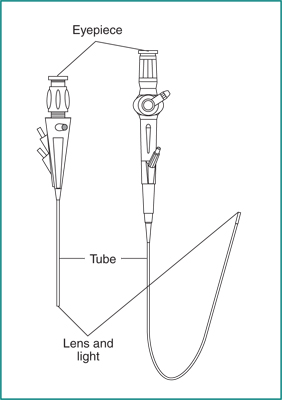
Treatment

The rashes caused by DH can be controlled with antibiotics such as dapsone. “What’s most interesting about DH is that the rash itself responds dramatically to dapsone, usually in 48 to 72 hours,” said Zone. This dramatic response—another hallmark of the disease—was once the basis for diagnosis, before skin biopsies became the norm. People who can’t tolerate dapsone may be given sulfapyridine or sulfamethoxypyridazine instead, although these drugs are less effective.

A strict gluten-free diet is the only treatment for the underlying celiac disease. Even with a gluten-free diet, dapsone or sulfapyridine therapy may need to be continued for 1–2 years to prevent further DH outbreaks. For people like Jones, who elected not to use dapsone, relief comes gradually as the diet does its job.

Jones’ circuitous path to a diagnosis is a common one for people with celiac disease. According to Zone, more than 95 percent of DH cases are misdiagnosed as eczema. Thankfully, once diagnosed, relief for most people with DH will come through treatment and strict adherence to a gluten-free diet.

This content is provided as a service of the National Institute of Diabetes and Digestive and Kidney Diseases (NIDDK), part of the National Institutes of Health. The NIDDK translates and disseminates research findings through its clearinghouses and education programs to increase knowledge and understanding about health and disease among patients, health professionals, and the public. Content produced by the NIDDK is carefully reviewed by NIDDK scientists and other experts.

Syndicated Content Details:
Source URL: https://www.niddk.nih.gov/Syndication/dermatitis-herpetiformis
Source Agency: National Institute of Diabetes and Digestive and Kidney Diseases (NIDDK)
Captured Date: 2015-12-05 06:53:00.0

Diabetes Diet, Eating, & Physical Activity

Nutrition and physical activity are important parts of a healthy lifestyle when you have diabetes. Along with other benefits, following a healthy meal plan and being active can help you keep your blood glucose level, also called blood sugar, in your target range. To manage your blood glucose, you need to balance what you eat and drink with physical activity and diabetes medicine, if you take any. What you choose to eat, how much you eat, and when you eat are all important in keeping your blood glucose level in the range that your health care team recommends.

Becoming more active and making changes in what you eat and drink can seem challenging at first. You may find it easier to start with small changes and get help from your family, friends, and health care team.

Eating well and being physically active most days of the week can help you

  • keep your blood glucose level, blood pressure, and cholesterol in your target ranges
  • lose weight or stay at a healthy weight
  • prevent or delay diabetes problems
  • feel good and have more energy

What foods can I eat if I have diabetes?

You may worry that having diabetes means going without foods you enjoy. The good news is that you can still eat your favorite foods, but you might need to eat smaller portions or enjoy them less often. Your health care team will help create a diabetes meal plan for you that meets your needs and likes.

The key to eating with diabetes is to eat a variety of healthy foods from all food groups, in the amounts your meal plan outlines.

The food groups are

  • vegetables
    • nonstarchy: includes broccoli, carrots, greens, peppers, and tomatoes
    • starchy: includes potatoes, corn, and green peas
  • fruits—includes oranges, melon, berries, apples, bananas, and grapes
  • grains—at least half of your grains for the day should be whole grains
    • includes wheat, rice, oats, cornmeal, barley, and quinoa
    • examples: bread, pasta, cereal, and tortillas
  • protein
    • lean meat
    • chicken or turkey without the skin
    • fish
    • eggs
    • nuts and peanuts
    • dried beans and certain peas, such as chickpeas and split peas
    • meat substitutes, such as tofu
  • dairy—nonfat or low fat

Learn more about the food groups at the U.S. Department of Agriculture’s (USDA) ChooseMyPlate.gov.

Eat foods with heart-healthy fats, which mainly come from these foods:

  • oils that are liquid at room temperature, such as canola and olive oil
  • nuts and seeds
  • heart-healthy fish such as salmon, tuna, and mackerel
  • avocado

Use oils when cooking food instead of butter, cream, shortening, lard, or stick margarine.

Photo of avocado, salmon, nuts, seeds, and olive oil.
Choose healthy fats, such as from nuts, seeds, and olive oil.

What foods and drinks should I limit if I have diabetes?

Foods and drinks to limit include

  • fried foods and other foods high in saturated fat and trans fat
  • foods high in salt, also called sodium
  • sweets, such as baked goods, candy, and ice cream
  • beverages with added sugars, such as juice, regular soda, and regular sports or energy drinks

Drink water instead of sweetened beverages. Consider using a sugar substitute in your coffee or tea.

If you drink alcohol, drink moderately—no more than one drink a day if you’re a woman or two drinks a day if you’re a man. If you use insulin or diabetes medicines that increase the amount of insulin your body makes, alcohol can make your blood glucose level drop too low. This is especially true if you haven’t eaten in a while. It’s best to eat some food when you drink alcohol.

When should I eat if I have diabetes?

Some people with diabetes need to eat at about the same time each day. Others can be more flexible with the timing of their meals. Depending on your diabetes medicines or type of insulin, you may need to eat the same amount of carbohydrates at the same time each day. If you take “mealtime” insulin, your eating schedule can be more flexible.

If you use certain diabetes medicines or insulin and you skip or delay a meal, your blood glucose level can drop too low. Ask your health care team when you should eat and whether you should eat before and after physical activity.

How much can I eat if I have diabetes?

Eating the right amount of food will also help you manage your blood glucose level and your weight. Your health care team can help you figure out how much food and how many calories you should eat each day. Look up how many calories are in what you eat and drink at the USDA’s Food-A-Pedia.

Weight-loss planning

If you are overweight or obese, work with your health care team to create a weight-loss plan.

These tools may help:

  • The Body Weight Planner can help you tailor your plans to reach and maintain your goal weight.
  • The SuperTracker lets you track your food, physical activity, and weight.

To lose weight, you need to eat fewer calories and replace less healthy foods with foods lower in calories, fat, and sugar.

If you have diabetes, are overweight or obese, and are planning to have a baby, you should try to lose any excess weight before you become pregnant. Learn more about planning for pregnancy if you have diabetes.

Meal plan methods

Two common ways to help you plan how much to eat if you have diabetes are the plate method and carbohydrate counting, also called carb counting. Check with your health care team about the method that’s best for you.

Plate method

The plate method helps you control your portion sizes. You don’t need to count calories. The plate method shows the amount of each food group you should eat. This method works best for lunch and dinner.

Use a 9-inch plate. Put nonstarchy vegetables on half of the plate; a meat or other protein on one-fourth of the plate; and a grain or other starch on the last one-fourth. Starches include starchy vegetables such as corn and peas. You also may eat a small bowl of fruit or a piece of fruit, and drink a small glass of milk as included in your meal plan.

Photo of a plate with cucumber and spinach on half of the plate, brown rice on one quarter of the plate, and baked chicken on the last quarter.
The plate method shows the amount of each food group you should eat.

You can find many different combinations of food and more details about using the plate method from the American Diabetes Association’s Create Your Plate.

Your daily eating plan also may include small snacks between meals.

Portion sizes

  • You can use everyday objects or your hand to judge the size of a portion.
  • 1 serving of meat or poultry is the palm of your hand or a deck of cards
  • 1 3-ounce serving of fish is a checkbook
  • 1 serving of cheese is six dice
  • 1/2 cup of cooked rice or pasta is a rounded handful or a tennis ball
  • 1 serving of a pancake or waffle is a DVD
  • 2 tablespoons of peanut butter is a ping-pong ball

Carbohydrate counting

Carbohydrate counting involves keeping track of the amount of carbohydrates you eat and drink each day. Because carbohydrates turn into glucose in your body, they affect your blood glucose level more than other foods do. Carb counting can help you manage your blood glucose level. If you take insulin, counting carbohydrates can help you know how much insulin to take.

The right amount of carbohydrates varies by how you manage your diabetes, including how physically active you are and what medicines you take, if any. Your health care team can help you create a personal eating plan based on carbohydrate counting.

The amount of carbohydrates in foods is measured in grams. To count carbohydrate grams in what you eat, you’ll need to

  • learn which foods have carbohydrates
  • read the Nutrition Facts food label, or learn to estimate the number of grams of carbohydrate in the foods you eat
  • add the grams of carbohydrate from each food you eat to get your total for each meal and for the day

Most carbohydrates come from starches, fruits, milk, and sweets. Try to limit carbohydrates with added sugars or those with refined grains, such as white bread and white rice. Instead, eat carbohydrates from fruit, vegetables, whole grains, beans, and low-fat or nonfat milk.

Photo of a bag of groceries containing fruit, vegetables, milk, and bread.
Choose healthy carbohydrates, such as fruit, vegetables, whole grains, beans, and low-fat milk, as part of your diabetes meal plan.

In addition to using the plate method and carb counting, you may want to visit a registered dietitian (RD) for medical nutrition therapy.

What is medical nutrition therapy?

Medical nutrition therapy is a service provided by an RD to create personal eating plans based on your needs and likes. For people with diabetes, medical nutrition therapy has been shown to improve diabetes management. Medicare pays for medical nutrition therapy for people with diabetes. If you have insurance other than Medicare, ask if it covers medical nutrition therapy for diabetes.

Will supplements and vitamins help my diabetes?

No clear proof exists that taking dietary supplements such as vitamins, minerals, herbs, or spices can help manage diabetes.1 You may need supplements if you cannot get enough vitamins and minerals from foods. Talk with your health care provider before you take any dietary supplement since some can cause side effects or affect how your medicines work.2

Why should I be physically active if I have diabetes?

Physical activity is an important part of managing your blood glucose level and staying healthy. Being active has many health benefits.

Physical activity

  • lowers blood glucose levels
  • lowers blood pressure
  • improves blood flow
  • burns extra calories so you can keep your weight down if needed
  • improves your mood
  • can prevent falls and improve memory in older adults
  • may help you sleep better

If you are overweight, combining physical activity with a reduced-calorie eating plan can lead to even more benefits. In the Look AHEAD: Action for Health in Diabetes study,1 overweight adults with type 2 diabetes who ate less and moved more had greater long-term health benefits compared to those who didn’t make these changes. These benefits included improved cholesterol levels, less sleep apnea, and being able to move around more easily.

Even small amounts of physical activity can help. Experts suggest that you aim for at least 30 minutes of moderate or vigorous physical activity 5 days of the week.3 Moderate activity feels somewhat hard, and vigorous activity is intense and feels hard. If you want to lose weight or maintain weight loss, you may need to do 60 minutes or more of physical activity 5 days of the week.3

Be patient. It may take a few weeks of physical activity before you see changes in your health.

How can I be physically active safely if I have diabetes?

Be sure to drink water before, during, and after exercise to stay well hydrated. The following are some other tips for safe physical activity when you have diabetes.

Photo of a man outdoors in exercise clothes drinking from a bottle of water.
Drink water when you exercise to stay well hydrated.

Plan ahead

Talk with your health care team before you start a new physical activity routine, especially if you have other health problems. Your health care team will tell you a target range for your blood glucose level and suggest how you can be active safely.

Your health care team also can help you decide the best time of day for you to do physical activity based on your daily schedule, meal plan, and diabetes medicines. If you take insulin, you need to balance the activity that you do with your insulin doses and meals so you don’t get low blood glucose.

Prevent low blood glucose

Because physical activity lowers your blood glucose, you should protect yourself against low blood glucose levels, also called hypoglycemia. You are most likely to have hypoglycemia if you take insulin or certain other diabetes medicines, such as a sulfonylurea. Hypoglycemia also can occur after a long intense workout or if you have skipped a meal before being active. Hypoglycemia can happen during or up to 24 hours after physical activity.

Planning is key to preventing hypoglycemia. For instance, if you take insulin, your health care provider might suggest you take less insulin or eat a small snack with carbohydrates before, during, or after physical activity, especially intense activity.4

You may need to check your blood glucose level before, during, and right after you are physically active.

Take care of your feet

People with diabetes may have problems with their feet because of poor blood flow and nerve damage that can result from high blood glucose levels. To help prevent foot problems, you should wear comfortable, supportive shoes and take care of your feet before, during, and after physical activity.

What physical activities should I do if I have diabetes?

Most kinds of physical activity can help you take care of your diabetes. Certain activities may be unsafe for some people, such as those with low vision or nerve damage to their feet. Ask your health care team what physical activities are safe for you. Many people choose walking with friends or family members for their activity.

Doing different types of physical activity each week will give you the most health benefits. Mixing it up also helps reduce boredom and lower your chance of getting hurt. Try these options for physical activity.

Add extra activity to your daily routine

If you have been inactive or you are trying a new activity, start slowly, with 5 to 10 minutes a day. Then add a little more time each week. Increase daily activity by spending less time in front of a TV or other screen. Try these simple ways to add physical activities in your life each day:

  • Walk around while you talk on the phone or during TV commercials.
  • Do chores, such as work in the garden, rake leaves, clean the house, or wash the car.
  • Park at the far end of the shopping center parking lot and walk to the store.
  • Take the stairs instead of the elevator.
  • Make your family outings active, such as a family bike ride or a walk in a park.

If you are sitting for a long time, such as working at a desk or watching TV, do some light activity for 3 minutes or more every half hour.5 Light activities include

  • leg lifts or extensions
  • overhead arm stretches
  • desk chair swivels
  • torso twists
  • side lunges
  • walking in place

Do aerobic exercise

Aerobic exercise is activity that makes your heart beat faster and makes you breathe harder. You should aim for doing aerobic exercise for 30 minutes a day most days of the week. You do not have to do all the activity at one time. You can split up these minutes into a few times throughout the day.

To get the most out of your activity, exercise at a moderate to vigorous level. Try

  • walking briskly or hiking
  • climbing stairs
  • swimming or a water-aerobics class
  • dancing
  • riding a bicycle or a stationary bicycle
  • taking an exercise class
  • playing basketball, tennis, or other sports

Talk with your health care team about how to warm up and cool down before and after you exercise.

Do strength training to build muscle

Strength training is a light or moderate physical activity that builds muscle and helps keep your bones healthy. Strength training is important for both men and women. When you have more muscle and less body fat, you’ll burn more calories. Burning more calories can help you lose and keep off extra weight.

You can do strength training with hand weights, elastic bands, or weight machines. Try to do strength training two to three times a week. Start with a light weight. Slowly increase the size of your weights as your muscles become stronger.

Photo of a smiling woman holding hand weights.
You can do strength training with hand weights, elastic bands, or weight machines.

Do stretching exercises

Stretching exercises are light or moderate physical activity. When you stretch, you increase your flexibility, lower your stress, and help prevent sore muscles.

You can choose from many types of stretching exercises. Yoga is a type of stretching that focuses on your breathing and helps you relax. Even if you have problems moving or balancing, certain types of yoga can help. For instance, chair yoga has stretches you can do when sitting in a chair or holding onto a chair while standing. Your health care team can suggest whether yoga is right for you.

References

This content is provided as a service of the National Institute of Diabetes and Digestive and Kidney Diseases (NIDDK), part of the National Institutes of Health. The NIDDK translates and disseminates research findings through its clearinghouses and education programs to increase knowledge and understanding about health and disease among patients, health professionals, and the public. Content produced by the NIDDK is carefully reviewed by NIDDK scientists and other experts.

Diabetes Inspidus

On this page:

What is diabetes insipidus?

Diabetes insipidus is a rare disorder that occurs when a person's kidneys pass an abnormally large volume of urine that is insipid—dilute and odorless. In most people, the kidneys pass about 1 to 2 quarts of urine a day. In people with diabetes insipidus, the kidneys can pass 3 to 20 quarts of urine a day. As a result, a person with diabetes insipidus may feel the need to drink large amounts of liquids.
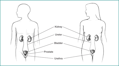
Diabetes insipidus and diabetes mellitus—which includes both type 1 and type 2 diabetes—are unrelated, although both conditions cause frequent urination and constant thirst. Diabetes mellitus causes high blood glucose, or blood sugar, resulting from the body's inability to use blood glucose for energy. People with diabetes insipidus have normal blood glucose levels; however, their kidneys cannot balance fluid in the body.

What are the kidneys and what do they do?

The kidneys are two bean-shaped organs, each about the size of a fist. They are located just below the rib cage, one on each side of the spine. Every day, the kidneys normally filter about 120 to 150 quarts of blood to produce about 1 to 2 quarts of urine, composed of wastes and extra fluid. The urine flows from the kidneys to the bladder through tubes called ureters. The bladder stores urine. When the bladder empties, urine flows out of the body through a tube called the urethra, located at the bottom of the bladder.

Frontal body diagram with kidneys and bladder
Every day, the kidneys normally filter about 120 to 150 quarts of blood to produce about 1 to 2 quarts of urine.

How is fluid regulated in the body?

A person's body regulates fluid by balancing liquid intake and removing extra fluid. Thirst usually controls a person’s rate of liquid intake, while urination removes most fluid, although people also lose fluid through sweating, breathing, or diarrhea. The hormone vasopressin, also called antidiuretic hormone, controls the fluid removal rate through urination. The hypothalamus, a small gland located at the base of the brain, produces vasopressin. The nearby pituitary gland stores the vasopressin and releases it into the bloodstream when the body has a low fluid level. Vasopressin signals the kidneys to absorb less fluid from the bloodstream, resulting in less urine. When the body has extra fluid, the pituitary gland releases smaller amounts of vasopressin, and sometimes none, so the kidneys remove more fluid from the bloodstream and produce more urine.

What are the types of diabetes insipidus?

The types of diabetes insipidus include

  • central
  • nephrogenic
  • dipsogenic
  • gestational

Each type of diabetes insipidus has a different cause.

Central Diabetes Insipidus

Central diabetes insipidus happens when damage to a person's hypothalamus or pituitary gland causes disruptions in the normal production, storage, and release of vasopressin. The disruption of vasopressin causes the kidneys to remove too much fluid from the body, leading to an increase in urination. Damage to the hypothalamus or pituitary gland can result from the following:

  • surgery
  • infection
  • inflammation
  • a tumor
  • head injury

Central diabetes insipidus can also result from an inherited defect in the gene that produces vasopressin, although this cause is rare. In some cases, the cause is unknown.

Nephrogenic Diabetes Insipidus

Nephrogenic diabetes insipidus occurs when the kidneys do not respond normally to vasopressin and continue to remove too much fluid from a person's bloodstream. Nephrogenic diabetes insipidus can result from inherited gene changes, or mutations, that prevent the kidneys from responding to vasopressin. Other causes of nephrogenic diabetes insipidus include

  • chronic kidney disease
  • certain medications, particularly lithium
  • low potassium levels in the blood
  • high calcium levels in the blood
  • blockage of the urinary tract

The causes of nephrogenic diabetes insipidus can also be unknown.

Dipsogenic Diabetes Insipidus

A defect in the thirst mechanism, located in a person's hypothalamus, causes dipsogenic diabetes insipidus. This defect results in an abnormal increase in thirst and liquid intake that suppresses vasopressin secretion and increases urine output. The same events and conditions that damage the hypothalamus or pituitary—surgery, infection, inflammation, a tumor, head injury—can also damage the thirst mechanism. Certain medications or mental health problems may predispose a person to dipsogenic diabetes insipidus.

Gestational Diabetes Insipidus

Gestational diabetes insipidus occurs only during pregnancy. In some cases, an enzyme made by the placenta—a temporary organ joining mother and baby—breaks down the mother's vasopressin. In other cases, pregnant women produce more prostaglandin, a hormone-like chemical that reduces kidney sensitivity to vasopressin. Most pregnant women who develop gestational diabetes insipidus have a mild case that does not cause noticeable symptoms. Gestational diabetes insipidus usually goes away after the mother delivers the baby; however, it may return if the mother becomes pregnant again.

What are the complications of diabetes insipidus?

The main complication of diabetes insipidus is dehydration if fluid loss is greater than liquid intake. Signs of dehydration include

  • thirst
  • dry skin
  • fatigue
  • sluggishness
  • dizziness
  • confusion
  • nausea

Severe dehydration can lead to seizures, permanent brain damage, and even death.

How is diabetes insipidus diagnosed?

A health care provider can diagnose a person with diabetes insipidus based on the following:

  • medical and family history
  • physical exam
  • urinalysis
  • blood tests
  • fluid deprivation test
  • magnetic resonance imaging (MRI)

Medical and Family History

Taking a medical and family history can help a health care provider diagnose diabetes insipidus. A health care provider will ask the patient to review his or her symptoms and ask whether the patient's family has a history of diabetes insipidus or its symptoms.

Physical Exam

A physical exam can help diagnose diabetes insipidus. During a physical exam, a health care provider usually examines the patient's skin and appearance, checking for signs of dehydration.

Urinalysis

Urinalysis tests a urine sample. A patient collects the urine sample in a special container at home, in a health care provider's office, or at a commercial facility. A health care provider tests the sample in the same location or sends it to a lab for analysis. The test can show whether the urine is dilute or concentrated. The test can also show the presence of glucose, which can distinguish between diabetes insipidus and diabetes mellitus. The health care provider may also have the patient collect urine in a special container over a 24-hour period to measure the total amount of urine produced by the kidneys.

Blood Tests

A blood test involves drawing a patient's blood at a health care provider’s office or a commercial facility and sending the sample to a lab for analysis. The blood test measures sodium levels, which can help diagnose diabetes insipidus and in some cases determine the type.

Fluid Deprivation Test

A fluid deprivation test measures changes in a patient’s body weight and urine concentration after restricting liquid intake. A health care provider can perform two types of fluid deprivation tests:

  • A short form of the deprivation test. A health care provider instructs the patient to stop drinking all liquids for a specific period of time, usually during dinner. The next morning, the patient will collect a urine sample at home. The patient then returns the urine sample to his or her health care provider or takes it to a lab where a technician measures the concentration of the urine sample.
  • A formal fluid deprivation test. A health care provider performs this test in a hospital to continuously monitor the patient for signs of dehydration. Patients do not need anesthesia. A health care provider weighs the patient and analyzes a urine sample. The health care provider repeats the tests and measures the patient's blood pressure every 1 to 2 hours until one of the following happens:
    • The patient's blood pressure drops too low or the patient has a rapid heartbeat when standing.
    • The patient loses 5 percent or more of his or her initial body weight.
    • Urine concentration increases only slightly in two to three consecutive measurements.
    At the end of the test, a health care provider will compare the patient's blood sodium, vasopressin levels, and urine concentration to determine whether the patient has diabetes insipidus. Sometimes, the health care provider may administer medications during the test to see if they increase a patient's urine concentration. In other cases, the health care provider may give the patient a concentrated sodium solution intravenously at the end of the test to increase the patient's blood sodium level and determine if he or she has diabetes insipidus.

Magnetic Resonance Imaging

Magnetic resonance imaging (MRI) is a test that takes pictures of the body's internal organs and soft tissues without using x-rays. A specially trained technician performs the procedure in an outpatient center or a hospital, and a radiologist—a doctor who specializes in medical imaging—interprets the images. A patient does not need anesthesia, although people with a fear of confined spaces may receive light sedation. An MRI may include an injection of a special dye, called contrast medium. With most MRI machines, the person lies on a table that slides into a tunnel-shaped device that may be open ended or closed at one end. Some MRI machines allow the patient to lie in a more open space. MRIs cannot diagnose diabetes insipidus. Instead, an MRI can show if the patient has problems with his or her hypothalamus or pituitary gland or help the health care provider determine if diabetes insipidus is the possible cause of the patient's symptoms.

How is diabetes insipidus treated?

The primary treatment for diabetes insipidus involves drinking enough liquid to prevent dehydration. A health care provider may refer a person with diabetes insipidus to a nephrologist—a doctor who specializes in treating kidney problems—or to an endocrinologist—a doctor who specializes in treating disorders of the hormone-producing glands. Treatment for frequent urination or constant thirst depends on the patient’s type of diabetes insipidus:

  • Central diabetes insipidus. A synthetic, or man-made, hormone called desmopressin treats central diabetes insipidus. The medication comes as an injection, a nasal spray, or a pill. The medication works by replacing the vasopressin that a patient’s body normally produces. This treatment helps a patient manage symptoms of central diabetes insipidus; however, it does not cure the disease.
  • Nephrogenic diabetes insipidus. In some cases, nephrogenic diabetes insipidus goes away after treatment of the cause. For example, switching medications or taking steps to balance the amount of calcium or potassium in the patient’s body may resolve the problem. Medications for nephrogenic diabetes insipidus include diuretics, either alone or combined with aspirin or ibuprofen. Health care providers commonly prescribe diuretics to help patients’ kidneys remove fluid from the body. Paradoxically, in people with nephrogenic diabetes insipidus, a class of diuretics called thiazides reduces urine production and helps patients’ kidneys concentrate urine. Aspirin or ibuprofen also helps reduce urine volume.
  • Dipsogenic diabetes insipidus. Researchers have not yet found an effective treatment for dipsogenic diabetes insipidus. People can try sucking on ice chips or sour candies to moisten their mouths and increase saliva flow, which may reduce the desire to drink. For a person who wakes multiple times at night to urinate because of dipsogenic diabetes insipidus, taking a small dose of desmopressin at bedtime may help. Initially, the health care provider will monitor the patient’s blood sodium levels to prevent hyponatremia, or low sodium levels in the blood.
  • Gestational diabetes insipidus. A health care provider can prescribe desmopressin for women with gestational diabetes insipidus. An expecting mother’s placenta does not destroy desmopressin as it does vasopressin. Most women will not need treatment after delivery.

Most people with diabetes insipidus can prevent serious problems and live a normal life if they follow the health care provider’s recommendations and keep their symptoms under control.

Eating, Diet, and Nutrition

Researchers have not found that eating, diet, and nutrition play a role in causing or preventing diabetes insipidus.

Points to Remember

  • Diabetes insipidus is a rare disorder that occurs when a person’s kidneys pass an abnormally large volume of urine that is insipid—dilute and odorless.
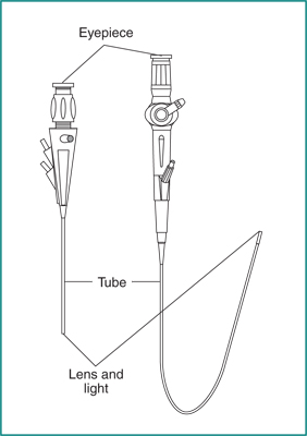
  • A person’s body regulates fluid by balancing liquid intake and removing extra fluid. Thirst usually controls a person’s rate of liquid intake, while urination removes most fluid, although people also lose fluid through sweating, breathing, or diarrhea. The hormone vasopressin, also called antidiuretic hormone, controls the fluid removal rate through urination.
  • The types of diabetes insipidus include central, nephrogenic, dipsogenic, and gestational. Each type of diabetes insipidus has a different cause.
  • The main complication of diabetes insipidus is dehydration if fluid loss is greater than liquid intake.
  • A health care provider can diagnose a person with diabetes insipidus based on a medical and family history, a physical exam, urinalysis, blood tests, a fluid deprivation test, and magnetic resonance imaging (MRI).
  • The primary treatment for diabetes insipidus involves drinking enough liquid to prevent dehydration.

Clinical Trials

The National Institute of Diabetes and Digestive and Kidney Diseases (NIDDK) and other components of the National Institutes of Health (NIH) conduct and support research into many diseases and conditions.
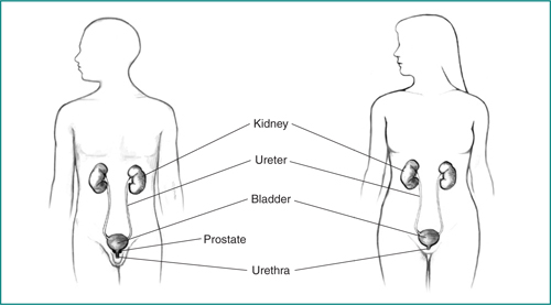
What are clinical trials, and are they right for you?

Clinical trials are part of clinical research and at the heart of all medical advances. Clinical trials look at new ways to prevent, detect, or treat disease. Researchers also use clinical trials to look at other aspects of care, such as improving the quality of life for people with chronic illnesses. Find out if clinical trials are right for you.

What clinical trials are open?

Clinical trials that are currently open and are recruiting can be viewed at www.ClinicalTrials.gov.

This content is provided as a service of the National Institute of Diabetes and Digestive and Kidney Diseases (NIDDK), part of the National Institutes of Health. The NIDDK translates and disseminates research findings through its clearinghouses and education programs to increase knowledge and understanding about health and disease among patients, health professionals, and the public. Content produced by the NIDDK is carefully reviewed by NIDDK scientists and other experts.

The NIDDK would like to thank:
Bessie Young, M.D., M.P.H., University of Washington; Dr. Joseph Verbalis, M.D, Georgetown University

Diabetes Tests & Diagnosis

Your health care professional can diagnose diabetes, prediabetes, and gestational diabetes through blood tests. The blood tests show if your blood glucose, also called blood sugar, is too high.

Do not try to diagnose yourself if you think you might have diabetes. Testing equipment that you can buy over the counter, such as a blood glucose meter, cannot diagnose diabetes.

Who should be tested for diabetes?

Anyone who has symptoms of diabetes should be tested for the disease. Some people will not have any symptoms but may have risk factors for diabetes and need to be tested. Testing allows health care professionals to find diabetes sooner and work with their patients to manage diabetes and prevent complications.

Testing also allows health care professionals to find prediabetes. Making lifestyle changes to lose a modest amount of weight if you are overweight may help you delay or prevent type 2 diabetes.

Photo of a man having blood drawn
Blood tests help health care professionals diagnose diabetes and prediabetes.

Type 1 diabetes

Most often, testing for occurs in people with diabetes symptoms. Doctors usually diagnose type 1 diabetes in children and young adults. Because type 1 diabetes can run in families, a study called TrialNet offers free testing to family members of people with the disease, even if they don’t have symptoms.

Type 2 diabetes

Experts recommend routine testing for type 2 diabetes if you

  • are age 45 or older
  • are between the ages of 19 and 44, are overweight or obese, and have one or more other diabetes risk factors
  • are a woman who had gestational diabetes1

Medicare covers the cost of diabetes tests for people with certain risk factors for diabetes. If you have Medicare, find out if you qualify for coverage. If you have different insurance, ask your insurance company if it covers diabetes tests.

Though type 2 diabetes most often develops in adults, children also can develop type 2 diabetes. Experts recommend testing children between the ages of 10 and 18 who are overweight or obese and have at least two other risk factors for developing diabetes.1

Gestational diabetes

All pregnant women who do not have a prior diabetes diagnosis should be tested for gestational diabetes. If you are pregnant, you will take a glucose challenge test between 24 and 28 weeks of pregnancy.1

What tests are used to diagnose diabetes and prediabetes?

Health care professionals most often use the fasting plasma glucose (FPG) test or the A1C test to diagnose diabetes. In some cases, they may use a random plasma glucose (RPG) test.

Fasting plasma glucose (FPG) test

The FPG blood test measures your blood glucose level at a single point in time. For the most reliable results, it is best to have this test in the morning, after you fast for at least 8 hours. Fasting means having nothing to eat or drink except sips of water.

A1C test

The A1C test is a blood test that provides your average levels of blood glucose over the past 3 months. Other names for the A1C test are hemoglobin A1C, HbA1C, glycated hemoglobin, and glycosylated hemoglobin test. You can eat and drink before this test. When it comes to using the A1C to diagnose diabetes, your doctor will consider factors such as your age and whether you have anemia or another problem with your blood.1 The A1C test is not accurate in people with anemia.

Your health care professional will report your A1C test result as a percentage, such as an A1C of 7 percent. The higher the percentage, the higher your average blood glucose levels.

People with diabetes also use information from the A1C test to help manage their diabetes.

Random plasma glucose (RPG) test

Sometimes health care professionals use the RPG test to diagnose diabetes when diabetes symptoms are present and they do not want to wait until you have fasted. You do not need to fast overnight for the RPG test. You may have this blood test at any time.

What tests are used to diagnose gestational diabetes?

Pregnant women may have the glucose challenge test, the oral glucose tolerance test, or both. These tests show how well your body handles glucose.

Glucose challenge test

If you are pregnant and a health care professional is checking you for gestational diabetes, you may first receive the glucose challenge test. Another name for this test is the glucose screening test. In this test, a health care professional will draw your blood 1 hour after you drink a sweet liquid containing glucose. You do not need to fast for this test. If your blood glucose is too high—135 to 140 or more—you may need to return for an oral glucose tolerance test while fasting.

Oral glucose tolerance test (OGTT)

The OGTT measures blood glucose after you fast for at least 8 hours. First, a health care professional will draw your blood. Then you will drink the liquid containing glucose. For diagnosing gestational diabetes, you will need your blood drawn every hour for 2 to 3 hours.

High blood glucose levels at any two or more blood test times during the OGTT—fasting, 1 hour, 2 hours, or 3 hours—mean you have gestational diabetes. Your health care team will explain what your OGTT results mean.

Health care professionals also can use the OGTT to diagnose type 2 diabetes and prediabetes in people who are not pregnant. The OGTT helps health care professionals detect type 2 diabetes and prediabetes better than the FPG test. However, the OGTT is a more expensive test and is not as easy to give. To diagnose type 2 diabetes and prediabetes, a health care professional will need to draw your blood 1 hour after you drink the liquid containing glucose and again after 2 hours.

What test numbers tell me if I have diabetes or prediabetes?

Each test to detect diabetes and prediabetes uses a different measurement. Usually, the same test method needs to be repeated on a second day to diagnose diabetes. Your doctor may also use a second test method to confirm that you have diabetes.

The following table helps you understand what your test numbers mean if you are not pregnant.

Diagnosis A1C (percent)  Fasting plasma glucose (FPG)a Oral glucose tolerance test (OGTT)ab Random plasma glucose test (RPG)a
Normal below 5.7 99 or below 139 or below  
Prediabetes 5.7 to 6.4 100 to 125 140 to 199  
Diabetes 6.5 or above 126 or above 200 or above 200 or above

Which tests help my health care professional know what kind of diabetes I have?

Even though the tests described here can confirm that you have diabetes, they can’t identify what type you have. Sometimes health care professionals are unsure if diabetes is type 1 or type 2. A rare type of diabetes that can occur in babies, called monogenic diabetes, can also be mistaken for type 1 diabetes. Treatment depends on the type of diabetes, so knowing which type you have is important.

To find out if your diabetes is type 1, your health care professional may look for certain autoantibodies. Autoantibodies are antibodies that mistakenly attack your healthy tissues and cells. The presence of one or more of several types of autoantibodies specific to diabetes is common in type 1 diabetes, but not in type 2 or monogenic diabetes. A health care professional will have to draw your blood for this test.

If you had diabetes while you were pregnant, you should get tested 6 to 12 weeks after your baby is born to see if you have type 2 diabetes.

References

This content is provided as a service of the National Institute of Diabetes and Digestive and Kidney Diseases (NIDDK), part of the National Institutes of Health. The NIDDK translates and disseminates research findings through its clearinghouses and education programs to increase knowledge and understanding about health and disease among patients, health professionals, and the public. Content produced by the NIDDK is carefully reviewed by NIDDK scientists and other experts.

Diabetes and Foot Problems

Foot problems are common in people with diabetes. You might be afraid you’ll lose a toe, foot, or leg to diabetes, or know someone who has, but you can lower your chances of having diabetes-related foot problems by taking care of your feet every day. Managing your blood glucose levels, also called blood sugar, can also help keep your feet healthy.

How can diabetes affect my feet?

Over time, diabetes may cause nerve damage, also called diabetic neuropathy, that can cause tingling and pain, and can make you lose feeling in your feet. When you lose feeling in your feet, you may not feel a pebble inside your sock or a blister on your foot, which can lead to cuts and sores. Cuts and sores can become infected.

Diabetes also can lower the amount of blood flow in your feet. Not having enough blood flowing to your legs and feet can make it hard for a sore or an infection to heal. Sometimes, a bad infection never heals. The infection might lead to gangrene.

Gangrene and foot ulcers that do not get better with treatment can lead to an amputation of your toe, foot, or part of your leg. A surgeon may perform an amputation to prevent a bad infection from spreading to the rest of your body, and to save your life. Good foot care is very important to prevent serious infections and gangrene.

Although rare, nerve damage from diabetes can lead to changes in the shape of your feet, such as Charcot’s foot. Charcot’s foot may start with redness, warmth, and swelling. Later, bones in your feet and toes can shift or break, which can cause your feet to have an odd shape, such as a “rocker bottom.”

Illustration of Charcot’s foot showing an enlarged sole of the foot with a rounded shape.
Charcot’s foot can cause your feet to have an odd shape, such as a “rocker bottom.”

What can I do to keep my feet healthy?

Work with your health care team to make a diabetes self-care plan, which is an action plan for how you will manage your diabetes. Your plan should include foot care. A foot doctor, also called a podiatrist, and other specialists may be part of your health care team.

Include these steps in your foot care plan:

Check your feet every day

You may have foot problems, but feel no pain in your feet. Checking your feet each day will help you spot problems early before they get worse. A good way to remember is to check your feet each evening when you take off your shoes. Also check between your toes. If you have trouble bending over to see your feet, try using a mirror to see them, or ask someone else to look at your feet.

Look for problems such as

  • cuts, sores, or red spots
  • swelling or fluid-filled blisters
  • ingrown toenails, in which the edge of your nail grows into your skin
  • corns or calluses, which are spots of rough skin caused by too much rubbing or pressure on the same spot
  • plantar warts, which are flesh-colored growths on the bottom of the feet
  • athlete’s foot
  • warm spots
Someone checking the bottom of their bare foot.

If you have certain foot problems that make it more likely you will develop a sore on your foot, your doctor may recommend taking the temperature of the skin on different parts of your feet. A “hot spot” can be the first sign that a blister or an ulcer is starting.

Cover a blister, cut, or sore with a bandage. Smooth corns and calluses as explained below.

Wash your feet every day

Wash your feet with soap in warm, not hot, water. Test the water to make sure it is not too hot. You can use a thermometer (90° to 95° F is safe) or your elbow to test the warmth of the water. Do not soak your feet because your skin will get too dry.

Photo of a person’s bare foot with hands examining the toes.

After washing and drying your feet, put talcum powder or cornstarch between your toes. Skin between the toes tends to stay moist. Powder will keep the skin dry to help prevent an infection.

Smooth corns and calluses gently

Thick patches of skin called corns or calluses can grow on the feet. If you have corns or calluses, talk with your foot doctor about the best way to care for these foot problems. If you have nerve damage, these patches can become ulcers.

Photo of a woman smoothing the bottom of her foot with a pumice stone.

If your doctor tells you to, use a pumice stone to smooth corns and calluses after bathing or showering. A pumice stone is a type of rock used to smooth the skin. Rub gently, only in one direction, to avoid tearing the skin.

Do NOT

  • cut corns and calluses
  • use corn plasters, which are medicated pads
  • use liquid corn and callus removers

Cutting and over-the counter corn removal products can damage your skin and cause an infection.

To keep your skin smooth and soft, rub a thin coat of lotion, cream, or petroleum jelly on the tops and bottoms of your feet. Do not put lotion or cream between your toes because moistness might cause an infection.

Trim your toenails straight across

Trim your toenails, when needed, after you wash and dry your feet. Using toenail clippers, trim your toenails straight across. Do not cut into the corners of your toenail. Gently smooth each nail with an emery board or nonsharp nail file. Trimming this way helps prevent cutting your skin and keeps the nails from growing into your skin.

Photo of someone clipping their toenails.

Have a foot doctor trim your toenails if

  • you cannot see, feel, or reach your feet
  • your toenails are thick or yellowed
  • your nails curve and grow into the skin

If you want to get a pedicure at a salon, you should bring your own nail tools to prevent getting an infection. You can ask your health care provider what other steps you can take at the salon to prevent infection.

Wear shoes and socks at all times

Wear shoes and socks at all times. Do not walk barefoot or in just socks – even when you are indoors. You could step on something and hurt your feet. You may not feel any pain and may not know that you hurt yourself.

Check the inside of your shoes before putting them on, to make sure the lining is smooth and free of pebbles or other objects.

Photo of a man feeling inside his shoe.

Make sure you wear socks, stockings, or nylons with your shoes to keep from getting blisters and sores. Choose clean, lightly padded socks that fit well. Socks with no seams are best.

Wear shoes that fit well and protect your feet. Here are some tips for finding the right type of shoes:

  • Walking shoes and athletic shoes are good for daily wear. They support your feet and allow them to “breathe.”
  • Do not wear vinyl or plastic shoes, because they do not stretch or “breathe.”
  • When buying shoes, make sure they feel good and have enough room for your toes. Buy shoes at the end of the day, when your feet are the largest, so that you can find the best fit.
  • If you have a bunion, or hammertoes, which are toes that curl under your feet, you may need extra-wide or deep shoes.1 Do not wear shoes with pointed toes or high heels, because they put too much pressure on your toes.
  • If your feet have changed shape, such as from Charcot’s foot, you may need special shoes or shoe inserts, called orthotics. You also may need inserts if you have bunions, hammertoes, or other foot problems.

When breaking in new shoes, only wear them for a few hours at first and then check your feet for areas of soreness.

Medicare Part B insurance and other health insurance programs may help pay for these special shoes or inserts. Ask your insurance plan if it covers your special shoes or inserts.

Protect your feet from hot and cold

If you have nerve damage from diabetes, you may burn your feet and not know you did. Take the following steps to protect your feet from heat:

  • Wear shoes at the beach and on hot pavement.
  • Put sunscreen on the tops of your feet to prevent sunburn.
  • Keep your feet away from heaters and open fires.
  • Do not put a hot water bottle or heating pad on your feet.
Photo of someone’s feet wearing shoes and walking on the sand.

Wear socks in bed if your feet get cold. In the winter, wear lined, waterproof boots to keep your feet warm and dry.

Keep the blood flowing to your feet

Try the following tips to improve blood flow to your feet:

  • Put your feet up when you are sitting.
  • Wiggle your toes for a few minutes throughout the day. Move your ankles up and down and in and out to help blood flow in your feet and legs.
  • Do not wear tight socks or elastic stockings. Do not try to hold up loose socks with rubber bands.
  • Be more physically active. Choose activities that are easy on your feet, such as walking, dancing, yoga or stretching, swimming, or bike riding.
  • Stop smoking.
Photo of a man sitting reading a book with his feet resting on footstool.

Smoking can lower the amount of blood flow to your feet. If you smoke, ask for help to stop. You can get help by calling the national quitline at 1-800-QUITNOW or 1-800-784-8669. For tips on quitting, go to SmokeFree.gov.

Get a foot check at every health care visit

Ask your health care team to check your feet at each visit. Take off your shoes and socks when you’re in the exam room so they will remember to check your feet. At least once a year, get a thorough foot exam, including a check of the feeling and pulses in your feet.

Photo of a doctor examining someone’s bare feet.

Get a thorough foot exam at each health care visit if you have

  • changes in the shape of your feet
  • loss of feeling in your feet
  • peripheral artery disease
  • had foot ulcers or an amputation in the past1

Ask your health care team to show you how to care for your feet.

When should I see my health care provider about foot problems?

Call your health care provider right away if you have

  • a cut, blister, or bruise on your foot that does not start to heal after a few days
  • skin on your foot that becomes red, warm, or painful—signs of a possible infection
  • a callus with dried blood inside of it,which often can be the first sign of a wound under the callus
  • a foot infection that becomes black and smelly—signs you might have gangrene

Ask your provider to refer you to a foot doctor, or podiatrist, if needed.

References

Clinical Trials

The National Institute of Diabetes and Digestive and Kidney Diseases (NIDDK) and other components of the National Institutes of Health (NIH) conduct and support research into many diseases and conditions.

What are clinical trials, and are they right for you?

Clinical trials are part of clinical research and at the heart of all medical advances. Clinical trials look at new ways to prevent, detect, or treat disease. Researchers also use clinical trials to look at other aspects of care, such as improving the quality of life for people with chronic illnesses. Find out if clinical trials are right for you.

What clinical trials are open?

Clinical trials that are currently open and are recruiting can be viewed at www.ClinicalTrials.gov.

This content is provided as a service of the National Institute of Diabetes and Digestive and Kidney Diseases (NIDDK), part of the National Institutes of Health. The NIDDK translates and disseminates research findings through its clearinghouses and education programs to increase knowledge and understanding about health and disease among patients, health professionals, and the public. Content produced by the NIDDK is carefully reviewed by NIDDK scientists and other experts.

Diabetes, Gum Disease, & Other Dental Problems

How can diabetes affect my mouth?

Too much glucose, also called sugar, in your blood from diabetes can cause pain, infection, and other problems in your mouth. Your mouth includes

  • your teeth
  • your gums
  • your jaw
  • tissues such as your tongue, the roof and bottom of your mouth, and the inside of your cheeks
Drawing of a woman’s facial profile with the jaw labeled. Inset shows teeth, gums, roof of the mouth, bottom of the mouth, tongue, and inside of cheek.

Glucose is present in your saliva—the fluid in your mouth that makes it wet. When diabetes is not controlled, high glucose levels in your saliva help harmful bacteria grow. These bacteria combine with food to form a soft, sticky film called plaque. Plaque also comes from eating foods that contain sugars or starches. Some types of plaque cause tooth decay or cavities. Other types of plaque cause gum disease and bad breath.

Image stating that high glucose levels equal an increase in plaque.  

Gum disease can be more severe and take longer to heal if you have diabetes. In turn, having gum disease can make your blood glucose hard to control.

What happens if I have plaque?

Plaque that is not removed hardens over time into tartar and collects above your gum line. Tartar makes it more difficult to brush and clean between your teeth. Your gums become red and swollen, and bleed easily—signs of unhealthy or inflamed gums, called gingivitis.

When gingivitis is not treated, it can advance to gum disease called periodontitis. In periodontitis, the gums pull away from the teeth and form spaces, called pockets, which slowly become infected. This infection can last a long time. Your body fights the bacteria as the plaque spreads and grows below the gum line. Both the bacteria and your body’s response to this infection start to break down the bone and the tissue that hold the teeth in place. If periodontitis is not treated, the gums, bones, and tissue that support the teeth are destroyed. Teeth may become loose and might need to be removed. If you have periodontitis, your dentist may send you to a periodontist, an expert in treating gum disease.

Drawing of a close-up view of teeth and healthy gums, and a drawing of a close-up view of teeth and gums with periodontitis.
Healthy gums                       Periodontitis

What are the most common mouth problems from diabetes?

The following chart shows the most common mouth problems from diabetes.

Problem What It Is Symptoms Treatment

gingivitis
  • unhealthy or inflamed gums
  • red, swollen, and bleeding gums
  • daily brushing and flossing
  • regular cleanings at the dentist

periodontitis
  • gum disease, which can change from mild to severe
  • red, swollen, and bleeding gums
  • gums that have pulled away from the teeth
  • long-lasting infection between the teeth and gums
  • bad breath that won’t go away
  • permanent teeth that are loose or moving away from one another
  • changes in the way your teeth fit together when you bite
  • sometimes pus between the teeth and gums
  • changes in the fit of dentures, which are teeth you can remove
  • deep cleaning at your dentist
  • medicine that your dentist prescribes
  • gum surgery in severe cases

thrush, called
candidiasis
  • the growth of a naturally occurring fungus that the body is unable to control
  • sore, white—or sometimes red—patches on your gums, tongue, cheeks, or the roof of your mouth
  • patches that have turned into open sores
  • medicine that your doctor or dentist prescribes to kill the fungus
  • cleaning dentures
  • removing dentures for part of the day or night, and soaking them in medicine that your doctor or dentist prescribes

dry mouth, called
xerostomia
  • a lack of saliva in your mouth, which raises your risk for tooth decay and gum disease
  • dry feeling in your mouth, often or all of the time
  • dry, rough tongue
  • pain in the mouth
  • cracked lips
  • mouth sores or infection
  • problems chewing, eating, swallowing, or talking
  • taking medicine to keep your mouth wet that your doctor or dentist prescribes
  • rinsing with a fluoride mouth rinse to prevent cavities
  • using sugarless gum or mints to increase saliva flow
  • taking frequent sips of water
  • avoiding tobacco, caffeine, and alcoholic beverages
  • using a humidifier, a device that raises the level of moisture in your home, at night
  • avoiding spicy or salty foods that may cause pain in a dry mouth

oral burning
  • a burning sensation inside the mouth caused by uncontrolled blood glucose levels
  • burning feeling in the mouth
  • dry mouth
  • bitter taste
  • symptoms may worsen throughout the day
  • seeing your doctor, who may change your diabetes medicine
  • once your blood glucose is under control, the oral burning will go away

More symptoms of a problem in your mouth are

  • a sore, or an ulcer, that does not heal
  • dark spots or holes in your teeth
  • pain in your mouth, face, or jaw that doesn’t go away
  • loose teeth
  • pain when chewing
  • a changed sense of taste or a bad taste in your mouth
  • bad breath that doesn’t go away when you brush your teeth

How will I know if I have mouth problems from diabetes?

Check your mouth for signs of problems from diabetes. If you notice any problems, see your dentist right away. Some of the first signs of gum disease are swollen, tender, or bleeding gums. Sometimes you won’t have any signs of gum disease. You may not know you have it until you have serious damage. Your best defense is to see your dentist twice a year for a cleaning and checkup.

Drawing of a man checking the inside of his mouth in the bathroom mirror for signs of problems from diabetes.
Check your mouth for signs of problems from diabetes.

How can I prepare for a visit to my dentist?

Plan ahead. Talk with your doctor and dentist before the visit about the best way to take care of your blood glucose during dental work.

You may be taking a diabetes medicine that can cause low blood glucose, also called hypoglycemia. If you take insulin or other diabetes medicines, take them and eat as usual before visiting the dentist. You may need to bring your diabetes medicines and your snacks or meal with you to the dentist’s office.

You may need to postpone any nonemergency dental work if your blood glucose is not under control.

If you feel nervous about visiting the dentist, tell your dentist and the staff about your feelings. Your dentist can adapt the treatment to your needs. Don’t let your nerves stop you from having regular checkups. Waiting too long to take care of your mouth may make things worse.

Drawing of a male patient seated in a dental chair and talking with his male dentist.
If you feel nervous about visiting the dentist, tell your dentist and the staff about your feelings.

If you feel nervous about visiting the dentist, tell your dentist and the staff about your feelings.

What if my mouth is sore after my dental work?

A sore mouth is common after dental work. If this happens, you might not be able to eat or chew the foods you normally eat for several hours or days. For guidance on how to adjust your usual routine while your mouth is healing, ask your doctor

  • what foods and drinks you should have
  • if you should change the time when you take your diabetes medicines
  • if you should change the dose of your diabetes medicines
  • how often you should check your blood glucose

How does smoking affect my mouth?

Smoking makes problems with your mouth worse. Smoking raises your chances of getting gum disease, oral and throat cancers, and oral fungal infections. Smoking also discolors your teeth and makes your breath smell bad.

Smoking and diabetes are a dangerous mix. Smoking raises your risk for many diabetes problems. If you quit smoking,

  • you will lower your risk for heart attack, stroke, nerve disease, kidney disease, and amputation
  • your cholesterol and blood pressure levels might improve
  • your blood circulation will improve

If you smoke, stop smoking. Ask for help so that you don’t have to do it alone. You can start by calling 1–800–QUITNOW or 1–800–784–8669.

No-smoking symbol. A lit cigarette inside a circle is crossed out by a heavy line.

How can I keep my mouth healthy?

You can keep your mouth healthy by taking these steps:

  • Keep your blood glucose numbers as close to your target as possible. Your doctor will help you set your target blood glucose numbers and teach you what to do if your numbers are too high or too low.
  • Eat healthy meals and follow the meal plan that you and your doctor or dietitian have worked out.
  • Brush your teeth at least twice a day with fluoride toothpaste. Fluoride protects against tooth decay.
    • Aim for brushing first thing in the morning, before going to bed, and after each meal and sugary or starchy snack.
    • Use a soft toothbrush.
    • Gently brush your teeth with the toothbrush angled towards the gum line.
    • Use small, circular motions.
    • Brush the front, back, and top of each tooth. Brush your tongue, too.
    • Change your toothbrush every 3 months or sooner if the toothbrush looks worn or the bristles spread out. A new toothbrush removes more plaque.
    Drawing of a toothbrush, toothpaste, and dental floss.
  • Drink water that contains added fluoride or ask your dentist about using a fluoride mouth rinse to prevent tooth decay.
  • Ask your dentist about using an anti-plaque or anti-gingivitis mouth rinse to control plaque or prevent gum disease.
  • Use dental floss to clean between your teeth at least once a day. Flossing helps prevent plaque from building up on your teeth. When flossing,
    • slide the floss up and down and then curve it around the base of each tooth under the gums
    • use clean sections of floss as you move from tooth to tooth
    Drawing of a woman flossing her lower teeth.
  • Another way of removing plaque between teeth is to use a dental pick or brush—thin tools designed to clean between the teeth. You can buy these picks at drug stores or grocery stores.
  • If you wear dentures, keep them clean and take them out at night. Have them adjusted if they become loose or uncomfortable.
  • Call your dentist right away if you have any symptoms of mouth problems.
  • See your dentist twice a year for a cleaning and checkup. Your dentist may suggest more visits if you need them.
    Drawing of a female dentist examining a male patient’s teeth.
    See your dentist twice a year for a cleaning and checkup.
  • Follow your dentist’s advice.
    • If your dentist tells you about a problem, take care of it right away.
    • Follow any steps or treatments from your dentist to keep your mouth healthy.
  • Tell your dentist that you have diabetes.
    • Tell your dentist about any changes in your health or medicines.
    • Share the results of some of your diabetes blood tests, such as the A1C test or the fasting blood glucose test.
    • Ask if you need antibiotics before and after dental treatment if your diabetes is uncontrolled.
  • If you smoke, stop smoking.
This information may contain content about medications and, when taken as prescribed, the conditions they treat. When prepared, this content included the most current information available. For updates or for questions about any medications, contact the U.S. Food and Drug Administration toll-free at 1-888-INFO-FDA (1-888-463-6332) or visit www.fda.gov. Consult your health care provider for more information.

This content is provided as a service of the National Institute of Diabetes and Digestive and Kidney Diseases (NIDDK), part of the National Institutes of Health. The NIDDK translates and disseminates research findings through its clearinghouses and education programs to increase knowledge and understanding about health and disease among patients, health professionals, and the public. Content produced by the NIDDK is carefully reviewed by NIDDK scientists and other experts.

Diabetes, Heart Disease, and Stroke

Having diabetes means that you are more likely to develop heart disease and have a greater chance of a heart attack or a stroke. People with diabetes are also more likely to have certain conditions, or risk factors, that increase the chances of having heart disease or stroke, such as high blood pressure or high cholesterol. If you have diabetes, you can protect your heart and health by managing your blood glucose, also called blood sugar, as well as your blood pressure and cholesterol. If you smoke, get help to stop.

What is the link between diabetes, heart disease, and stroke?

Over time, high blood glucose from diabetes can damage your blood vessels and the nerves that control your heart and blood vessels. The longer you have diabetes, the higher the chances that you will develop heart disease.1

People with diabetes tend to develop heart disease at a younger age than people without diabetes. In adults with diabetes, the most common causes of death are heart disease and stroke. Adults with diabetes are nearly twice as likely to die from heart disease or stroke as people without diabetes.2

The good news is that the steps you take to manage your diabetes also help to lower your chances of having heart disease or stroke.

What else increases my chances of heart disease or stroke if I have diabetes?

If you have diabetes, other factors add to your chances of developing heart disease or having a stroke.

Smoking

Smoking raises your risk of developing heart disease. If you have diabetes, it is important to stop smoking because both smoking and diabetes narrow blood vessels. Smoking also increases your chances of developing other long-term problems such as lung disease. Smoking also can damage the blood vessels in your legs and increase the risk of lower leg infections, ulcers, and amputation.

High blood pressure

If you have high blood pressure, your heart must work harder to pump blood. High blood pressure can strain your heart, damage blood vessels, and increase your risk of heart attack, stroke, eye problems, and kidney problems.

Abnormal cholesterol levels

Cholesterol is a type of fat produced by your liver and found in your blood. You have two kinds of cholesterol in your blood: LDL and HDL.

LDL, often called “bad” cholesterol, can build up and clog your blood vessels. High levels of LDL cholesterol raise your risk of developing heart disease.

Another type of blood fat, triglycerides, also can raise your risk of heart disease when the levels are higher than recommended by your health care team.

Obesity and belly fat

Being overweight or obese can affect your ability to manage your diabetes and increase your risk for many health problems, including heart disease and high blood pressure. If you are overweight, a healthy eating plan with reduced calories often will lower your glucose levels and reduce your need for medications.

Excess belly fat around your waist, even if you are not overweight, can raise your chances of developing heart disease.

Health care professional measures a man’s waistline.
Even if you are a normal weight, excess belly fat can raise your risk for heart disease.

You have excess belly fat if your waist measures

  • more than 40 inches and you are a man
  • more than 35 inches and you are a woman

Learn how to correctly measure your waist.

Family history of heart disease

A family history of heart disease may also add to your chances of developing heart disease. If one or more of your family members had a heart attack before age 50, you may have an even higher chance of developing heart disease.3

You can’t change whether heart disease runs in your family, but if you have diabetes, it’s even more important to take steps to protect yourself from heart disease and decrease your chances of having a stroke.

How can I lower my chances of a heart attack or stroke if I have diabetes?

Taking care of your diabetes is important to help you take care of your heart. You can lower your chances of having a heart attack or stroke by taking the following steps to manage your diabetes to keep your heart and blood vessels healthy.

Manage your diabetes ABCs

Knowing your diabetes ABCs will help you manage your blood glucose, blood pressure, and cholesterol. Stopping smoking if you have diabetes is also important to lower your chances for heart disease.

A is for the A1C test. The A1C test shows your average blood glucose level over the past 3 months. This is different from the blood glucose checks that you do every day. The higher your A1C number, the higher your blood glucose levels have been during the past 3 months. High levels of blood glucose can harm your heart, blood vessels, kidneys, feet, and eyes.

The A1C goal for many people with diabetes is below 7 percent. Some people may do better with a slightly higher A1C goal. Ask your health care team what your goal should be.

B is for blood pressure. Blood pressure is the force of your blood against the wall of your blood vessels. If your blood pressure gets too high, it makes your heart work too hard. High blood pressure can cause a heart attack or stroke and damage your kidneys and eyes.

The blood pressure goal for most people with diabetes is below 140/90 mm Hg. Ask what your goal should be.

C is for cholesterol. You have two kinds of cholesterol in your blood: LDL and HDL. LDL or “bad” cholesterol can build up and clog your blood vessels. Too much bad cholesterol can cause a heart attack or stroke. HDL or “good” cholesterol helps remove the “bad” cholesterol from your blood vessels.

Ask your health care team what your cholesterol numbers should be. If you are over 40 years of age, you may need to take medicine such as a statin to lower your cholesterol and protect your heart. Some people with very high LDL (“bad”) cholesterol may need to take medicine at a younger age.

S is for stop smoking. Not smoking is especially important for people with diabetes because both smoking and diabetes narrow blood vessels, so your heart has to work harder.

If you quit smoking

  • you will lower your risk for heart attack, stroke, nerve disease, kidney disease, eye disease, and amputation
  • your blood glucose, blood pressure, and cholesterol levels may improve
  • your blood circulation will improve
  • you may have an easier time being physically active

If you smoke or use other tobacco products, stop. Ask for help so you don’t have to do it alone. You can start by calling the national quitline at 1-800-QUITNOW or 1-800-784-8669. For tips on quitting, go to Smokefree.gov.

Ask your health care team about your goals for A1C, blood pressure, and cholesterol, and what you can do to reach these goals.

Develop or maintain healthy lifestyle habits

Developing or maintaining healthy lifestyle habits can help you manage your diabetes and prevent heart disease.

  • Follow your healthy eating plan.
  • Make physical activity part of your routine.
  • Stay at or get to a healthy weight
  • Get enough sleep.

Learn more about these tips to manage diabetes.

Smiling couple walking in a park.
Develop or maintain healthy lifestyle habits. Physical activity can help you manage your diabetes and may help you cope with stress.

Watch a video about what you can do to keep your heart healthy.

Learn to manage stress

Managing diabetes is not always easy. Feeling stressed, sad, or angry is common when you are living with diabetes. You may know what to do to stay healthy but may have trouble sticking with your plan over time. Long-term stress can raise your blood glucose and blood pressure, but you can learn ways to lower your stress. Try deep breathing, gardening, taking a walk, doing yoga, meditating, doing a hobby, or listening to your favorite music. Learn more about healthy ways to cope with stress.

Take medicine to protect your heart

Medicines may be an important part of your treatment plan. Your doctor will prescribe medicine based on your specific needs. Medicine may help you

  • meet your A1C (blood glucose), blood pressure, and cholesterol goals.
  • reduce your risk of blood clots, heart attack, or stroke.
  • treat angina, or chest pain that is often a symptom of heart disease. (Angina can also be an early symptom of a heart attack.)

Ask your doctor whether you should take aspirin. Aspirin is not safe for everyone. Your doctor can tell you whether taking aspirin is right for you and exactly how much to take.

Statins can reduce the risk of having a heart attack or stroke in some people with diabetes. Statins are a type of medicine often used to help people meet their cholesterol goals. Talk with your doctor to find out whether taking a statin is right for you.

Talk with your doctor if you have questions about your medicines. Before you start a new medicine, ask your doctor about possible side effects and how you can avoid them. If the side effects of your medicine bother you, tell your doctor. Don’t stop taking your medicines without checking with your doctor first.

Woman taking her medicine.
Take medicines as prescribed.

How do doctors diagnose heart disease in diabetes?

Doctors diagnose heart disease in diabetes based on

  • your symptoms
  • your medical and family history
  • how likely you are to have heart disease
  • a physical exam
  • results from tests and procedures

Tests used to monitor your diabetes—A1C, blood pressure, and cholesterol—help your doctor decide whether it is important to do other tests to check your heart health.

Doctor using a stethoscope to examine a patient.
Your health care provider will do a physical exam.

What are the warning signs of heart attack and stroke?

Call 9-1-1 right away if you have warning signs of a heart attack:

  • pain or pressure in your chest that lasts longer than a few minutes or goes away and comes back
  • pain or discomfort in one or both of your arms or shoulders; or your back, neck, or jaw
  • shortness of breath
  • sweating or light-headedness
  • indigestion or nausea (feeling sick to your stomach)
  • feeling very tired

Treatment works best when it is given right away. Warning signs can be different in different people. You may not have all of these symptoms.

If you have angina, it’s important to know how and when to seek medical treatment.

Women sometimes have nausea and vomiting, feel very tired (sometimes for days), and have pain in the back, shoulders, or jaw without any chest pain.

People with diabetes-related nerve damage may not notice any chest pain.

Call 9-1-1 right away if you have warning signs of a stroke, including sudden

  • weakness or numbness of your face, arm, or leg on one side of your body
  • confusion, or trouble talking or understanding
  • dizziness, loss of balance, or trouble walking
  • trouble seeing out of one or both eyes
  • sudden severe headache

If you have any one of these warning signs, call 9-1-1. You can help prevent permanent damage by getting to a hospital within an hour of a stroke.

Red button with “911 Emergency”
Call 9-1-1 if you have the warning signs of a heart attack or stroke. Treatment works best when given right away.

References

Clinical Trials

The National Institute of Diabetes and Digestive and Kidney Diseases (NIDDK) and other components of the National Institutes of Health (NIH) conduct and support research into many diseases and conditions.

What are clinical trials, and are they right for you?

Clinical trials are part of clinical research and at the heart of all medical advances. Clinical trials look at new ways to prevent, detect, or treat disease. Researchers also use clinical trials to look at other aspects of care, such as improving the quality of life for people with chronic illnesses. Find out if clinical trials are right for you.

What clinical trials are open?

Clinical trials that are currently open and are recruiting can be viewed at www.ClinicalTrials.gov.

This content is provided as a service of the National Institute of Diabetes and Digestive and Kidney Diseases (NIDDK), part of the National Institutes of Health. The NIDDK translates and disseminates research findings through its clearinghouses and education programs to increase knowledge and understanding about health and disease among patients, health professionals, and the public. Content produced by the NIDDK is carefully reviewed by NIDDK scientists and other experts.

The NIDDK would like to thank:

John B. Buse, M.D., Ph.D., Chief, Division of Endocrinology; Director, NC Translational and Clinical Sciences Institute; University of North Carolina School of Medicine

Diabetic Kidney Disease

What is diabetic kidney disease?

Diabetic kidney disease is a type of kidney disease caused by diabetes.

Diabetes is the leading cause of kidney disease. About 1 out of 4 adults with diabetes has kidney disease.1

The main job of the kidneys is to filter wastes and extra water out of your blood to make urine. Your kidneys also help control blood pressure and make hormones that your body needs to stay healthy.

Illustration of an upper body showing the location of the kidneys.
Your kidneys are located in the middle of your back, just below your rib cage.

When your kidneys are damaged, they can’t filter blood like they should, which can cause wastes to build up in your body. Kidney damage can also cause other health problems.

Kidney damage caused by diabetes usually occurs slowly, over many years. You can take steps to protect your kidneys and to prevent or delay kidney damage.

Watch a video about Diabetes and Kidney Disease.

What are other names for diabetic kidney disease?

Diabetic kidney disease is also called DKD, chronic kidney disease, CKD, kidney disease of diabetes, or diabetic nephropathy.

How does diabetes cause kidney disease?

High blood glucose, also called blood sugar, can damage the blood vessels in your kidneys. When the blood vessels are damaged, they don’t work as well. Many people with diabetes also develop high blood pressure, which can also damage your kidneys. Learn more about high blood pressure and kidney disease.

What increases my chances of developing diabetic kidney disease?

Having diabetes for a longer time increases the chances that you will have kidney damage. If you have diabetes, you are more likely to develop kidney disease if your

  • blood glucose is too high
  • blood pressure is too high

African Americans, American Indians, and Hispanics/Latinos develop diabetes, kidney disease, and kidney failure at a higher rate than Caucasians.

You are also more likely to develop kidney disease if you have diabetes and

  • smoke
  • don’t follow your diabetes eating plan
  • eat foods high in salt
  • are not active
  • are overweight
  • have heart disease
  • have a family history of kidney failure
A woman having her blood pressure checked by a health care professional.
If you have diabetes, you are more likely to develop kidney disease if you have high blood pressure.

How can I tell if I have diabetic kidney disease?

Most people with diabetic kidney disease do not have symptoms. The only way to know if you have diabetic kidney disease is to get your kidneys checked.

Health care professionals use blood and urine tests to check for diabetic kidney disease. Your health care professional will check your urine for albumin and will also do a blood test to see how well your kidneys are filtering your blood.

You should get tested every year for kidney disease if you

One vial with a blood sample and another vial with a urine sample.
Health care professionals use blood and urine tests to check for kidney disease.

How can I keep my kidneys healthy if I have diabetes?

The best way to slow or prevent diabetes-related kidney disease is to try to reach your blood glucose and blood pressure goals. Healthy lifestyle habits and taking your medicines as prescribed can help you achieve these goals and improve your health overall.

Reach your blood glucose goals

Your health care professional will test your A1C. The A1C is a blood test that shows your average blood glucose level over the past 3 months. This is different from the blood glucose checks that you may do yourself. The higher your A1C number, the higher your blood glucose levels have been during the past 3 months.

The A1C goal for many people with diabetes is below 7 percent. Ask your health care team what your goal should be. Reaching your goal numbers will help you protect your kidneys.

To reach your A1C goal, your health care professional may ask you to check your blood glucose levels. Work with your health care team to use the results to guide decisions about food, physical activity, and medicines. Ask your health care team how often you should check your blood glucose level.

Man measuring the level of glucose in his blood.
Protect your kidneys by keeping your blood glucose under control.

Control your blood pressure

Blood pressure is the force of your blood against the wall of your blood vessels. High blood pressure makes your heart work too hard. It can cause heart attack, stroke, and kidney disease.

Your health care team will also work with you to help you set and reach your blood pressure goal. The blood pressure goal for most people with diabetes is below 140/90 mm Hg. Ask your health care team what your goal should be.

Man having his blood pressure checked by a health care professional.
Protect your kidneys by keeping your blood pressure under control.

Medicines that lower blood pressure can also help slow kidney damage. Two types of blood pressure medicines, ACE inhibitors and ARBs, play a special role in protecting your kidneys. Each has been found to slow kidney damage in people with diabetes who have high blood pressure and DKD. The names of these medicines end in –pril or –sartan. ACE inhibitors and ARBs are not safe for women who are pregnant.

Develop or maintain healthy lifestyle habits

Healthy lifestyle habits can help you reach your blood glucose and blood pressure goals. Following the steps below will also help you keep your kidneys healthy:

  • Stop smoking.
  • Work with a dietitian to develop a diabetes meal plan and limit salt and sodium.
  • Make physical activity part of your routine.
  • Stay at or get to a healthy weight.
  • Get enough sleep. Aim for 7 to 8 hours of sleep each night.

Learn more about these tips to manage diabetes.

Take medicines as prescribed

Medicines may be an important part of your treatment plan. Your health care professional will prescribe medicine based on your specific needs. Medicine can help you meet your blood glucose and blood pressure goals. You may need to take more than one kind of medicine to control your blood pressure.

Couple talking with a pharmacist.
Talk to your health care professional or pharmacist about all of the medicines you take, including over-the-counter medicines.

How can I cope with the stress of managing my diabetes?

Managing diabetes isn’t always easy. Feeling stressed, sad, or angry is common when you are living with diabetes. You may know what to do to stay healthy but may have trouble sticking with your plan over time. Long-term stress can raise your blood glucose and blood pressure, but you can learn ways to lower your stress. Try deep breathing, gardening, taking a walk, doing yoga, meditating, doing a hobby, or listening to your favorite music. Learn more about healthy ways to cope with stress.

Does diabetic kidney disease get worse over time?

Kidney damage from diabetes can get worse over time. However, you can take steps to keep your kidneys healthy and help slow kidney damage to prevent or delay kidney failure. Kidney failure means that your kidneys have lost most of their ability to function—less than 15 percent of normal kidney function. However, most people with diabetes and kidney disease don’t end up with kidney failure.

If your kidneys become damaged as a result of diabetes, learn how to manage kidney disease.

References

This content is provided as a service of the National Institute of Diabetes and Digestive and Kidney Diseases (NIDDK), part of the National Institutes of Health. The NIDDK translates and disseminates research findings through its clearinghouses and education programs to increase knowledge and understanding about health and disease among patients, health professionals, and the public. Content produced by the NIDDK is carefully reviewed by NIDDK scientists and other experts.

The NIDDK would like to thank:
Ian de Boer, MD, MS, Associate Professor, Division of Nephrology, University of Washington, Associate Director, Kidney Research Institute

Diarrhea

Definition & Facts

What is diarrhea?

Diarrhea is loose, watery stools three or more times a day. Diarrhea may be acute, persistent, or chronic:

  • Acute diarrhea is a common problem that typically lasts 1 or 2 days and goes away on its own.

  • Persistent diarrhea lasts longer than 2 weeks and less than 4 weeks.

  • Chronic diarrhea lasts at least 4 weeks. Chronic diarrhea symptoms may be continual or may come and go.

How common is diarrhea?

Diarrhea is a common problem. Acute diarrhea is more common than persistent or chronic diarrhea. Researchers estimate that about 179 million cases of acute diarrhea occur in the United States each year.1

What are the complications of diarrhea?

Dehydration

Diarrhea may cause dehydration, which means your body lacks enough fluid and electrolytes to work properly. Your body loses more fluid and electrolytes in loose stools than solid stools. See a list of the symptoms of dehydration.

Malabsorption

Diarrhea may cause malabsorption. If people do not absorb enough nutrients from the food they eat, they may become malnourished. Certain conditions that cause chronic diarrhea—such as infections, food allergies and intolerances, and certain digestive tract problems—may also cause malabsorption. See a list of the symptoms of malabsorption.

References


Symptoms & Causes

What are the symptoms of diarrhea?

The main symptom of diarrhea is passing loose, watery stools three or more times a day.

People with diarrhea may also have one or more of the following symptoms:

People with diarrhea caused by some infections may also have one or more of the following symptoms:

  • bloody stools
  • fever and chills
  • light-headedness and dizziness
  • vomiting

Diarrhea may cause dehydration and malabsorption.

What are the symptoms of dehydration and malabsorption?

Dehydration and malabsorption can be serious complications of diarrhea. Their symptoms in adults, infants, toddlers, and young children are as follows.

Dehydration

Symptoms of dehydration in adults may include:

  • thirst
  • urinating less than usual
  • feeling tired
  • dark-colored urine
  • dry mouth
  • decreased skin turgor, meaning that when your skin is pinched and released, the skin does not flatten back to normal right away
  • sunken eyes or cheeks
  • light-headedness or fainting

Signs of dehydration in infants, toddlers, and young children may include

  • thirst
  • urinating less than usual, or no wet diapers for 3 hours or more
  • lack of energy
  • dry mouth
  • no tears when crying
  • decreased skin turgor
  • sunken eyes, cheeks, or soft spot in the skull

Malabsorption

Symptoms of malabsorption in adults may include

  • bloating
  • changes in appetite
  • gas
  • loose, greasy, foul-smelling bowel movements
  • weight loss

Symptoms of malabsorption in infants, toddlers, and young children may include

  • bloating
  • changes in appetite
  • gas
  • loose, greasy, foul-smelling bowel movements
  • weight loss or poor weight gain

What causes diarrhea?

Acute and persistent diarrhea may have causes that are different from those of chronic diarrhea. In many cases, doctors do not find the cause of diarrhea. Most diarrhea goes away on its own within 4 days, and finding the cause is not necessary.

Acute and persistent diarrhea

The most common causes of acute and persistent diarrhea are infections, travelers’ diarrhea, and side effects of medicines.

Infections

Three types of infections that cause diarrhea include

Infections in the digestive tract that spread through foods or drinks are called foodborne illnesses.

Infections lasting more than 2 weeks and less than 4 weeks can cause persistent diarrhea.

Travelers’ diarrhea

Travelers’ diarrhea is caused by eating food or drinking water contaminated with bacteria, viruses, or parasites. Travelers’ diarrhea is most often acute. However, some parasites cause diarrhea that lasts longer. Travelers’ diarrhea can be a problem for people traveling to developing countries.

Man and woman having picnic lunch outside
Travelers’ diarrhea can be a problem for people traveling to developing countries.

Side effects of medicines

Many medicines may cause diarrhea. Medicines that may cause diarrhea include antibiotics, antacids containing magnesium, and medicines used to treat cancer.

Chronic diarrhea

Some infections, food allergies and intolerances, digestive tract problems, abdominal surgery, and long-term use of medicines can cause chronic diarrhea.

Infections

Some infections from bacteria and parasites that cause diarrhea do not go away quickly without treatment. Also, after an infection, people may have problems digesting carbohydrates such as lactose or proteins in foods such as cow’s milk, milk products, or soy. Problems digesting carbohydrates or proteins can prolong diarrhea.

Food allergies and intolerances

Allergies to foods such as cow’s milk, soy, cereal grains, eggs, and seafood may cause chronic diarrhea.

Lactose intolerance is a common condition that may cause diarrhea after eating foods or drinking liquids that contain milk or milk products.

Fructose intolerance is a condition that may cause diarrhea after eating foods or drinking liquids that contain fructose, a sugar found in fruits, fruit juices, and honey. Fructose is added to many foods and soft drinks as a sweetener called high-fructose corn syrup.

Sugar alcohols such as sorbitol, mannitol, and xylitol may cause diarrhea in some people. Sugar-free candies and gum often include these sugar alcohols.

Digestive tract problems

Digestive tract problems that may cause chronic diarrhea include

Abdominal surgery

You may develop chronic diarrhea after abdominal surgery. Abdominal surgery is an operation on the appendix, gallbladder, large intestine, liver, pancreas, small intestine, spleen, or stomach.

Long-term use of medicines

Medicines that must be taken for a long time may cause chronic diarrhea. Some medicines, such as antibiotics, can change the normal gut flora and increase your chances of infection with Clostridium difficile, a bacterium that can cause chronic diarrhea.


Diagnosis

How do doctors find the cause of diarrhea?

Doctors do not typically need to find a cause of acute diarrhea. If your diarrhea lasts longer than 4 days or you have symptoms such as fever or bloody stools, your doctor may need to find the cause. Your doctor may use information from your medical and family history, a physical exam, or tests to find the cause of your diarrhea.

Medical and family history

Your doctor will ask for information about your symptoms, such as

  • how long you have had diarrhea
  • how much stool you have passed
  • how often you have diarrhea
  • how your stool looks, such as color or consistency
  • whether you have other symptoms along with diarrhea

Your doctor will ask about the foods you eat and the beverages you drink. If your doctor suspects you have a food allergy or intolerance, he or she may recommend that you change what you eat to see if your symptoms improve.

Your doctor will also ask about

  • current and past medical conditions
  • prescription and over-the-counter medicines
  • recent contact with other people who are sick
  • recent travel to developing countries

Your doctor may ask whether anyone in your family has a history of conditions that cause chronic diarrhea, such as celiac disease, Crohn’s disease, irritable bowel syndrome, lactose intolerance, and ulcerative colitis.

Male doctor talking to male patient sitting on an examining table.
Your doctor may use information from your medical and family history, a physical exam, or tests to find the cause of your diarrhea.

Physical exam

During a physical exam, your doctor may

  • check your blood pressure and pulse for signs of dehydration
  • examine your body for signs of fever or dehydration
  • use a stethoscope to listen to sounds in your abdomen
  • tap on your abdomen to check for tenderness or pain

Sometimes, doctors perform a digital rectal exam. Your doctor will have you bend over a table or lie on your side while holding your knees close to your chest. After putting on a glove, the doctor will slide a lubricated finger into your anus to check for blood in your stool.

What tests do doctors use to find the cause of diarrhea?

Your doctor may use the following tests to help find the cause of your diarrhea.

Stool test

Stool tests can show the presence of blood, bacteria, or parasites; or signs of diseases and disorders. A health care professional will give you a container for catching and storing the stool. You will receive instructions on where to send or take the container for analysis.

Blood tests

A health care professional may take a blood sample to test for certain diseases or disorders that can cause diarrhea.

Hydrogen breath test

This test is used to diagnose lactose intolerance by measuring the amount of hydrogen in your breath. Normally, little hydrogen is detectable in your breath. With lactose intolerance, undigested lactose produces high levels of hydrogen in your breath. For this test, you will drink a beverage that contains a known amount of lactose. You will then breathe into a balloon-type container that measures your breath hydrogen level. If the hydrogen level is high, your doctor will diagnose lactose intolerance.

Fasting tests

To find out if a food intolerance or allergy is causing your diarrhea, your doctor may ask you to avoid foods with lactose, carbohydrates, wheat, or other ingredients to see whether your diarrhea responds to a change in diet.

Endoscopy

Your doctor may use endoscopy to look inside your body to help find the cause of your diarrhea. Endoscopic procedures include


Treatment

How can I treat my acute diarrhea?

In most cases, you can treat your acute diarrhea with over-the-counter medicines such as loperamide (Imodium) and bismuth subsalicylate (Pepto-Bismol, Kaopectate). Doctors generally do not recommend using over-the-counter medicines for people who have bloody stools or fever—signs of infection with bacteria or parasites. If your diarrhea lasts more than 2 days, see a doctor right away.

Young woman sitting on a sofa wrapped in a blanket and holding a large mug.
In most cases, you can treat acute diarrhea with over-the-counter medicines.

When you have acute diarrhea, you may lose your appetite for a short time. When your appetite returns, you can go back to eating your normal diet. Learn more about eating when you have diarrhea.

How can I treat my child’s acute diarrhea?

Over-the-counter medicines to treat acute diarrhea in adults can be dangerous for infants, toddlers, and young children. Talk to a doctor before giving your child an over-the-counter medicine. If your child’s diarrhea lasts more than 24 hours, see a doctor right away.

You can give your child his or her usual age-appropriate diet. You can give your infant breast milk or formula as usual.

How do doctors treat persistent and chronic diarrhea?

How doctors treat persistent and chronic diarrhea depends on the cause. Doctors may prescribe antibiotics and medicines that target parasites to treat bacterial or parasitic infections. Doctors may also prescribe medicines to treat some of the conditions that cause chronic diarrhea, such as Crohn’s disease, irritable bowel syndrome, or ulcerative colitis. How doctors treat chronic diarrhea in children also depends on the cause.

Doctors may recommend probiotics. Probiotics are live microorganisms, most often bacteria, that are similar to microorganisms you normally have in your digestive tract. Researchers are still studying the use of probiotics to treat diarrhea.

For safety reasons, talk with your doctor before using probiotics or any other complementary or alternative medicines or practices. If your doctor recommends probiotics, talk with him or her about how much probiotics you should take and for how long.

How can I prevent diarrhea?

You can prevent certain types of diarrhea, such as those caused by infections—including rotavirus and traveler’s diarrhea—and foodborne illnesses.

Infections

You can reduce your chances of getting or spreading infections that can cause diarrhea by washing your hands thoroughly with soap and warm water for 15 to 30 seconds

  • after using the bathroom
  • after changing diapers
  • before and after handling or preparing food

Rotavirus, which causes viral gastroenteritis, was the most common cause of diarrhea in infants before rotavirus vaccines became available. The vaccines have reduced the number of cases of rotavirus and hospitalizations due to rotavirus among children in the United States.1

Two oral vaccines are approved to protect children from rotavirus infections:

  • rotavirus vaccine, live, oral, pentavalent (RotaTeq). Doctors give infants this vaccine in three doses: at 2 months of age, 4 months of age, and 6 months of age.
  • rotavirus vaccine, live, oral (Rotarix). Doctors give infants this vaccine in two doses: at 2 months of age and at 4 months of age.

For the rotavirus vaccine to be effective, infants should receive all doses by 8 months of age. Infants 15 weeks of age or older who have never received the rotavirus vaccine should not start the series.

Parents or caregivers of infants should discuss rotavirus vaccination with a doctor.

Travelers’ diarrhea

To reduce the chances of getting travelers’ diarrhea when traveling to developing countries, avoid

  • drinking tap water
  • using tap water to make ice, prepare foods or drinks, or brush your teeth
  • drinking juice or milk or eating milk products that have not been pasteurized—heated to kill harmful microbes—viruses, bacteria, and parasites
  • eating food from street vendors
  • eating meat, fish, or shellfish that is raw, undercooked, or not served hot
  • eating raw vegetables and most raw fruits

You can drink bottled water, soft drinks, and hot drinks such as coffee or tea made with boiling water.

If you are worried about travelers’ diarrhea, talk with your doctor before traveling. Doctors may recommend taking antibiotics before and during a trip to help prevent travelers’ diarrhea. Early treatment with antibiotics can shorten a case of travelers’ diarrhea.

Foodborne illnesses

You can prevent foodborne illnesses that cause diarrhea by properly storing, cooking, cleaning, and handling foods.

How can I treat or prevent dehydration caused by diarrhea?

To treat or prevent dehydration, you need to replace lost fluids and electrolytes—called rehydration therapy—especially if you have acute diarrhea. Although drinking plenty of water is important in treating and preventing dehydration, you should also drink liquids that contain electrolytes, such as the following:

  • broths
  • caffeine-free soft drinks
  • fruit juices
  • sports drinks

If you are an older adult or have a weak immune system, you should also drink oral rehydration solutions, such as Pedialyte, Naturalyte, Infalyte, or CeraLyte. Oral rehydration solutions are liquids that contain glucose and electrolytes. You can make oral rehydration solutions at home (PDF, 90KB) .

How can I treat or prevent my child’s dehydration caused by diarrhea?

To treat or prevent dehydration, give your child liquids that contain electrolytes. You can also give your child an oral rehydration solution, such as Pedialyte, Naturalyte, Infalyte, or CeraLyte, as directed. Talk to a doctor about giving these solutions to your infant.

References


Eating, Diet, & Nutrition

What should I eat if I have diarrhea?

If you have diarrhea, you may lose your appetite for a short time. In most cases, when your appetite returns, you can go back to eating your normal diet. Parents and caretakers should give children with diarrhea their usual age-appropriate diet and give infants breast milk or formula.

Mother feeding a bottle to an infant.
Parents and caretakers should give children with diarrhea their usual age-appropriate diet and give infants breast milk or formula.

Your doctor may recommend changing your diet to treat some causes of chronic diarrhea, such as lactose intolerance or celiac disease.

What should I avoid eating if I have diarrhea?

You should avoid foods that may make your diarrhea worse, such as

  • alcoholic beverages
  • drinks and foods containing caffeine
  • dairy products such as milk, cheese, and ice cream
  • fatty and greasy foods
  • drinks and foods containing fructose
  • fruits such as apples, peaches, and pears
  • spicy foods
  • diet drinks and sugarless gum and candies containing sweeteners such as sorbitol, mannitol, and xylitol

Research shows that following a restricted diet does not help treat diarrhea in most cases. Most experts do not recommend fasting or following a restricted diet when you have diarrhea.


Clinical Trials

The National Institute of Diabetes and Digestive and Kidney Diseases (NIDDK) and other components of the National Institutes of Health (NIH) conduct and support research into many diseases and conditions.

What are clinical trials, and are they right for you?

Clinical trials are part of clinical research and at the heart of all medical advances. Clinical trials look at new ways to prevent, detect, or treat disease. Researchers also use clinical trials to look at other aspects of care, such as improving the quality of life for people with chronic illnesses. Find out if clinical trials are right for you.

What clinical trials are open?

Clinical trials that are currently open and are recruiting can be viewed at www.ClinicalTrials.gov.


This content is provided as a service of the National Institute of Diabetes and Digestive and Kidney Diseases (NIDDK), part of the National Institutes of Health. The NIDDK translates and disseminates research findings through its clearinghouses and education programs to increase knowledge and understanding about health and disease among patients, health professionals, and the public. Content produced by the NIDDK is carefully reviewed by NIDDK scientists and other experts.

Dieting & Gallstones

Introduction

According to estimates, as many as 20 million Americans have gallstones—solid deposits that may form in the gallbladder. Most people with gallstones do not know that they have them and experience no symptoms (signs that a disease is present). Others may have symptoms like pain and nausea in the abdomen (the part of the body that holds the stomach, intestines, and other organs), often after meals. In some cases, gallstones may cause serious health problems that require the gallbladder to be removed.

Although it is not clear what causes gallstones, many factors may increase your chances of having problems related to gallstones. These factors include having too much body fat, especially around your waist, and losing weight very quickly. This fact sheet will tell you more about gallstones, how they are linked to obesity and dieting, and how you may help prevent this very common health problem.

What are gallstones?

Gallstones are hard crystals that may form in the gallbladder, a small pear-shaped organ located on the right side of the abdomen, under the liver. The gallbladder helps the body digest foods by storing and releasing bile into the small intestine. Bile, a liquid made in the liver, has bile salts and other substances that help break down fats so they can be digested.

Gallstones form when substances in the bile join together to form crystals. These crystals lodge in the inner lining of the gallbladder, growing into gallstones over time. Gallstones can vary from the size of little pebbles to as large as golf balls. The gallbladder may have one or more gallstones of different sizes.

Liver with Pancreatic duct

In the United States, most gallstones are made of cholesterol, a type of fat that is created in the liver and is also found in foods from animal sources, such as dairy products, eggs, meat, and poultry. Cholesterol gallstones are yellow in color. Other types of gallstones, called pigment stones, are made up of other substances in the bile.

How can I tell if I have gallstones?

Most people who have gallstones have no signs. These gallstones are called "silent gallstones" and do not need to be treated.

In some people, a gallstone may become stuck in the narrow canal, or duct, that carries bile from the gallbladder to the small intestine. The blockage may cause the gallbladder, ducts, or more rarely, the liver or pancreas to become inflamed.

Signs of gallstones or a gallstone attack include these:

  • severe pain in the upper-right side of the abdomen that starts suddenly and lasts from 30 minutes to many hours
  • pain under the right shoulder or in the right shoulder blade
  • indigestion after eating foods high in fat or protein, including desserts and fried foods

Gallstone attacks often take place during the night.

Symptoms of a serious gallbladder attack

You may want to seek help right away if you have any of these symptoms:

  • abdominal pain that lasts more than 5 hours
  • clay-colored stools
  • fever or chills
  • nausea and vomiting
  • yellowish color of the skin or of the whites of the eyes

What causes gallstones?

What causes gallstones is not clear. Most gallstones are made up of cholesterol, a type of fat made in the liver and obtained from some foods.

Gallstones may form when:

  • the liver releases too much cholesterol into the bile
  • there are not enough bile salts in the bile to dissolve the cholesterol
  • there are other substances in the bile that cause the cholesterol to form crystals
  • the gallbladder does not empty completely or often enough, which concentrates the bile

Gallstones are more common among women and adults ages 40 and older than among other groups. The female sex hormone estrogen may help explain why gallstones are more common among women than among men. Estrogen may increase the amount of cholesterol in the bile and decrease gallbladder movement, which may lead to gallstones.

Other factors that may increase your chances of developing gallstones are these:

  • diabetes
  • family history of gallstones
  • high triglycerides (a type of fat in the blood)
  • lack of physical activity
  • low HDL (good) cholesterol
  • obesity, particularly a large waist size
  • pregnancy
  • rapid weight loss

Some drugs may also increase your chances of getting gallstones. Among them are drugs that have estrogen, such as birth control pills and hormone replacement therapy (medicine that may be given to some women to address problems related to menopause). Taking drugs that lower cholesterol levels in the blood may also make it more likely that you will develop gallstones, as some of these drugs may make the liver release more cholesterol into the bile.

How may obesity increase my chances of getting gallstones?

Being overweight or obese may increase your chances of having gallstones, especially if you are female. Researchers have found that people who are obese may produce high levels of cholesterol. This may produce bile having more cholesterol than it can dissolve. When this happens, gallstones can form. People who are obese may also have large gallbladders that do not work well. Some studies have shown that men and women who carry large amounts of fat around their waist may be more likely to develop gallstones than those who carry fat around their hips and thighs.

Although rapid weight loss may increase your chances of developing gallstones (see the next section), obesity may be a bigger problem. In addition to gallstones, obesity is linked to many serious health problems, including diabetes, heart disease, stroke, and certain types of cancer.

For those who are overweight or obese, even a small weight loss of 10 percent of body weight over a period of 6 months can improve health. In addition, weight loss may bring other benefits such as better mood, increased energy, and positive self-image.

How may rapid weight loss increase my chances of getting gallstones?

Losing weight very quickly may increase your chances of forming gallstones. If you have silent gallstones, you may also be more likely to develop symptoms. People who lose more than 3 pounds per week may have a greater chance of getting gallstones than those who lose weight more slowly.

Some ways of treating obesity, such as weight-loss surgery and very low-calorie diets (VLCDs), may increase your chances of developing gallstones by promoting rapid weight loss.

Weight-loss surgery is an operation on the stomach and/or intestines to help people lose weight by limiting food intake and/or by affecting how food is digested.

A very low-calorie diet is a very restrictive diet that uses a commercially prepared formula providing about 800 calories per day. A health care provider closely supervises these types of diets.

Several factors may increase your chances of having problems with gallstones after weight-loss surgery or a VLCD. They include:

  • existing gallstones before your surgery or VLCD, especially if they are causing symptoms
  • a large amount of excess weight before the surgery or VLCD
  • very rapid weight loss after the surgery or VLCD

Your chances of developing gallstones may vary by type of treatment. Diets or surgeries that cause very rapid weight loss may be more likely to lead to gallstone problems than diets or surgeries that lead to slower weight loss.

If you are starting a VLCD or having weight-loss surgery, talk to your health care provider about how to reduce your chances of getting gallstones.

Is weight cycling a problem?

Weight cycling, or losing and regaining weight repeatedly, may also lead to gallstones. The more weight you lose and regain during a cycle, the greater your chances of developing gallstones.

When trying to lose weight on your own, stay away from "crash diets" that promise to help you drop the pounds quickly. Aim for losing weight at a slower pace and keeping it off over time.

How may I safely lose weight and reduce my chances of getting gallstones?

Losing weight at a slow pace may make it less likely that you will develop gallstones. Depending on your starting weight, experts recommend losing about 1/2 to 2 pounds per week.

When making healthy food choices to help you lose weight, you can choose food that may also lower your chances of developing gallstones.

Experts recommend the following:

  • Eat more foods high in fiber, like brown rice, oats, and whole wheat bread.
  • Eat fewer refined grains and less sugar.
  • Eat healthy fats, like fish oil and olive oil, to help your gallbladder contract and empty on a regular basis.

Regular physical activity, which may improve your health, is also related to a reduced chance of developing gallstones. To lose weight or prevent weight gain, aim for 300 minutes (5 hours) of moderately intense aerobic activity each week. Aerobic activity uses your large muscles (back, chest, and legs), increases your heart rate, and may make you breathe harder. To sustain weight loss, you may need at least 60 to 90 minutes a day.

If you are thinking about starting an eating and physical activity plan to lose weight, talk with your health care provider first. Together, you can discuss various eating and physical activity programs, your medical history, and the benefits and risks of losing weight, including the chances of developing gallstones.

How are gallstones treated?

Silent gallstones are usually left alone and sometimes disappear on their own. Gallstones that are causing symptoms are usually treated.

The most common way to treat gallstones that are causing symptoms is to remove the organ. This operation is called a cholecystectomy. In most cases, surgeons can use a laparoscope, a thin, lighted tube that shows them what is inside your abdomen. The surgery is done while you are under general anesthesia (asleep and pain-free). The surgeon makes small cuts in your abdomen to insert the surgical tools and take out the gallbladder.

Most people go home on the same day or the day after this surgery. If there were problems during your surgery, or if you have bleeding, a lot of pain, or a fever, you may need to stay in the hospital longer. In general, you can expect to go home once you are able to eat and drink without pain and are able to walk without help. It may take about a week for you to fully recover.

If surgery is not a good option for you, your health care provider may give you drugs to dissolve your gallstones. However, this approach may take months or years to dissolve the gallstones. In addition, you may develop gallstones again.

Researchers are looking into other treatments for gallstone problems, including drugs that affect how your body uses cholesterol. Your health care provider can help determine which option is best for you.

Research

The National Institute of Diabetes and Digestive and Kidney Diseases (NIDDK) conducts and supports a broad range of basic and clinical obesity research. More information about obesity research is available at http://www.obesityresearch.nih.gov.

Clinical Trials

The National Institute of Diabetes and Digestive and Kidney Diseases (NIDDK) and other components of the National Institutes of Health (NIH) conduct and support research into many diseases and conditions.

What are clinical trials, and are they right for you?

Clinical trials are part of clinical research and at the heart of all medical advances. Clinical trials look at new ways to prevent, detect, or treat disease. Researchers also use clinical trials to look at other aspects of care, such as improving the quality of life for people with chronic illnesses. Find out if clinical trials are right for you.

What clinical trials are open?

Clinical trials that are currently open and are recruiting can be viewed at www.ClinicalTrials.gov.

This content is provided as a service of the National Institute of Diabetes and Digestive and Kidney Diseases (NIDDK), part of the National Institutes of Health. The NIDDK translates and disseminates research findings through its clearinghouses and education programs to increase knowledge and understanding about health and disease among patients, health professionals, and the public. Content produced by the NIDDK is carefully reviewed by NIDDK scientists and other experts.

The NIDDK would like to thank:
James Everhart, M.D., M.P.H., Chief, Epidemiology and Clinical Trials Branch, NIDDK; Van S. Hubbard, M.D., Ph.D., Director, NIH Division of Nutrition Research Coordination; and Susan Z. Yanovski, M.D., Co-Director, Office of Obesity Research, NIDDK for reviewing this fact sheet.

Dumping Syndrome

What is dumping syndrome?

Dumping syndrome occurs when food, especially sugar, moves too fast from the stomach to the duodenum—the first part of the small intestine—in the upper gastrointestinal (GI) tract. This condition is also called rapid gastric emptying. Dumping syndrome has two forms, based on when symptoms occur:

  • early dumping syndrome—occurs 10 to 30 minutes after a meal
  • late dumping syndrome—occurs 2 to 3 hours after a meal

What is the GI tract?

The GI tract is a series of hollow organs joined in a long, twisting tube from the mouth to the anus—the opening where stool leaves the body. The body digests food using the movement of muscles in the GI tract, along with the release of hormones and enzymes. The upper GI tract includes the mouth, esophagus, stomach, duodenum, and small intestine. The esophagus carries food and liquids from the mouth to the stomach. The stomach slowly pumps the food and liquids into the intestine, which then absorbs needed nutrients. Two digestive organs, the liver and the pancreas, produce digestive juices that reach the small intestine through small tubes called ducts.

The last part of the GI tract—called the lower GI tract—consists of the large intestine and anus. The large intestine is about 5 feet long in adults and absorbs water and any remaining nutrients from partially digested food passed from the small intestine. The large intestine then changes waste from liquid to a solid matter called stool. Stool passes from the colon to the rectum. The rectum is located between the last part of the colon—called the sigmoid colon—and the anus. The rectum stores stool prior to a bowel movement. During a bowel movement, stool moves from the rectum to the anus.

Illustration of the digestive tract within an outline of the top half of a human body.
The upper GI tract includes the mouth, esophagus, stomach, duodenum, and small intestine. The lower GI tract consists of the large intestine—which includes the colon and rectum—and anus.

What causes dumping syndrome?

Dumping syndrome is caused by problems with the storage of food particles in the stomach and emptying of particles into the duodenum. Early dumping syndrome results from rapid movement of fluid into the intestine following a sudden addition of a large amount of food from the stomach. Late dumping syndrome results from rapid movement of sugar into the intestine, which raises the body's blood glucose level and causes the pancreas to increase its release of the hormone insulin. The increased release of insulin causes a rapid drop in blood glucose levels, a condition known as hypoglycemia, or low blood sugar.

Who is more likely to develop dumping syndrome?

People who have had surgery to remove or bypass a significant part of the stomach are more likely to develop dumping syndrome. Some types of gastric surgery, such as bariatric surgery, reduce the size of the stomach. As a result, dietary nutrients pass quickly into the small intestine. Other conditions that impair how the stomach stores and empties itself of food, such as nerve damage caused by esophageal surgery, can also cause dumping syndrome.

What are the symptoms of dumping syndrome?

The symptoms of early and late dumping syndrome are different and vary from person to person. Early dumping syndrome symptoms may include

  • nausea
  • vomiting
  • abdominal pain and cramping
  • diarrhea
  • feeling uncomfortably full or bloated after a meal
  • sweating
  • weakness
  • dizziness
  • flushing, or blushing of the face or skin
  • rapid or irregular heartbeat

The symptoms of late dumping syndrome may include

  • hypoglycemia
  • sweating
  • weakness
  • rapid or irregular heartbeat
  • flushing
  • dizziness

About 75 percent of people with dumping syndrome report symptoms of early dumping syndrome and about 25 percent report symptoms of late dumping syndrome. Some people have symptoms of both types of dumping syndrome.1

How is dumping syndrome diagnosed?

A health care provider will diagnose dumping syndrome primarily on the basis of symptoms. A scoring system helps differentiate dumping syndrome from other GI problems. The scoring system assigns points to each symptom and the total points result in a score. A person with a score above 7 likely has dumping syndrome.

The following tests may confirm dumping syndrome and exclude other conditions with similar symptoms:

  • A modified oral glucose tolerance test checks how well insulin works with tissues to absorb glucose. A health care provider performs the test during an office visit or in a commercial facility and sends the blood samples to a lab for analysis. The person should fast—eat or drink nothing except water—for at least 8 hours before the test. The health care provider will measure blood glucose concentration, hematocrit—the amount of red blood cells in the blood—pulse rate, and blood pressure before the test begins. After the initial measurements, the person drinks a glucose solution. The health care provider repeats the initial measurements immediately and at 30-minute intervals for up to 180 minutes. A health care provider often confirms dumping syndrome in people with
    • low blood sugar between 120 and 180 minutes after drinking the solution
    • an increase in hematocrit of more than 3 percent at 30 minutes
    • a rise in pulse rate of more than 10 beats per minute after 30 minutes
  • A gastric emptying scintigraphy test involves eating a bland meal—such as eggs or an egg substitute—that contains a small amount of radioactive material. A specially trained technician performs this test in a radiology center or hospital, and a radiologist—a doctor who specializes in medical imaging—interprets the results. Anesthesia is not needed. An external camera scans the abdomen to locate the radioactive material. The radiologist measures the rate of gastric emptying at 1, 2, 3, and 4 hours after the meal. The test can help confirm a diagnosis of dumping syndrome.

The health care provider may also examine the structure of the esophagus, stomach, and upper small intestine with the following tests:

  • An upper GI endoscopy involves using an endoscope—a small, flexible tube with a light—to see the upper GI tract. A gastroenterologist—a doctor who specializes in digestive diseases—performs the test at a hospital or an outpatient center. The gastroenterologist carefully feeds the endoscope down the esophagus and into the stomach and duodenum. A small camera mounted on the endoscope transmits a video image to a monitor, allowing close examination of the intestinal lining. A person may receive general anesthesia or a liquid anesthetic that is gargled or sprayed on the back of the throat. If the person receives general anesthesia, a health care provider will place an intravenous (IV) needle in a vein in the arm. The test may show ulcers, swelling of the stomach lining, or cancer.
  • An upper GI series examines the small intestine. An x-ray technician performs the test at a hospital or an outpatient center and a radiologist interprets the images. Anesthesia is not needed. No eating or drinking is allowed before the procedure, as directed by the health care staff. During the procedure, the person will stand or sit in front of an x-ray machine and drink barium, a chalky liquid. Barium coats the small intestine, making signs of a blockage or other complications of gastric surgery show up more clearly on x rays.

A person may experience bloating and nausea for a short time after the test. For several days afterward, barium liquid in the GI tract causes white or light-colored stools. A health care provider will give the person specific instructions about eating and drinking after the test.

How is dumping syndrome treated?

Treatment for dumping syndrome includes changes in eating, diet, and nutrition; medication; and, in some cases, surgery. Many people with dumping syndrome have mild symptoms that improve over time with simple dietary changes.

Eating, Diet, and Nutrition

The first step to minimizing symptoms of dumping syndrome involves changes in eating, diet, and nutrition, and may include

  • eating five or six small meals a day instead of three larger meals
  • delaying liquid intake until at least 30 minutes after a meal
  • increasing intake of protein, fiber, and complex carbohydrates—found in starchy foods such as oatmeal and rice
  • avoiding simple sugars such as table sugar, which can be found in candy, syrup, sodas, and juice beverages
  • increasing the thickness of food by adding pectin or guar gum—plant extracts used as thickening agents

Some people find that lying down for 30 minutes after meals also helps reduce symptoms.

Medication

A health care provider may prescribe octreotide acetate (Sandostatin) to treat dumping syndrome symptoms. The medication works by slowing gastric emptying and inhibiting the release of insulin and other GI hormones. Octreotide comes in short- and long-acting formulas. The short-acting formula is injected subcutaneously—under the skin—or intravenously—into a vein—two to four times a day. A health care provider may perform the injections or may train the patient or patient's friend or relative to perform the injections. A health care provider injects the long-acting formula into the buttocks muscles once every 4 weeks. Complications of octreotide treatment include increased or decreased blood glucose levels, pain at the injection site, gallstones, and fatty, foul-smelling stools.

Surgery

A person may need surgery if dumping syndrome is caused by previous gastric surgery or if the condition is not responsive to other treatments. For most people, the type of surgery depends on the type of gastric surgery performed previously. However, surgery to correct dumping syndrome often has unsuccessful results.

Points to Remember

  • Dumping syndrome occurs when food, especially sugar, moves too fast from the stomach to the duodenum—the first part of the small intestine—in the upper gastrointestinal (GI) tract.
  • Dumping syndrome has two forms, based on when symptoms occur:
    • early dumping syndrome—occurs 10 to 30 minutes after a meal
    • late dumping syndrome—occurs 2 to 3 hours after a meal
  • People who have had surgery to remove or bypass a significant part of the stomach are more likely to develop dumping syndrome. Other conditions that impair how the stomach stores and empties itself of food, such as nerve damage caused by esophageal surgery, can also cause dumping syndrome.
  • Early dumping syndrome symptoms include
    • nausea
    • vomiting
    • abdominal pain and cramping
    • diarrhea
    • feeling uncomfortably full or bloated after a meal
    • sweating
    • weakness
    • dizziness
    • flushing, or blushing of the face or skin
    • rapid or irregular heartbeat
  • The symptoms of late dumping syndrome include
    • hypoglycemia
    • sweating
    • weakness
    • rapid or irregular heartbeat
    • flushing
    • dizziness
  • Treatment for dumping syndrome includes changes in eating, diet, and nutrition; medication; and, in some cases, surgery. Many people with dumping syndrome have mild symptoms that improve over time with simple dietary changes.

References

Clinical Trials

The National Institute of Diabetes and Digestive and Kidney Diseases (NIDDK) and other components of the National Institutes of Health (NIH) conduct and support research into many diseases and conditions.

What are clinical trials, and are they right for you?

Clinical trials are part of clinical research and at the heart of all medical advances. Clinical trials look at new ways to prevent, detect, or treat disease. Researchers also use clinical trials to look at other aspects of care, such as improving the quality of life for people with chronic illnesses. Find out if clinical trials are right for you.

What clinical trials are open?

Clinical trials that are currently open and are recruiting can be viewed at www.ClinicalTrials.gov.

This content is provided as a service of the National Institute of Diabetes and Digestive and Kidney Diseases (NIDDK), part of the National Institutes of Health. The NIDDK translates and disseminates research findings through its clearinghouses and education programs to increase knowledge and understanding about health and disease among patients, health professionals, and the public. Content produced by the NIDDK is carefully reviewed by NIDDK scientists and other experts.

The NIDDK would like to thank:
Thomas Ziegler, M.D., Emory University Hospital

Syndicated Content Details:
Source URL: https://www.niddk.nih.gov/Syndication/dumping-syndrome
Source Agency: National Institute of Diabetes and Digestive and Kidney Diseases (NIDDK)
Captured Date: 2015-12-05 06:55:00.0

Eating Right

You may need to change what you eat to manage your chronic kidney disease (CKD). Work with a registered dietitian to develop a meal plan that includes foods that you enjoy eating while maintaining your kidney health.

The steps below will help you eat right as you manage your kidney disease. The first three steps (1-3) are important for all people with kidney disease. The last two steps (4-5) may become important as your kidney function goes down.

The first steps to eating right

Step 1: Choose and prepare foods with less salt and sodium

Why? To help control your blood pressure. Your diet should contain less than 2,300 milligrams of sodium each day.

  • Buy fresh food often. Sodium (a part of salt) is added to many prepared or packaged foods you buy at the supermarket or at restaurants.
  • Cook foods from scratch instead of eating prepared foods, “fast” foods, frozen dinners, and canned foods that are higher in sodium. When you prepare your own food, you control what goes into it.
  • Use spices, herbs, and sodium-free seasonings in place of salt.
  • Check for sodium on the Nutrition Facts label of food packages. A Daily Value of 20 percent or more means the food is high in sodium.
  • Try lower-sodium versions of frozen dinners and other convenience foods.
  • Rinse canned vegetables, beans, meats, and fish with water before eating.

Look for food labels with words like sodium free or salt free; or low, reduced, or no salt or sodium; or unsalted or lightly salted.

An example of a Nutrition Facts food label that shows a Percent Daily Value of 5 percent of sodium per serving.
Look for sodium on the food label. A food label showing a Percent Daily Value of 5% or less is low sodium. Also look for the amount of saturated and trans fats listed on the label.

Step 2: Eat the right amount and the right types of protein

Why? To help protect your kidneys. When your body uses protein, it produces waste. Your kidneys remove this waste. Eating more protein than you need may make your kidneys work harder.

  • Eat small portions of protein foods.
  • Protein is found in foods from plants and animals. Most people eat both types of protein. Talk to your dietitian about how to choose the right combination of protein foods for you.

Animal-protein foods:

  • Chicken
  • Fish
  • Meat
  • Eggs
  • Dairy

A cooked portion of chicken, fish, or meat is about 2 to 3 ounces or about the size of a deck of cards. A portion of dairy foods is ½ cup of milk or yogurt, or one slice of cheese.

Plant-protein foods:

  • Beans
  • Nuts
  • Grains

A portion of cooked beans is about ½ cup, and a portion of nuts is ¼ cup. A portion of bread is a single slice, and a portion of cooked rice or cooked noodles is ½ cup.

Step 3: Choose foods that are healthy for your heart

Why? To help keep fat from building up in your blood vessels, heart, and kidneys. To help keep fat from building up in your blood vessels, heart, and kidneys.

  • Grill, broil, bake, roast, or stir-fry foods, instead of deep frying.
  • Cook with nonstick cooking spray or a small amount of olive oil instead of butter.
  • Trim fat from meat and remove skin from poultry before eating.
  • Try to limit saturated and trans fats. Read the food label.

Heart-healthy foods:

  • Lean cuts of meat, such as loin or round
  • Poultry without the skin
  • Fish
  • Beans
  • Vegetables
  • Fruits
  • Low-fat or fat-free milk, yogurt, and cheese

Learn more about heart-healthy eating.

A photo of an older couple making a heart-healthy meal.
Choose heart-healthy foods to help protect your blood vessels, heart, and kidneys.

Limit alcohol
Drink alcohol only in moderation: no more than one drink per day if you are a woman, and no more than two if you are a man. Drinking too much alcohol can damage the liver, heart, and brain and cause serious health problems. Ask your health care provider how much alcohol you can drink safely.

The next steps to eating right

As your kidney function goes down, you may need to eat foods with less phosphorus and potassium. Your health care provider will use lab tests to check phosphorus and potassium levels in your blood, and you can work with your dietitian to adjust your meal plan. More information is provided in the NIDDK health topic, Nutrition for Advanced Chronic Kidney Disease.

Step 4: Choose foods and drinks with less phosphorus

Why? To help protect your bones and blood vessels. When you have CKD, phosphorus can build up in your blood. Too much phosphorus in your blood pulls calcium from your bones, making your bones thin, weak, and more likely to break. High levels of phosphorus in your blood can also cause itchy skin, and bone and joint pain.

  • Many packaged foods have added phosphorus. Look for phosphorus—or for words with “PHOS”—on ingredient labels.
  • Deli meats and some fresh meat and poultry can have added phosphorus. Ask the butcher to help you pick fresh meats without added phosphorus.

Your health care provider may talk to you about taking a phosphate binder with meals to lower the amount of phosphorus in your blood. A phosphate binder is a medicine that acts like a sponge to soak up, or bind, phosphorus while it is in the stomach. Because it is bound, the phosphorus does not get into your blood. Instead, your body removes the phosphorus through your stool.

Step 5: Choose foods with the right amount of potassium

Why? To help your nerves and muscles work the right way. Problems can occur when blood potassium levels are too high or too low. Damaged kidneys allow potassium to build up in your blood, which can cause serious heart problems. Your food and drink choices can help you lower your potassium level, if needed.

  • Salt substitutes can be very high in potassium. Read the ingredient label. Check with your provider about using salt substitutes.
  • Drain canned fruits and vegetables before eating.
Foods Lower in Potassium
  • Apples, peaches
  • Carrots, green beans
  • White bread and pasta
  • White rice
  • Rice milk (not enriched)
  • Cooked rice and wheat cereals, grits
  • Apple, grape, or cranberry juice
Foods Higher in Potassium
  • Oranges, bananas, and orange juice
  • Potatoes, tomatoes
  • Brown and wild rice
  • Bran cereals
  • Dairy foods
  • Whole-wheat bread and pasta
  • Beans and nuts
Foods Lower in Phosphorus
  • Fresh fruits and vegetables
  • Breads, pasta, rice
  • Rice milk (not enriched)
  • Corn and rice cereals
  • Light-colored sodas/pop, such as lemon-lime or homemade iced tea
Foods Higher in Phosphorus
  • Meat, poultry, fish
  • Bran cereals and oatmeal
  • Dairy foods
  • Beans, lentils, nuts
  • Dark-colored sodas/pop, fruit punch, some bottled or canned iced teas that have added phosphorus

Some medicines also can raise your potassium level. Your health care provider may adjust the medicines you take.

View tips for people with chronic kidney disease:

This content is provided as a service of the National Institute of Diabetes and Digestive and Kidney Diseases (NIDDK), part of the National Institutes of Health. The NIDDK translates and disseminates research findings through its clearinghouses and education programs to increase knowledge and understanding about health and disease among patients, health professionals, and the public. Content produced by the NIDDK is carefully reviewed by NIDDK scientists and other experts.

Eating, Diet, & Nutrition

Can I help prevent kidney stones by changing what I eat or drink?

Drinking enough liquid, mainly water, is the most important thing you can do to prevent kidney stones. Unless you have kidney failure, many health care professionals recommend that you drink six to eight, 8-ounce glasses a day. Talk with a health care professional about how much liquid you should drink.

Studies have shown that the Dietary Approaches to Stop Hypertension (DASH) diet can reduce the risk of kidney stones. Learn more about the DASH diet.2

Studies have shown that being overweight increases your risk of kidney stones. A dietitian can help you plan meals to help you lose weight.

Does the type of kidney stone I had affect food choices I should make?

Yes. If you have already had kidney stones, ask your health care professional which type of kidney stone you had. Based on the type of kidney stone you had, you may be able to prevent kidney stones by making changes in how much sodium, animal protein, calcium, or oxalate is in the food you eat.

You may need to change what you eat and drink for these types of kidney stones:

A dietitian who specializes in kidney stone prevention can help you plan meals to prevent kidney stones. Find a dietitian who can help you.

Calcium Oxalate Stones

Reduce oxalate

If you’ve had calcium oxalate stones, you may want to avoid these foods to help reduce the amount of oxalate in your urine:

  • nuts and nut products
  • peanuts—which are legumes, not nuts, and are high in oxalate
  • rhubarb
  • spinach
  • wheat bran

Talk with a health care professional about other food sources of oxalate and how much oxalate should be in what you eat.

Reduce sodium

Your chance of developing kidney stones increases when you eat more sodium. Sodium is a part of salt. Sodium is in many canned, packaged, and fast foods. It is also in many condiments, seasonings, and meats.

Talk with a health care professional about how much sodium should be in what you eat. See tips to reduce your sodium intake.

Limit animal protein

Eating animal protein may increase your chances of developing kidney stones.

A health care professional may tell you to limit eating animal protein, including

  • beef, chicken, and pork, especially organ meats
  • eggs
  • fish and shellfish
  • milk, cheese, and other dairy products

Although you may need to limit how much animal protein you eat each day, you still need to make sure you get enough protein. Consider replacing some of the meat and animal protein you would typically eat with beans, dried peas, and lentils, which are plant-based foods that are high in protein and low in oxalate.

Talk with a health care professional about how much total protein you should eat and how much should come from animal or plant-based foods.

Get enough calcium from foods

Even though calcium sounds like it would be the cause of calcium stones, it’s not. In the right amounts, calcium can block other substances in the digestive tract that may cause stones. Talk with a health care professional about how much calcium you should eat to help prevent getting more calcium oxalate stones and to support strong bones. It may be best to get calcium from low-oxalate, plant-based foods such as calcium-fortified juices, cereals, breads, some kinds of vegetables, and some types of beans. Ask a dietitian or other health care professional which foods are the best sources of calcium for you.

Calcium Phosphate Stones

Reduce sodium

Your chance of developing kidney stones increases when you eat more sodium. Sodium is a part of salt. Sodium is in many canned, packaged, and fast foods. It is also in many condiments, seasonings, and meats.

Talk with a health care professional about how much sodium should be in what you eat. See tips to reduce your sodium intake.

Limit animal protein

Eating animal protein may increase your chances of developing kidney stones.

A health care professional may tell you to limit eating animal protein, including

  • beef, chicken, and pork, especially organ meats
  • eggs
  • fish and shellfish
  • milk, cheese, and other dairy products

Although you may need to limit how much animal protein you have each day, you still need to make sure you get enough protein. Consider replacing some of the meat and animal protein you would typically eat with some of these plant-based foods that are high in protein:

  • legumes such as beans, dried peas, lentils, and peanuts
  • soy foods, such as soy milk, soy nut butter, and tofu
  • nuts and nut products, such as almonds and almond butter, cashews and cashew butter, walnuts, and pistachios
  • sunflower seeds

Talk with a health care professional about how much total protein you should eat and how much should come from animal or plant-based foods.

Get enough calcium from foods

Even though calcium sounds like it would be the cause of calcium stones, it’s not. In the right amounts, calcium can block other substances in the digestive tract that may lead to stones. Talk with a health care professional about how much calcium you should eat to help prevent getting more calcium phosphate stones and to support strong bones. It may be best to get calcium from plant-based foods such as calcium-fortified juices, cereals, breads, some kinds of vegetables, and some types of beans. Ask a dietitian or other health care professional which foods are the best sources of calcium for you.

Uric Acid Stones

Limit animal protein

Eating animal protein may increase your chances of developing kidney stones.

A health care professional may tell you to limit eating animal protein, including

  • beef, chicken, and pork, especially organ meats
  • eggs
  • fish and shellfish
  • milk, cheese, and other dairy products

Although you may need to limit how much animal protein you have each day, you still need to make sure you get enough protein. Consider replacing some of the meat and animal protein you would typically eat with some of these plant-based foods that are high in protein:

  • legumes such as beans, dried peas, lentils, and peanuts
  • soy foods, such as soy milk, soy nut butter, and tofu
  • nuts and nut products, such as almonds and almond butter, cashews and cashew butter, walnuts, and pistachios
  • sunflower seeds

Talk with a health care professional about how much total protein you should eat and how much should come from animal or plant-based foods.

Losing weight if you are overweight is especially important for people who have had uric acid stones.

Cystine Stones

Drinking enough liquid, mainly water, is the most important lifestyle change you can make to prevent cystine stones. Talk with a health care professional about how much liquid you should drink.

References

This content is provided as a service of the National Institute of Diabetes and Digestive and Kidney Diseases (NIDDK), part of the National Institutes of Health. The NIDDK translates and disseminates research findings through its clearinghouses and education programs to increase knowledge and understanding about health and disease among patients, health professionals, and the public. Content produced by the NIDDK is carefully reviewed by NIDDK scientists and other experts.

Endoscopic Retrograde Cholangiopancreatography (ERCP)

What is ERCP?

Endoscopic retrograde cholangiopancreatography (ERCP) is a procedure that combines upper gastrointestinal (GI) endoscopy and x-rays to treat problems of the bile and pancreatic ducts.

What are the bile and pancreatic ducts?

Your bile ducts are tubes that carry bile from your liver to your gallbladder and duodenum. Your pancreatic ducts are tubes that carry pancreatic juice from your pancreas to your duodenum. Small pancreatic ducts empty into the main pancreatic duct. Your common bile duct and main pancreatic duct join before emptying into your duodenum.

Illustration of the liver, pancreas, duodenum, gallbladder, and bile ducts, including the common bile duct, pancreatic ducts, and pain pancreatic duct.

Why do doctors use ERCP?

Doctors use ERCP to treat problems of the bile and pancreatic ducts. Doctors also use ERCP to diagnose problems of the bile and pancreatic ducts if they expect to treat problems during the procedure. For diagnosis alone, doctors may use noninvasive tests—tests that do not physically enter the body—instead of ERCP. Noninvasive tests such as magnetic resonance cholangiopancreatography (MRCP)—a type of magnetic resonance imaging (MRI)—are safer and can also diagnose many problems of the bile and pancreatic ducts.

Doctors perform ERCP when your bile or pancreatic ducts have become narrowed or blocked because of

How do I prepare for ERCP?

To prepare for ERCP, talk with your doctor, arrange for a ride home, and follow your doctor’s instructions.

Talk with your doctor

You should talk with your doctor about any allergies and medical conditions you have and all prescribed and over-the-counter medicines, vitamins, and supplements you take, including

Your doctor may ask you to temporarily stop taking medicines that affect blood clotting or interact with sedatives. You typically receive sedatives during ERCP to help you relax and stay comfortable.

Tell your doctor if you are, or may be, pregnant. If you are pregnant and need ERCP to treat a problem, the doctor performing the procedure may make changes to protect the fetus from x-rays. Research has found that ERCP is generally safe during pregnancy.1

Arrange for a ride home

For safety reasons, you can’t drive for 24 hours after ERCP, as the sedatives or anesthesia used during the procedure needs time to wear off. You will need to make plans for getting a ride home after ERCP.

Don’t eat, drink, smoke, or chew gum

To see your upper GI tract clearly, you doctor will most likely ask you not to eat, drink, smoke, or chew gum during the 8 hours before ERCP.

How do doctors perform ERCP?

Doctors who have specialized training in ERCP perform this procedure at a hospital or an outpatient center. An intravenous (IV) needle will be placed in your arm to provide a sedative. Sedatives help you stay relaxed and comfortable during the procedure. A health care professional will give you a liquid anesthetic to gargle or will spray anesthetic on the back of your throat. The anesthetic numbs your throat and helps prevent gagging during the procedure. The health care staff will monitor your vital signs and keep you as comfortable as possible. In some cases, you may receive general anesthesia.

You’ll be asked to lie on an examination table. The doctor will carefully feed the endoscope down your esophagus, through your stomach, and into your duodenum. A small camera mounted on the endoscope will send a video image to a monitor. The endoscope pumps air into your stomach and duodenum, making them easier to see.

During ERCP, the doctor

  • locates the opening where the bile and pancreatic ducts empty into the duodenum
  • slides a thin, flexible tube called a catheter through the endoscope and into the ducts
  • injects a special dye, also called contrast medium, into the ducts through the catheter to make the ducts more visible on x-rays
  • uses a type of x-ray imaging, called fluoroscopy, to examine the ducts and look for narrowed areas or blockages

The doctor may pass tiny tools through the endoscope to

  • open blocked or narrowed ducts.
  • break up or remove stones.
  • perform a biopsy or remove tumors in the ducts.
  • insert stents—tiny tubes that a doctor leaves in narrowed ducts to hold them open. A doctor may also insert temporary stents to stop bile leaks that can occur after gallbladder surgery.

The procedure most often takes between 1 and 2 hours.

What should I expect after ERCP?

After ERCP, you can expect the following:

  • You will most often stay at the hospital or outpatient center for 1 to 2 hours after the procedure so the sedation or anesthesia can wear off. In some cases, you may need to stay overnight in the hospital after ERCP.
  • You may have bloating or nausea for a short time after the procedure.
  • You may have a sore throat for 1 to 2 days.
  • You can go back to a normal diet once your swallowing has returned to normal.
  • You should rest at home for the remainder of the day.

Following the procedure, you—or a friend or family member who is with you if you’re still groggy—will receive instructions on how to care for yourself after the procedure. You should follow all instructions.

A doctor talking with a patient.
You will receive instructions on how to care for yourself after ERCP. You should follow all instructions.

Some results from ERCP are available right away after the procedure. After the sedative has worn off, the doctor will share results with you or, if you choose, with your friend or family member.

If the doctor performed a biopsy, a pathologist will examine the biopsy tissue. Biopsy results take a few days or longer to come back.

What are the risks of ERCP?

The risks of ERCP include complications such as the following:

  • pancreatitis
  • infection of the bile ducts or gallbladder
  • excessive bleeding, called hemorrhage
  • an abnormal reaction to the sedative, including respiratory or cardiac problems
  • perforation in the bile or pancreatic ducts, or in the duodenum near the opening where the bile and pancreatic ducts empty into it
  • tissue damage from x-ray exposure
  • death, although this complication is rare

Research has found that these complications occur in about 5 to 10 percent of ERCP procedures.2 People with complications often need treatment at a hospital.

References

This content is provided as a service of the National Institute of Diabetes and Digestive and Kidney Diseases (NIDDK), part of the National Institutes of Health. The NIDDK translates and disseminates research findings through its clearinghouses and education programs to increase knowledge and understanding about health and disease among patients, health professionals, and the public. Content produced by the NIDDK is carefully reviewed by NIDDK scientists and other experts.

The NIDDK would like to thank:
Christopher E. Forsmark, M.D., University of Florida College of Medicine

Erectile Dysfunction (ED)

Definition & Facts

What is erectile dysfunction?

Erectile dysfunction (ED) is a condition in which you are unable to get or keep an erection firm enough for satisfactory sexual intercourse. ED can be a short-term or long-term problem. You have ED when you

  • can get an erection sometimes, but not every time you want to have sex
  • can get an erection, but it does not last long enough for fulfilling or satisfactory sex
  • are unable to get an erection at any time

Health care professionals, such as primary care providers and urologists, often can treat ED. Although ED is very common, it is not a normal part of aging. Talk with a health care professional if you have any ED symptoms. ED could be a sign of a more serious health problem.

You may find it embarrassing and difficult to talk with a health care professional about ED. However, remember that a healthy sex life can improve your quality of life and is part of a healthy life overall. Health care professionals, especially urologists, are trained to speak to people about many kinds of sexual problems.

Does erectile dysfunction have another name?

ED is sometimes called impotence, but health care professionals use this term less often now so it won’t be confused with other, nonmedical meanings of the word.

How common is erectile dysfunction?

ED is very common. It affects about 30 million men in the United States.1

A man and a woman walk on a beach.
Although erectile dysfunction (ED) is very common, it is not a normal part of aging; talk with your health care professional about treatment.

Who is more likely to develop erectile dysfunction?

You are more likely to develop ED if you

  • are older
  • have certain diseases or conditions
  • take certain medicines
  • have certain psychological or emotional issues
  • have certain health-related factors or behaviors, such as overweight or smoking

Read about how certain factors can cause or contribute to ED.

What are the complications of erectile dysfunction?

Complications of ED may include

  • an unfulfilled sex life
  • a loss of intimacy between you and a partner, resulting in a strained relationship
  • depression, anxiety, and low self-esteem
  • being unable to get a partner pregnant

Depression, anxiety, and low self-esteem can also contribute to ED, creating a cycle of health problems.

References


Symptoms & Causes

What are the symptoms of erectile dysfunction?

Symptoms of ED include

  • being able to get an erection sometimes, but not every time you want to have sex
  • being able to get an erection, but not having it last long enough for sex
  • being unable to get an erection at any time

ED is often a symptom of another health problem or health-related factor.

A man having trouble sleeping.
Erectile dysfunction (ED) is often a symptom of another health problem.

What causes erectile dysfunction?

Many different factors affecting your vascular system, nervous system, and endocrine system can cause or contribute to ED.

Although you are more likely to develop ED as you age, aging does not cause ED. ED can be treated at any age.

Certain diseases and conditions

The following diseases and conditions can lead to ED:

Men who have diabetes are two to three times more likely to develop ED than men who do not have diabetes. Read more about diabetes and sexual and urologic problems.

Taking certain medicines

ED can be a side effect of many common medicines, such as

View a list of specific medicines that may cause ED.

Certain psychological or emotional issues

Psychological or emotional factors may make ED worse. You may develop ED if you have one or more of the following:

  • fear of sexual failure
  • anxiety
  • depression
  • guilt about sexual performance or certain sexual activities
  • low self-esteem
  • stress—about sexual performance, or stress in your life in general

Certain health-related factors and behaviors

The following health-related factors and behaviors may contribute to ED:

  • smoking
  • drinking too much alcohol
  • using illegal drugs
  • being overweight
  • not being physically active

Diagnosis

How do doctors diagnose erectile dysfunction?

A doctor, such as a urologist, diagnoses erectile dysfunction (ED) with a medical and sexual history, and a mental health and physical exam. You may find it difficult to talk with a health care professional about ED. However, remember that a healthy sex life is part of a healthy life. The more your doctor knows about you, the more likely he or she can help treat your condition.

Medical and sexual history

Taking a medical and sexual history is one of the first things a doctor will do to help diagnose ED. He or she will ask you to provide information, such as

  • how you would rate your confidence that you can get and keep an erection
  • how often your penis is firm enough for intercourse when you have erections from sexual stimulation
  • how often you are able to maintain an erection during sexual intercourse
  • how often you find sexual intercourse satisfying
  • if you have an erection when you wake up in the morning
  • how you would rate your level of sexual desire
  • how often you’re able to climax, or orgasm, and ejaculate
  • any surgeries or treatments that may have damaged your nerves or blood vessels near the penis
  • any prescription or over-the-counter medicines you take
  • if you use illegal drugs, drink alcohol, or smoke

This information will help your doctor understand your ED problem. The medical history can reveal diseases and treatments that lead to ED. Reviewing your sexual activity can help your doctor diagnose problems with sexual desire, erection, climax, or ejaculation.

A health care professional takes a man’s blood pressure.
A doctor will take a medical and sexual history to help diagnose the cause of your erectile dysfunction (ED).

Mental health and physical exam

A health care professional may ask you some personal questions and use a questionnaire to help diagnose any psychological or emotional issues that may be leading to ED. The health care professional may also ask your sexual partner questions about your relationship and how it may affect your ED.

He or she also will perform a physical exam to help diagnose the causes of ED. During the physical exam, a health care professional most often checks your

  • penis to find out if it’s sensitive to touch. If the penis lacks sensitivity, a problem in the nervous system may be the cause.
  • penis’s appearance for the source of the problem. For example, Peyronie’s disease causes the penis to bend or curve when erect.
  • body for extra hair or breast enlargement, which can point to hormonal problems.
  • blood pressure.
  • pulse in your wrist and ankles to see if you have a problem with circulation.

Lab tests

Blood tests can uncover possible causes of ED, such as diabetes, atherosclerosis, chronic kidney disease, and hormonal problems.

Imaging tests

A technician most often performs a Doppler ultrasound in a doctor’s office or an outpatient center. The ultrasound can detect poor blood flow through your penis. The technician passes a handheld device lightly over your penis to measure blood flow. Color images on a computer screen show the speed and direction blood is flowing through a blood vessel. A radiologist or urologist interprets the images. During this exam, a health care professional may inject medicine into your penis to create an erection.

Other tests

Nocturnal erection test. During a nocturnal, or nighttime, erection test, you wear a plastic, ring-like device around your penis to test whether you have erections during the night while you sleep. This test usually takes place at home or in a special sleep lab. A more involved version of this test uses an electronic monitoring device that will record how firm the erections are, the number of erections, and how long they last.

Each night during deep sleep, a man normally has three to five erections. If you have erections during either type of test, it shows that you are physically able to have an erection and that the cause of your ED is more likely a psychological or emotional issue. If you do not have an erection during either test, your ED is more likely due to a physical cause.

Injection test. During an injection test, also called intracavernosal injection, a health care professional will inject a medicine into your penis to cause an erection. In some cases, a health care professional may insert the medicine into your urethra instead. The health care professional will evaluate how full your penis becomes and how long your erection lasts. Either test helps the health care professional find the cause for your ED. The tests most often take place in a health care professional’s office.


Treatment

How can I treat erectile dysfunction?

You can work with a health care professional to treat an underlying cause of your erectile dysfunction (ED). Choosing an ED treatment is a personal decision. However, you also may benefit from talking with your partner about which treatment is best for you as a couple.

Lifestyle changes

Your health care professional may suggest that you make lifestyle changes to help reduce or improve ED. You can

You can seek help from a health professional if you have trouble making these changes on your own.

Go to counseling

Talk with your doctor about going to a counselor if psychological or emotional issues are affecting your ED. A counselor can teach you how to lower your anxiety or stress related to sex. Your counselor may suggest that you bring your partner to counseling sessions to learn how to support you. As you work on relieving your anxiety or stress, a doctor can focus on treating the physical causes of ED.

 A woman and man talk with each other and a counselor.
Your counselor may suggest that you bring your partner to counseling sessions to learn how to support you.

How do doctors treat erectile dysfunction?

Change your medicines

If a medicine you need for another health condition is causing ED, your doctor may suggest a different dose or different medicine. Never stop taking a medicine without speaking with your doctor first. Read about which medicines make it more likely that you’ll develop ED.

Prescribe medicines you take by mouth

A health care professional may prescribe you an oral medicine, or medicine you take by mouth, such as one of the following, to help you get and maintain an erection:

All of these medicines work by relaxing smooth muscles and increasing blood flow in the penis during sexual stimulation. You should not take any of these medicines to treat ED if you are taking nitrates to treat a heart condition. Nitrates widen and relax your blood vessels. The combination can lead to a sudden drop in blood pressure, which may cause you to become faint or dizzy, or fall, leading to possible injuries.

Also talk to your health care professional if you are taking alpha-blockers to treat prostate enlargement. The combination of alpha-blockers and ED medicines also could cause a sudden drop in blood pressure.

A health care professional may prescribe testosterone if you have low levels of this hormone in your blood. Although taking testosterone may help your ED, it is often unhelpful if your ED is caused by circulatory or nerve problems. Taking testosterone also may lead to side effects, including a high red blood cell count and problems urinating.

Testosterone treatment also has not been proven to help ED associated with age-related or late-onset hypogonadism. Do not take testosterone therapy that hasn’t been prescribed by your doctor. Testosterone therapy can affect how your other medicines work and can cause serious side effects.

A man seated taking oral medicine with water.
A health care professional may prescribe you an oral medicine to help you get and maintain an erection.

Prescribe injectable medicines and suppositories

Many men get stronger erections by injecting a medicine called alprostadil into the penis, causing it to become filled with blood. Oral medicines can improve your response to sexual stimulation, but they do not trigger an automatic erection like injectable medicines do.

Instead of injecting a medicine, some men insert a suppository of alprostadil into the urethra. A suppository is a solid piece of medicine that you insert into your body where it dissolves. A health care professional will prescribe a prefilled applicator for you to insert the pellet about an inch into your urethra. An erection will begin within 8 to 10 minutes and may last 30 to 60 minutes.

Discuss alternative medicines

Some men say certain alternative medicines taken by mouth can help them get and maintain an erection. However, not all “natural” medicines or supplements are safe. Combinations of certain prescribed and alternative medicines could cause major health problems. To help ensure coordinated and safe care, discuss your use of alternative medicines, including use of vitamin and mineral supplements, with a health care professional. Also, never order a medicine online without talking with your doctor.

A health care professional listens to a male patient.
To help ensure coordinated and safe care, discuss your use of alternative medicines, including use of vitamin and mineral supplements, with a health care professional.

How will side effects of erectile dysfunction medicines affect me?

ED medicines that you take by mouth, through an injection, or as a pellet in the urethra can have side effects, including a lasting erection known as priapism. Call a health care professional right away if an erection lasts 4 hours or longer.

A small number of men have vision or hearing loss after taking oral ED medicines. Call your health care professional right away if you develop these problems.

Prescribe a vacuum device

A vacuum device causes an erection by pulling blood into the penis. The device has three parts:

  • a plastic tube, which you put around your penis
  • a pump, which draws air out of the tube, creating a vacuum
  • an elastic ring, which you move from the end of the tube to the base of your penis as you remove the tube

The elastic ring maintains the erection during intercourse by preventing blood from flowing back into your body. The elastic ring can remain in place up to 30 minutes. Remove the ring after that time to bring back normal circulation and to prevent skin irritation.

You may find that using a vacuum device requires some practice or adjustment. Using the device may make your penis feel cold or numb and have a purple color. You also may have bruising on your penis. However, the bruises are most often painless and disappear in a few days. Vacuum devices may weaken ejaculation but, in most cases, the devices do not affect the pleasure of climax, or orgasm.

A black and white illustration of a man using a vacuum device on his penis. The device includes an elastic ring, a pump, and a cylinder.
A vacuum device causes an erection by pulling blood into the penis.

Recommend Surgery

For most men, surgery should be a last resort. Talk with your doctor about whether surgery is right for you. A urologist performs surgery at a surgical center or hospital to

  • implant a device to make the penis erect
  • rebuild arteries to increase blood flow to the penis

Implanted devices. Implanted devices, known as prostheses, can help many men with ED have an erection. Implants are typically placed by a urologist. The two types of devices are

  • inflatable implants, which make your penis longer and wider using a pump in the scrotum
  • malleable implants, which are rods that allow you to manually adjust the position of your penis

You usually can leave the hospital the day of or day after the surgery. You should be able to use the implant 4 to 6 weeks after the surgery.

Once you have either implant, you must use the device to get an erection. Possible problems with implants include breaking and infection.

Artery reconstruction. Surgery to repair arteries can reverse ED caused by blockages that stop blood flow to the penis. Usually men younger than 30 are the best candidates for this type of surgery.


Prevention

What steps can I take to prevent erectile dysfunction?

You can help prevent many of the causes of erectile dysfunction (ED).

Quit smoking

If you smoke, get help quitting. Smoking is linked to heart and blood vessel disease, which can lead to ED. Even when heart and blood vessel disease and other possible causes of ED are taken into account, smoking still increases the chances that you will have ED.

Follow a healthy eating plan

To help maintain erectile function, choose whole-grain foods, low-fat dairy foods, fruits and vegetables, and lean meats. Avoid foods high in fat, especially saturated fat, and sodium. Follow a healthy eating plan to help aim for a healthy weight, and control your blood pressure and diabetes. Controlling your blood pressure and diabetes may help prevent ED.

Also, avoid drinking too much alcohol. If you are having trouble cutting out alcohol, see a counselor who has expert knowledge in treating people who drink too much.

Men and women eat a healthy meal together.
Take steps to prevent erectile dysfunction (ED): quit smoking, follow a healthy eating plan, maintain a healthy weight, and be physically active.

Maintain a healthy weight to prevent diabetes and high blood pressure

Maintaining a healthy weight also can help delay the start of diabetes and keep your blood pressure down. Talk with your doctor about how to prevent diabetes—or manage the disease if you already have it. Get regular checkups to measure your blood pressure.

If you need to lose weight, talk with your health care provider about how to lose weight safely. Ask for a referral to a dietitian who can help you plan healthy meals to lose weight. Losing weight may help reduce inflammation, increase testosterone levels, and increase self-esteem, all of which may help prevent ED. If you are at a healthy weight for your height, maintain that weight through healthy eating and physical activity.

Be physically active

Physical activity increases blood flow through your body, including the penis. Talk with a health care professional before starting new activities. Beginners should start slow, with easier activities such as walking at a normal pace or gardening. You can gradually work up to harder activities, such as walking briskly or swimming. Aim for at least 30 minutes of activity most days of the week.

Avoid using illegal drugs

Using illegal drugs may prevent you from getting or keeping an erection. For instance, some illegal drugs may prevent you from becoming aroused or feeling other sensations. Using illegal drugs may mask other psychological, emotional, or physical factors that may be causing your ED. Talk with your health care provider if you think you need help with drug abuse.


Clinical Trials

The National Institute of Diabetes and Digestive and Kidney Diseases (NIDDK) and other components of the National Institutes of Health (NIH) conduct and support research into many diseases and conditions.

What are clinical trials, and are they right for you?

Clinical trials are part of clinical research and at the heart of all medical advances. Clinical trials look at new ways to prevent, detect, or treat disease. Researchers also use clinical trials to look at other aspects of care, such as improving the quality of life for people with chronic illnesses. Find out if clinical trials are right for you.

What clinical trials are open?

Clinical trials that are currently open and are recruiting can be viewed at www.ClinicalTrials.gov.


This content is provided as a service of the National Institute of Diabetes and Digestive and Kidney Diseases (NIDDK), part of the National Institutes of Health. The NIDDK translates and disseminates research findings through its clearinghouses and education programs to increase knowledge and understanding about health and disease among patients, health professionals, and the public. Content produced by the NIDDK is carefully reviewed by NIDDK scientists and other experts.

Falla de los rinones: Cómo escoger el mejor tratamiento

Introducción

Los riñones filtran los desechos de la sangre y regulan otras funciones del cuerpo. Cuando sus riñones fallan, usted necesita un tratamiento que haga el trabajo que sus riñones normalmente hacen.

Si padece insuficiencia renal deberá tomar algunas decisiones sobre su tratamiento. Tal vez opte por no recibir ningún tratamiento. Si decide recibir tratamiento, entre sus opciones está la hemodiálisis, que usa una máquina para filtrar la sangre fuera de su cuerpo; la diálisis peritoneal, que usa el revestimiento de su abdomen para filtrar la sangre dentro de su cuerpo; y el trasplante renal, en el que se coloca un riñón nuevo en su cuerpo. Cada tratamiento tiene ventajas y desventajas. Su decisión en cuanto al tratamiento tendrá un impacto importante en su vida diaria, por ejemplo, en su capacidad de conservar su empleo si usted trabaja. Usted es quien puede decidir qué es lo más importante. Leer este librito constituye una buena forma de conocer sus opciones para que pueda tomar una decisión informada. Y, si descubre que la opción que escoge no se adapta bien a su vida, puede cambiar de tratamiento. Con ayuda de los profesionales de la salud, sus familiares y amigos, usted podrá vivir una vida plena y activa.

Cuando los riñones fallan

Los riñones sanos filtran la sangre eliminando el exceso de líquido, minerales y desechos. Los riñones también producen hormonas que mantienen los huesos fuertes y la sangre sana. Cuando los riñones fallan, los desechos dañinos se acumulan en el cuerpo, la presión arterial puede elevarse, y el cuerpo puede retener el exceso de líquido y no producir suficientes glóbulos rojos. Cuando esto ocurre, usted necesita tratamiento para reemplazar la tarea de sus riñones.

Opción de tratamiento: hemodiálisis

Propósito

La hemodiálisis depura y filtra la sangre usando una máquina para eliminar temporalmente los desechos peligrosos del cuerpo, y el exceso de sal y de agua. La hemodiálisis ayuda a controlar la presión arterial y ayuda a que el cuerpo mantenga el equilibrio adecuado de sustancias químicas importantes, tales como el potasio, el sodio, el calcio y el bicarbonato.

La diálisis puede reemplazar parte de las funciones de los riñones. También son necesarios los medicamentos, las dietas especiales y la restricción en el consumo de líquidos. Lo que puede comer y beber, y la cantidad de medicamentos que necesitará dependerán del tratamiento que elija.

Una diagrama del circuito extracorpóreo de la hemodiálisis. Las etiquetas se~alan la sangre extraída para filtración, el monitor de presión arterial, el bombeo de sangre, el bombeo de heparina para prevenir la coagulación, el dializador, el monitor de presión del flujo de entrada al dializador, el sujetador del detector de aire, el monitor de presión venosa, el purgador de aire y el detector de aire y la sangre filtrada regresando al cuerpo.
Hemodiálisis.

Cómo funciona la hemodiálisis

La hemodiálisis utiliza un filtro especial llamado dializador que funciona como un riñón artificial para filtrar la sangre. El dializador es un cilindro conectado a la máquina de hemodiálisis. Durante el tratamiento, su sangre llega a través de unos tubos hasta el dializador, el cual filtra los desechos y elimina el exceso de sal y agua. Luego, la sangre limpia fluye a través de otro conjunto de tubos y vuelve a entrar en su cuerpo. La máquina de hemodiálisis vigila el flujo de sangre y elimina los desechos del dializador.

La hemodiálisis por lo general se realiza tres veces a la semana. Cada tratamiento dura de 3 a 5 horas o más. Durante el tratamiento, usted puede leer, escribir, dormir, conversar o mirar televisión.

Cómo prepararse

Será necesario crear un acceso a su torrente sanguíneo varios meses antes de su primer tratamiento de hemodiálisis. Podría tener que permanecer una noche en el hospital,pero a muchos pacientes se les realiza el acceso sin hospitalización. Este acceso proporciona una manera eficaz para que la sangre se transporte desde su cuerpo hasta el dializador y de vuelta al cuerpo sin causar molestias. Los dos tipos principales de acceso son una fístula y un injerto.

Una ilustración de un antebrazo con una fístula arteriovenosa. Las flechas muestran la dirección del flujo de sangre. Dos agujas se insertan en la fístula. Las etiquetas explican que una aguja lleva la sangre hacia la máquina del dializador. La otra regresa la sangre desde la máquina del dializador.
Fístula arteriovenosa.
  • Un cirujano hace una fístula usando los vasos sanguíneos del paciente; se conecta una arteria directamente a una vena, por lo general en el antebrazo. El aumento en el flujo sanguíneo hace que la vena aumente de tamaño y se fortalezca de manera que se pueda usar para inserciones repetidas de aguja. Este tipo de acceso es el preferido. Podría demorar varias semanas en estar listo para usarse.
    Ilustración de un brazo con un injerto en bucle. Las etiquetas se~alan hacia la arteria y la vena. Un tubo encorvado, etiquetado como injerto de bucle, se conecta con la arteria hacia la vena. Las flechas se~alan la dirección del flujo de sangre desde la arteria hacia la vena.
    Injerto.
  • Un injerto conecta una arteria a una vena usando un tubo sintético. No necesita desarrollarse como la fístula, por lo que se puede usar más pronto después de colocarlo. Sin embargo, un injerto tiene más probabilidades de tener problemas de infección y coagulación.

Antes de la diálisis, se colocan agujas en el acceso para extraer la sangre.

Si su enfermedad renal ha progresado rápidamente, usted podría no tener el tiempo necesario para hacerse el acceso vascular permanente antes de comenzar los tratamientos de hemodiálisis. Podría tener que usar un catéter (un tubo pequeño y flexible que se inserta en una vena de su cuello, pecho o pierna cerca de la ingle) como acceso temporal. Algunas personas usan un catéter como acceso a largo plazo también. Los catéteres que se usarán por más de unas tres semanas deben colocarse debajo de la piel para mayor comodidad y menos complicaciones.

Para obtener más información sobre el acceso vascular, consulte la hoja informativa titulada Vascular Access for Hemodialysis del National Institute of Diabetes and Digestive and Kidney Diseases, NIDDK por sus siglas, que en español se llama Instituto Nacional de la Diabetes y las Enfermedades Digestivas y Renales. (Esta publicación sólo está disponible en inglés.)

Ilustración de una catéter intravenoso para el acceso temporal de la hemodiálisis. El catéter se encuentra insertado a través de la piel y cerca de la clavícula.
Catéter para acceso temporal.

Quién realiza la hemodiálisis

Por lo general, la hemodiálisis es realizada en un centro de diálisis por técnicos capacitados en el cuidado del paciente que son supervisados por enfermeros. Medicare paga tres tratamientos de hemodiálisis por semana. Si elige realizar el tratamiento en un centro, se la hará durante un horario fijo, tres veces a la semana, los lunes, miércoles y viernes o los martes, jueves y sábado. Si no obtiene el horario de su preferencia, puede solicitar que lo pongan en una lista de espera para el horario que prefiera. En casos especiales, puede que intercambie su horario con otra persona. Tendrá que planificar bien su programa de diálisis si trabaja o tiene hijos que cuidar. Algunos centros ofrecen tratamientos de diálisis durante la noche dentro del centro. Este tratamiento se realiza durante un periodo de tiempo mayor durante la noche, mientras duerme en el centro. Este tipo de diálisis reduce las limitaciones en cuanto a la alimentación y el consumo de líquidos, y además lo ofrece más tiempo durante los días para trabajar, cuidar a sus hijos y realizar pasatiempos y otras actividades.

Puede elegir aprender a hacerse sus propios tratamientos de hemodiálisis en su hogar. Cuando usted es el único paciente, es posible hacerse tratamientos de diálisis más frecuentes o duraderos que casi reemplazan el trabajo normal que realizan los riñones sanos. La hemodiálisis diurna en casa (DHHD por sus siglas en inglés) se realiza de 5 a 7 días a la semana durante 2 ó 3 horas por sesión y usted organiza el horario. Si su plan de seguro médico fuera a pagar más de tres tratamientos, usted podría hacerse los tratamientos breves durante las mañanas o en las tardes. La hemodiálisis nocturna en casa (NHHD por sus siglas en inglés) se realiza de 3 a 6 noches por semana mientras duerme. Ya sea que elija la DHHD o la NHHD, esto le permitirá una alimentación y un consumo de líquidos normal, con menos medicamentos para la presión arterial y otros problemas de salud. La mayoría de los programas piden que los pacientes que se hacen la hemodiálisis en casa cuenten con un ayudante capacitado durante los tratamientos. Aprender a hacerse la hemodiálisis es como aprender a conducir un automóvil: toma algunas semanas y, al principio, es alarmante pero después se vuelve una rutina. El centro de diálisis proporciona la máquina y la capacitación, así como ayuda por teléfono las 24 horas si tiene alguna pregunta o problema. Las máquinas nuevas para la diálisis en casa son más pequeñas y fáciles de usar que las máquinas que se utilizan en los centros.

Usted tiene opciones de centros de diálisis y muchas ciudades tienen más de un centro para elegir. Puede visitar un centro para saber si existen los tratamientos que desea o el horario que necesita. Algunos centros le permiten el uso de computadoras portátiles o teléfonos celulares o que reciba visitas, otros no. Medicare cuenta con una lista de todos los centros en los Estados Unidos en su sitio web en www.medicare.gov/dialysisfacilitycompare/search.html (en inglés), la cual muestra las calificaciones de calidad de cada centro. Su plan de salud puede contar con una lista de los centros adonde puede acudir. Si elige una opción de tratamiento en un centro, ésta deberá estar cerca de su hogar a fin de reducir su tiempo de viaje. Si se hace el tratamiento en casa, una vez que ya esté capacitado, sólo deberá acudir al centro una vez al mes. De manera que, el centro puede estar tan lejos como usted desee para viajar una vez al mes.

Posibles complicaciones

Los problemas de acceso vascular son la razón más común para la hospitalización entre personas que reciben el tratamiento de hemodiálisis. Algunos de los problemas comunes son infección, bloqueo por coagulación y mala circulación. Estos problemas pueden impedir que sus tratamientos den resultado. Podría tener que someterse a repetidas cirugías para lograr un acceso que funcione adecuadamente.

Otros problemas pueden ser causados por cambios rápidos en el agua y el equilibrio químico de su cuerpo durante el tratamiento. Los calambres musculares y la hipotensión (una disminución repentina de la presión arterial) son dos efectos secundarios comunes. La hipotensión puede hacerle sentir débil, mareado o mal del estómago.

Probablemente necesite unos cuantos meses para adaptarse a la hemodiálisis. Los efectos secundarios a menudo se pueden tratar rápida y fácilmente, por lo que debe informar a su médico y al personal de diálisis de cualquier efecto secundario que sufra. Puede evitar muchos efectos secundarios si lleva una dieta adecuada, limita el consumo de líquidos y toma sus medicamentos según las instrucciones.

Dieta para la hemodiálisis

La hemodiálisis y una dieta adecuada contribuyen a reducir los desechos que se acumulan en la sangre. En todos los centros de diálisis hay un dietista disponible para ayudarle a planificar sus comidas según las instrucciones de su médico. Cuando escoja alimentos recuerde

  • consumir cantidades equilibradas de alimentos altos en proteína como la carne, el pollo y el pescado.
  • controlar la cantidad de potasio que coma. El potasio es un mineral que se encuentra en: los sustitutos de la sal; algunas frutas como bananas y naranjas; los vegetales; el chocolate; y las nueces. Consumir demasiado potasio puede ser peligroso para el corazón.
  • limitar la cantidad de líquidos que bebe. Cuando los riñones no están funcionando, el agua se acumula rápidamente en el cuerpo. El exceso de líquido hace que sus tejidos se hinchen y puede provocar presión arterial alta, problemas cardíacos, calambres y baja presión arterial durante la diálisis.
  • evitar la sal. Los alimentos salados le dan sed y hacen que el cuerpo retenga agua.
  • limitar el consumo de alimentos como la leche, el queso, las nueces, los frijoles secos y las sodas/gaseosas oscuras. Estos alimentos contienen grandes cantidades de fósforo mineral. Tener demasiado fósforo en la sangre saca el calcio de los huesos, lo que los debilita, los hace frágiles y puede causar artritis. Para prevenir problemas con los huesos, su médico podría darle medicamentos especiales, los cuales debe tomar con las comidas todos los días según las instrucciones.

Para obtener más información sobre cómo escoger los alimentos adecuados, consulte el librito del NIDDK titulado Coma bien para sentirse bien durante el tratamiento de hemodiálisis.

Ventajas y desventajas

Todas las personas responden de manera diferente a situaciones similares. Lo que puede ser un factor negativo para unos podría ser positivo para otros. Consulte la siguiente lista sobre las ventajas y desventajas generales de la hemodiálisis realizada en un centro y la realizada en el hogar.

Cómo trabajar con su equipo de profesionales médicos

Preguntas que quizás quiera hacer:

  • ¿Es la hemodiálisis la mejor opción de tratamiento para mí? ¿Por qué?
  • Si recibo el tratamiento en un centro, ¿puedo escoger el centro?
  • ¿Qué debo buscar en un centro de diálisis?
  • ¿Me verá mi nefrólogo en el centro de diálisis?
  • ¿Qué se siente en la hemodiálisis?
  • ¿Qué es la diálisis de autocuidado?
  • ¿Está disponible en mi área la hemodiálisis en el hogar? ¿Cuánto tiempo toma aprender? ¿Quién nos capacitará a mi ayudante y a mí?
  • ¿Qué tipo de acceso vascular es el mejor para mí?
  • Como paciente de hemodiálisis, ¿podré seguir trabajando? ¿Puedo recibir tratamiento por la noche?
  • ¿Cuánto ejercicio debo hacer?
  • ¿Quién formará parte de mi equipo de profesionales médicos? ¿Cómo me pueden ayudar estas personas?
  • ¿Con quién puedo conversar sobre finanzas, sexualidad o inquietudes familiares?
  • ¿Cómo y dónde puedo conversar con otras personas que han tenido que hacer frente a esta decisión?

Hemodiálisis en un centro

Ventajas

  • Las instalaciones están ampliamente disponibles.
  • Hay profesionales capacitados que están junto a usted todo el tiempo.
  • Puede llegar a conocer a otros pacientes.
  • No necesita un ayudante ni guardar equipo en su casa.

Desventajas

  • Los tratamientos son programados por el centro y son relativamente fijos.
  • Usted debe viajar al centro para recibir tratamiento.
  • Este tratamiento requiere la dieta más estricta y la mayor restricción de líquidos de todos.
  • Deberá tomar y pagar más medicamentos.
  • Es posible que sufra cambios en el estado de ánimo con más frecuencia.
  • Podría tomar algunas horas para sentirse mejor después del tratamiento.

Hemodiálisis en el hogar

Ventajas

  • Puede hacerla en los momentos que usted escoja, pero debe hacerla con la frecuencia que le indique el médico.
  • No tiene que viajar al centro.
  • Usted logra la sensación de independencia y control sobre su tratamiento.
  • Las máquinas más recientes requieren menos espacio.
  • Sufrirá cambios en el estado de ánimo con menos frecuencia.
  • La hemodiálisis en casa afecta menos su horario de trabajo que el tratamiento en un centro.
  • Su dieta y consumo de líquidos serán más parecidos a lo normal para usted.
  • Puede llevar con usted las máquinas nuevas: en viajes en auto, en remolques o en aviones.
  • Puede pasar más tiempo con sus seres queridos.

Desventajas

  • Debe tener un ayudante.
  • Ayudar con los tratamientos podría causar estrés a su familia.
  • Usted y su ayudante necesitan capacitación.
  • Usted necesita espacio para guardar la máquina y los suministros en la casa.
  • Es posible que necesite solicitar un permiso para ausentarse de su trabajo a fin de recibir su capacitación.
  • Deberá aprender a introducirse las agujas para diálisis.
  • Las formas diurna y nocturna de hemodiálisis en el hogar aún no se ofrecen en todos los lugares.

Para obtener más información sobre la hemodiálisis, consulte el librito del NIDDK titulado Métodos de tratamiento para la insuficiencia renal: Hemodiálisis. También puede consultar la tabla que resume tres opciones de tratamiento.

Opción de tratamiento: diálisis peritoneal

Propósito

La diálisis peritoneal es otro procedimiento que elimina los desechos, los químicos y el exceso de agua de su cuerpo. Este tipo de diálisis usa el revestimiento del abdomen, o barriga, para filtrar la sangre. Este revestimiento se llama membrana peritoneal y actúa como un riñón artificial.

Cómo funciona la diálisis peritoneal

Una mezcla de minerales y azúcar disuelta en agua, llamada solución de diálisis, se transporta por un catéter hasta llegar a su abdomen. El azúcar llamada dextrosa saca los desechos, las sustancias químicas y el exceso de agua de los diminutos vasos sanguíneos que hay en su membrana peritoneal y los lleva a la solución de diálisis. Después de varias horas, la solución usada se drena de su abdomen a través de un tubo, llevando con ella los desechos de su sangre. Luego su abdomen vuelve a llenarse con una solución de diálisis recién preparada, y el ciclo se repite. El proceso de drenar y volver a llenar se llama intercambio.

Un diagrama de un paciente recibiendo diálisis peritoneal. Las etiquetas se~alan la solución de diálisis, el catéter, el peritoneo y la cavidad abdominal. La solución de diálisis gotea mediante el catéter hacia una bolsa de plástico en la cavidad abdominal.
Diálisis peritoneal.

Cómo prepararse

Antes de su primer tratamiento, un cirujano le coloca un catéter en el abdomen o el pecho. El catéter tiende a funcionar mejor si tiene el tiempo adecuado (generalmente de 10 días a 2 ó 3 semanas) para que el sitio de inserción sane. La planificación de su acceso de diálisis puede mejorar el éxito del tratamiento. Este catéter se queda ahí permanentemente para ayudar a transportar la solución de diálisis desde y hacia su abdomen.

Tipos de diálisis peritoneal

Hay tres tipos de diálisis peritoneales disponibles

  • Diálisis peritoneal ambulatoria continua (CAPD por sus siglas en inglés)
    La CAPD no requiere máquina y se puede hacer en cualquier lugar limpio y bien iluminado. Con la CAPD, su sangre siempre se está limpiando. La solución de diálisis pasa desde una bolsa plástica a través del catéter hasta su abdomen, donde se queda durante varias horas con el catéter sellado. El período que la solución de diálisis está en su abdomen se llama "tiempo de permanencia". Luego usted drena la solución de diálisis en una bolsa vacía para desecharla. Entonces vuelve a llenar el abdomen con una solución de diálisis fresca para que el proceso de limpieza vuelva a comenzar. Con la CAPD, la solución de diálisis permanece en el abdomen por un tiempo de permanencia de 4 a 6 horas o más. El proceso de drenar la solución de diálisis usada y de reemplazarla con una solución fresca toma de 30 a 40 minutos. La mayoría de la gente cambia la solución de diálisis al menos cuatro veces al día y duerme con la solución en el abdomen por la noche. Con la CAPD, no es necesario despertar para hacer tareas de diálisis durante la noche.
  • Diálisis peritoneal continua asistida por un ciclador (CCPD por sus siglas en inglés)
    La CCPD utiliza una máquina llamada ciclador para llenar y vaciar el abdomen de tres a cinco veces durante la noche mientras usted duerme. En la mañana, usted comienza un intercambio con un tiempo de permanencia que dura todo el día. Podría hacerse un intercambio adicional a media tarde sin el ciclador para aumentar la cantidad de desechos extraídos y para reducir la cantidad de líquidos que se quedan en su cuerpo.
  • Combinación de CAPD y CCPD
    Si usted pesa más de 175 libras (79.5 kilos) o si su peritoneo filtra los desechos lentamente, podría necesitar una combinación de CAPD y CCPD para obtener la dosis correcta de diálisis. Por ejemplo, algunas personas usan un ciclador por la noche pero también se hacen un intercambio durante el día. Otras hacen cuatro intercambios durante el día y usan un miniciclador para hacer uno o más intercambios durante la noche. Usted trabajará con su equipo de profesionales médicos para determinar el mejor programa para usted.

Quién realiza la diálisis peritoneal

Ambos tipos de diálisis peritoneal por lo general las realiza el paciente sin ayuda de nadie. La CAPD es una forma de autotratamiento que no necesita máquina. Sin embargo, con la CCPD, usted necesita una máquina para drenar y volver a llenar el abdomen.

Posibles complicaciones

El problema más común de la diálisis peritoneal es una peritonitis, una grave infección abdominal. Esta infección puede ocurrir si la apertura por donde entra el catéter en su cuerpo se infecta o si ocurre contaminación cuando el catéter se conecta o desconecta de las bolsas. Las infecciones son menos frecuentes en catéteres presternales, que se colocan en el pecho. La peritonitis requiere un tratamiento con antibióticos ordenado por su médico.

Para evitar una peritonitis, usted debe tener cuidado de seguir los procedimientos exactamente y aprender a reconocer los primeros signos de peritonitis, entre los que se incluyen fiebre, un color desacostumbrado o turbio del líquido utilizado, y enrojecimiento o dolor alrededor del catéter. Informe de estos signos a su médico o enfermera de inmediato para que se pueda tratar la peritonitis rápidamente y evitar otros problemas.

Dieta para la diálisis peritoneal

Una dieta para la diálisis peritoneal es ligeramente distinta de una dieta para hemodiálisis en un centro.

  • Aún necesitará limitar el consumo de sal y líquidos, pero podría comer más de cada uno de ellos en comparación con la hemodiálisis en un centro.
  • Debe consumir más proteína.
  • Podría tener diferentes restricciones para el potasio. Es posible que hasta tenga que consumir alimentos con alto contenido de potasio.
  • Podría necesitar reducir la cantidad de calorías que consume porque hay calorías en el líquido de diálisis que podrían hacerle aumentar de peso.

Su médico y un dietista que se especialice en ayudar a las personas que padecen insuficiencia renal podrán ayudarle a planificar sus comidas.

Ventajas y desventajas

Cada tipo de diálisis peritoneal tiene ventajas y desventajas.

Cómo trabajar con su equipo de profesionales médicos

Preguntas que quizás quiera hacer:

  • ¿Es la diálisis peritoneal la mejor opción de tratamiento para mí? ¿Por qué? Si la respuesta es "sí", ¿cuál tipo es el mejor?
  • ¿Cuánto tiempo me va a tomar aprender a hacer la diálisis peritoneal?
  • ¿Qué se siente en la diálisis peritoneal?
  • ¿De qué forma afectará la diálisis peritoneal mi presión arterial?
  • ¿Cómo sabré si tengo peritonitis? ¿Cómo se trata?
  • Como paciente de diálisis peritoneal, ¿podré seguir trabajando?
  • ¿Cuánto ejercicio debo hacer?
  • ¿Dónde guardo los suministros?
  • ¿Con qué frecuencia consulto a mi médico?
  • ¿Quién formará parte de mi equipo de profesionales médicos? ¿Cómo me pueden ayudar estas personas?
  • ¿A quién llamo si tengo problemas?
  • ¿Con quién puedo conversar sobre finanzas, sexualidad o inquietudes familiares?
  • ¿Cómo y dónde puedo conversar con otras personas que han tenido que hacer frente a esta decisión?

Diálisis peritoneal

CAPD

Ventajas

  • Puede hacerla solo.
  • Puede hacerla en momentos que usted escoja siempre y cuando lleve a cabo los intercambios requeridos cada día.
  • Puede hacerla en muchos lugares.
  • No necesita máquina.
  • No sufrirá los cambios en el estado de ánimo que sufren muchos pacientes que reciben hemodiálisis.
  • No tendrá que viajar a un centro tres veces a la semana.

Desventajas

  • Puede interrumpir sus actividades diarias.
  • Es un tratamiento continuo, y todos los intercambios deben llevarse a cabo los siete días de la semana.

CCPD

Ventajas

  • Puede hacerla por la noche principalmente mientras duerme.
  • No tiene que llevar a cabo intercambios durante el día.

Desventajas

  • Necesita una máquina.
  • Su movimiento por la noche está limitado por su conexión al ciclador.

Para obtener más información sobre la diálisis peritoneal, consulte el librito del NIDDK titulado Treatment Methods for Kidney Failure: Peritoneal Dialysis. (Esta publicación sólo está disponible en inglés). También puede consultar la tabla que resume tres opciones de tratamiento.

La diálisis no es una cura

La hemodiálisis y la diálisis peritoneal son tratamientos que ayudan a reemplazar el trabajo que hacían sus riñones. Estos tratamientos le ayudan a sentirse mejor y a vivir más tiempo, pero no curan la insuficiencia renal. A pesar de que los pacientes que padecen insuficiencia renal ahora viven más tiempo que nunca, con el pasar de los años la enfermedad renal puede causar problemas tales como enfermedades del corazón y de los huesos, artritis, daño nervioso, infertilidad y desnutrición. Estos problemas no desaparecen con la diálisis, pero los médicos ahora tienen maneras nuevas y mejores de prevenirlos o tratarlos. Debe conversar con su médico sobre estas complicaciones y sus tratamientos.

Opción de tratamiento: trasplante renal

Propósito

El trasplante renal consiste en colocar quirúrgicamente un riñón sano de otra persona en su cuerpo. El riñón donado hace una porción suficiente del trabajo que solían hacer sus dos riñones para mantenerlo sano y asintomático.

Cómo funciona el trasplante renal

Un cirujano coloca el riñón nuevo dentro de su abdomen bajo y conecta la arteria y la vena del riñón nuevo a su arteria y vena. Su sangre fluye a través del riñón donado, lo que produce orina, igual que lo hacían sus propios riñones cuando estaban sanos. Es posible que el riñón nuevo comience a trabajar de inmediato o puede que necesite hasta unas cuantas semanas para producir orina. A menos que sus propios riñones le estén causando una infección o presión arterial alta, se quedan en su sitio.

Un diagrama que muestra la localización de un ri~ón donado en la parte inferior del abdomen. Los ri~ones enfermos se quedan en la parte superior del abdomen. Las etiquetas se~alan a los ri~ones enfermos, la arteria, la vena, el ri~ón trasplantado, el uréter trasplantado y la vejiga.
Trasplante renal.

Cómo prepararse

El proceso de trasplante tiene muchos pasos. Primero, hable con su médico porque el trasplante no es para todo el mundo. Usted podría tener una afección que haría peligroso el trasplante o hacer que el éxito sea improbable.

Puede que reciba un riñón de un donante fallecido (una persona que haya muerto hace poco) o de un donante vivo. Un donante vivo puede ser o no pariente, por lo general un cónyuge o amigo. Si usted no tiene un donante vivo, le colocan en una lista de espera para recibir un riñón de un donante fallecido. La espera de un riñón de un donante fallecido puede durar varios años.

El equipo de trasplante considera tres factores para emparejar riñones con posibles receptores. Estos factores contribuyen a predecir si el sistema inmunológico de su cuerpo aceptará o rechazará el nuevo riñón.

  • Tipo de sangre. Su tipo de sangre (A, B, AB u O) debe ser compatible con el del donante. El tipo de sangre es el factor de compatibilidad más importante.
  • Antígenos leucocitarios humanos (HLA por sus siglas en inglés). Sus células llevan seis HLA importantes, tres heredados de cada uno de sus padres. Los miembros de la misma familia tienen más probabilidades de ser perfectamente compatibles. Usted podría recibir un riñón si los HLA no son perfectamente compatibles, siempre y cuando su tipo de sangre sea compatible con el tipo de sangre del donante del órgano y otras pruebas no muestren problemas de compatibilidad.
  • Antígenos de compatibilidad cruzada. La última prueba que se hace antes de implantar un órgano es la de compatibilidad cruzada. Se mezcla una pequeña muestra de su sangre con una muestra de la sangre del donante del órgano en un tubo para ver si se produce una reacción. Si no se produce ninguna reacción, el resultado se llama compatibilidad cruzada negativa y la operación de trasplante puede proceder.

El tiempo que toma un trasplante renal

El tiempo que tenga que esperar para la donación de un riñón varía. Puesto que no hay suficientes donantes fallecidos por cada persona que necesita un trasplante, a usted se le deberá poner en lista de espera. Sin embargo, si un donante voluntario le da un riñón, el trasplante se puede programar tan pronto como ambos estén preparados. Evitar la larga espera es una ventaja importante de la donación de un donante vivo.

La cirugía toma de 3 a 4 horas. La hospitalización por lo general es una semana. Después de que usted sale del hospital, hará consultas regulares de seguimiento.

En una donación de un donante vivo, el donante probablemente tendrá que permanecer en el hospital aproximadamente la misma cantidad de tiempo. Sin embargo, una técnica nueva para extraer un riñón para fines de donación utiliza una incisión pequeña y posiblemente hará que el donante pueda salir del hospital a los 2 ó 3 días.

Entre 85 y 90 por ciento de los trasplantes de donantes fallecidos están funcionando un año después de la cirugía. Los trasplantes de parientes vivos a menudo dan mejor resultado que los trasplantes de donantes no emparentados o fallecidos porque generalmente la compatibilidad es más estrecha.

Posibles complicaciones

El trasplante es lo que más se acerca a la cura. Pero por muy buena que sea la compatibilidad, es posible que su cuerpo rechace su nuevo riñón. Una causa común de rechazo es no tomar los medicamentos tal como fueron recetados.

Su médico le dará medicamentos llamados inmunosupresores para ayudarle a impedir que el sistema inmunológico ataque al riñón, un proceso llamado rechazo. Deberá tomar inmunosupresores todos los días durante todo el tiempo en que el riñón esté funcionando. Sin embargo, a veces ni siquiera estos medicamentos pueden impedir que su cuerpo rechace el riñón nuevo. Si esto sucede, usted deberá regresar a alguna forma de diálisis y posiblemente esperar otro trasplante.

Los inmunosupresores debilitan su sistema inmunitario, lo cual puede producir infecciones. Algunos medicamentos también podrían cambiar su apariencia. Puede que la cara se le ponga más llena; tal vez aumente de peso o le pueda salir acné o vello facial. No obstante, no todos los pacientes tienen estos problemas, y la dieta y el maquillaje pueden ayudar.

Los inmunosupresores funcionan porque disminuyen la capacidad de funcionamiento de las células inmunitarias.

En algunos pacientes la reducción de la inmunidad por prolongados períodos puede aumentar el riesgo de padecer cáncer. Algunos inmunosupresores pueden causar cataratas, diabetes, exceso de ácido estomacal, presión arterial alta y enfermedades óseas. Cuando se usan por cierto tiempo, estos medicamentos podrían causar daño hepático o renal en unos cuantos pacientes.

Dieta para el trasplante renal

La dieta para los pacientes que van a recibir un trasplante es menos limitada que para los pacientes de diálisis, aunque es posible que usted todavía deba reducir el consumo de algunos alimentos. Su dieta probablemente cambie a medida que sus medicamentos, valores sanguíneos, peso y presión arterial cambien.

  • Tal vez tenga que contar calorías. Su medicamento podría aumentarle el apetito y hacerle aumentar de peso.
  • Tal vez tenga que comer menos sal. Sus medicamentos podrían hacer que su cuerpo retenga sodio, lo cual origina la presión arterial alta.

Ventajas y desventajas

El trasplante renal tiene ventajas y desventajas. Vea la lista a continuación.

Trasplante renal

Ventajas

  • Un riñón trasplantado trabaja igual que un riñón normal.
  • Usted podría sentirse más sano y "más normal".
  • Tiene menos restricciones en la dieta.
  • No necesitará diálisis.
  • Los pacientes que pasan con éxito por el proceso de selección tienen mayor probabilidad de vivir una vida más larga.

Desventajas

  • Exige una cirugía mayor.
  • Es posible que tenga que esperar a que haya un donante.
  • Puede que su cuerpo rechace el riñón nuevo, por lo que un trasplante tal vez no dure toda una vida.
  • Deberá tomar inmunosupresores, lo que puede causar complicaciones.

Cómo trabajar con su equipo de profesionales médicos

Preguntas que quizás quiera hacer:

  • ¿Es el trasplante la mejor opción de tratamiento para mí? ¿Por qué?
  • ¿Cuáles son mis probabilidades de tener un trasplante exitoso?
  • ¿Cómo averiguo si un familiar o amigo puede donar?
  • ¿Cuáles son los riesgos para un familiar o amigo que dona?
  • Si un familiar o amigo no hace la donación, ¿cómo me pongo en una lista de espera para recibir un riñón? ¿Cuánto tiempo voy a tener que esperar?
  • ¿Qué síntomas causa el rechazo?
  • ¿Por cuánto tiempo funciona un trasplante?
  • ¿Qué efectos secundarios causan los inmunosupresores?
  • ¿Quién formará parte de mi equipo de profesionales médicos? ¿Cómo me pueden ayudar estas personas?
  • ¿Con quién puedo conversar sobre finanzas, sexualidad o inquietudes familiares?
  • ¿Cómo o dónde puedo conversar con otras personas que han tenido que hacer frente a esta decisión?

Para obtener más información sobre el trasplante, consulte el librito del NIDDK titulado Treatment Methods for Kidney Failure: Kidney Transplantation. (Esta publicación sólo está disponible en inglés en este momento.) También puede consultar la tabla que resume tres opciones de tratamiento.

Opción de tratamiento: rehusar o descontinuar el tratamiento

Para muchas personas, la diálisis y el trasplante no solamente prolongan la vida, sino que también mejoran su calidad de vida. Para otras personas que tienen graves enfermedades además de la insuficiencia renal, la diálisis podría parecer una carga que sólo prolonga el sufrimiento. Usted tiene derecho a rehusar la diálisis o a descontinuarla. Tal vez quiera hablar con su cónyuge, familia, consejero religioso o trabajador social cuando tome esta decisión.

Si deja los tratamientos de diálisis o se niega a comenzarlos, podría vivir unos cuantos días o varias semanas, dependiendo de su salud y de la eficiencia de su función renal restante. Su médico le puede dar medicamentos para que se sienta más cómodo durante este tiempo. Puede comenzar o reanudar sus tratamientos si cambia de opinión sobre el rechazo a la diálisis.

Incluso si está satisfecho con su calidad de vida durante el tratamiento de diálisis, debe pensar en las circunstancias que podrían hacerle detener los tratamientos de diálisis. En algún momento de una crisis médica, usted podría perder la capacidad de expresar sus deseos a su médico. Una instrucción anticipada ("advanced directive" en inglés) es una declaración o documento en que usted da instrucciones para detener el tratamiento o para recibirlo, dependiendo de sus deseos y de las circunstancias específicas.

Una instrucción anticipada puede ser un testamento vital, un documento que detalla las condiciones en las que usted querría rechazar el tratamiento. Usted puede afirmar que desea que su equipo de profesionales médicos use todos los medios disponibles para mantenerle con vida. O puede dar instrucciones para que le retiren de la diálisis si llega a un estado permanente en que no responde o cae en un coma del cual no se va a despertar. Además de la diálisis, otros tratamientos que mantienen la vida y que usted puede elegir o rechazar son

  • resucitación cardiopulmonar (CPR por sus siglas en inglés)
  • alimentación por medio de tubos
  • respiración mecánica o artificial
  • antibióticos
  • cirugía
  • transfusiones de sangre

Otra forma de instrucción anticipada es lo que se llama un poder legal duradero para decisiones médicas o un poder para la atención médica. En este tipo de instrucción anticipada, usted designa a una persona para que tome decisiones sobre su atención médica por usted si llega un momento en el que usted no es capaz de tomarlas. Asegúrese de que la persona a la que usted nombre entienda sus valores y esté dispuesta a cumplir sus instrucciones.

Cada estado tiene sus propias leyes que rigen las instrucciones anticipadas. Usted puede obtener un formulario de una instrucción médica anticipada que sea válido en su estado en el National Hospice and Palliative Care Organization, que en español se llama Organización Nacional de Hospicios y Cuidados Paliativos (vea la sección Cómo obtener más información).

Cómo pagar el tratamiento de la insuficiencia renal

El tratamiento de la insuficiencia renal es costoso, pero Medicaid y Medicare pagan gran parte del costo, por lo general hasta el 80 por ciento. Con frecuencia, los seguros privados o los programas estatales pagan el resto. Para obtener más información, consulte la hoja informativa del NIDDK titulada Ayuda financiera para el tratamiento de la insuficiencia renal. (Esta publicación sólo está disponible en inglés en este momento.)

Algunas cosas que debe recordar

  • Los riñones filtran desechos de la sangre y regulan otras funciones del cuerpo.
  • Cuando los riñones fallan, se necesita tratamiento para reemplazar la función que los riñones normalmente desempeñan.
  • Las tres opciones de tratamiento son la hemodiálisis, la diálisis peritoneal y el trasplante renal.
  • La opción que escoja afectará su dieta, su capacidad para seguir trabajando y otros aspectos de la vida diaria.
  • Tiene derecho a rehusar o descontinuar el tratamiento si así lo decide.
  • Medicare y Medicaid pagan la mayor parte del tratamiento para la insuficiencia renal.

Lectura adicional

Si desea aprender más sobre la insuficiencia renal y su tratamiento, puede que le interese leer las siguientes publicaciones.

AAKP Patient Plan
Una serie de libritos y boletines informativos que comprenden diferentes fases del aprendizaje sobre la insuficiencia renal, la elección de un tratamiento y la adaptación a los cambios. American Association of Kidney Patients
3505 East Frontage Road, Suite 315
Tampa, FL 33607
Teléfono: 1-800-749-2257
Correo electrónico: info@aakp.org
Internet: www.aakp.org

Cobertura Medicare de Diálisis Renal y Servicios de Trasplante de Riñón
Número de publicación CMS-10128
U.S. Department of Health and Human Services Centers for Medicare & Medicaid Services
7500 Security Boulevard
Baltimore, MD 21244-1850
Teléfono: 1-800-MEDICARE (633-4227)
TDD: 1-877-486-2048
Internet: www.medicare.gov/Pubs/pdf/10128-S-Medicare-Coverage-ESRD.pdf (PDF, 3.8 MB)

Folletos educativos del National Kidney Foundation (NKF) para pacientes (que incluyen materiales basados en la Dialysis Outcomes Quality Initiative del NKF)
National Kidney Foundation
30 East 33rd Street, Suite 1100
New York, NY 10016
Teléfono: 1-800-622-9010 ó 212-889-2210
Internet: www.kidney.org

Boletines informativos y revistas

Family Focus Newsletter (publicación trimestral)
National Kidney Foundation
30 East 33rd Street, Suite 1100
New York, NY 10016
Teléfono: 1-800-622-9010 ó 212-889-2210
Internet: www.kidney.org

Renalife (publicación trimestral)
American Association of Kidney Patients
3505 East Frontage Road, Suite 315
Tampa, FL 33607
Teléfono: 1-800-749-2257
Correo electrónico: info@aakp.org
Internet: www.aakp.org

Ensayos Clínicos

El Instituto Nacional de Diabetes y Enfermedades Digestivas y Renales (NIDDK por sus siglas en inglés) y otras organizaciones del Instituto Nacional de Salud (NIH) conducen y ayudan en la investigación de muchas enfermedades y condiciones médicas.

Que son ensayos clínicos, y serian una buena opción para usted?

Los ensayos clínicos son parte de la investigación clínica y a la raíz de todos avances médicos. Los ensayos clínicos buscan maneras nuevas de prevenir, detectar o tratar enfermedades. Los investigadores también usan los ensayos clínicos para estudiar otros aspectos de la atención clínica, como la manera de mejorar la calidad de vida de las personas con enfermedades crónicas. Averigüe si los ensayos clínicos son adecuados para usted.

Que ensayos clínicos están disponibles?

Para más información sobre los ensayos clínicos que están disponibles y están reclutando visite: www.ClinicalTrials.gov.

Acerca de la serie sobre insuficiencia renal

Usted y su médico trabajarán juntos para elegir el tratamiento que sea mejor para usted. La serie de publicaciones sobre insuficiencia renal puede ayudarle a enterarse de los problemas específicos a los que se enfrentará. Estas publicaciones son del National Institute of Diabetes and Digestive and Kidney Diseases, NIDDK por sus siglas, que en español se llama Instituto Nacional de la Diabetes y las Enfermedades Digestivas y Renales.

Libritos

Hojas de información

Aprender todo lo que pueda sobre su tratamiento le ayudará a convertirse en un miembro importante de su equipo de profesionales de la salud.

This content is provided as a service of the National Institute of Diabetes and Digestive and Kidney Diseases (NIDDK), part of the National Institutes of Health. The NIDDK translates and disseminates research findings through its clearinghouses and education programs to increase knowledge and understanding about health and disease among patients, health professionals, and the public. Content produced by the NIDDK is carefully reviewed by NIDDK scientists and other experts.

El NIDDK quiere agradecer a:
William Owen Jr., M.D., Duke University Medical Center; Richard D. Swartz, M.D., University of Michigan Health System; Kim Bayer, M.A., R.D., L.D., BMA Dialysis; Cora Benedicto, R.N., Gambro Health Care

Financial Help for Diabetes Care

How costly is diabetes management and treatment?

Diabetes management and treatment is expensive. According to the American Diabetes Association (ADA), the average cost of health care for a person with diabetes is $13,741 a year—more than twice the cost of health care for a person without diabetes.1

Many people who have diabetes need help paying for their care. For those who qualify, a variety of government and nongovernment programs can help cover health care expenses. This publication is meant to help people with diabetes and their family members find and access such resources.

1American Diabetes Association. Economic costs of diabetes in the U.S. in 2012. Diabetes Care. 2013;36(4):1033–1046.

What is health insurance?

Health insurance helps pay for medical care, including the cost of diabetes care. Health insurance options include the following:

  • private health insurance, which includes group and individual health insurance
  • government health insurance, such as Medicare, Medicaid, the Children’s Health Insurance Program (CHIP), TRICARE, and veterans’ health care programs

Starting in 2014, the Affordable Care Act (ACA) prevents insurers from denying coverage or charging higher premiums to people with preexisting conditions, such as diabetes. The ACA also requires most people to have health insurance or pay a fee. Some people may be exempt from this fee. Read more about the ACA at HealthCare.gov or call 1–800–318–2596, TTY 1–855–889–4325.

Key Terms

Some terms listed here have many meanings; only those meanings that relate to the financial and medical aspects of diabetes and its management and treatment are included.

affiliation period: a period of time that must pass before health insurance coverage provided by a health maintenance organization (HMO) becomes effective.

coinsurance: an amount a person may still need to pay after a deductible for health care. The amount is most often a percent, such as 20 percent.

coordination period: if a person has more than one health plan, a coordination period is used to figure out which plan pays first and for how long. For example, if a person has an employer group plan and Medicare, the employer group plan is the first payer for the first 30 months the person is eligible for Medicare.

copay (or copayment): an amount a person may have to pay for health care. A copay is often a set fee. A person might pay $10 or $20 for a health care provider’s visit or prescription.

deductible: an amount a person must pay for health care or prescriptions before the health plan(s) will pay.

network: a group of health care providers that gives members a discount. Some plans pay for health care and prescriptions only if received from a network provider.

out of network: health care providers who are not in a plan’s network. In some health plans, health care and prescriptions cost more if received from these providers.

premium: an amount a person must pay periodically—monthly or quarterly—for Medicare, other health plan, or drug plan coverage.

primary payer: the health plan that pays medical bills first, before bills can be sent to a secondary payer.

secondary payer: the health plan that pays medical bills second, after the primary payer has paid its portion.

social worker: a person with special training to help people solve problems in their daily lives, especially people with disabilities or low incomes. A social worker may help with financial and employment issues.

waiting period: the time that must pass before coverage can become effective for an employee or a dependent, who is otherwise eligible for coverage under a job-based health plan.

What is private health insurance?

Insurance companies sell private health insurance plans. Two types of private health insurance are

  • Group health insurance. People may be eligible to purchase group health insurance through their employer or union or through a family member’s employer or union. Other organizations, such as professional or alumni organizations, may also offer group health insurance.
  • Individual health insurance. People may purchase individual health insurance for themselves and their families. The website HealthCare.gov provides information about individual insurance plans. The website also provides a search function, called the Health Insurance Marketplace, to find health insurance options by state. Depending on their income and family size, some people may qualify for lower-cost premiums through the Health Insurance Marketplace. People can select or change individual health insurance plans during the open enrollment period each year. HealthCare.gov lists open enrollment period dates. The website also provides information about life events that may allow people to enroll outside the open enrollment period.

Employers may have a waiting period before an employee and his or her family members can enroll in the company health plan. Under the ACA, the waiting period can be no longer than 90 days. Certain health plans called health maintenance organizations (HMOs) may have an affiliation period—a time that must pass before health insurance coverage becomes effective. An affiliation period can be no longer than 3 months.

The ACA expanded coverage of preventive services. For example, adults with sustained high blood pressure may have access to diabetes screening at no cost. Adults and children may have access to obesity screening and counseling at no cost.

Each state’s insurance regulatory office, sometimes called the state insurance department or commission, provides more information about health insurance laws. This office can also help identify an insurance company that offers individual coverage. The National Association of Insurance Commissioners’ website, www.naic.org/state_web_map.htm , provides a membership list with contact information and a link to the website for each state’s insurance regulatory office.

The ADA also provides information about health insurance options at www.diabetes.org/living-with-diabetes/health-insurance.

Keeping Group Health Insurance after Leaving a Job

When leaving a job, a person may be able to continue the group health insurance provided by his or her employer for up to 18 months under a federal law called the Consolidated Omnibus Budget Reconciliation Act, or COBRA. Although people pay more for group health insurance through COBRA than they did as employees, group coverage may be cheaper than individual coverage. People who have a disability before becoming eligible for COBRA or who are determined by the Social Security Administration to be disabled within the first 60 days of COBRA coverage may be able to extend COBRA coverage an additional 11 months, for up to 29 months of coverage. COBRA may also cover young adults who were insured under a parent’s policy after they have reached the age limit and are trying to obtain their own insurance.

Read more at www.dol.gov/dol/topic/health-plans/cobra.htm or call the U.S. Department of Labor at 1–866–4–USA–DOL (1–866–487–2365).

If a person doesn’t qualify for coverage or if COBRA coverage has expired, other options may be available:

  • Some states require employers to offer conversion policies, in which people stay with their insurance company and buy individual coverage.
  • Some professional and alumni organizations offer group coverage for members.
  • Some insurance companies offer short-term stopgap policies designed for people who are between jobs. However, these policies may not meet ACA requirements. For example, they may not cover preexisting conditions.
  • People can purchase individual health insurance policies.

Each state’s insurance regulatory office can provide more information about these and other options. Information about consumer health plans is also available at the U.S. Department of Labor’s website at www.dol.gov/dol/topic/health-plans/consumerinfhealth.htm.

What is Medicare?

Medicare is a federal health insurance program that pays health care costs for eligible people who are

  • age 65 or older
  • under age 65 with certain disabilities
  • of any age with end-stage renal disease—total and permanent kidney failure that requires a kidney transplant or blood-filtering treatments called dialysis

What health plans does Medicare offer?

Medicare has four parts:

  • Part A (hospital insurance) covers inpatient care, skilled nursing home residence, hospice care, and home health care. Part A has no premium for those who have paid enough Medicare taxes. A premium is an amount a person must pay periodically—monthly or quarterly—for Medicare, other health plan, or drug plan coverage. Part A does have a deductible, an amount a person must pay for health care or prescriptions before the health plan will pay. A person must pay a daily amount for hospital stays that last longer than 60 days.
  • Part B (medical insurance) covers services from health care providers, outpatient care, home health care, durable medical equipment, and some preventative services. Part B has a monthly premium based on a person’s income. Rates change each year. After a person pays the deductible each year, Part B pays 80 percent for most covered services as a primary payer. The billing staff of the service provider—hospital or clinic—can calculate how much a person will owe.
  • Part C (Medicare Advantage Plans) are part of Medicare and are sometimes called MA Plans. Medicare must approve Medicare Advantage Plans. Each Medicare Advantage Plan must cover Part A and Part B services and may cover other services, too. Medicare Advantage Plans may have Part D prescription coverage. If not, a person can buy a Part D plan separately. Medicare Advantage Plans are not all the same. A person who is thinking of choosing a Medicare Advantage Plan should ask about the rules of the plan. The rules may specify which health care providers or hospitals a person may use. The plan may require a referral from a primary care provider to see a specialist. The plan may not cover medical expenses incurred during travel. How much a person has to pay out-of-pocket each year will vary by plan. People who have a Medicare Advantage Plan cannot have a Medigap plan to help pay out of-pocket costs. See the section on Medigap.

    Four types of Medicare Advantage Plans are available:
    • HMOs
    • preferred provider organizations (PPOs)
    • private fee for service plans
    • special needs plans for certain groups
  • Part D (prescription drug coverage) has a premium and covers some medications. Private insurance companies offer different Part D plans approved by Medicare. Costs and coverage vary by plan. A person who has few assets and earns less than 150 percent of the federal poverty level may qualify for extra help to pay Part D premiums and medication costs. People can apply for this help by calling the Social Security Administration, visiting www.socialsecurity.gov to apply online, visiting their local Social Security office, or contacting their state medical assistance (Medicaid) office. People can find the current-year guidelines at www.aspe.hhs.gov/poverty or by calling Social Security at 1–800–772–1213, TTY 1–800–325–0778. People can find information and applications for Part D plans at www.medicare.gov. A person can also apply for Part D with an insurance company that sells one of these plans.

Other Medicare health plans are for certain groups, such as frail people living in the community and people with multiple chronic illnesses, and include hospital and medical coverage. Some pay for prescribed medications, too. State health insurance programs—called Medicaid—partially finance and administer these services. The plans include the following:

  • Medicare Cost Plans are HMOs, like the ones offered as Medicare Advantage plans, only out-of-network providers are paid as if the policyholder had Original Medicare. Original Medicare is Medicare Part A and Part B.
  • Program of All-Inclusive Care for the Elderly (PACE) combines medical, social, and long-term care services for frail people who live and get health care in the community.
  • Medicare Innovation Projects are special projects that test improvements in Medicare coverage, payment, and quality of care.

Read more about Medicare Cost Plans and Demonstration or Pilot Programs on the state Medicaid website at www.medicaid.gov or call 1–800–MEDICARE (1–800–633–4227). State Medicaid offices can provide more information about PACE. See the section on Medicaid.

Does Medicare cover diabetes services and supplies?

Medicare helps pay for the diabetes services, supplies, and equipment listed below and for some preventive services for people who are at risk for diabetes. However, coinsurance or deductibles may apply. A person must have Medicare Part B or Medicare Part D to receive these covered services and supplies.

Medicare Part B helps pay for

  • diabetes screening tests for people at risk of developing diabetes
  • diabetes self-management training
  • diabetes supplies such as glucose monitors, test strips, and lancets
  • insulin pumps and insulin if used with an insulin pump
  • counseling to help people who are obese lose weight
  • flu and pneumonia shots
  • foot exams and treatment for people with diabetes
  • eye exams to check for glaucoma and diabetic retinopathy
  • medical nutrition therapy services for people with diabetes or kidney disease, when referred by a health care provider
  • therapeutic shoes or inserts, in some cases

Medicare Part D helps pay for

  • diabetes medications
  • insulin, excluding insulin used with an insulin pump
  • diabetes supplies such as needles and syringes for injecting insulin

People who are in a Medicare Advantage Plan or other Medicare health plan should check their plan’s membership materials and call for details about how the plan provides the diabetes services, supplies, and medications covered by Medicare.

Read more at www.medicare.gov/Pubs/pdf/11022-Medicare-Diabetes-Coverage.pdf (PDF, 1,023 KB) or call 1–800–MEDICARE (1–800–633–4227) to request the free booklet Medicare’s Coverage of Diabetes Supplies & Services.

Where can a person find more information about Medicare?

A person can find more information about Medicare by

  • visiting the Medicare website
  • calling 1–800–MEDICARE

Medicare website. Read more about Medicare at www.medicare.gov, the official U.S. Government website for people with Medicare. The website has a full range of information about Medicare, including free publications such as Medicare & You, which is the official Government handbook about Medicare, and Medicare Basics—A Guide for Families and Friends of People with Medicare.

Through the Medicare website, people can also

  • find out if they are eligible for Medicare and when they can enroll
  • learn about their Medicare health plan options
  • find out what Medicare covers
  • find a Medicare Prescription Drug Plan
  • compare Medicare health plan options in their area
  • find a health care provider who participates in Medicare
  • get information about the quality of care provided by hospitals, home health agencies, and dialysis facilities

Calling Medicare. Calling 1–800–MEDICARE (1–800–633–4227) is another way to get help with Medicare questions, order free publications, and more. Help is available 24 hours a day, every day, and is available in English, Spanish, and other languages. TTY users should call 1–877–486–2048.

Access Personal Medicare Information

People who enroll in Medicare can register with www.MyMedicare.gov, a secure online service, and use the site to access their personal Medicare information at any time. People can view their claims and order history, and see a description of covered preventive services.

What is Medigap?

A Medigap plan, also known as a Medicare supplement plan, can help pay what Original Medicare does not pay for covered services. Insurance companies sell Medigap coverage. People who have a Medicare Advantage plan cannot also have a Medigap plan. A person can buy a Medigap policy from any insurance company licensed to sell the policy in the person’s home state.

For people who are 65 and older, federal law says that in the first 6 months a person has Part B, companies cannot deny an application or limit payment for anything Original Medicare covers. Some states make insurance companies sell at least one Medigap coverage plan to those under 65 with Medicare. State insurance offices can explain the plans in their state. Find local offices on a map at www.naic.org/state_web_map.htm.

What other federal programs can help?

The following federal programs can provide more resources for people with diabetes:

  • Department of Veterans Affairs (VA)
  • TRICARE
  • The Indian Health Service
  • The Hill-Burton Free and Reduced-Cost Health Care Program
  • Bureau of Primary Health Care
  • Social Security Administration
  • Social Security Disability Insurance (SSDI)
  • Supplemental Security Income (SSI)
  • Women, Infants, and Children (WIC)

The VA runs hospitals and clinics that serve veterans who have service-related health problems or who simply need financial aid. Read more at www.va.gov/healthbenefits/online or call 1–877–222–8387.

TRICARE—the health care program serving uniformed service members, retirees, and their families worldwide—is available to people who are

  • active duty service members
  • military retirees
  • family members of an active duty service member or a military retiree
  • members of the National Guard/Reserves on active duty for 30 days
  • family members of someone who is in the National Guard/Reserves on active duty for 30 days

TRICARE for Life is a specific TRICARE plan that offers secondary coverage for people who have Medicare Part A and Part B. Read more about TRICARE and access phone numbers for its four regions at www.tricare.mil .

The Indian Health Service may help members of federally recognized American Indian or Alaska Native tribes. Read more on the Indian Health Service website at www.IHS.gov. American Indians or Alaska Natives may also be eligible for help from public, private, and state programs.

The Hill-Burton Free and Reduced-Cost Health Care Program can help people who are uninsured and need help with the cost of hospital care. Although the program originally provided hospitals with federal grants for modernization, today it provides free or reduced-fee medical services to people with low incomes. The U.S. Department of Health and Human Services administers the program. Read more at www.hrsa.gov/gethealthcare/affordable/hillburton or call 1–800–638–0742 (1–800–492–0359 in Maryland).

The Bureau of Primary Health Care, a service of the Health Resources and Services Administration (HRSA), offers primary and preventive health care to medically underserved populations through community health centers. For people with no insurance, the Bureau bases fees for care on family size and income. To find local health centers, call 1–888–ASK–HRSA (1–888–275–4772) and ask for a directory, or visit http://findahealthcenter.hrsa.gov.

The Social Security Administration can provide information about eligibility for Medicare. People can contact the agency at 1–800–772–1213, visit the agency website at www.socialsecurity.gov, or check with their local Social Security office to learn if they are eligible for Medicare.

The Social Security Administration also provides the following programs:

  • SSDI is a federal insurance plan that pays a monthly amount to people who cannot work. People earn SSDI work credits when they pay Social Security taxes. A person must have enough credits based on age to qualify. Then, if an illness or injury prohibits a person from working for at least a year, SSDI payments may be an option. A chart shows how many work credits a person needs at www.socialsecurity.gov/retire2/credits3.htm.
  • SSI is a federal safety net program that pays a monthly amount to disabled children and adults who earn little and have few assets. A person who gets SSI may be able to get food stamps and Medicaid, too.

Read more about both SSDI and SSI and how to apply at www.ssa.gov or by calling 1–800–772–1213, TTY 1–800–325–0778.

WIC provides the following services to low-income pregnant, breastfeeding, and postpartum women, as well as infants and children up to age 5 who are at nutritional risk:

  • supplemental foods
  • health care referrals
  • nutrition education
  • breastfeeding information

The U.S. Department of Agriculture administers the program. Applicants must meet residential, financial need, and nutrition risk criteria to be eligible for assistance. Having gestational diabetes is considered a medically based nutrition risk and would qualify a woman for assistance through the WIC program if she meets the financial need requirements and has lived in a particular state the required amount of time. The WIC website provides a page of contact information for each state and for American Indian and Alaska Native tribes. Read more at www.fns.usda.gov/wic or call the WIC’s national headquarters at 703–305–2062.

What are Medicaid and the Children’s Health Insurance Program?

Medicaid is a state health insurance program for those with low incomes and few assets. Each state runs its own program. The Federal Government requires that Medicaid programs cover a specific set of services; however, states can choose to cover more services in addition to the ones required. A person may have Medicaid alone or Medicare and Medicaid. If a person has both types of coverage, Medicare pays first and Medicaid pays second. Medicaid may pay for things Medicare does not. A person can apply for Medicaid at a city or county department of social services office. The state medical assistance (Medicaid) office can help people find out whether they qualify for Medicaid and can provide more information about Medicaid programs. A social worker can also explain a state’s Medicaid program and help a person apply.

To contact a state Medicaid office, people can

  • search for Medicaid information for a state at www.medicaid.gov or call 1–877–267–2323
  • search online or check the government pages of the phone book for the local department of human services or department of social services

CHIP gives free or low-cost Medicaid to children whose parents earn too much for Medicaid, though not enough to pay for a health plan. CHIP may also provide assistance to parents. CHIP is a federal and state program. Read more at www.insurekidsnow.gov or call 1–877–543–7669.

What other state programs can help?

The following state programs can provide more resources for people with diabetes:

  • Medicare Savings Programs
  • State Health Insurance Assistance Programs (SHIPs)
  • State Pharmaceutical Assistance Programs (SPAPs)

Medicare Savings Programs. Some states may pay Medicare premiums, deductibles, and coinsurance if a person has low income and few assets. A city or county department of social services can determine whether a person is eligible.

SHIP. SHIPs get money from the Federal Government to give free health insurance advice to those with Medicare. SHIP counselors can help people choose a Medicare health plan or a Medicare Prescription Drug Plan. A person can find a SHIP counselor at www.shiptalk.org . A person who needs more health insurance should talk with a SHIP counselor or a social worker.

SPAP. Several states have SPAPs that help certain people pay for prescription drugs. Each SPAP makes its own rules about how to provide drug coverage to its members. Read more about each state’s SPAP at www.medicare.gov/pharmaceutical-assistance-program/state-programs.aspx, or call Medicare or the state’s SHIP.

What local resources can help?

Many local governments have public health departments that can help people who need medical care. The local county or city government’s health and human services office can provide further information. Local resources such as the following charitable groups may offer financial help for some expenses related to diabetes:

In many areas, nonprofit or special-interest groups, such as those listed above, can sometimes provide financial assistance or help with fundraising. Religious organizations also may offer assistance. In addition, some local governments may have special trusts set up to help people in need. The local library or local city or county government’s health and human services office may provide more information about such groups.

The NIDDK gathered information from various agencies and organizations to provide the most comprehensive and helpful information possible. Changes may occur in these programs from the time the NIDDK published this information. Please contact each organization directly for the most up-to-date information. The NIDDK welcomes corrections and updates to the information on this page. Please send updates to healthinfo@niddk.nih.gov.

How can a person save money on diabetes medications and medical supplies?

People should talk with their health care providers if they have problems paying for diabetes medications. Some people do not fill prescriptions or take less medication than what a provider prescribes in order to save money; however, health care providers advise against taking less than the prescribed amount of medication. Less expensive generic medications for diabetes, blood pressure, and cholesterol are available. If a health care provider prescribes medications that a person cannot afford, the person should ask the health care provider about cheaper alternatives.

Health care providers may also be able to assist people who need help paying for their medications and diabetes testing supplies, such as glucose test strips, by providing free samples or referring them to local programs. Drug companies that sell insulin or diabetes medications often have patient assistance programs. Each patient assistance program has its own eligibility criteria.

The websites below provide links to programs that can help patients determine if they qualify for the different types of assistance and find free or low-cost health care. People can also search these websites for needed diabetes testing supplies by using keywords such as “glucose test strips” or the names of specific diabetes medications.

  • The Partnership for Prescription Assistance website at www.PPARx.org lists more than 475 programs that help pay for medications. The drug companies that produce medications provide many of these programs. People can find programs and apply for help by calling 1–888–477–2669.
  • NeedyMeds is a nonprofit group that helps people find programs that help pay for medications. The NeedyMeds website at www.NeedyMeds.org allows the user to search a list of programs by medication or manufacturer name. Some of the forms to apply are online.
  • RxAssist has a website at www.rxassist.org that provides information about drug company programs, state programs, discount drug cards, copay help, and more.
  • Rx Outreach is a nonprofit pharmacy that provides affordable medications to people in need. The Rx Outreach website at www.rxoutreach.org provides information about the medications offered and how to apply.
  • The National Council on Aging provides benefit information for seniors with limited income and resources at www.benefitscheckup.org .

HRSA offers a free nylon filament—similar to a bristle on a hairbrush—to check feet for nerve damage. Order the filament, with instructions for use, at www.hrsa.gov/leap or call 1–888–ASK–HRSA (1–888–275–4772, TTY 1–877–489–4772).

Also, some programs for the homeless may be able to provide help. A person can contact a local homeless shelter for more information about how to obtain free medications and medical supplies. People can access the number or location of the nearest homeless shelter online or in the phone book under “Human Service Organizations” or “Social Service Organizations.”

Where can a person find help paying for prosthetic care?

People who have had an amputation may need assistance in paying their rehabilitation expenses and the cost of a prosthesis. The following organizations provide financial assistance or information about finding resources for people who need prosthetic care:

Amputee Coalition
900 East Hill Avenue, Suite 290
Knoxville, TN 37915
Phone: 1–888–AMP–KNOW (1–888–267–5669)
TTY: 865–525–4512
Internet: www.amputee-coalition.org

Limbs for Life Foundation
218 East Main Street
Oklahoma City, OK 73104
Phone: 1-888-235-5462 or 405-605-5462
Fax: 405-843-5123
Email: admin@limbsforlife.org
Internet: www.limbsforlife.org

Where can a person find help paying for kidney dialysis and transplantation?

Kidney failure, also called end-stage renal disease, is a complication of diabetes. People of any age with kidney failure can get Medicare if they meet certain criteria. More information is provided in the NIDDK health topic, Financial Help for Treatment of Kidney Failure.

What services are available for children attending school or adults attending college?

College students who have diabetes-related disabilities may face both the costs of tuition and additional expenses. These costs may include special equipment and disability-related medical expenses not covered by insurance. Some special equipment and support services may be available at the educational institution, or through community organizations, the state vocational rehabilitation agency, or specific disability organizations.

The HEATH Resource Center, an online clearinghouse on postsecondary education for individuals with disabilities, offers information about sources of financial aid. Contact the clearinghouse at

The George Washington University Graduate School of Education and Human Development
HEATH Resource Center at the National Youth Transitions Center
2134 G Street NW, Suite 308
Washington, D.C. 20052–0001
Email: AskHEATH@gwu.edu
Internet: www.heath.gwu.edu

What is assistive technology and what organizations might provide assistance?

Assistive technology is any device that assists, adapts, or helps to rehabilitate someone with a disability so he or she may function more safely, effectively, and independently at home, at work, and in the community. Assistive technology may include

  • computers with features that make them accessible to people with disabilities
  • adaptive equipment, such as wheelchairs
  • bathroom modifications, such as grab bars or shower seats

The following organizations may be able to provide information, awareness, and training in the use of technology to assist people with disabilities:

Alliance for Technology Access
1119 Old Humboldt Road
Jackson, TN 38305
Phone: 1–800–914–3017 or 731–554–5ATA (731–554–5282)
TTY: 731–554–5284
Fax: 731–554–5283
Email: atainfo@ataccess.org

National Assistive Technology Technical Assistance Partnership
1700 North Moore Street, Suite 1540
Arlington, VA 22209–1903
Phone: 703–524–6686
Fax: 703–524–6630
TTY: 703–524–6639
Email: resnaTA@resna.org
Internet: www.resnaprojects.org/nattap

United Cerebral Palsy
1825 K Street NW, Suite 600
Washington, D.C. 20006
Phone: 1–800–872–5827 or 202–776–0406
Internet: www.ucp.org/resources/assistive-technology

Points to Remember

  • Diabetes management and treatment is expensive. Many people who have diabetes need help paying for their care. For those who qualify, a variety of government and nongovernment programs can help cover health care expenses.
  • Health insurance helps pay for medical care, including the cost of diabetes care. Health insurance options include private health insurance and government health insurance.
  • Insurance companies sell private health insurance plans. Two types of private health insurance are group health insurance and individual health insurance.
  • Medicare is a federal health insurance program that pays health care costs for eligible people who are age 65 or older, under age 65 with certain disabilities, or of any age with end-stage renal disease.
  • Medicaid is a state health insurance program for those with low incomes and few assets. Each state runs its own program.
  • The Children’s Health Insurance Program (CHIP) gives free or low-cost Medicaid to children whose parents earn too much for Medicaid, though not enough to pay for a health plan.
  • Many local governments have public health departments that can help people who need medical care. Local resources such as charitable groups may offer financial help for some expenses related to diabetes.
  • People should talk with their health care providers if they have problems paying for diabetes medications. Less expensive generic medications for diabetes, blood pressure, and cholesterol are available. If a health care provider prescribes medications that a person cannot afford, the person should ask the health care provider about cheaper alternatives.
  • Health care providers may also be able to assist people who need help paying for their medications and diabetes testing supplies, such as glucose test strips, by providing free samples or referring them to local programs. Drug companies that sell insulin or diabetes medications often have patient assistance programs.

Clinical Trials

The National Institute of Diabetes and Digestive and Kidney Diseases (NIDDK) and other components of the National Institutes of Health (NIH) conduct and support research into many diseases and conditions.

What are clinical trials, and are they right for you?
Clinical trials are part of clinical research and at the heart of all medical advances. Clinical trials look at new ways to prevent, detect, or treat disease. Researchers also use clinical trials to look at other aspects of care, such as improving the quality of life for people with chronic illnesses. Find out if clinical trials are right for you.

What clinical trials are open?
Clinical trials that are currently open and are recruiting can be viewed at www.ClinicalTrials.gov.

This content is provided as a service of the National Institute of Diabetes and Digestive and Kidney Diseases (NIDDK), part of the National Institutes of Health. The NIDDK translates and disseminates research findings through its clearinghouses and education programs to increase knowledge and understanding about health and disease among patients, health professionals, and the public. Content produced by the NIDDK is carefully reviewed by NIDDK scientists and other experts.

The NIDDK would like to thank:
Thomas Dudley, M.S., R.N., Centers for Medicare & Medicaid Services

Financial Help for Treatment of Kidney Failure

How costly is kidney failure treatment?

Kidney failure treatment—hemodialysis, peritoneal dialysis, and kidney transplantation—is costly, and most people need financial help. The average cost to Medicare per person in 2011 was1

  • almost $88,000 for hemodialysis, a treatment for kidney failure that filters blood outside the body
  • more than $71,000 for peritoneal dialysis, a treatment for kidney failure that uses the lining of a person’s abdominal cavity as a filter
  • almost $33,000 for a transplant, surgery to place a healthy kidney from someone who has just died or a living donor, usually a family member, into a person’s body

Financial help is available from the Federal Government and other sources. In 1972, the U.S. Congress passed a law that allows most people with kidney failure coverage by Medicare.

Health care providers often bill private health plans for additional costs. States and other sources may also help. A person can live years with kidney failure, so getting help to pay for treatment is important.

Key Terms

Some terms listed here have many meanings; only those meanings that relate to the financial and medical aspects of kidney failure and its treatment are included.

coinsurance: an amount a person may still need to pay after a deductible for health care. The amount is most often a percent, such as 20 percent.

coordination period: if a person has more than one health plan, a coordination period is used to figure out which plan pays first and for how long. For example, if a person has an employer group plan and Medicare, the employer group plan is the first payer for the first 30 months the person is eligible for Medicare.

copay (or copayment): an amount a person may have to pay for health care. A copay is often a set fee. A person might pay $10 or $20 for a health care provider’s visit or prescription.

deductible: an amount a person must pay for health care or prescriptions before the health plan(s) will pay.

dialysis: the process of filtering wastes and extra fluid from the body by means other than the kidneys. The two forms of dialysis are hemodialysis and peritoneal dialysis. See also “hemodialysis” and “peritoneal dialysis.”

ESRD (end-stage renal disease): the legal term for permanent kidney failure. Renal means kidney.

hemodialysis: a treatment for kidney failure that filters wastes and extra fluid from the body. Hemodialysis uses a machine outside the person’s body to circulate a person’s blood through a filter, called a dialyzer.

kidney transplant: surgery to place a healthy kidney from someone who has just died or a living donor, usually a family member, into a person’s body to take over the job of the failing kidney.

network: a group of health care providers that gives members a discount. In some plans, health care and prescriptions are paid only if received from a network provider.

out of network: health care providers who are not in a plan’s network. In some health plans, health care and prescriptions cost more if received from these providers.

peritoneal dialysis: a treatment for kidney failure that uses the lining of a person’s abdominal cavity to filter blood.

premium: an amount a person must pay periodically—monthly or quarterly—for Medicare, other health plan, or drug plan coverage.

primary payer: the health plan that pays medical bills first, before bills can be sent to a secondary payer.

secondary payer: the health plan that pays medical bills second, after the primary payer has paid its portion.

social worker: a person who is trained to help people solve problems in their daily lives, especially people with disabilities or low incomes. A social worker may help with financial and employment issues. Dialysis clinics and transplant centers must have a social worker to help their patients.

What is Medicare?

Medicare is a federal insurance program that pays health care costs for eligible people who are

  • age 65 or older
  • under age 65 with certain disabilities
  • of any age with ESRD

How can a person get Medicare for kidney failure?

To get Medicare for kidney failure, a person must meet two conditions:

  1. Be on dialysis or have a kidney transplant for ESRD
  2. Have paid enough taxes—or be the spouse or child of someone who has—through
    • Social Security
    • Railroad Retirement Board
    • Government employment

What health plans does Medicare offer?

Three main types of Medicare health plans are available:

  1. Original Medicare has three parts:
    • Part A (hospital) covers inpatient care, kidney transplantation, skilled nursing home residence, and hospice care. Part A has no premium for those who have paid enough Medicare taxes. A premium is an amount a person must pay periodically—monthly or quarterly—for Medicare, other health plan, or drug plan coverage. Part A does have a deductible, an amount a person must pay for health care or prescriptions before the health plan will pay. A person must pay a daily amount for hospital stays that last longer than 60 days.
    • Part B (outpatient) covers most dialysis treatments and supplies, health care provider fees, and anti-rejection medications for transplant. Part B has a monthly premium based on a person’s income. Rates change each year. After a person pays the deductible each year, Part B pays 80 percent for most covered services as a primary payer. The billing staff of the service provider—hospital or clinic—can calculate how much a person will owe.
    • Part D (medications) has a premium and covers some medications. Private insurance companies offer different Part D plans approved by Medicare. Costs and coverage vary by plan. A person who has few assets and earns less than 150 percent of the federal poverty level may qualify for extra help to pay Part D premiums and medication costs. The current-year guidelines can be found at www.aspe.hhs.gov or by calling Social Security at 1–800–772–1213. Information and applications for Part D plans can be found at www.medicare.gov. A person can also apply for Part D with an insurance company that sells one of these plans.
  2. Medicare Advantage plans, also called Part C, are sold by insurance companies. Medicare must approve Medicare Advantage plans. Each Medicare Advantage plan must cover Part A and Part B services and may cover other services, too. Medicare Advantage plans may have Part D medication coverage. If not, a person can buy a Part D plan separately. Medicare Advantage plans are not all the same. A person with failing kidneys who is thinking of choosing a Medicare Advantage plan should ask about the rules of the plan. The rules may specify which health care providers or hospitals a person may use. The plan may require a referral from a primary care provider to see a specialist. The plan may not cover medical expenses incurred during travel.

    How much a person has to pay out-of-pocket each year will vary by plan. People who have a Medicare Advantage plan cannot have a Medigap plan to help pay out-of-pocket costs. See the section on Medigap.

    Four types of Medicare Advantage plans are available:

    • health maintenance organizations (HMOs)
    • preferred provider organizations (PPOs)
    • private fee for service plans
    • special needs plans for certain groups

    Those already on dialysis cannot join most Medicare Advantage plans. However, a person who had a Medicare Advantage plan before kidney failure can keep the plan. In some regions, special needs plans are designed for those on dialysis. A person can call 1–800–MEDICARE (1–800–633–4227) to learn if region-specific special needs plans are available for those on dialysis.

  3. Other Medicare health plans are for certain groups, such as frail people living in the community, and people with multiple chronic illnesses and include hospital and medical coverage. Some pay for prescribed medications, too. Some are partially financed and administered by state health insurance programs—called Medicaid. The plans include the following:
    • Medicare Cost Plans are HMOs, like the ones offered as Medicare Advantage plans, only out-of-network providers are paid as if the policy holder had original Medicare.
    • Program of All-Inclusive Care for the Elderly (PACE) combines medical, social, and long-term care services for frail people who live and get health care in the community.
    • Medicare Innovation Projects are special projects that test improvements in Medicare coverage, payment, and quality of care.

More information about Medicare Cost Plans and Demonstration or Pilot Programs can be found on the state Medicaid website at www.medicaid.gov or by calling 1–800–MEDICARE (1–800–633–4227). State Medicaid offices can provide more information about PACE. See the section on Medicaid.

Where can a person enroll in Medicare?

A person can apply for Medicare online at www.ssa.gov or at a local Social Security office. Social Security’s toll-free number is 1–800–772–1213, TTY 1–800–325–0778. A person can call to set up a time to meet with someone at a local office and apply.

When does Medicare start for people with ESRD?

A person with ESRD can apply for Medicare at the start of dialysis or at the time of a kidney transplant. The Medicare start date depends on the type of treatment:

  • Home dialysis, including peritoneal dialysis and hemodialysis at home. Medicare can start the first month of dialysis only if a person trains for home dialysis.
  • Kidney transplant. Medicare can start the same month as the transplant. In some cases, Medicare could start up to 2 months earlier if the patient is admitted to the hospital and the transplant is delayed. For example, if a patient is admitted to the hospital for a transplant in March and the transplant is delayed until May, payment still begins in March. A transplant financial counselor can provide more information.
  • In-center dialysis. Medicare will not start until the fourth month of in-center dialysis. For example, if a person starts dialysis in a clinic in July and does not train for home dialysis, Medicare will not start to pay until October 1.
July August September October
First month of dialysis Second month of dialysis Third month of dialysis Fourth month of dialysis. Medicare coverage begins.

When does Medicare end for people with ESRD?

Medicare continues for as long as a person is on dialysis and pays a premium, even for people who have jobs. Medicare will end for the following two reasons:

  1. Kidney failure is the only reason a person had Medicare and the person gets a kidney transplant. If the new kidney works and the person no longer needs dialysis, Medicare ends 3 years after the transplant surgery.
  2. A person’s kidneys get better and the person no longer needs dialysis. Medicare will end 12 months after a person stops dialysis, unless the person returns to dialysis or gets a transplant within those 12 months. Medicare will not end if the person is eligible because of age or disability.

How does Medicare pay if a person has other health insurance?

The rules for which plan pays first depend on which other health insurance a person has.

*This time period is called the “Medicare secondary payer coordination period.”

Type of Insurance Plan Who Pays First
Individual plan—not provided by an employer Medicare always pays first. An individual plan always pays second.
Employer or union group plan With kidney failure, the employer or union group plan pays first for 30 months* after a person is eligible for Medicare because of kidney failure. The 30-month clock starts whether the person enrolls in Medicare or not. However, the person will eventually have to enroll in Medicare. After the 30 months, Medicare pays first. The employer or union group plan may pay all or part of the rest.

Having Medicare Part B plus another health plan can limit what a person pays out-of-pocket for health care. In some cases, Medicare can limit how much a health care provider charges for services. If a person does not have Part B, the health care provider or dialysis clinic can bill the individual or group health plan and the person at a much higher rate. The person may have to pay what the plan does not pay.

In some situations, a person who has other insurance may be able to save money by not enrolling in Part B until the 30-month coordination period is over. The person would not have to pay the Part B premiums during that period.

Should a person with other insurance wait to start paying for Part B?

  • YES, if: A person’s plan pays 100 percent of all health care costs during that time.
    • If a person waits to enroll in both Part A and B, enrollment for both can happen at any time.
    • If a person takes Part A—waiting to start Part B—the person can only enroll once a year from January 1 through March 31 and Part B will not start until July 1. A person should enroll in Part B in time to prevent a gap in coverage. For example, if the 30-month coordination period ends April 30, an employer or union group plan can stop paying first on May 1. If a person enrolled in Part B by March 31, Part B will not start paying first until July 1. The person will have large bills for May and June.
  • NO, if: The person has to pay yearly deductibles, copays, or coinsurance, which are fees not covered by the insurance plan. Medicare may or may not pay those fees. However, having Medicare limits what a dialysis clinic can charge. The premium for Part B usually costs less than paying deductibles, copays, or coinsurance.

Where can I get more information about Medicare’s kidney failure treatment coverage?

These booklets from Medicare offer more information about Medicare’s kidney failure treatment coverage:

  • Medicare Coverage of Kidney Dialysis & Kidney Transplant Services
  • Medicare for Children with End-Stage Renal Disease

When does an employer or union group plan start to pay?

An employer or union group plan may help pay treatment and prescribed medication costs under the following circumstances:

  • Before Medicare starts to pay for dialysis, the employer or union group plan may cover most or all costs.
  • During the 30-month coordination period, the employer or union group plan pays the larger part of the costs first, and then Medicare pays its portion of the remaining costs.
  • After the 30-month coordination period is over, Medicare pays the larger portion first, and then the employer or union group plan pays its portion of the remaining costs.

People with kidney failure should read their health insurance policy carefully to make sure it covers kidney failure treatment. If they have questions about their benefits, they should ask their insurance agent or employer benefits counselor.

What other federal programs can help?

The following federal programs can help cover the cost of kidney failure treatment:

  • Social Security Disability Insurance (SSDI). SSDI is a federal insurance plan that pays a monthly amount to people who cannot work. People earn SSDI work credits when they pay Social Security taxes. A person must have enough credits based on age to qualify. Then, if an illness or injury prohibits a person from working for at least a year, SSDI payments may be an option. A chart shows how many work credits a person needs at www.ssa.gov.
  • Supplemental Security Income (SSI). SSI is a federal safety net program that pays a monthly amount to disabled children and adults who earn little and have few assets. A person who gets SSI may be able to get food stamps and Medicaid, too.

More information about both SSDI and SSI and how to apply can be found at www.ssa.gov or by calling 1–800–772–1213, TTY 1–800–325–0778.

What state programs can help?

State programs such as Medicaid, Medigap, and Medicare Savings Programs are funded in part by the Federal Government. However, states provide the services and decide who receives help.

Medicaid. Medicaid is a state program for those with low incomes and few assets. Each state runs its own program. The Federal Government requires that Medicaid programs cover a specific set of services; however, states can choose to cover more services in addition to the ones required. A person may have Medicaid alone or Medicare and Medicaid. If a person has both types of coverage, Medicare pays first and Medicaid pays second. Medicaid may pay for things Medicare does not. A person can apply for Medicaid at a city or county Department of Social Services office. A social worker can explain a state’s Medicaid program and help a person apply. More information about Medicaid can be found at www.medicaid.gov.

Medigap, also known as Medicare supplement. A Medigap plan can help pay what Original Medicare does not pay for covered services. Insurance companies sell Medigap coverage. People who have a Medicare Advantage plan cannot also have a Medigap plan.

For people who are 65 and older, federal law says that in the first 6 months a person has Part B, companies cannot deny an application or limit payment for anything Original Medicare covers based on the person’s health. Some states make insurance companies sell at least one Medigap coverage plan to those under 65 with Medicare. State insurance offices can explain the plans in their state. Local offices can be found by clicking on a map at www.naic.org.

Medicare Savings Programs. Some states may pay Medicare premiums, deductibles, and coinsurance if a person has low income and few assets. A city or county Department of Social Services can determine whether a person is eligible.

Kidney-specific state assistance programs. Some states use state funds to help pay for specific kidney-related costs. A dialysis or transplant clinic social worker or a social worker in a local Department of Social Services can help a person determine if a state has a kidney program.

State Health Insurance Assistance Program (SHIP). SHIPs get money from the Federal Government to give free health insurance advice to those with Medicare. A person who needs more health insurance should talk with a SHIP counselor or a social worker.

What programs can help special populations?

Many programs can help specific populations such as U.S. veterans; military service members, retirees, and families; American Indians and Alaska Natives; children; people with disabilities; job seekers with kidney failure; kidney transplant recipients; and living organ donors.

U.S. Veterans

For U.S. veterans, the U.S. Department of Veterans Affairs (VA) can help pay some health costs. Veterans can see if they qualify and apply online at www.va.gov/healthbenefits. A veteran can learn more at a local VA hospital or by calling 1–877–222–8387. Some VA hospitals provide dialysis. Others contract with dialysis clinics to give veterans better access to treatment. The VA also has its own network of transplant centers.

Military Service Members, Retirees, and Families

A person may be able to get TRICARE—the health care program serving uniformed service members, retirees, and their families worldwide—if that person is

  • an active duty service member
  • a military retiree
  • a family member of an active duty service member or military retiree
  • a member of the National Guard/Reserves on active duty for 30 days
  • a family member of someone who is in the National Guard/Reserves on active duty for 30 days

TRICARE for Life is a specific TRICARE plan that offers secondary coverage for people who have Medicare Part A and Part B.

Information about TRICARE and phone numbers for its four regions can be found at www.tricare.mil.

American Indians and Alaska Natives

The Indian Health Service may help members of federally recognized American Indian or Alaska Native tribes. More information is available on the Indian Health Service website at www.IHS.gov. An American Indian or Alaska Native may also be eligible for help from public, private, and state programs.

Children

The Children’s Health Insurance Program (CHIP) gives free or low-cost Medicaid to children whose parents earn too much for Medicaid, though not enough to pay for a health plan. CHIP may also provide assistance to parents. CHIP is a federal and state program. More information can be found at www.insurekidsnow.gov or by calling 1–877–543–7669.

People with Disabilities

A person can find state and local help for people with disabilities at www.disability.gov. On this website, a person can learn more about civil rights, community life, education, emergency planning, work, health, housing, technology, and transportation. A social worker can refer a person to federal, state, and local resources.

Job Seekers with Kidney Failure

Laws such as the Americans with Disabilities Act protect people with certain disabilities, including kidney failure. A person with kidney failure may find job training and job seeking information through

  • state vocational rehabilitation programs
  • private employment networks that contract with Social Security to help people with disabilities get jobs

More information about Social Security work incentives, the “Ticket to Work” program, and who can help, including vocational rehabilitation programs and employment networks, can be found at https://yourtickettowork.com.

Kidney Transplant Recipients

The United Network for Organ Sharing (UNOS) has a website called Transplant Living at www.transplantliving.org. More information about UNOS is provided in the NIDDK health topic, Treatment Methods for Kidney Failure: Transplantation.

Living Organ Donors

The National Living Donor Assistance Program may help those who want to donate an organ, though cannot afford to. Donors should let the transplant center know they want to apply for help to pay travel and living costs. More information about this program can be found at www.livingdonorassistance.org.

The Federal Government, some state governments, and private companies give donors medical leave. Some states give tax deductions or credits to living donors.

What private organizations can help?

Private organizations include charities and foundations. A few exist specifically to help people with kidney disease and kidney failure, such as the

  • American Kidney Fund. The American Kidney Fund gives small grants to U.S. dialysis and transplant patients based on need. The American Kidney Fund has grants to help pay health plan premiums. A social worker can help a person apply for assistance. The American Kidney Fund depends on donations, so there may be times when funds are low. More information can be found at www.kidneyfund.org.
  • National Kidney Foundation. Local National Kidney Foundation offices may help with nutrition, transportation, medications, and scholarships. A social worker can help a person apply for assistance. The National Kidney Foundation depends on donations, so there may be times when funds are low. More information can be found at www.kidney.org or by calling 1–800–622–9010.

How can a person save money on medications?

Medicare recommends the following ways to save money on medications:

  • Ask a health care provider or pharmacist about generic and lower-cost medications.
  • Ask charities for help.
  • Look into Medicaid and other state medication programs.
  • Apply for Part D “extra help” from Social Security.
  • Research patient assistance programs. A person will need to fill out a form to apply. Forms ask about income and health plan(s). A social worker may be able to help fill out the forms if needed. The following patient assistance programs may help people who cannot afford their medications:
    • State Pharmaceutical Assistance Programs are available in 22 states and one territory. More information is available at www.medicare.gov.
    • The Partnership for Prescription Assistance website at www.pparx.org lists more than 475 programs that help pay for medications. Many of these programs are provided by the drug companies that produce medications. People can find programs and apply for help by calling 1–888–477–2669.
    • NeedyMeds is a nonprofit group that helps people find programs that help pay for medications. The NeedyMeds website at www.NeedyMeds.org allows the user to search a list of programs by medication or manufacturer name. Some of the forms to apply are online.
    • RxAssist has a website at www.rxassist.org that provides information about drug company programs, state programs, discount drug cards, copay help, and more.

Points to Remember

  • Kidney failure treatment—hemodialysis, peritoneal dialysis, and kidney transplantation—is costly, and most people need financial help.
  • In 1972, the U.S. Congress passed a law that allows most people with kidney failure coverage by Medicare.
  • A social worker is a person who is trained to help people solve problems in their daily lives, especially people with disabilities or low incomes. A social worker may help with financial and employment issues. Dialysis clinics and transplant centers must have a social worker to help their patients.
  • Medicare is a federal insurance program that pays health care costs for eligible people.
  • Having Medicare Part B plus another health plan can limit what a person pays out-of-pocket for health care.
  • People with kidney failure should read their health insurance policy carefully to be sure it covers kidney failure treatment. If they have questions about their benefits, they should ask their insurance agent or employer benefits counselor.
  • State programs such as Medicaid, Medigap, and Medicare Savings Programs are funded in part by the Federal Government. However, states provide the services and decide who receives help.
  • State Health Insurance Assistance Programs (SHIPs) get money from the Federal Government to give free health insurance advice to those with Medicare.
  • Many programs can help specific populations such as U.S. veterans, American Indians, children, and people with disabilities.
  • Laws such as the Americans with Disabilities Act protect people with certain disabilities, including kidney failure.
  • A person with kidney failure may find job training and job seeking information.
  • The United Network for Organ Sharing (UNOS) has a website called Transplant Living at www.transplantliving.org.
  • Private organizations include charities and foundations. A few exist specifically to help people with kidney disease and kidney failure.
  • Patient assistance programs may help people who cannot afford their medications.

References

Clinical Trials

The National Institute of Diabetes and Digestive and Kidney Diseases (NIDDK) and other components of the National Institutes of Health (NIH) conduct and support research into many diseases and conditions.

What are clinical trials, and are they right for you?

Clinical trials are part of clinical research and at the heart of all medical advances. Clinical trials look at new ways to prevent, detect, or treat disease. Researchers also use clinical trials to look at other aspects of care, such as improving the quality of life for people with chronic illnesses. Find out if clinical trials are right for you.

What clinical trials are open?

Clinical trials that are currently open and are recruiting can be viewed at www.ClinicalTrials.gov.

About the Kidney Failure Series

You and your doctor will work together to choose a treatment that's best for you. The publications of the NIDDK Kidney Failure Series can help you learn about the specific issues you will face.

Booklets

Fact Sheets

Learning as much as you can about your treatment will help make you an important member of your health care team.

This content is provided as a service of the National Institute of Diabetes and Digestive and Kidney Diseases (NIDDK), part of the National Institutes of Health. The NIDDK translates and disseminates research findings through its clearinghouses and education programs to increase knowledge and understanding about health and disease among patients, health professionals, and the public. Content produced by the NIDDK is carefully reviewed by NIDDK scientists and other experts.

The NIDDK would like to thank:
Thomas Dudley, Maria Ciccanti, Eva Fung, Emerson Carvalho, and Kelly Anderson, Centers for Medicare & Medicaid Services; Leslie Pionke, M.S.W., Inova Transplant Center; Cindy Cornejo, M.S.W., DaVita Dialysis Clinic; Beth Witten, M.S.W., A.C.S.W., L.S.C.S.W., Medical Education Institute; Dori Schatell, M.S., Medical Education Institute; Amy E. Witten, B.A., Missouri Kidney Program’s Center for Chronic Kidney Disease Education

Foodborne Illnesses

What are foodborne illnesses?

Foodborne illnesses are infections or irritations of the gastrointestinal (GI) tract caused by food or beverages that contain harmful bacteria, parasites, viruses, or chemicals. The GI tract is a series of hollow organs joined in a long, twisting tube from the mouth to the anus. Common symptoms of foodborne illnesses include vomiting, diarrhea, abdominal pain, fever, and chills.

Most foodborne illnesses are acute, meaning they happen suddenly and last a short time, and most people recover on their own without treatment. Rarely, foodborne illnesses may lead to more serious complications. Each year, an estimated 48 million people in the United States experience a foodborne illness. Foodborne illnesses cause about 3,000 deaths in the United States annually.1

What causes foodborne illnesses?

The majority of foodborne illnesses are caused by harmful bacteria and viruses.2 Some parasites and chemicals also cause foodborne illnesses.

Bacteria

Bacteria are tiny organisms that can cause infections of the GI tract. Not all bacteria are harmful to humans.

Some harmful bacteria may already be present in foods when they are purchased. Raw foods including meat, poultry, fish and shellfish, eggs, unpasteurized milk and dairy products, and fresh produce often contain bacteria that cause foodborne illnesses. Bacteria can contaminate food—making it harmful to eat—at any time during growth, harvesting or slaughter, processing, storage, and shipping.

Foods may also be contaminated with bacteria during food preparation in a restaurant or home kitchen. If food preparers do not thoroughly wash their hands, kitchen utensils, cutting boards, and other kitchen surfaces that come into contact with raw foods, cross-contamination—the spread of bacteria from contaminated food to uncontaminated food—may occur.

If hot food is not kept hot enough or cold food is not kept cold enough, bacteria may multiply. Bacteria multiply quickly when the temperature of food is between 40 and 140 degrees. Cold food should be kept below 40 degrees and hot food should be kept above 140 degrees. Bacteria multiply more slowly when food is refrigerated, and freezing food can further slow or even stop the spread of bacteria. However, bacteria in refrigerated or frozen foods become active again when food is brought to room temperature. Thoroughly cooking food kills bacteria.

Many types of bacteria cause foodborne illnesses. Examples include

  • Salmonella, a bacterium found in many foods, including raw and undercooked meat, poultry, dairy products, and seafood. Salmonella may also be present on egg shells and inside eggs.
  • Campylobacter jejuni (C. jejuni), found in raw or undercooked chicken and unpasteurized milk.
  • Shigella, a bacterium spread from person to person. These bacteria are present in the stools of people who are infected. If people who are infected do not wash their hands thoroughly after using the bathroom, they can contaminate food that they handle or prepare. Water contaminated with infected stools can also contaminate produce in the field.
  • Escherichia coli (E. coli), which includes several different strains, only a few of which cause illness in humans. E. coli O157:H7 is the strain that causes the most severe illness. Common sources of E. coli include raw or undercooked hamburger, unpasteurized fruit juices and milk, and fresh produce.
  • Listeria monocytogenes (L. monocytogenes), which has been found in raw and undercooked meats, unpasteurized milk, soft cheeses, and ready-to-eat deli meats and hot dogs.
  • Vibrio, a bacterium that may contaminate fish or shellfish.
  • Clostridium botulinum (C. botulinum), a bacterium that may contaminate improperly canned foods and smoked and salted fish.

Viruses

Viruses are tiny capsules, much smaller than bacteria, that contain genetic material. Viruses cause infections that can lead to sickness. People can pass viruses to each other. Viruses are present in the stool or vomit of people who are infected. People who are infected with a virus may contaminate food and drinks, especially if they do not wash their hands thoroughly after using the bathroom.

Common sources of foodborne viruses include

  • food prepared by a person infected with a virus
  • shellfish from contaminated water
  • produce irrigated with contaminated water

Common foodborne viruses include

  • norovirus, which causes inflammation of the stomach and intestines
  • hepatitis A, which causes inflammation of the liver

Parasites

Parasites are tiny organisms that live inside another organism. In developed countries such as the United States, parasitic infections are relatively rare.

Cryptosporidium parvum and Giardia intestinalis are parasites that are spread through water contaminated with the stools of people or animals who are infected. Foods that come into contact with contaminated water during growth or preparation can become contaminated with these parasites. Food preparers who are infected with these parasites can also contaminate foods if they do not thoroughly wash their hands after using the bathroom and before handling food.

Trichinella spiralis is a type of roundworm parasite. People may be infected with this parasite by consuming raw or undercooked pork or wild game.

Chemicals

Harmful chemicals that cause illness may contaminate foods such as

  • fish or shellfish, which may feed on algae that produce toxins, leading to high concentrations of toxins in their bodies. Some types of fish, including tuna and mahi mahi, may be contaminated with bacteria that produce toxins if the fish are not properly refrigerated before they are cooked or served.
  • certain types of wild mushrooms.
  • unwashed fruits and vegetables that contain high concentrations of pesticides.

Who gets foodborne illnesses?

Anyone can get a foodborne illness. However, some people are more likely to develop foodborne illnesses than others, including

  • infants and children
  • pregnant women and their fetuses
  • older adults
  • people with weak immune systems

These groups also have a greater risk of developing severe symptoms or complications of foodborne illnesses.

What are the symptoms of foodborne illnesses?

Symptoms of foodborne illnesses depend on the cause. Common symptoms of many foodborne illnesses include

  • vomiting
  • diarrhea or bloody diarrhea
  • abdominal pain
  • fever
  • chills

Symptoms can range from mild to serious and can last from a few hours to several days.

C. botulinum and some chemicals affect the nervous system, causing symptoms such as

  • headache
  • tingling or numbness of the skin
  • blurred vision
  • weakness
  • dizziness
  • paralysis

What are the complications of foodborne illnesses?

Foodborne illnesses may lead to dehydration, hemolytic uremic syndrome (HUS), and other complications. Acute foodborne illnesses may also lead to chronic—or long lasting—health problems.

Dehydration

When someone does not drink enough fluids to replace those that are lost through vomiting and diarrhea, dehydration can result. When dehydrated, the body lacks enough fluid and electrolytes—minerals in salts, including sodium, potassium, and chloride—to function properly. Infants, children, older adults, and people with weak immune systems have the greatest risk of becoming dehydrated.

Signs of dehydration are

  • excessive thirst
  • infrequent urination
  • dark-colored urine
  • lethargy, dizziness, or faintness

Signs of dehydration in infants and young children are

  • dry mouth and tongue
  • lack of tears when crying
  • no wet diapers for 3 hours or more
  • high fever
  • unusually cranky or drowsy behavior
  • sunken eyes, cheeks, or soft spot in the skull

Also, when people are dehydrated, their skin does not flatten back to normal right away after being gently pinched and released.

Severe dehydration may require intravenous fluids and hospitalization. Untreated severe dehydration can cause serious health problems such as organ damage, shock, or coma—a sleeplike state in which a person is not conscious.

HUS

Hemolytic uremic syndrome is a rare disease that mostly affects children younger than 10 years of age. HUS develops when E. coli bacteria lodged in the digestive tract make toxins that enter the bloodstream. The toxins start to destroy red blood cells, which help the blood to clot, and the lining of the blood vessels.

In the United States, E. coli O157:H7 infection is the most common cause of HUS, but infection with other strains of E. coli, other bacteria, and viruses may also cause HUS. A recent study found that about 6 percent of people with E. coli O157:H7 infections developed HUS. Children younger than age 5 have the highest risk, but females and people age 60 and older also have increased risk.3

Symptoms of E. coli O157:H7 infection include diarrhea, which may be bloody, and abdominal pain, often accompanied by nausea, vomiting, and fever. Up to a week after E. coli symptoms appear, symptoms of HUS may develop, including irritability, paleness, and decreased urination. HUS may lead to acute renal failure, which is a sudden and temporary loss of kidney function. HUS may also affect other organs and the central nervous system. Most people who develop HUS recover with treatment. Research shows that in the United States between 2000 and 2006, fewer than 5 percent of people who developed HUS died of the disorder. Older adults had the highest mortality rate—about one-third of people age 60 and older who developed HUS died.3

Studies have shown that some children who recover from HUS develop chronic complications, including kidney problems, high blood pressure, and diabetes.

Other Complications

Some foodborne illnesses lead to other serious complications. For example, C. botulinum and certain chemicals in fish and seafood can paralyze the muscles that control breathing. L. monocytogenes can cause spontaneous abortion or stillbirth in pregnant women.

Research suggests that acute foodborne illnesses may lead to chronic disorders, including

  • reactive arthritis, a type of joint inflammation that usually affects the knees, ankles, or feet. Some people develop this disorder following foodborne illnesses caused by certain bacteria, including C. jejuni and Salmonella. Reactive arthritis usually lasts fewer than 6 months, but this condition may recur or become chronic arthritis.4
  • irritable bowel syndrome (IBS), a disorder of unknown cause that is associated with abdominal pain, bloating, and diarrhea or constipation or both. Foodborne illnesses caused by bacteria increase the risk of developing IBS.5
  • Guillain-Barré syndrome, a disorder characterized by muscle weakness or paralysis that begins in the lower body and progresses to the upper body. This syndrome may occur after foodborne illnesses caused by bacteria, most commonly C. jejuni. Most people recover in 6 to 12 months.6

A recent study found that adults who had recovered from E. coli O157:H7 infections had increased risks of high blood pressure, kidney problems, and cardiovascular disease.7

When should people with foodborne illnesses see a health care provider?

People with any of the following symptoms should see a health care provider immediately:

  • signs of dehydration
  • prolonged vomiting that prevents keeping liquids down
  • diarrhea for more than 2 days in adults or for more than 24 hours in children
  • severe pain in the abdomen or rectum
  • a fever higher than 101 degrees
  • stools containing blood or pus
  • stools that are black and tarry
  • nervous system symptoms
  • signs of HUS

If a child has a foodborne illness, parents or guardians should not hesitate to call a health care provider for advice.

How are foodborne illnesses diagnosed?

To diagnose foodborne illnesses, health care providers ask about symptoms, foods and beverages recently consumed, and medical history. Health care providers will also perform a physical examination to look for signs of illness.

Diagnostic tests for foodborne illnesses may include a stool culture, in which a sample of stool is analyzed in a laboratory to check for signs of infections or diseases. A sample of vomit or a sample of the suspected food, if available, may also be tested. A health care provider may perform additional medical tests to rule out diseases and disorders that cause symptoms similar to the symptoms of foodborne illnesses.

If symptoms of foodborne illnesses are mild and last only a short time, diagnostic tests are usually not necessary.

How are foodborne illnesses treated?

The only treatment needed for most foodborne illnesses is replacing lost fluids and electrolytes to prevent dehydration.

Over-the-counter medications such as loperamide (Imodium) and bismuth subsalicylate (Pepto-Bismol and Kaopectate) may help stop diarrhea in adults. However, people with bloody diarrhea—a sign of bacterial or parasitic infection—should not use these medications. If diarrhea is caused by bacteria or parasites, over-the-counter medications may prolong the problem. Medications to treat diarrhea in adults can be dangerous for infants and children and should only be given with a health care provider’s guidance.

If the specific cause of the foodborne illness is diagnosed, a health care provider may prescribe medications, such as antibiotics, to treat the illness.

Hospitalization may be required to treat lifethreatening symptoms and complications, such as paralysis, severe dehydration, and HUS.

Eating, Diet, and Nutrition

The following steps may help relieve the symptoms of foodborne illnesses and prevent dehydration in adults:

  • drinking plenty of liquids such as fruit juices, sports drinks, caffeine-free soft drinks, and broths to replace fluids and electrolytes
  • sipping small amounts of clear liquids or sucking on ice chips if vomiting is still a problem
  • gradually reintroducing food, starting with bland, easy-to-digest foods such as rice, potatoes, toast or bread, cereal, lean meat, applesauce, and bananas
  • avoiding fatty foods, sugary foods, dairy products, caffeine, and alcohol until recovery is complete

Infants and children present special concerns. Infants and children are likely to become dehydrated more quickly from diarrhea and vomiting because of their smaller body size. The following steps may help relieve symptoms and prevent dehydration in infants and children:

  • giving oral rehydration solutions such as Pedialyte, Naturalyte, Infalyte, and CeraLyte to prevent dehydration
  • giving food as soon as the child is hungry
  • giving infants breast milk or fullstrength formula, as usual, along with oral rehydration solutions

Older adults and adults with weak immune systems should also drink oral rehydration solutions to prevent dehydration.

How are foodborne illnesses prevented?

Foodborne illnesses can be prevented by properly storing, cooking, cleaning, and handling foods.

  • Raw and cooked perishable foods—foods that can spoil—should be refrigerated or frozen promptly. If perishable foods stand at room temperature for more than 2 hours, they may not be safe to eat. Refrigerators should be set at 40 degrees or lower and freezers should be set at 0 degrees.
  • Foods should be cooked long enough and at a high enough temperature to kill the harmful bacteria that cause illnesses. A meat thermometer should be used to ensure foods are cooked to the appropriate internal temperature:
    • 145 degrees for roasts, steaks, and chops of beef, veal, pork, and lamb, followed by 3 minutes of rest time after the meat is removed from the heat source
    • 160 degrees for ground beef, veal, pork, and lamb
    • 165 degrees for poultry
  • Cold foods should be kept cold and hot foods should be kept hot.
  • Fruits and vegetables should be washed under running water just before eating, cutting, or cooking. A produce brush can be used under running water to clean fruits and vegetables with firm skin.
  • Raw meat, poultry, seafood, and their juices should be kept away from other foods.
  • People should wash their hands for at least 20 seconds with warm, soapy water before and after handling raw meat, poultry, fish, shellfish, produce, or eggs. People should also wash their hands after using the bathroom, changing diapers, or touching animals.
  • Utensils and surfaces should be washed with hot, soapy water before and after they are used to prepare food. Diluted bleach—1 teaspoon of bleach to 1 quart of hot water—can also be used to sanitize utensils and surfaces.

More information about preventing foodborne illnesses is available at www.foodsafety.gov.

Traveler’s Diarrhea

People who visit certain foreign countries are at risk for traveler’s diarrhea, which is caused by eating food or drinking water contaminated with bacteria, viruses, or parasites. Traveler’s diarrhea can be a problem for people traveling to developing countries in Africa, Asia, Latin America, and the Caribbean. Visitors to Canada, most European countries, Japan, Australia, and New Zealand do not face much risk for traveler’s diarrhea.

To prevent traveler’s diarrhea, people traveling from the United States to developing countries should avoid

  • drinking tap water, using tap water to brush their teeth, or using ice made from tap water
  • drinking unpasteurized milk or milk products
  • eating raw fruits and vegetables, including lettuce and fruit salads, unless they peel the fruits or vegetables themselves
  • eating raw or rare meat and fish
  • eating meat or shellfish that is not hot when served
  • eating food from street vendors

Travelers can drink bottled water, bottled soft drinks, and hot drinks such as coffee or tea.

People concerned about traveler’s diarrhea should talk with a health care provider before traveling. The health care provider may recommend that travelers bring medication with them in case they develop diarrhea during their trip. Health care providers may advise some people—especially people with weakened immune systems—to take antibiotics before and during a trip to help prevent traveler’s diarrhea. Early treatment with antibiotics can shorten a bout of traveler’s diarrhea.

Points to Remember

  • Foodborne illnesses are infections or irritations of the gastrointestinal (GI) tract caused by food or beverages that contain harmful bacteria, parasites, viruses, or chemicals.
  • Anyone can get a foodborne illness. However, some people are more likely to develop foodborne illnesses than others, including infants and children, pregnant women and their fetuses, older adults, and people with weakened immune systems.
  • Symptoms of foodborne illnesses depend on the cause. Common symptoms of many foodborne illnesses include vomiting, diarrhea or bloody diarrhea, abdominal pain, fever, and chills.
  • Foodborne illnesses may lead to dehydration, hemolytic uremic syndrome (HUS), and other complications. Acute foodborne illnesses may also lead to chronic—or long lasting—health problems.
  • The only treatment needed for most foodborne illnesses is replacing lost fluids and electrolytes to prevent dehydration.
  • Foodborne illnesses can be prevented by properly storing, cooking, cleaning, and handling foods.

References

Clinical Trials

The National Institute of Diabetes and Digestive and Kidney Diseases (NIDDK) and other components of the National Institutes of Health (NIH) conduct and support research into many diseases and conditions.

What are clinical trials, and are they right for you?

Clinical trials are part of clinical research and at the heart of all medical advances. Clinical trials look at new ways to prevent, detect, or treat disease. Researchers also use clinical trials to look at other aspects of care, such as improving the quality of life for people with chronic illnesses. Find out if clinical trials are right for you.

What clinical trials are open?

Clinical trials that are currently open and are recruiting can be viewed at www.ClinicalTrials.gov.

This content is provided as a service of the National Institute of Diabetes and Digestive and Kidney Diseases (NIDDK), part of the National Institutes of Health. The NIDDK translates and disseminates research findings through its clearinghouses and education programs to increase knowledge and understanding about health and disease among patients, health professionals, and the public. Content produced by the NIDDK is carefully reviewed by NIDDK scientists and other experts.

The NIDDK would like to thank:
Howard Trachtman, M.D., Long Island Jewish Medical Center; Peter McNally, M.D., American College of Gastroenterology; Howard Sutter, U.S. Food and Drug Administration; Peter McNally, M.D., American College of Gastroenterology; Howard Sutter, U.S. Food and Drug Administration; Paul N. Maton, M.D., Digestive Disease Specialists, Oklahoma City, OK

Syndicated Content Details:
Source URL: https://www.niddk.nih.gov/Syndication/foodborne-illnesses
Source Agency: National Institute of Diabetes and Digestive and Kidney Diseases (NIDDK)
Captured Date: 2015-12-05 06:49:00.0

Gallstones

What are gallstones?

Gallstones are hard particles that develop in the gallbladder. The gallbladder is a small, pear-shaped organ located in the upper right abdomen—the area between the chest and hips—below the liver.

Gallstones can range in size from a grain of sand to a golf ball. The gallbladder can develop a single large gallstone, hundreds of tiny stones, or both small and large stones. Gallstones can cause sudden pain in the upper right abdomen. This pain, called a gallbladder attack or biliary colic, occurs when gallstones block the ducts of the biliary tract.

What is the biliary tract?

The biliary tract consists of the gallbladder and the bile ducts. The bile ducts carry bile and other digestive enzymes from the liver and pancreas to the duodenum—the first part of the small intestine.

The liver produces bile—a fluid that carries toxins and waste products out of the body and helps the body digest fats and the fat-soluble vitamins A, D, E, and K. Bile mostly consists of cholesterol, bile salts, and bilirubin. Bilirubin, a reddish-yellow substance, forms when hemoglobin from red blood cells breaks down. Most bilirubin is excreted through bile.

Illustration of the biliary system, with the liver, gallbladder, duodenum, pancreatic duct, common bile duct, pancreas, cystic duct, and hepatic ducts labeled.
The biliary tract

The bile ducts of the biliary tract include the hepatic ducts, the common bile duct, the pancreatic duct, and the cystic duct. The gallbladder stores bile. Eating signals the gallbladder to contract and empty bile through the cystic duct and common bile duct into the duodenum to mix with food.

What causes gallstones?

Imbalances in the substances that make up bile cause gallstones. Gallstones may form if bile contains too much cholesterol, too much bilirubin, or not enough bile salts. Scientists do not fully understand why these imbalances occur. Gallstones also may form if the gallbladder does not empty completely or often enough.

The two types of gallstones are cholesterol and pigment stones:

  • Cholesterol stones, usually yellow-green in color, consist primarily of hardened cholesterol. In the United States, more than 80 percent of gallstones are cholesterol stones.1
  • Pigment stones, dark in color, are made of bilirubin.

Who is at risk for gallstones?

Certain people have a higher risk of developing gallstones than others:2

  • Women are more likely to develop gallstones than men. Extra estrogen can increase cholesterol levels in bile and decrease gallbladder contractions, which may cause gallstones to form. Women may have extra estrogen due to pregnancy, hormone replacement therapy, or birth control pills.
  • People over age 40 are more likely to develop gallstones than younger people.
  • People with a family history of gallstones have a higher risk.
  • American Indians have genetic factors that increase the amount of cholesterol in their bile. In fact, American Indians have the highest rate of gallstones in the United States—almost 65 percent of women and 30 percent of men have gallstones.
  • Mexican Americans are at higher risk of developing gallstones.

Other factors that affect a person’s risk of gallstones include2

  • Obesity. People who are obese, especially women, have increased risk of developing gallstones. Obesity increases the amount of cholesterol in bile, which can cause stone formation.
  • Rapid weight loss. As the body breaks down fat during prolonged fasting and rapid weight loss, the liver secretes extra cholesterol into bile. Rapid weight loss can also prevent the gallbladder from emptying properly. Low-calorie diets and bariatric surgery—surgery that limits the amount of food a person can eat or digest—lead to rapid weight loss and increased risk of gallstones.
  • Diet. Research suggests diets high in calories and refined carbohydrates and low in fiber increase the risk of gallstones. Refined carbohydrates are grains processed to remove bran and germ, which contain nutrients and fiber. Examples of refined carbohydrates include white bread and white rice.
  • Certain intestinal diseases. Diseases that affect normal absorption of nutrients, such as Crohn’s disease, are associated with gallstones.
  • Metabolic syndrome, diabetes, and insulin resistance. These conditions increase the risk of gallstones. Metabolic syndrome also increases the risk of gallstone complications. Metabolic syndrome is a group of traits and medical conditions linked to being overweight or obese that puts people at risk for heart disease and type 2 diabetes.

More information about these conditions is provided in the NIDDK health topic, Insulin Resistance and Prediabetes.

Pigment stones tend to develop in people who have
  • cirrhosis—a condition in which the liver slowly deteriorates and malfunctions due to chronic, or long lasting, injury
  • infections in the bile ducts
  • severe hemolytic anemias—conditions in which red blood cells are continuously broken down, such as sickle cell anemia

What are the symptoms and complications of gallstones?

Many people with gallstones do not have symptoms. Gallstones that do not cause symptoms are called asymptomatic, or silent, gallstones. Silent gallstones do not interfere with the function of the gallbladder, liver, or pancreas.

If gallstones block the bile ducts, pressure increases in the gallbladder, causing a gallbladder attack. The pain usually lasts from 1 to several hours.1 Gallbladder attacks often follow heavy meals, and they usually occur in the evening or during the night.

Gallbladder attacks usually stop when gallstones move and no longer block the bile ducts. However, if any of the bile ducts remain blocked for more than a few hours, complications can occur. Complications include inflammation, or swelling, of the gallbladder and severe damage or infection of the gallbladder, bile ducts, or liver.

A gallstone that becomes lodged in the common bile duct near the duodenum and blocks the pancreatic duct can cause gallstone pancreatitis—inflammation of the pancreas.

Left untreated, blockages of the bile ducts or pancreatic duct can be fatal.

When should a person talk with a health care provider about gallstones?

People who think they have had a gallbladder attack should notify their health care provider. Although these attacks usually resolve as gallstones move, complications can develop if the bile ducts remain blocked.

People with any of the following symptoms during or after a gallbladder attack should see a health care provider immediately:

  • abdominal pain lasting more than 5 hours
  • nausea and vomiting
  • fever—even a low-grade fever—or chills
  • yellowish color of the skin or whites of the eyes, called jaundice
  • tea-colored urine and light-colored stools

These symptoms may be signs of serious infection or inflammation of the gallbladder, liver, or pancreas.

How are gallstones diagnosed?

A health care provider will usually order an ultrasound exam to diagnose gallstones. Other imaging tests may also be used.

  • Ultrasound exam. Ultrasound uses a device, called a transducer, that bounces safe, painless sound waves off organs to create an image of their structure. A specially trained technician performs the procedure in a health care provider’s office, outpatient center, or hospital, and a radiologist—a doctor who specializes in medical imaging—interprets the images. Anesthesia is not needed. If gallstones are present, they will be visible in the image. Ultrasound is the most accurate method to detect gallstones.
  • Computerized tomography (CT) scan. A CT scan is an x ray that produces pictures of the body. A CT scan may include the injection of a special dye, called contrast medium. CT scans use a combination of x rays and computer technology to create three-dimensional (3-D) images. CT scans require the person to lie on a table that slides into a tunnel-shaped device where the x rays are taken. An x-ray technician performs the procedure in an outpatient center or hospital, and a radiologist interprets the images. Anesthesia is not needed. CT scans can show gallstones or complications, such as infection and blockage of the gallbladder or bile ducts. However, CT scans can miss gallstones that are present.
  • Magnetic resonance imaging (MRI). MRI machines use radio waves and magnets to produce detailed pictures of the body’s internal organs and soft tissues without using x rays. A specially trained technician performs the procedure in an outpatient center or hospital, and a radiologist interprets the images. Anesthesia is not needed, though people with a fear of confined spaces may receive light sedation. An MRI may include the injection of contrast medium. With most MRI machines, the person lies on a table that slides into a tunnel-shaped device that may be open ended or closed at one end; some newer machines allow the person to lie in a more open space. MRIs can show gallstones in the ducts of the biliary system.
  • Cholescintigraphy. Cholescintigraphy—also called a hydroxyl iminodiacetic acid scan, HIDA scan, or hepatobiliary scan—uses an unharmful radioactive material to produce pictures of the biliary system. In cholescintigraphy, the person lies on an exam table and a health care provider injects a small amount of unharmful radioactive material into a vein in the person’s arm. The health care provider may also inject a substance that causes the gallbladder to contract. A special camera takes pictures of the radioactive material as it moves through the biliary system. A specially trained technician performs the procedure in an outpatient center or hospital, and a radiologist interprets the images. Anesthesia is not needed. Cholescintigraphy is used to diagnose abnormal contractions of the gallbladder or obstruction of the bile ducts.
  • Endoscopic retrograde cholangiopancreatography (ERCP). ERCP uses an x ray to look into the bile and pancreatic ducts. After lightly sedating the person, the health care provider inserts an endoscope—a small, flexible tube with a light and a camera on the end—through the mouth into the duodenum and bile ducts. The endoscope is connected to a computer and video monitor. The health care provider injects contrast medium through the tube into the bile ducts, which makes the ducts show up on the monitor. The health care provider performs the procedure in an outpatient center or hospital. ERCP helps the health care provider locate the affected bile duct and the gallstone. The stone is captured in a tiny basket attached to the endoscope and removed. This test is more invasive than other tests and is used selectively.

Health care providers also use blood tests to look for signs of infection or inflammation of the bile ducts, gallbladder, pancreas, or liver. A blood test involves drawing blood at a health care provider’s office or commercial facility and sending the sample to a lab for analysis.

Gallstone symptoms may be similar to those of other conditions, such as appendicitis, ulcers, pancreatitis, and gastroesophageal reflux disease.

Sometimes, silent gallstones are found when a person does not have any symptoms. For example, a health care provider may notice gallstones when performing ultrasound for a different reason.

How are gallstones treated?

If gallstones are not causing symptoms, treatment is usually not needed. However, if a person has a gallbladder attack or other symptoms, a health care provider will usually recommend treatment. A person may be referred to a gastroenterologist—a doctor who specializes in digestive diseases—for treatment. If a person has had one gallbladder attack, more episodes will likely follow.

The usual treatment for gallstones is surgery to remove the gallbladder. If a person cannot undergo surgery, nonsurgical treatments may be used to dissolve cholesterol gallstones. A health care provider may use ERCP to remove stones in people who cannot undergo surgery or to remove stones from the common bile duct in people who are about to have gallbladder removal surgery.

Surgery

Surgery to remove the gallbladder, called cholecystectomy, is one of the most common operations performed on adults in the United States.

The gallbladder is not an essential organ, which means a person can live normally without a gallbladder. Once the gallbladder is removed, bile flows out of the liver through the hepatic and common bile ducts and directly into the duodenum, instead of being stored in the gallbladder.

Surgeons perform two types of cholecystectomy:

  • Laparoscopic cholecystectomy. In a laparoscopic cholecystectomy, the surgeon makes several tiny incisions in the abdomen and inserts a laparoscope—a thin tube with a tiny video camera attached. The camera sends a magnified image from inside the body to a video monitor, giving the surgeon a close-up view of organs and tissues. While watching the monitor, the surgeon uses instruments to carefully separate the gallbladder from the liver, bile ducts, and other structures. Then the surgeon removes the gallbladder through one of the small incisions. Patients usually receive general anesthesia.

    Most cholecystectomies are performed with laparoscopy. Many laparoscopic cholecystectomies are performed on an outpatient basis, meaning the person is able to go home the same day. Normal physical activity can usually be resumed in about a week.3

  • Open cholecystectomy. An open cholecystectomy is performed when the gallbladder is severely inflamed, infected, or scarred from other operations. In most of these cases, open cholecystectomy is planned from the start. However, a surgeon may perform an open cholecystectomy when problems occur during a laparoscopic cholecystectomy. In these cases, the surgeon must switch to open cholecystectomy as a safety measure for the patient.

    To perform an open cholecystectomy, the surgeon creates an incision about 4 to 6 inches long in the abdomen to remove the gallbladder.4 Patients usually receive general anesthesia. Recovery from open cholecystectomy may require some people to stay in the hospital for up to a week. Normal physical activity can usually be resumed after about a month.3

A small number of people have softer and more frequent stools after gallbladder removal because bile flows into the duodenum more often. Changes in bowel habits are usually temporary; however, they should be discussed with a health care provider.

Though complications from gallbladder surgery are rare, the most common complication is injury to the bile ducts. An injured common bile duct can leak bile and cause a painful and possibly dangerous infection. One or more additional operations may be needed to repair the bile ducts. Bile duct injuries occur in less than 1 percent of cholecystectomies.5

Nonsurgical Treatments for Cholesterol Gallstones

Nonsurgical treatments are used only in special situations, such as when a person with cholesterol stones has a serious medical condition that prevents surgery. Gallstones often recur within 5 years after nonsurgical treatment.6

Two types of nonsurgical treatments can be used to dissolve cholesterol gallstones:

  • Oral dissolution therapy. Ursodiol (Actigall) and chenodiol (Chenix) are medications that contain bile acids that can dissolve gallstones. These medications are most effective in dissolving small cholesterol stones. Months or years of treatment may be needed to dissolve all stones.
  • Shock wave lithotripsy. A machine called a lithotripter is used to crush the gallstone. The lithotripter generates shock waves that pass through the person’s body to break the gallstone into smaller pieces. This procedure is used only rarely and may be used along with ursodiol.

Eating, Diet, and Nutrition

Factors related to eating, diet, and nutrition that increase the risk of gallstones include

  • obesity
  • rapid weight loss
  • diets high in calories and refined carbohydrates and low in fiber

People can decrease their risk of gallstones by maintaining a healthy weight through proper diet and nutrition.

Ursodiol can help prevent gallstones in people who rapidly lose weight through low-calorie diets or bariatric surgery. People should talk with their health care provider or dietitian about what diet is right for them.

Points to Remember

  • Gallstones are hard particles that develop in the gallbladder.
  • Imbalances in the substances that make up bile cause gallstones. Gallstones may form if bile contains too much cholesterol, too much bilirubin, or not enough bile salts. Scientists do not fully understand why these imbalances occur.
  • Women, people over age 40, people with a family history of gallstones, American Indians, and Mexican Americans have a higher risk of developing gallstones.
  • Many people with gallstones do not have symptoms. Gallstones that do not cause symptoms are called asymptomatic, or silent, gallstones.
  • If gallstones block the bile ducts, pressure increases in the gallbladder, causing a gallbladder attack.
  • Gallbladder attacks often follow heavy meals, and they usually occur in the evening or during the night.
  • Gallstone symptoms may be similar to those of other conditions.
  • If gallstones are not causing symptoms, treatment is usually not needed. However, if a person has a gallbladder attack or other symptoms, a health care provider will usually recommend treatment.
  • The usual treatment for gallstones is surgery to remove the gallbladder. If a person cannot undergo surgery, nonsurgical treatments may be used to dissolve cholesterol gallstones. A health care provider may use endoscopic retrograde cholangiopancreatography (ERCP) to remove stones in people who cannot undergo surgery or to remove stones from the common bile duct in people who are about to have gallbladder removal surgery.
  • The gallbladder is not an essential organ, which means a person can live normally without a gallbladder. Once the gallbladder is removed, bile flows out of the liver through the hepatic and common bile ducts and directly into the duodenum, instead of being stored in the gallbladder.

References

Clinical Trials

The National Institute of Diabetes and Digestive and Kidney Diseases (NIDDK) and other components of the National Institutes of Health (NIH) conduct and support research into many diseases and conditions.

What are clinical trials, and are they right for you?

Clinical trials are part of clinical research and at the heart of all medical advances. Clinical trials look at new ways to prevent, detect, or treat disease. Researchers also use clinical trials to look at other aspects of care, such as improving the quality of life for people with chronic illnesses. Find out if clinical trials are right for you.

What clinical trials are open?

Clinical trials that are currently open and are recruiting can be viewed at www.ClinicalTrials.gov.

This content is provided as a service of the National Institute of Diabetes and Digestive and Kidney Diseases (NIDDK), part of the National Institutes of Health. The NIDDK translates and disseminates research findings through its clearinghouses and education programs to increase knowledge and understanding about health and disease among patients, health professionals, and the public. Content produced by the NIDDK is carefully reviewed by NIDDK scientists and other experts.

The NIDDK would like to thank:
Michael G. Sarr, M.D., Mayo Clinic

Syndicated Content Details:
Source URL: https://niddk.nih.gov/Syndication/gallstones
Source Agency: National Institute of Diabetes and Digestive and Kidney Diseases (NIDDK)
Captured Date: 2015-12-05 06:52:00.0

Gas in the Digestive Tract

Definition & Facts

What is gas?

Gas is air in your digestive tract. Gas leaves your body through your mouth when you burp or through your anus when you pass gas.

Flatulence is excess gas in your stomach or intestines that can cause bloating and flatus. Flatus, gas that leaves your body through your anus, can contain small amounts of sulfur. Flatus that contains more sulfur has more of an odor.

Learn more about your digestive tract and how it works.

How common is gas?

Everyone has gas. People may think that they burp or pass gas too often and that they have too much gas. Having too much gas is uncommon.

Who is more likely to get gas?

Certain conditions may cause you to have more gas or to have more symptoms when you have a normal amount of gas in your digestive tract. People who swallow more air or eat certain foods may be more likely to have more gas.


Symptoms & Causes

What are the symptoms of gas?

The most common gas symptoms include burping, passing gas, bloating, and pain or discomfort in your abdomen. Gas symptoms vary from person to person.

Burping

Burping, or belching, once in a while, especially during and after meals, is normal. If you burp a lot, you may be swallowing too much air and releasing it before the air enters your stomach.

Passing gas

Passing gas around 13 to 21 times a day is normal.1

Bloating

Bloating is a feeling of fullness or swelling in your abdomen. Bloating most often occurs during or after a meal.

Pain or discomfort in your abdomen

You may feel pain or discomfort in your abdomen when gas does not move through your intestines normally.

When should I talk with a doctor about my gas symptoms?

You should talk with your doctor if

  • gas symptoms bother you
  • your symptoms change suddenly
  • you have other symptoms with gas—such as constipation, diarrhea, or weight loss

What causes gas?

Gas normally enters your digestive tract when you swallow air and when bacteria in your large intestine break down certain undigested foods. You may have more gas in your digestive tract if you swallow more air or eat certain foods.

Swallowed air

Everyone swallows a small amount of air when eating and drinking. You swallow more air when you

  • chew gum
  • drink carbonated, or fizzy, drinks
  • eat or drink too fast
  • smoke
  • suck on hard candy
  • wear loose-fitting dentures

Swallowed air that doesn’t leave your stomach by burping moves into your intestines and passes through your anus.

Stick of gum out of wrapper
Glass of soda with ice and straw
Set of four colorful hard candies
You swallow more air when you chew gum; drink carbonated, or fizzy, drinks; or suck on hard candy.

Bacteria in your large intestine

Your stomach and small intestine don’t fully digest some of the carbohydrates—sugars, starches, and fiber—in the food you eat. Undigested carbohydrates will pass to your large intestine, which contains bacteria. These bacteria break down undigested carbohydrates and create gas in the process.

What foods, drinks, or products cause gas?

A variety of foods, drinks, and products can cause gas. See the following table for examples.

Table 1. Examples of foods, drinks, and products that can cause gas
Foods
Vegetables
asparagus
artichokes
black beans
broccoli
brussels sprouts
cabbage
cauliflower
kidney beans
mushrooms
navy beans
onions
pinto beans
Fruits
apples
peaches
pears

Whole Grains
bran
whole wheat
Milk Products
cheese
ice cream
yogurt

Packaged Foods with Lactose
bread
cereal
salad dressing
Drinks
apple juice
pear juice
carbonated drinks
drinks with high-fructose corn syrup
fruit drinks (such as fruit punch)
milk
Products
Sugar-free Products with Sorbitol, Mannitol, or Xylitol
candies
gum

Dietary Supplements and Additives
certain types of fiber, such as inulin and fructo-oligosaccharide, that may be added to processed foods to replace fat or sugar fiber supplements

What conditions cause excess gas or increase gas symptoms?

Some conditions can cause you to have more gas than usual or have more symptoms when you have gas. These conditions include the following:

Small intestinal bacterial overgrowth

Small intestinal bacterial overgrowth is an increase in the number of bacteria or a change in the type of bacteria in your small intestine. These bacteria can produce extra gas and may also cause diarrhea and weight loss. Small intestinal bacterial overgrowth is most often a complication of other conditions.

IBS

Irritable bowel syndrome (IBS) is a group of symptoms—including pain or discomfort in your abdomen and changes in your bowel movement patterns—that occur together. IBS can affect how gas moves through your intestines. You may also feel bloated due to increased sensitivity to normal amounts of gas.

GERD

Gastroesophageal reflux disease (GERD) is a chronic condition that occurs when stomach contents flow back up into your esophagus. People with GERD may burp a lot to relieve discomfort.

Problems digesting carbohydrates

Problems digesting carbohydrates that can lead to gas and bloating include

  • lactose intolerance, a condition in which you have digestive symptoms such as bloating, gas, or diarrhea after eating or drinking milk or milk products.
  • dietary fructose intolerance, a condition in which you have digestive symptoms such as bloating, gas, or diarrhea after consuming foods that contain fructose.
  • celiac disease, an immune disorder in which you cannot tolerate gluten, a protein found in wheat, rye, barley, and some products such as lip balm and cosmetics. If you have celiac disease, gluten damages the lining of your small intestine.

Conditions that affect how gas moves through your intestines

Conditions that affect how gas moves through your intestines can lead to problems with gas and bloating. These conditions include dumping syndrome, abdominal adhesions, abdominal hernias, and conditions that can cause an intestinal obstruction such as colon cancer or ovarian cancer.

References


Diagnosis

How do doctors diagnose the cause of gas?

Doctors may diagnose the causes of excess gas or increased gas symptoms with a medical history and physical exam.

If your doctor thinks you may have a condition that causes excess gas or increases gas symptoms, he or she may order more tests.

Medical history

For a medical history, your doctor will ask about

  • your symptoms
  • your eating habits
  • prescription and over-the-counter medicines you take
  • current and past medical conditions

Your doctor may ask you to keep a diary of the food you eat and when your gas symptoms occur. Your diary may show specific foods that are causing gas. Reviewing your diary may also help your doctor find out if you have more gas or are more sensitive to normal amounts of gas.

Bowl of strawberries on desk on top of a blank page of a journal book.
Your doctor may ask you to keep a diary of the food you eat and when your gas symptoms occur.

Physical exam

During a physical exam, a doctor typically

  • checks for bloating or swelling in your abdomen
  • listens to sounds within your abdomen using a stethoscope
  • taps on your abdomen to check for tenderness or pain

Treatment

How can I reduce or prevent excess gas?

To reduce or prevent excess gas and gas symptoms, your doctor may suggest the following:

Swallow less air

Your doctor may suggest that you take steps to swallow less air. For example, eat more slowly, avoid gum and hard candies, and don’t use a straw. If you wear dentures, check with your dentist to make sure they fit correctly. Swallowing less air may help ease gas symptoms, especially if you burp a lot.

Quit smoking

If you smoke, quit smoking. Your doctor can help you find ways to quit smoking. Studies show that people who get help quitting have a better chance of succeeding.

Woman breaking a cigarette in half
If you smoke, quit.

Change your diet

To reduce gas, your doctor may suggest you eat smaller, more frequent meals and eat less of the foods that give you gas. Learn more about changing your diet to reduce gas.

Take medicines

Some over-the-counter medicines may reduce gas or gas symptoms:

  • Alpha-galactosidase (Beano, Gas-Zyme 3x) contains the enzyme the body lacks to digest sugars in beans, grains, and many vegetables. You can take this enzyme just before eating to break down gas-producing sugars. Doctors recommend the enzyme for adults and for children ages 12 and older.
  • Simethicone (Gas-X, Mylanta Gas) can relieve gas-related bloating and pain or discomfort in your abdomen by helping gas pass through your digestive tract. Doctors may recommend simethicone for infants and children.
  • Lactase tablets and drops are available for people with lactose intolerance. The lactase enzyme digests the lactose in the food or drink and reduces the chances of developing symptoms such as bloating, gas, or diarrhea. Lactose-free and lactose-reduced milk and milk products are available at most supermarkets and are identical nutritionally to regular milk and milk products. Check with your doctor before using lactase products. Some people, such as children younger than age 3 and pregnant and breastfeeding women, may not be able to take these products.

For safety reasons, talk with your doctor before using supplements or any complementary or alternative medicines or medical practices.

Your doctor may prescribe medicines to help reduce gas or gas symptoms, especially if you have small intestinal bacterial overgrowth or irritable bowel syndrome.


Eating, Diet, & Nutrition

What should I avoid eating to reduce gas?

You may be able to reduce gas by avoiding or eating less of the foods that give you gas. You can keep a food diary to help figure out which foods give you gas and how much of the gas-producing foods you can handle.

You may try avoiding or limiting

  • carbonated, or fizzy, drinks
  • fried and high-fat foods
  • high-fiber foods for a few weeks and then slowly increasing your daily fiber intake
  • sugar

If your doctor diagnoses you with celiac disease, your doctor will recommend a gluten-free diet. Most people with celiac disease see a big improvement in their symptoms when they follow a gluten-free diet.

If your doctor diagnoses you with lactose intolerance, your doctor may recommend limiting how much lactose you eat or drink. Many people can manage the symptoms of lactose intolerance by changing their diet.

If your doctor diagnoses you with irritable bowel syndrome, your doctor may recommend trying a special diet—called Low FODMAP or FODMAP. If you follow this diet, you avoid or eat less of certain foods—called high FODMAP foods—that contain carbohydrates that are hard to digest. Examples of high FODMAP foods include certain fruits and vegetables, dairy products, wheat and rye products, and foods that contain certain types of sweeteners.

Doctor writing data on a clipboard while sitting in front of a patient with folded hands.
Talk with your doctor about which changes to your diet are best.

Clinical Trials

The National Institute of Diabetes and Digestive and Kidney Diseases (NIDDK) and other components of the National Institutes of Health (NIH) conduct and support research into many diseases and conditions.

What are clinical trials and are they right for you?

Clinical trials are part of clinical research and at the heart of all medical advances. Clinical trials look at new ways to prevent, detect, or treat disease. Researchers also use clinical trials to look at other aspects of care, such as improving the quality of life for people with chronic illnesses. Find out if clinical trials are right for you.

What clinical trials are open?

Clinical trials that are currently open and are recruiting can be viewed at www.ClinicalTrials.gov.?


This content is provided as a service of the National Institute of Diabetes and Digestive and Kidney Diseases (NIDDK), part of the National Institutes of Health. The NIDDK translates and disseminates research findings through its clearinghouses and education programs to increase knowledge and understanding about health and disease among patients, health professionals, and the public. Content produced by the NIDDK is carefully reviewed by NIDDK scientists and other experts.

Gastritis

What is gastritis?

Gastritis is a condition in which the stomachlining—known as the mucosa—is inflamed, or swollen. The stomach lining contains glands that produce stomach acid and an enzyme called pepsin. The stomach acid breaks down food and pepsin digests protein. A thick layer of mucus coats the stomach lining and helps prevent the acidic digestive juice from dissolving the stomach tissue. When the stomach lining is inflamed, it produces less acid and fewer enzymes. However, the stomach lining also produces less mucus and other substances that normally protect the stomach lining from acidic digestive juice.

Gastritis may be acute or chronic:

  • Acute gastritis starts suddenly and lasts for a short time.
  • Chronic gastritis is long lasting. If chronic gastritis is not treated, it may last for years or even a lifetime.

Gastritis can be erosive or nonerosive:

  • Erosive gastritis can cause the stomach lining to wear away, causing erosions—shallow breaks in the stomach lining—or ulcers—deep sores in the stomach lining.
  • Nonerosive gastritis causes inflammation in the stomach lining; however, erosions or ulcers do not accompany nonerosive gastritis.
Drawing of the digestive tract with labels for the mouth, esophagus, stomach, duodenum, and small intestine.
A health care provider may refer a person with gastritis to a gastroenterologist—a doctor who specializes in digestive diseases.

What causes gastritis?

Common causes of gastritis include

  • Helicobacter pylori (H. pylori) infection
  • damage to the stomach lining, which leads to reactive gastritis
  • an autoimmune response

H. pylori infection. H. pylori is a type of bacteria—organisms that may cause an infection. H. pylori infection

  • causes most cases of gastritis
  • typically causes nonerosive gastritis
  • may cause acute or chronic gastritis

H. pylori infection is common, particularly in developing countries, and the infection often begins in childhood. Many people who are infected with H. pylori never have any symptoms. Adults are more likely to show symptoms when symptoms do occur.

Researchers are not sure how the H. pylori infection spreads, although they think contaminated food, water, or eating utensils may transmit the bacteria. Some infected people have H. pylori in their saliva, which suggests that infection can spread through direct contact with saliva or other body fluids.

More information about Peptic Ulcer Disease and H. pylori is provided in the NIDDK health topic, Peptic Ulcer Disease.

Damage to the stomach lining, which leads to reactive gastritis. Some people who have damage to the stomach lining can develop reactive gastritis.

Reactive gastritis

  • may be acute or chronic
  • may cause erosions
  • may cause little or no inflammation

Reactive gastritis may also be called reactive gastropathy when it causes little or no inflammation.

The causes of reactive gastritis may include

  • nonsteroidal anti-inflammatory drugs (NSAIDs), a type of over-the-counter medication. Aspirin and ibuprofen are common types of NSAIDs.
  • drinking alcohol.
  • using cocaine.
  • exposure to radiation or having radiation treatments.
  • reflux of bile from the small intestine into the stomach. Bile reflux may occur in people who have had part of their stomach removed.
  • a reaction to stress caused by traumatic injuries, critical illness, severe burns, and major surgery. This type of reactive gastritis is called stress gastritis.

An autoimmune response. In autoimmune gastritis, the immune system attacks healthy cells in the stomach lining. The immune system normally protects people from infection by identifying and destroying bacteria, viruses, and other potentially harmful foreign substances. Autoimmune gastritis is chronic and typically nonerosive.

Less common causes of gastritis may include

  • Crohn's disease, which causes inflammation and irritation of any part of the gastrointestinal (GI) tract.
  • sarcoidosis, a disease that causes inflammation that will not go away. The chronic inflammation causes tiny clumps of abnormal tissue to form in various organs in the body. The disease typically starts in the lungs, skin, and lymph nodes.
  • allergies to food, such as cow's milk and soy, especially in children.
  • infections with viruses, parasites, fungi, and bacteria other than H. pylori, typically in people with weakened immune systems.

What are the signs and symptoms of gastritis?

Some people who have gastritis have pain or discomfort in the upper part of the abdomen—the area between the chest and hips. However, many people with gastritis do not have any signs and symptoms. The relationship between gastritis and a person's symptoms is not clear. The term “gastritis” is sometimes mistakenly used to describe any symptoms of pain or discomfort in the upper abdomen.

When symptoms are present, they may include

  • upper abdominal discomfort or pain
  • nausea
  • vomiting

What are the complications of chronic and acute gastritis?

The complications of chronic gastritis may include

  • peptic ulcers. Peptic ulcers are sores involving the lining of the stomach or duodenum, the first part of the small intestine. NSAID use and H. pylori gastritis increase the chance of developing peptic ulcers.
  • atrophic gastritis. Atrophic gastritis happens when chronic inflammation of the stomach lining causes the loss of the stomach lining and glands. Chronic gastritis can progress to atrophic gastritis.
  • anemia. Erosive gastritis can cause chronic bleeding in the stomach, and the blood loss can lead to anemia. Anemia is a condition in which red blood cells are fewer or smaller than normal, which prevents the body's cells from getting enough oxygen. Red blood cells contain hemoglobin, an iron-rich protein that gives blood its red color and enables the red blood cells to transport oxygen from the lungs to the tissues of the body. Research suggests that H. pylori gastritis and autoimmune atrophic gastritis can interfere with the body's ability to absorb iron from food, which may also cause anemia. Read more about anemia at www.nhlbi.nih.gov.
  • vitamin B12 deficiency and pernicious anemia. People with autoimmune atrophic gastritis do not produce enough intrinsic factor. Intrinsic factor is a protein made in the stomach and helps the intestines absorb vitamin B12. The body needs vitamin B12 to make red blood cells and nerve cells. Poor absorption of vitamin B12 may lead to a type of anemia called pernicious anemia. Read more about pernicious anemia at www.nhlbi.nih.gov.
  • growths in the stomach lining. Chronic gastritis increases the chance of developing benign, or noncancerous, and malignant, or cancerous, growths in the stomach lining. Chronic H. pylori gastritis increases the chance of developing a type of cancer called gastric mucosa-associated lymphoid tissue (MALT) lymphoma. Read more about MALT lymphoma and gastric cancer at www.cancer.gov.

In most cases, acute gastritis does not lead to complications. In rare cases, acute stress gastritis can cause severe bleeding that can be life threatening.

How is gastritis diagnosed?

A health care provider diagnoses gastritis based on the following:

  • medical history
  • physical exam
  • upper GI endoscopy
  • other tests

Medical History

Taking a medical history may help the health care provider diagnose gastritis. He or she will ask the patient to provide a medical history. The history may include questions about chronic symptoms and travel to developing countries.

Physical Exam

A physical exam may help diagnose gastritis. During a physical exam, a health care provider usually

  • examines a patient's body
  • uses a stethoscope to listen to sounds in the abdomen
  • taps on the abdomen checking for tenderness or pain

Upper Gastrointestinal Endoscopy

Upper GI endoscopy is a procedure that uses an endoscope—a small, flexible camera with a light—to see the upper GI tract. A health care provider performs the test at a hospital or an outpatient center. The health care provider carefully feeds the endoscope down the esophagus and into the stomach and duodenum. The small camera built into the endoscope transmits a video image to a monitor, allowing close examination of the GI lining. A health care provider may give a patient a liquid anesthetic to gargle or may spray anesthetic on the back of the patient's throat before inserting the endoscope. A health care provider will place an intravenous (IV) needle in a vein in the arm to administer sedation. Sedatives help patients stay relaxed and comfortable. The test may show signs of inflammation or erosions in the stomach lining.

The health care provider can use tiny tools passed through the endoscope to perform biopsies. A biopsy is a procedure that involves taking a piece of tissue for examination with a microscope by a pathologist—a doctor who specializes in examining tissues to diagnose diseases. A health care provider may use the biopsy to diagnose gastritis, find the cause of gastritis, and find out if chronic gastritis has progressed to atrophic gastritis. More information is provided in the NIDDK health topic, Upper GI Endoscopy.

Other Tests

A health care provider may have a patient complete other tests to identify the cause of gastritis or any complications. These tests may include the following:

  • Upper GI series. Upper GI series is an x-ray exam that provides a look at the shape of the upper GI tract. An x-ray technician performs this test at a hospital or an outpatient center, and a radiologist—a doctor who specializes in medical imaging—interprets the images. This test does not require anesthesia. A patient should not eat or drink before the procedure, as directed by the health care provider. Patients should check with their health care provider about what to do to prepare for an upper GI series. During the procedure, the patient will stand or sit in front of an x-ray machine and drink barium, a chalky liquid. Barium coats the esophagus, stomach, and small intestine so the radiologist and health care provider can see these organs' shapes more clearly on x-rays. A patient may experience bloating and nausea for a short time after the test. For several days afterward, barium liquid in the GI tract may cause white or light-colored stools. A health care provider will give the patient specific instructions about eating and drinking after the test. More information is provided in the NIDDK health topic, Upper GI Series.
  • Blood tests. A health care provider may use blood tests to check for anemia or H. pylori. A health care provider draws a blood sample during an office visit or at a commercial facility and sends the sample to a lab for analysis.
  • Stool test. A health care provider may use a stool test to check for blood in the stool, another sign of bleeding in the stomach, and for H. pylori infection. A stool test is an analysis of a sample of stool. The health care provider will give the patient a container for catching and storing the stool. The patient returns the sample to the health care provider or a commercial facility that will send the sample to a lab for analysis.
  • Urea breath test. A health care provider may use a urea breath test to check for H. pylori infection. The patient swallows a capsule, liquid, or pudding that contains urea—a waste product the body produces as it breaks down protein. The urea is “labeled” with a special carbon atom. If H. pylori are present, the bacteria will convert the urea into carbon dioxide. After a few minutes, the patient breathes into a container, exhaling carbon dioxide. A nurse or technician will perform this test at a health care provider's office or a commercial facility and send the samples to a lab. If the test detects the labeled carbon atoms in the exhaled breath, the health care provider will confirm an H. pylori infection in the GI tract.

How is gastritis treated?

Health care providers treat gastritis with medications to

  • reduce the amount of acid in the stomach
  • treat the underlying cause

Reduce the Amount of Acid in the Stomach

The stomach lining of a person with gastritis may have less protection from acidic digestive juice. Reducing acid can promote healing of the stomach lining. Medications that reduce acid include

  • antacids, such as Alka-Seltzer, Maalox, Mylanta, Rolaids, and Riopan. Many brands use different combinations of three basic salts—magnesium, aluminum, and calcium—along with hydroxide or bicarbonate ions to neutralize stomach acid. Antacids, however, can have side effects. Magnesium salt can lead to diarrhea, and aluminum salt can cause constipation. Magnesium and aluminum salts are often combined in a single product to balance these effects. Calcium carbonate antacids, such as Tums, Titralac, and Alka-2, can cause constipation.
  • H2 blockers, such as cimetidine (Tagamet HB), famotidine (Pepcid AC), nizatidine (Axid AR), and ranitidine (Zantac 75). H2 blockers decrease acid production. They are available in both over-the-counter and prescription strengths.
  • proton pump inhibitors (PPIs) include omeprazole (Prilosec, Zegerid), lansoprazole (Prevacid), dexlansoprazole (Dexilant), pantoprazole (Protonix), rabeprazole (AcipHex), and esomeprazole (Nexium). PPIs decrease acid production more effectively than H2 blockers. All of these medications are available by prescription. Omeprazole and lansoprazole are also available in over-the-counter strength.

Treat the Underlying Cause

Depending on the cause of gastritis, a health care provider may recommend additional treatments.

  • Treating H. pylori infection with antibiotics is important, even if a person does not have symptoms from the infection. Curing the infection often cures the gastritis and decreases the chance of developing complications, such as peptic ulcer disease, MALT lymphoma, and gastric cancer.
  • Avoiding the cause of reactive gastritis can provide some people with a cure. For example, if prolonged NSAID use is the cause of the gastritis, a health care provider may advise the patient to stop taking the NSAIDs, reduce the dose, or change pain medications.
  • Health care providers may prescribe medications to prevent or treat stress gastritis in a patient who is critically ill or injured. Medications to protect the stomach lining include sucralfate (Carafate), H2 blockers, and PPIs. Treating the underlying illness or injury most often cures stress gastritis.
  • Health care providers may treat people with pernicious anemia due to autoimmune atrophic gastritis with vitamin B12 injections.

How can gastritis be prevented?

People may be able to reduce their chances of getting gastritis by preventing H. pylori infection. No one knows for sure how H. pylori infection spreads, so prevention is difficult. To help prevent infection, health care providers advise people to

  • wash their hands with soap and water after using the bathroom and before eating
  • eat food that has been washed well and cooked properly
  • drink water from a clean, safe source

Eating, Diet, and Nutrition

Researchers have not found that eating, diet, and nutrition play a major role in causing or preventing gastritis.

Points to Remember

  • Gastritis is a condition in which the stomach lining—known as the mucosa—is inflamed, or swollen.
  • Common causes of gastritis include Helicobacter pylori (H. pylori) infection, damage to the stomach lining, and an autoimmune response.
  • Some people who have gastritis have pain or discomfort in the upper part of the abdomen. However, many people with gastritis do not have any signs and symptoms.
  • Erosive gastritis may cause ulcers or erosions in the stomach lining that can bleed. A person with any signs or symptoms of bleeding in the stomach should call or see a health care provider right away.
  • A health care provider diagnoses gastritis based on a medical history, a physical exam, upper GI endoscopy, and other tests.
  • Health care providers treat gastritis with medications to reduce the amount of acid in the stomach and treat the underlying cause.

Clinical Trials

The National Institute of Diabetes and Digestive and Kidney Diseases (NIDDK) and other components of the National Institutes of Health (NIH) conduct and support research into many diseases and conditions.

What are clinical trials, and are they right for you?

Clinical trials are part of clinical research and at the heart of all medical advances. Clinical trials look at new ways to prevent, detect, or treat disease. Researchers also use clinical trials to look at other aspects of care, such as improving the quality of life for people with chronic illnesses. Find out if clinical trials are right for you.

What clinical trials are open?

Clinical trials that are currently open and are recruiting can be viewed at www.ClinicalTrials.gov.

This content is provided as a service of the National Institute of Diabetes and Digestive and Kidney Diseases (NIDDK), part of the National Institutes of Health. The NIDDK translates and disseminates research findings through its clearinghouses and education programs to increase knowledge and understanding about health and disease among patients, health professionals, and the public. Content produced by the NIDDK is carefully reviewed by NIDDK scientists and other experts.

The NIDDK would like to thank:
Lindsay Jones, M.D., and Brooks D. Cash, M.D., F.A.C.P., F.A.C.G., National Naval Medical Center (now at the University of South Alabama College of Medicine)

Acid Reflux (GER & GERD) in Adults

Definition & Facts

What is GER?

Gastroesophageal reflux (GER) happens when your stomach contents come back up into your esophagus.

Stomach acid that touches the lining of your esophagus can cause heartburn, also called acid indigestion.

Does GER have another name?

Doctors also refer to GER as

  • acid indigestion
  • acid reflux
  • acid regurgitation
  • heartburn
  • reflux

How common is GER?

Having GER once in a while is common.

What is GERD?

Gastroesophageal reflux disease (GERD) is a more serious and long-lasting form of GER.

What is the difference between GER and GERD?

GER that occurs more than twice a week for a few weeks could be GERD. GERD can lead to more serious health problems over time. If you think you have GERD, you should see your doctor.

How common is GERD?

GERD affects about 20 percent of the U.S. population.1

Who is more likely to have GERD?

Anyone can develop GERD, some for unknown reasons. You are more likely to have GERD if you are

What are the complications of GERD?

Without treatment, GERD can sometimes cause serious complications over time, such as

Esophagitis

Esophagitis is inflammation in the esophagus. Adults who have chronic esophagitis over many years are more likely to develop precancerous changes in the esophagus.

Esophageal stricture

An esophageal stricture happens when your esophagus becomes too narrow. Esophageal strictures can lead to problems with swallowing.

Respiratory problems

With GERD you might breathe stomach acid into your lungs. The stomach acid can then irritate your throat and lungs, causing respiratory problems, such as

  • asthma—a long-lasting disease in your lungs that makes you extra sensitive to things that you’re allergic to
  • chest congestion, or extra fluid in your lungs
  • a dry, long-lasting cough or a sore throat
  • hoarseness—the partial loss of your voice
  • laryngitis—the swelling of your voice box that can lead to a short-term loss of your voice
  • pneumonia—an infection in one or both of your lungs—that keeps coming back
  • wheezing—a high-pitched whistling sound when you breathe

Barrett’s esophagus

GERD can sometimes cause Barrett’s esophagus. A small number of people with Barrett’s esophagus develop a rare yet often deadly type of cancer of the esophagus.

If you have GERD, talk with your doctor about how to prevent or treat long-term problems.

References


Symptoms & Causes

What are the symptoms of GER and GERD?

If you have gastroesophageal reflux (GER), you may taste food or stomach acid in the back of your mouth.

The most common symptom of gastroesophageal reflux disease (GERD) is regular heartburn, a painful, burning feeling in the middle of your chest, behind your breastbone, and in the middle of your abdomen. Not all adults with GERD have heartburn.

Other common GERD symptoms include

  • bad breath
  • nausea
  • pain in your chest or the upper part of your abdomen
  • problems swallowing or painful swallowing
  • respiratory problems
  • vomiting
  • the wearing away of your teeth

Some symptoms of GERD come from its complications, including those that affect your lungs.

What causes GER and GERD?

GER and GERD happen when your lower esophageal sphincter becomes weak or relaxes when it shouldn’t, causing stomach contents to rise up into the esophagus. The lower esophageal sphincter becomes weak or relaxes due to certain things, such as

  • increased pressure on your abdomen from being overweight, obese, or pregnant
  • certain medicines, including
    • those that doctors use to treat asthma—a long-lasting disease in your lungs that makes you extra sensitive to things that you’re allergic to
    • calcium channel blockers—medicines that treat high blood pressure
    • antihistamines—medicines that treat allergy symptoms
    • painkillers
    • sedatives—medicines that help put you to sleep
    • antidepressants—medicines that treat depression
  • smoking, or inhaling secondhand smoke

A hiatal hernia can also cause GERD. Hiatal hernia is a condition in which the opening in your diaphragm lets the upper part of the stomach move up into your chest, which lowers the pressure in the esophageal sphincter.

When should I seek a doctor’s help?

You should see a doctor if you have persistent GER symptoms that do not get better with over-the-counter medications or change in your diet.

Call a doctor right away if you

  • vomit large amounts
  • have regular projectile, or forceful, vomiting
  • vomit fluid that is
    • green or yellow
    • looks like coffee grounds
    • contains blood
  • have problems breathing after vomiting
  • have pain in the mouth or throat when you eat
  • have problems swallowing or painful swallowing

Diagnosis

How do doctors diagnose GER?

In most cases, your doctor diagnoses gastroesophageal reflux (GER) by reviewing your symptoms and medical history. If your symptoms don’t improve with lifestyle changes and medications, you may need testing.

How do doctors diagnose GERD?

If your GER symptoms don’t improve, if they come back frequently, or if you have trouble swallowing, your doctor may recommend testing you for gastroesophageal reflux disease (GERD).

Your doctor may refer you to a gastroenterologist to diagnose and treat GERD.

What tests do doctors use to diagnose GERD?

Several tests can help a doctor diagnose GERD. Your doctor may order more than one test to make a diagnosis.

Upper gastrointestinal (GI) endoscopy and biopsy

In an upper GI endoscopy, a gastroenterologist, surgeon, or other trained health care professional uses an endoscope to see inside your upper GI tract. This procedure takes place at a hospital or an outpatient center.

An intravenous (IV) needle will be placed in your arm to provide a sedative. Sedatives help you stay relaxed and comfortable during the procedure. In some cases, the procedure can be performed without sedation. You will be given a liquid anesthetic to gargle or spray anesthetic on the back of your throat. The doctor carefully feeds the endoscope down your esophagus and into your stomach and duodenum. A small camera mounted on the endoscope sends a video image to a monitor, allowing close examination of the lining of your upper GI tract. The endoscope pumps air into your stomach and duodenum, making them easier to see.

The doctor may perform a biopsy with the endoscope by taking a small piece of tissue from the lining of your esophagus. You won’t feel the biopsy. A pathologist examines the tissue in a lab.

In most cases, the procedure only diagnoses GERD if you have moderate to severe symptoms.

Read more about upper GI endoscopy.

Upper GI series

An upper GI series looks at the shape of your upper GI tract.

An x-ray technician performs this procedure at a hospital or an outpatient center. A radiologist reads and reports on the x-ray images. You don’t need anesthesia. A health care professional will tell you how to prepare for the procedure, including when to stop eating and drinking.

During the procedure, you will stand or sit in front of an x-ray machine and drink barium to coat the inner lining of your upper GI tract. The x-ray technician takes several x-rays as the barium moves through your GI tract. The upper GI series can’t show GERD in your esophagus; rather, the barium shows up on the x-ray and can find problems related to GERD, such as

You may have bloating and nausea for a short time after the procedure. For several days afterward, you may have white or light-colored stools from the barium. A health care professional will give you instructions about eating, drinking, and taking your medicines after the procedure.

Esophageal pH and impedance monitoring

The most accurate procedure to detect acid reflux is esophageal pH and impedance monitoring. Esophageal pH and impedance monitoring measures the amount of acid in your esophagus while you do normal things, such as eating and sleeping.

A gastroenterologist performs this procedure at a hospital or an outpatient center as a part of an upper GI endoscopy. Most often, you can stay awake during the procedure.

A gastroenterologist will pass a thin tube through your nose or mouth into your stomach. The gastroenterologist will then pull the tube back into your esophagus and tape it to your cheek. The end of the tube in your esophagus measures when and how much acid comes up your esophagus. The other end of the tube attaches to a monitor outside your body that records the measurements.

You will wear a monitor for the next 24 hours. You will return to the hospital or outpatient center to have the tube removed.

This procedure is most useful to your doctor if you keep a diary of when, what, and how much food you eat and your GERD symptoms are after you eat. The gastroenterologist can see how your symptoms, certain foods, and certain times of day relate to one another. The procedure can also help show whether acid reflux triggers any respiratory symptoms.

Bravo wireless esophageal pH monitoring

Bravo wireless esophageal pH monitoring also measures and records the pH in your esophagus to determine if you have GERD. A doctor temporarily attaches a small capsule to the wall of your esophagus during an upper endoscopy. The capsule measures pH levels in the esophagus and transmits information to a receiver. The receiver is about the size of a pager, which you wear on your belt or waistband.

You will follow your usual daily routine during monitoring, which usually lasts 48 hours. The receiver has several buttons on it that you will press to record symptoms of GERD such as heartburn. The nurse will tell you what symptoms to record. You will be asked to maintain a diary to record certain events such as when you start and stop eating and drinking, when you lie down, and when you get back up.

To prepare for the test talk to your doctor about medicines you are taking. He or she will tell you whether you can eat or drink before the procedure. After about seven to ten days the capsule will fall off the esophageal lining and pass through your digestive tract.

Esophageal manometry

Esophageal manometry measures muscle contractions in your esophagus. A gastroenterologist may order this procedure if you’re thinking about anti-reflux surgery.

The gastroenterologist can perform this procedure during an office visit. A health care professional will spray a liquid anesthetic on the back of your throat or ask you to gargle a liquid anesthetic.

The gastroenterologist passes a soft, thin tube through your nose and into your stomach. You swallow as the gastroenterologist pulls the tube slowly back into your esophagus. A computer measures and records the pressure of muscle contractions in different parts of your esophagus.

The procedure can show if your symptoms are due to a weak sphincter muscle. A doctor can also use the procedure to diagnose other esophagus problems that might have symptoms similar to heartburn. A health care professional will give you instructions about eating, drinking, and taking your medicines after the procedure.


Treatment

How do you control GER and GERD?

You may be able to control gastroesophageal reflux (GER) and gastroesophageal reflux disease (GERD) by

  • not eating or drinking items that may cause GER, such as greasy or spicy foods and alcoholic drinks
  • not overeating
  • not eating 2 to 3 hours before bedtime
  • losing weight if you’re overweight or obese
  • quitting smoking and avoiding secondhand smoke
  • taking over-the-counter medicines, such as Maalox, or Rolaids

How do doctors treat GERD?

Depending on the severity of your symptoms, your doctor may recommend lifestyle changes, medicines, surgery, or a combination.

Lifestyle changes

Making lifestyle changes can reduce your GER and GERD symptoms. You should

  • lose weight, if needed.
  • wear loose-fitting clothing around your abdomen. Tight clothing can squeeze your stomach area and push acid up into your esophagus.
  • stay upright for 3 hours after meals. Avoid reclining and slouching when sitting.
  • sleep on a slight angle. Raise the head of your bed 6 to 8 inches by safely putting blocks under the bedposts. Just using extra pillows will not help.
  • quit smoking and avoid secondhand smoke.

Over-the-counter and prescription medicines

You can buy many GERD medicines without a prescription. However, if you have symptoms that will not go away, you should see your doctor.

All GERD medicines work in different ways. You may need a combination of GERD medicines to control your symptoms.

Antacids. Doctors often first recommend antacids to relieve heartburn and other mild GER and GERD symptoms. Antacids include over-the-counter medicines such as

Antacids can have side effects, including diarrhea and constipation.

H2 blockers. H2 blockers decrease acid production. They provide short-term or on-demand relief for many people with GER and GERD symptoms. They can also help heal the esophagus, although not as well as other medicines. You can buy H2 blockers over-the-counter or your doctor can prescribe one. Types of H2 blockers include

If you get heartburn after eating, your doctor may recommend that you take an antacid and an H2 blocker. The antacid neutralizes stomach acid, and the H2 blocker stops your stomach from creating acid. By the time the antacid stops working, the H2 blocker has stopped the acid.

Proton pump inhibitors (PPIs). PPIs lower the amount of acid your stomach makes. PPIs are better at treating GERD symptoms than H2 blockers.2 They can heal the esophageal lining in most people with GERD. Doctors often prescribe PPIs for long-term GERD treatment.

However, studies show that people who take PPIs for a long time or in high doses are more likely to have hip, wrist, and spinal fractures. You need to take these medicines on an empty stomach so that your stomach acid can make them work.

Several types of PPIs are available by a doctor’s prescription, including

Talk with your doctor about taking lower-strength omeprazole or lansoprazole, sold over the counter.

Prokinetics. Prokinetics help your stomach empty faster. Prescription prokinetics include

Both of these medicines have side effects, including

  • nausea
  • diarrhea
  • fatigue, or feeling tired
  • depression
  • anxiety
  • delayed or abnormal physical movement

Prokinetics can cause problems if you mix them with other medicines, so tell your doctor about all the medicines you’re taking.

Antibiotics. Antibiotics, including erythromycin, can help your stomach empty faster. Erythromycin has fewer side effects than prokinetics; however, it can cause diarrhea.

Surgery

Your doctor may recommend surgery if your GERD symptoms don’t improve with lifestyle changes or medicines. You’re more likely to develop complications from surgery than from medicines.

Fundoplication is the most common surgery for GERD. In most cases, it leads to long-term reflux control.

A surgeon performs fundoplication using a laparoscope, a thin tube with a tiny video camera. During the operation, a surgeon sews the top of your stomach around your esophagus to add pressure to the lower end of your esophagus and reduce reflux. The surgeon performs the operation at a hospital. You receive general anesthesia and can leave the hospital in 1 to 3 days. Most people return to their usual daily activities in 2 to 3 weeks.

Endoscopic techniques, such as endoscopic sewing and radiofrequency, help control GERD in a small number of people. Endoscopic sewing uses small stitches to tighten your sphincter muscle. Radiofrequency creates heat lesions, or sores, that help tighten your sphincter muscle. A surgeon performs both operations using an endoscope at a hospital or an outpatient center, and you receive general anesthesia.

The results for endoscopic techniques may not be as good as those for fundoplication. Doctors don’t use endoscopic techniques often.

References


Eating, Diet, & Nutrition

How can your diet help prevent or relieve GER or GERD?

You can prevent or relieve your symptoms from gastroesophageal reflux (GER) or gastroesophageal reflux disease (GERD) by changing your diet. You may need to avoid certain foods and drinks that make your symptoms worse. Other dietary changes that can help reduce your symptoms include

  • decreasing fatty foods
  • eating small, frequent meals instead of three large meals

What should I avoid eating if I have GER or GERD?

Avoid eating or drinking the following items that may make GER or GERD worse:

  • chocolate
  • coffee
  • peppermint
  • greasy or spicy foods
  • tomatoes and tomato products
  • alcoholic drinks

What can I eat if I have GER or GERD?

Eating healthy and balanced amounts of different types of foods is good for your overall health. For more information about eating a balanced diet, visit Choose My Plate.

If you’re overweight or obese, talk with your doctor or a dietitian about dietary changes that can help you lose weight and decrease your GERD symptoms.


Clinical Trials

The National Institute of Diabetes and Digestive and Kidney Diseases (NIDDK) and other components of the National Institutes of Health (NIH) conduct and support basic and clinical research into many digestive disorders.

What are clinical trials and are they right for you?

Clinical trials are part of clinical research and at the heart of all medical advances. Clinical trials look at new ways to prevent, detect, or treat disease. Researchers also use clinical trials to look at other aspects of care, such as improving the quality of life for people with chronic illnesses. Find out if clinical trials are right for you.

What clinical trials are open?

Clinical trials that are currently open and are recruiting can be viewed at www.ClinicalTrials.gov.?


This content is provided as a service of the National Institute of Diabetes and Digestive and Kidney Diseases (NIDDK), part of the National Institutes of Health. The NIDDK translates and disseminates research findings through its clearinghouses and education programs to increase knowledge and understanding about health and disease among patients, health professionals, and the public. Content produced by the NIDDK is carefully reviewed by NIDDK scientists and other experts.

Acid Reflux (GER & GERD) in Children & Teens

Definition & Facts

What is GER?

Gastroesophageal reflux (GER) happens when stomach contents come back up into the esophagus.

Stomach acid that touches the lining of the esophagus can cause heartburn, also called acid indigestion.

Does GER have another name?

Doctors also refer to GER as:

  • acid indigestion
  • acid reflux
  • acid regurgitation
  • heartburn
  • reflux

How common is GER in children and teens?

Occasional GER is common in children and teens—ages 2 to 19—and doesn’t always mean that they have gastroesophageal reflux disease (GERD).1

What is GERD?

GERD is a more serious and long-lasting form of GER in which acid reflux irritates the esophagus.

What is the difference between GER and GERD?

GER that occurs more than twice a week for a few weeks could be GERD. GERD can lead to more serious health problems over time. If you think your child or teen has GERD, you should take him or her to see a doctor or a pediatrician.

How common is GERD in children and teens?

Up to 25 percent of children and teens have symptoms of GERD, although GERD is more common in adults.1

What are the complications of GERD in children and teens?

Without treatment, GERD can sometimes cause serious complications over time, such as:

Esophagitis

Esophagitis may lead to ulcerations, a sore in the lining of the esophagus.

Esophageal Stricture

An esophageal stricture happens when a person’s esophagus becomes too narrow. Esophageal strictures can lead to problems with swallowing.

Respiratory Problems

A child or teen with GERD might breathe stomach acid into his or her lungs. The stomach acid can then irritate his or her throat and lungs, causing respiratory problems or symptoms, such as

  • asthma—a long-lasting lung disease that makes a child or teen extra sensitive to things that he or she is allergic to
  • chest congestion, or extra fluid in the lungs
  • a dry, long-lasting cough or a sore throat
  • hoarseness—the partial loss of a child or teen’s voice
  • laryngitis—the swelling of a child or teen’s voice box that can lead to a short-term loss of his or her voice
  • pneumonia—an infection in one or both lungs—that keeps coming back
  • wheezing—a high-pitched whistling sound that happens while breathing

A pediatrician should monitor children and teens with GERD to prevent or treat long-term problems.

References


Symptoms & Causes

What are the symptoms of GER and GERD in children and teens?

If a child or teen has gastroesophageal reflux (GER), he or she may taste food or stomach acid in the back of the mouth.

Symptoms of gastroesophageal reflux disease (GERD) in children and teens can vary depending on their age. The most common symptom of GERD in children 12 years and older is regular heartburn, a painful, burning feeling in the middle of the chest, behind the breastbone, and in the middle of the abdomen. In many cases, children with GERD who are younger than 12 don’t have heartburn.

Other common GERD symptoms include

  • bad breath
  • nausea
  • pain in the chest or the upper part of the abdomen
  • problems swallowing or painful swallowing
  • respiratory problems
  • vomiting
  • the wearing away of teeth

What causes GER and GERD in children and teens?

GER and GERD happen when a child or teen’s lower esophageal sphincter becomes weak or relaxes when it shouldn’t, causing stomach contents to rise up into the esophagus. The lower esophageal sphincter becomes weak or relaxes due to certain things, such as

  • increased pressure on the abdomen from being overweight, obese, or pregnant
  • certain medicines, including
    • those used to treat asthma—a long-lasting disease in the lungs that makes a child or teen extra sensitive to things that he or she is allergic to
    • antihistamines—medicines that treat allergy symptoms
    • painkillers
    • sedatives—medicines that help put someone to sleep
    • antidepressants—medicines that treat depression
  • smoking,which is more likely with teens than younger children, or inhaling secondhand smoke

Other reasons a child or teen develops GERD include

  • previous esophageal surgery
  • having a severe developmental delay or neurological condition, such as cerebral palsy

When should I seek a doctor's help?

Call a doctor right away if your child or teen

  • vomits large amounts
  • has regular projectile, or forceful, vomiting
  • vomits fluid that is
    • green or yellow
    • looks like coffee grounds
    • contains blood
  • has problems breathing after vomiting
  • has mouth of throat pain when he or she eats
  • has problems swallowing or pain when swallowing
  • refuses food repeatedly, causing weight loss or poor growth
  • shows signs of dehydration, such as no tears when he or shes cries

Diagnosis

How do doctors diagnose GER in children and teens?

In most cases, a doctor diagnoses gastroesophageal reflux (GER) by reviewing a child or teen’s symptoms and medical history. If symptoms of GER do not improve with lifestyle changes and anti-reflux medicines, he or she may need testing.

How do doctors diagnose GERD in children and teens?

If a child or teen’s GER symptoms do not improve, if they come back frequently, or he or she has trouble swallowing, the doctor may recommend testing for gastroesophageal reflux disease (GERD).

The doctor may refer the child or teen to a pediatric gastroenterologist to diagnose and treat GERD.

What tests do doctors use to diagnose GERD?

Several tests can help a doctor diagnose GERD. A doctor may order more than one test to make a diagnosis.

Upper GI Series

An upper GI series looks at the shape of the child or teen’s upper GI tract.

During the procedure, the child or teen will drink liquid contrast (barium or gastrograffin) to coat the lining of the upper GI tract. The x-ray technician takes several x-rays as the contrast moves through the GI tract. The technician or radiologist will often change the position of the child or teen to get the best view of the GI tract. They may press on the child’s abdomen during the x-ray procedure.

The upper GI series can’t show mild irritation in the esophagus. It can find problems related to GERD, such as esophageal strictures, or problems with the anatomy that may cause symptoms of GERD.

Children or teens may have bloating and nausea for a short time after the procedure. For several days afterward, they may have white or light-colored stools from the barium. A health care professional will give you specific instructions about the child or teen’s eating and drinking after the procedure.

Esophageal pH and impedance monitoring

The most accurate procedure to detect acid reflux is esophageal pH and impedance monitoring. Esophageal pH and impedance monitoring measures the amount of acid or liquid in a child or teen’s esophagus while he or she does normal things, such as eating and sleeping.

This procedure takes place at a hospital or outpatient center. A nurse or physician places a thin flexible tube through the child or teen’s nose into the stomach. The tube is then pulled back into the esophagus and taped to the child or teen’s cheek. The end of the tube in the esophagus measures when and how much acid comes up into the esophagus. The other end of the tube attaches to a monitor outside his or her body that records the measurements. The placement of the tube is sometimes done while a child is sedated after an upper endoscopy, but can be done while a child is fully awake.

The child or teen will wear a monitor for the next 24 hours. He or she will return to the hospital or outpatient center to have the tube removed. Children may need to stay in the hospital for the esophageal pH and impedancemonitoring.

This procedure is most useful to the doctor if you keep a diary of when, what, and how much food the child or teen eats and his or her GERD symptoms after eating. The gastroenterologist can see how the symptoms, certain foods, and certain times of day relate to one another. The procedure can also help show whether acid reflux triggers any respiratory symptoms the child or teen might have.

Upper Gastro Intestinal (GI) endoscopy and biopsy

In an upper GI endoscopy, a gastroenterologist, surgeon, or other trained health care professional uses an endoscope to see inside a child or teen’s upper GI tract. This procedure takes place at a hospital or an outpatient center.

An intravenous (IV) needle will be placed in the child or teen’s arm to give him or her medicines that keep him or her relaxed and comfortable during the procedure. They may be given a liquid anesthetic to gargle or spray anesthetic on the back of his or her throat. The doctor carefully feeds the endoscope down the child or teen’s esophagus then into the stomach and duodenum. A small camera mounted on the endoscope sends a video image to a monitor, allowing close examination of the lining of the upper GI tract. The endoscope pumps air into the child or teen’s stomach and duodenum, making them easier to see.

The doctor may perform a biopsy with the endoscope by taking small pieces of tissue from the lining of the child or teen’s esophagus, stomach, or duodenum. He or she won’t feel the biopsy. A pathologist examines the tissue in a lab.

In most cases, the procedure only diagnoses GERD if the child or teen has moderate to severe symptoms.


Treatment

How do doctors treat GER and GERD in children and teens?

You can help control a child or teen’s gastroesophageal reflux (GER) or gastroesophageal reflux disease (GERD) by having him or her

How do doctors treat GERD in children and teens?

Depending on the severity of the child’s symptoms, a doctor may recommend lifestyle changes, medicines, or surgery.

Lifestyle changes

Helping a child or teen make lifestyle changes can reduce his or her GERD symptoms. A child or teen should

  • lose weight, if needed.
  • eat smaller meals
  • avoid high-fat foods
  • wear loose-fitting clothing around the abdomen. Tight clothing can squeeze the stomach area and push the acid up into the esophagus.
  • stay upright for 3 hours after meals and avoid reclining and slouching when sitting.
  • sleep at a slight angle. Raise the head of the child or teen’s bed 6 to 8 inches by safely putting blocks under the bedposts. Just using extra pillows will not help.
  • If a teen smokes, help them quit smoking and avoid secondhand smoke.

Over-the-counter and prescription medicines

If a child or teen has symptoms that won’t go away, you should take him or her to see a doctor. The doctor can prescribe medicine to relieve his or her symptoms. Some medicines are available over the counter.

All GERD medicines work in different ways. A child or teen may need a combination of GERD medicines to control symptoms.

Antacids

Doctors often first recommend antacids to relieve GER and other mild GERD symptoms. A doctor will tell you which over-the-counter antacids to give a child or teen, such as

Antacids can have side effects, including diarrhea and constipation. Don’t give your child or teen over-the-counter antacids without first checking with his or her doctor.

H2 blockers

H2 blockers decrease acid production. They provide short-term or on-demand relief for many people with GERD symptoms. They can also help heal the esophagus, although not as well as other medicines. If a doctor recommends an H2 blocker for the child or teen, you can buy them over the counter or a doctor can prescribe one. Types of H2 blockers include

If a child or teen develops heartburn after eating, his or her doctor may prescribe an antacid and an H2 blocker. The antacids neutralize stomach acid, and the H2 blockers stop the stomach from creating acid. By the time the antacids wear off, the H2 blockers are controlling the acid in the stomach.

Don’t give your child or teen over-the-counter H2 blockers without first checking with his or her doctor.

Proton pump inhibitors (PPIs)

PPIs lower the amount of acid the stomach makes. PPIs are better at treating GERD symptoms than H2 blockers.2 They can heal the esophageal lining in most people with GERD. Doctors often prescribe PPIs for long-term GERD treatment.

However, studies show that people who take PPIs for a long time or in high doses are more likely to have hip, wrist, and spinal fractures. A child or teen should take these medicines on an empty stomach so that his or her stomach acid can make them work correctly.

Several types of PPIs are available by a doctor’s prescription, including

Talk with the child or teen’s doctor about taking lower-strength omeprazole or lansoprazole, sold over the counter. Don’t give a child or teen over-the-counter PPIs without first checking with his or her doctor.

Prokinetics

Prokinetics help the stomach empty faster. Prescription prokinetics include

Both these medicines have side effects, including

  • nausea
  • diarrhea
  • fatigue, or feeling tired
  • depression
  • anxiety
  • delayed or abnormal physical movement

Prokinetics can cause problems if a child or teen mixes them with other medicines, so tell the doctor about all the medicines he or she is taking.

Antibiotics

Antibiotics, including erythromycin, can help the stomach empty faster. Erythromycin has fewer side effects than prokinetics; however, it can cause diarrhea.

Surgery

A pediatric gastroenterologist may recommend surgery if a child or teen’s GERD symptoms don’t improve with lifestyle changes or medicines. A child or teen is more likely to develop complications from surgery than from medicines.

Fundoplication is the most common surgery for GERD. In most cases, it leads to long-term reflux control.

A surgeon performs fundoplication using a laparoscope, a thin tube with a tiny video camera. During the operation, a surgeon sews the top of the stomach around the esophagus to add pressure to the lower end of the esophagus and reduce reflux.

The surgeon performs the operation at a hospital. The child or teen receives general anesthesia and can leave the hospital in 1 to 3 days. Most children and teens return to their usual daily activities in 2 to 3 weeks.

Endoscopic techniques, such as endoscopic sewing and radiofrequency, help control GERD in a small number of people. Endoscopic sewing uses small stitches to tighten the sphincter muscle. Radiofrequency creates heat lesions, or sores, that help tighten the sphincter muscle. A surgeon performs both operations using an endoscope at a hospital or an outpatient center, and the child or teen receives general anesthesia.

The results for endoscopic techniques may not be as good as those for fundoplication. Doctors don’t use endoscopic techniques.

References


Eating, Diet, & Nutrition

How can diet help prevent or relieve GER or GERD in children and teens?

You can help a child or teen prevent or relieve their symptoms from gastroesophageal reflux (GER) or gastroesophageal reflux disease (GERD) by changing their diet. He or she may need to avoid certain foods and drinks that make his or her symptoms worse. Other dietary changes that can help reduce the child or teen’s symptoms include

  • decreasing fatty foods
  • eating small, frequent meals instead of three large meals

What should a child or teen with GERD avoid eating or drinking?

He or she should avoid eating or drinking the following items that may make GER or GERD worse

  • chocolate
  • coffee
  • peppermint
  • greasy or spicy foods
  • tomatoes and tomato products

What can a child or teen eat if they have GERD?

Eating healthy and balanced amounts of different types of foods is good for your child or teen’s overall health. For more information about eating a balanced diet, visit Choose My Plate.

If your child or teen is overweight or obese, talk with a doctor or dietitian about dietary changes that can help with losing weight and decreasing the GERD symptoms.


Clinical Trials

The National Institute of Diabetes and Digestive and Kidney Diseases (NIDDK) and other components of the National Institutes of Health (NIH) conduct and support basic and clinical research into many diseases and conditions.

What are clinical trials and what role do children play in research?

Clinical trials are research studies involving people of all ages. Clinical trials look at safe and effective new ways to prevent, detect, or treat disease. Researchers also use clinical trials to look at other aspects of care, such as improving quality of life. Research involving children helps scientists

  • identify care that is best for a child
  • find the best dose of medicines
  • find treatments for conditions that only affect children
  • treat conditions that behave differently in children
  • understand how treatment affects a growing child’s body

Find out more about clinical trials and children.

What clinical trials are open?

Clinical trials that are currently open and are recruiting can be viewed at www.ClinicalTrials.gov.??


This content is provided as a service of the National Institute of Diabetes and Digestive and Kidney Diseases (NIDDK), part of the National Institutes of Health. The NIDDK translates and disseminates research findings through its clearinghouses and education programs to increase knowledge and understanding about health and disease among patients, health professionals, and the public. Content produced by the NIDDK is carefully reviewed by NIDDK scientists and other experts.

Acid Reflux (GER & GERD) in Infants

Definition & Facts

What is GER?

Gastroesophageal reflux (GER) happens when stomach contents come back up into the esophagus. Infants—babies younger than 2 years—with GER spit up liquid mostly made of saliva and stomach acid. Stomach acid that touches the lining of the infant’s esophagus can cause heartburn, also called acid indigestion.

Does GER have another name?

Doctors also refer to GER as

  • acid indigestion
  • acid reflux
  • acid regurgitation
  • heartburn
  • reflux

How common is GER in infants?

GER is common in infants. About half of all infants spit up, or regurgitate, many times a day in the first 3 months of their lives. In most cases, infants stop spitting up between the ages of 12 and 14 months.1

What is GERD?

GERD is a more serious and long-lasting form of GER in which acid reflux irritates the esophagus.

What is the difference between GER and GERD?

Infants with symptoms that prevent them from feeding or those with GER that lasts more than 12 to 14 months may actually have GERD. If you think your infant has GERD, you should take him or her to see a doctor or a pediatrician.

How common is GERD in infants?

GERD is common in infants. Two-thirds of 4-month-olds have symptoms of GERD. By 1 year old, up to 10 percent of infants have symptoms of GERD.1

References


Symptoms & Causes

What are the symptoms of GERD in infants?

The main symptom of gastroesophageal reflux disease (GERD) in infants is spitting up more than they normally do. Infants with GERD can also have some or all of the following recurring symptoms:

  • arching of the back, often during or right after feeding
  • colic—crying that lasts for more than 3 hours a day with no medical cause
  • coughing
  • gagging or trouble swallowing
  • irritability, particularly after feeding
  • pneumonia—an infection in one or both of the lungs
  • poor feeding or refusal to feed
  • poor growth and malnutrition
  • poor weight gain
  • trouble breathing
  • vomiting
  • weight loss
  • wheezing—a high-pitched whistling sound that happens while breathing

What causes GER & GERD in infants?

Gastroesophageal reflux (GER) happens when an infant’s lower esophageal sphincter is not fully developed, and the muscle lets the stomach contents back up the esophagus. Once the stomach contents move up into the esophagus, the infant will regurgitate, or spit up. Once an infant’s sphincter muscle fully develops, he or she should no longer spit up.

GERD happens when an infant’s lower esophageal sphincter muscle becomes weak or relaxes when it shouldn’t. This weakness or relaxation lets the stomach contents come back up into the esophagus.

When should I seek a doctor's help?

Call a doctor right away if an infant

  • vomits large amounts
  • has regular projectile, or forceful, vomiting, particularly in infants younger than 2 months
  • vomits fluid that is
    • green or yellow
    • looks like coffee grounds
    • contains blood
  • has problems breathing after vomiting or spitting up
  • often refuses feedings, causing weight loss or poor growth
  • cries 3 or more hours a day and is more irritable than usual
  • shows signs of dehydration, such as having dry diapers or extreme fussiness

Diagnosis

How do doctors diagnose GER in infants?

In most cases, a doctor diagnoses gastroesophageal reflux (GER) by reviewing an infant’s symptoms and medical history. If symptoms of GER do not improve with feeding changes and anti-reflux medicines, he or she may need testing.

How do doctors diagnose GERD in infants?

The doctor may recommend testing for gastroesophageal reflux disease (GERD) if

  • an infant’s symptoms don’t improve
  • he or she is not gaining weight
  • he or she is having lung problems

The doctor may refer the infant to a pediatric gastroenterologist to diagnose and treat GERD.

What tests do doctors use to diagnose GERD in infants?

Several tests can help a doctor diagnose GERD. A doctor may order more than one test to make a diagnosis.

Upper gastro intestinal (GI) endoscopy and biopsy

In an upper GI endoscopy, a gastroenterologist, surgeon, or other trained health care professional uses an endoscope to see inside an infant’s upper GI tract. This procedure takes place at a hospital or an outpatient center. A health care professional will use an upper GI endoscopy especially if an infant has growth or breathing problems.

An intravenous (IV) needle is placed into one of the veins in the infant’s arms, hands, or feet to give him or her medicines to keep him or her relaxed during the endoscopy procedure. The infant will receive extra oxygen throughout the procedure. The health care professional carefully feeds the endoscope down the infant’s esophagus and into the stomach and duodenum. A small camera mounted on the endoscope sends a video image to a monitor, allowing close examination of the lining of the upper GI tract. The endoscope pumps air into the infant’s GI tract, making them easier to see.

The doctor may perform a biopsy with the endoscope by taking a small piece of tissue from the lining of the infant’s esophagus. He or she won’t feel the biopsy. A pathologist examines the tissue in a lab.

In most cases, the procedure only diagnoses GERD if the infant has moderate to severe symptoms

Upper GI series

An upper GI series looks at the shape of an infant’s upper GI tract.

An x-ray technician performs this procedure at a hospital or an outpatient center. A radiologist reads and reports on the x-ray images. The infant doesn’t need anesthesia. If possible, you shouldn’t feed the infant before the procedure. Check with the doctor about what to do to prepare the infant for an upper GI series.

During the procedure, a health care professional will give the infant liquid contrast (barium) in a bottle or mixed with food to coat the inner lining of the upper GI tract. The x-ray technician takes several x-rays as the contrast moves through the GI tract. The technician or radiologist will often change the position of the infant to get the best view of the GI tract. The barium shows up on the x-ray and can help find problems related to GERD.

For several days afterward, the infant may have white or light-colored stools from the barium. A health care professional will give you specific instructions about the infant’s feeding and drinking after the procedure.

Esophageal pH and impedance monitoring

The most accurate procedure to detect acid reflux is esophageal pH and impedance monitoring. Esophageal pH and impedance monitoring measures the amount of acid or liquid in an infant’s esophagus while he or she does normal things, such as eating and sleeping.

This procedure takes place at a hospital or outpatient center. A nurse or physician places a thin flexible tube through the infant’s nose into the stomach. The tube is then pulled back into the esophagus and is secured in place with tape to the infant’s cheek. The end of the tube in the esophagus measures when and how much acid or liquid comes into the esophagus from the stomach. The other end of the tube attaches to a monitor outside his or her body that records the measurements. The placement of the tube is sometimes done while a child is sedated after an upper endoscopy, but can be done while an infant is fully awake.

Most infants will stay overnight in the hospital for 24 hours after the tube is placed.

This procedure is most useful to the doctor if you keep a diary of when, what, and how much food the infant eats and his or her GERD symptoms after feeding. The gastroenterologist can see how the symptoms, certain foods, and certain times of day relate to one another. The procedure can also show whether or not reflux triggers any breathing problems.


Treatment

How do doctors treat GER in infants?

In most cases, gastroesophageal reflux (GER) in infants goes away before it becomes gastroesophageal reflux disease (GERD), so doctors don’t treat GER in infants.

How do doctors treat GERD in infants?

Treatment for GERD depends on an infant’s symptoms and age and may involve feeding changes, medicines, or surgery.

Feeding changes

A doctor may first recommend treating an infant’s GERD by changing the way you feed him or her. The doctor may suggest that you

  • add up to 1 tablespoon of rice cereal for every 2 ounces of formula in the infant’s bottles. If the mixture is too thick, you can change the nipple size or cut a little “x” in the nipple to make the opening larger. Do not change formulas unless the doctor tells you to.
  • add rice cereal to breast milk stored in a bottle for breastfed babies.
  • burp infants after they have 1 to 2 ounces of formula, or burp breastfed infants after nursing from each breast.
  • avoid overfeeding infants. Follow the amount of formula or breast milk recommended.
  • hold infants upright for 30 minutes after feedings.
  • try putting infants on a hydrolyzed protein formula for 2 to 4 weeks if the doctor thinks he or she may be sensitive to milk protein. The protein content of this type of formula is already broken down or “predigested.”

Over-the-counter and prescription medicines

A doctor may recommend medicines that treat GERD by decreasing the amount of acid in the infant’s stomach. The doctor will only prescribe a medicine if the infant still has regular GERD symptoms and if

  • you have tried making feeding changes
  • the infant has problems sleeping or feeding
  • the infant does not grow properly

The doctor will often prescribe a medicine on a trial basis and will explain any possible complications. You shouldn’t give an infant any medicines unless told to do so by a doctor.

H2 blockers. H2 blockers decrease acid production. They provide short-term or on-demand relief for infants with GERD symptoms. They can also help heal the esophagus.

A doctor may prescribe an H2 blocker, such as

Proton pump inhibitors (PPIs). PPIs lower the amount of acid the infant’s stomach makes. PPIs are better at treating GERD symptoms than H2 blockers.2 They can heal the esophageal lining in infants. Doctors often prescribe PPIs for long-term GERD treatment.

An infant needs to be given these medicines on an empty stomach so that his or her stomach acid can make them work.

Several types of PPIs are available by a doctor’s prescription, including

Surgery

A pediatric gastroenterologist will only use surgery to treat GERD in infants in severe cases. Infants must have severe breathing problems or a physical problem that causes GERD symptoms for surgery to be an option.

References


Eating, Diet, & Nutrition

How can diet prevent or relieve GER and GERD in infants?

An infant’s doctor will first suggest feeding changes if the infant is not growing well or has malnutrition.

If feeding changes don’t help an infant’s GERD symptoms, the doctor may suggest a higher-calorie formula or tube feedings. For tube feedings, a doctor places a feeding tube through an infant’s nose or mouth and into the stomach. An infant feeds from food, liquids, and medicines through the tube.


Clinical Trials

The National Institute of Diabetes and Digestive and Kidney Diseases (NIDDK) and other components of the National Institutes of Health (NIH) conduct and support basic and clinical research into many diseases and conditions.

What are clinical trials and what role do children play in research?

Clinical trials are research studies involving people of all ages. Clinical trials look at safe and effective new ways to prevent, detect, or treat disease. Researchers also use clinical trials to look at other aspects of care, such as improving quality of life. Research involving children helps scientists

  • identify care that is best for a child
  • find the best dose of medicines
  • find treatments for conditions that only affect children
  • treat conditions that behave differently in children
  • understand how treatment affects a growing child’s body

Find out more about clinical trials and children.

What clinical trials are open?

Clinical trials that are currently open and are recruiting can be viewed at www.ClinicalTrials.gov.??


This content is provided as a service of the National Institute of Diabetes and Digestive and Kidney Diseases (NIDDK), part of the National Institutes of Health. The NIDDK translates and disseminates research findings through its clearinghouses and education programs to increase knowledge and understanding about health and disease among patients, health professionals, and the public. Content produced by the NIDDK is carefully reviewed by NIDDK scientists and other experts.

Gastrointestinal Bleeding

Definition & Facts

What is GI bleeding?

Gastrointestinal (GI) bleeding is any type of bleeding that starts in your GI tract, also called your digestive tract. GI bleeding is a symptom of a disease or condition, rather than a disease or condition itself.

Acute GI bleeding is sudden and can sometimes be severe. Chronic GI bleeding is slight bleeding that can last a long time or may come and go.

Learn more about your digestive system and how it works.

Does GI bleeding have another name?

GI bleeding is also called bleeding in the digestive tract, upper GI bleeding, or lower GI bleeding. The upper GI tract and lower GI tract are different areas of your GI tract.

How common is GI bleeding?

Every year, about 100,000 people in the United States go to the hospital for upper GI bleeding.1 About 20 to 33 percent of GI bleeding episodes in Western countries are in the lower GI tract.2

Who is more likely to have GI bleeding?

Men are twice as likely as women to have upper GI bleeding.3

Female nurse speaking to gentlemen and going over medical charts.
Men are twice as likely as women to have upper GI bleeding.

References


Symptoms & Causes

What are the symptoms of GI bleeding?

Symptoms of gastrointestinal (GI) bleeding may include

  • black or tarry stool
  • bright red blood in vomit
  • cramps in the abdomen
  • dark or bright red blood mixed with stool
  • dizziness or faintness
  • feeling tired
  • paleness
  • shortness of breath
  • vomit that looks like coffee grounds
  • weakness

Acute bleeding symptoms

You may go into shock if you have acute bleeding. Acute bleeding is an emergency condition. Symptoms of shock include

  • a drop in blood pressure
  • little or no urination
  • a rapid pulse
  • unconsciousness

If you have any symptoms of shock, you or someone should call 911 right away.

Chronic bleeding symptoms

You may develop anemia if you have chronic bleeding. Symptoms of anemia may include feeling tired and shortness of breath, which can develop over time.

Some people may have occult bleeding. Occult bleeding may be a symptom of inflammation or a disease such as colorectal cancer. A simple lab test can detect occult blood in your stool.

What causes GI bleeding?

Many conditions can cause GI bleeding. A doctor can try to find the cause of your bleeding by finding its source. The following conditions, which are listed in alphabetical order, include possible causes of GI bleeding:

Angiodysplasia. Angiodysplasia is when you have abnormal or enlarged blood vessels in your GI tract. These blood vessels can become fragile and bleed.

Benign tumors and cancer. Benign tumors and cancer in the esophagus, stomach, colon, or rectum may cause bleeding when they weaken the lining of the GI tract. A benign tumor is an abnormal tissue growth that is not cancerous.

Colitis. Ulcers in the large intestine are a complication of colitis. Ulcerative colitis is an inflammatory bowel disease that can cause GI bleeding.

Colon polyps. Colon polyps can cause GI bleeding. You can have more than one colon polyp at a time. Some types of polyps may be cancerous or can become cancerous.

Diverticular disease. Diverticular disease can cause GI bleeding when small pouches, or sacs, form and push outward through weak spots in your colon wall.

Esophageal varices. Esophageal varices can cause GI bleeding. Esophageal varices are usually related to a chronic liver condition called cirrhosis.

Esophagitis. The most common cause of esophagitis is gastroesophageal reflux (GER). GER happens when your lower esophageal sphincter is weak or relaxes when it should not. Stomach acid can damage your esophagus and cause sores and bleeding.

Gastritis. Some common causes of gastritis include

If untreated, gastritis can lead to ulcers or worn-away areas of the stomach lining that can bleed in your GI tract.

Hemorrhoids or anal fissures. Hemorrhoids can cause GI bleeding. Constipation and straining during bowel movements cause hemorrhoids to swell. Hemorrhoids cause itching, pain, and sometimes bleeding in your anus or lower rectum. Anal fissures are small tears that also can cause itching, tearing, or bleeding in your anus.

Mallory-Weiss tears. Severe vomiting may cause Mallory-Weiss tears, which can cause GI bleeding. You can have more than one Mallory-Weiss tear at a time.

Peptic Ulcers. The bacteria Helicobacter pylori (H. pylori) and use of NSAIDs can cause peptic ulcers. Peptic ulcers can wear away your mucosa and cause GI bleeding.

Pills and a pill bottle on its side on top of a table.
Nonsteroidal anti-inflammatory drugs (NSAIDs) can cause peptic ulcers, which can cause GI bleeding

Diagnosis

How do doctors diagnose GI bleeding?

To diagnose gastrointestinal (GI) bleeding, a doctor will first find the site of the bleeding based on your medical history—including what medicines you are taking—and family history, a physical exam, and diagnostic tests.

Physical exam

During a physical exam, a doctor most often

  • examines your body
  • listens to sounds in your abdomen using a stethoscope
  • taps on specific areas of your body

Diagnostic tests

Depending on your symptoms, your doctor will order one or more diagnostic tests to confirm whether you have GI bleeding and, if so, to help find the source of the bleeding.

What tests do doctors use to diagnose GI bleeding?

Your doctor may perform the following tests to help diagnose the cause of your GI bleeding.

Lab tests

Lab tests to help diagnose the cause of your GI bleeding include:

Stool tests. A stool test is the analysis of a sample of stool. Your doctor will give you a container for catching and storing the stool. You will receive instructions on where to send or take the kit for analysis. Stool tests can show occult bleeding.

Blood tests. A health care professional may take a blood sample from you and send the sample to a lab to test. The blood test can help determine the extent of your bleeding and whether you have anemia.

Gastric lavage

A gastric lavage is a procedure in which a doctor passes a tube through your nose or mouth into your stomach to remove your stomach contents to determine the possible location of your GI bleeding. A doctor may also use gastric lavage to help prepare for another diagnostic test or, most often, for acute, severe bleeding. The doctor performs this procedure in an outpatient center or a hospital. You most often receive a liquid anesthetic to numb your throat.

Endoscopy

Endoscopy procedures involve a doctor examining a hollow passage in your body using a special instrument. An endoscopy procedure may help your doctor see if and where you have GI bleeding and the bleeding’s cause. Doctors most often use upper GI endoscopy and colonoscopy to test for acute GI bleeding in the upper and lower GI tracts.4,5

Upper GI endoscopy. In an upper GI endoscopy, your doctor feeds an endoscope down your esophagus and into your stomach and duodenum. A trained specialist performs the procedure at a hospital or an outpatient center. You most often receive a liquid anesthetic to numb your throat and a light sedative to help you stay relaxed and comfortable during the procedure.

Enteroscopy. During an enteroscopy, a doctor examines your small intestine with a special, longer endoscope.

Capsule endoscopy. In capsule endoscopy, you swallow a capsule containing a tiny camera that allows your doctor to see inside your GI tract. You don’t need anesthesia for this procedure. The test begins in a doctor’s office, where you swallow the capsule. As the capsule passes through your GI tract, the camera will record images that your doctor later downloads and reviews. The camera capsule leaves your body during a bowel movement.

Colonoscopy. Colonoscopy is a procedure in which a doctor uses a long, flexible, narrow tube with a light and tiny camera on one end, called a colonoscope or scope, to look inside your rectum and colon. A trained specialist performs a colonoscopy in a hospital or an outpatient center. You will receive sedatives, anesthesia, or pain medicine during the procedure. The doctor can see and treat any GI bleeding during a colonoscopy.

Flexible sigmoidoscopy. Flexible sigmoidoscopy is a procedure in which a doctor uses a flexible, narrow tube with a light and tiny camera on one end, called a sigmoidoscope or scope, to look inside your rectum and sigmoid colon and treat any bleeding. A trained specialist performs a flexible sigmoidoscopy at a doctor’s office, a hospital, or an outpatient center. You do not need anesthesia for this procedure.

Illustration of an upper GI endoscopy procedure
An upper GI endoscopy procedure may help your doctor see if and where you have GI bleeding and the bleeding’s cause

Imaging tests

To help find the cause of your GI bleeding, your doctor may order one or more of the following imaging tests. You do not need anesthesia for these tests.

Abdominal CT scan. An abdominal computerized tomography (CT) scan uses a combination of x-rays and computer technology to create images of your GI tract. An x-ray technician performs the procedure in an outpatient center or a hospital. A radiologist reads and reports on the images.

Lower GI series. A lower GI series is a procedure in which a doctor uses x-rays and a chalky liquid called barium to view your large intestine. An x-ray technician and a radiologist perform a lower GI series at a hospital or an outpatient center.

Upper GI series. An upper GI series is a procedure in which a doctor uses x-rays, fluoroscopy, and a chalky liquid called barium to view your upper GI tract. An x-ray technician and a radiologist perform an upper GI series at a hospital or an outpatient center.

Angiogram. An angiogram is a special kind of x-ray in which a radiologist threads a catheter through your large arteries. The radiologist performs the procedure and interprets the images in a hospital or an outpatient center. You may receive a light sedative to help you stay relaxed and comfortable during the procedure.

Radionuclide scan. A radionuclide scan can help your doctor find the cause of your GI bleeding. A specially trained technician performs this scan in an outpatient center or a hospital. The technician injects a mixture of your blood and radioactive material into your body to highlight the area in your body that is bleeding. The dose of radioactive chemicals is small, so the chance of it causing damage to your cells is low. A special camera takes pictures that highlight the radioactive material.

Image of a woman entering a CT Scan machine.
To help find the cause of your GI bleeding, your doctor may order one or more imaging tests, such as an abdominal CT scan.

Procedures to examine the GI tract

If none of the other tests help your doctor diagnose the source of your GI bleeding, a surgeon may perform one of the following operations to examine your GI tract:

Laparotomy. During a laparotomy, a surgeon will make a single cut in your abdomen and explore the abdomen. During the operation, the surgeon can treat the problems that cause the bleeding. You will receive general anesthesia.

Laparoscopy. During a laparoscopy, a surgeon uses a laparoscope to make several small cuts in your abdomen. The surgeon inserts special tools and a camera to try to locate and treat the source of the bleeding. You will receive general anesthesia.

References


Treatment

How do doctors treat GI bleeding?

Treatment of gastrointestinal (GI) bleeding depends on the cause and location of your bleeding.

Treatment during a diagnostic procedure

During an upper GI endoscopy, a lower GI endoscopy, a colonoscopy, a flexible sigmoidoscopy, or a laparotomy, a doctor can stop the bleeding in your GI tract. He or she can stop the bleeding by inserting tools through an endoscope, colonoscope, or sigmoidoscope to

  • inject medicines into the bleeding site
  • treat the bleeding site and surrounding tissue with a heat probe, an electric current, or a laser
  • close affected blood vessels with a band or clip

During an angiogram, a radiologist can inject medicines or other materials into blood vessels to stop some types of bleeding.

Learn more about the procedures doctors use to diagnose GI bleeding.

Illustration of a torso and over top of that illustration, an illustration of a doctor conducting a colonoscopy
During certain diagnostic procedures, such as a colonoscopy, a doctor can stop GI bleeding

Medicines

When infections or ulcers cause bleeding in your GI tract, health care professionals prescribe medicines to treat the problem.

Surgery

When a person has severe acute bleeding or bleeding that does not stop, a surgeon may need to perform a laparoscopy or a laparotomy to stop the bleeding.

How can I prevent GI bleeding?

Doctors can prevent GI bleeding by treating the conditions that cause the bleeding. You can prevent some of the causes of bleeding in your GI tract by


Eating, Diet, & Nutrition

What should I eat if I have GI bleeding?

If you have a history of gastrointestinal (GI) bleeding from diverticular disease, anal fissures, or hemorrhoids, you should follow the diet your health care professional recommends.

What should I avoid if I have GI bleeding?

If you have had bleeding from peptic ulcers or gastritis, you can help prevent GI bleeding by avoiding alcoholic drinks and smoking. Alcohol and smoking can increase stomach acids and lead to ulcers. Do not drink alcoholic beverages or smoke if you have GI bleeding.


Clinical Trials

The National Institute of Diabetes and Digestive and Kidney Diseases (NIDDK) and other components of the National Institutes of Health (NIH) conduct and support research into many diseases and conditions.

What are clinical trials and are they right for you?

Clinical trials are part of clinical research and at the heart of all medical advances. Clinical trials look at new ways to prevent, detect, or treat disease. Researchers also use clinical trials to look at other aspects of care, such as improving the quality of life for people with chronic illnesses. Find out if clinical trials are right for you.

What clinical trials are open?

Clinical trials that are currently open and are recruiting can be viewed at www.ClinicalTrials.gov.?


This content is provided as a service of the National Institute of Diabetes and Digestive and Kidney Diseases (NIDDK), part of the National Institutes of Health. The NIDDK translates and disseminates research findings through its clearinghouses and education programs to increase knowledge and understanding about health and disease among patients, health professionals, and the public. Content produced by the NIDDK is carefully reviewed by NIDDK scientists and other experts.

Gastroparesis

What is gastroparesis?

Gastroparesis, also called delayed gastric emptying, is a disorder that slows or stops the movement of food from the stomach to the small intestine. Normally, the muscles of the stomach, which are controlled by the vagus nerve, contract to break up food and move it through the gastrointestinal (GI) tract. The GI tract is a series of hollow organs joined in a long, twisting tube from the mouth to the anus. The movement of muscles in the GI tract, along with the release of hormones and enzymes, allows for the digestion of food. Gastroparesis can occur when the vagus nerve is damaged by illness or injury and the stomach muscles stop working normally. Food then moves slowly from the stomach to the small intestine or stops moving altogether.

Drawing of the digestive tract with the stomach, small intestine, and jejunum labeled.
Gastroparesis slows or stops the movement of food from the stomach to the small intestine.

What causes gastroparesis?

Most people diagnosed with gastroparesis have idiopathic gastroparesis, which means a health care provider cannot identify the cause, even with medical tests. Diabetes is the most common known cause of gastroparesis. People with diabetes have high levels of blood glucose, also called blood sugar. Over time, high blood glucose levels can damage the vagus nerve. Other identifiable causes of gastroparesis include intestinal surgery and nervous system diseases such as Parkinson’s disease or multiple sclerosis. For reasons that are still unclear, gastroparesis is more commonly found in women than in men.

What are the symptoms of gastroparesis?

The most common symptoms of gastroparesis are nausea, a feeling of fullness after eating only a small amount of food, and vomiting undigested food—sometimes several hours after a meal. Other symptoms of gastroparesis include

  • gastroesophageal reflux (GER), also called acid reflux or acid regurgitation—a condition in which stomach contents flow back up into the esophagus, the organ that connects the mouth to the stomach
  • pain in the stomach area
  • abdominal bloating
  • lack of appetite

Symptoms may be aggravated by eating greasy or rich foods, large quantities of foods with fiber—such as raw fruits and vegetables—or drinking beverages high in fat or carbonation. Symptoms may be mild or severe, and they can occur frequently in some people and less often in others. The symptoms of gastroparesis may also vary in intensity over time in the same individual. Sometimes gastroparesis is difficult to diagnose because people experience a range of symptoms similar to those of other diseases.

How is gastroparesis diagnosed?

Gastroparesis is diagnosed through a physical exam, medical history, blood tests, tests to rule out blockage or structural problems in the GI tract, and gastric emptying tests. Tests may also identify a nutritional disorder or underlying disease. To rule out any blockage or other structural problems, the health care provider may perform one or more of the following tests:

  • Upper gastrointestinal (GI) endoscopy. This procedure involves using an endoscope—a small, flexible tube with a light—to see the upper GI tract, which includes the esophagus, stomach, and duodenum—the first part of the small intestine. The test is performed at a hospital or outpatient center by a gastroenterologist—a doctor who specializes in digestive diseases. The endoscope is carefully fed down the esophagus and into the stomach and duodenum. A small camera mounted on the endoscope transmits a video image to a monitor, allowing close examination of the intestinal lining. A person may receive a liquid anesthetic that is gargled or sprayed on the back of the throat. An intravenous (IV) needle is placed in a vein in the arm if general anesthesia is given. The test may show blockage or large bezoars—solid collections of food, mucus, vegetable fiber, hair, or other material that cannot be digested in the stomach—that are sometimes softened, dissolved, or broken up during an upper GI endoscopy.
  • Upper GI series. An upper GI series may be done to look at the small intestine. The test is performed at a hospital or outpatient center by an x-ray technician, and the images are interpreted by a radiologist—a doctor who specializes in medical imaging. Anesthesia is not needed. No eating or drinking is allowed for 8 hours before the procedure, if possible. If the person has diabetes, a health care provider may give different instructions about fasting before the test. During the procedure, the person will stand or sit in front of an x-ray machine and drink barium, a chalky liquid. Barium coats the small intestine, making signs of gastroparesis show up more clearly on x rays. Gastroparesis is likely if the x ray shows food in the stomach after fasting. A person may experience bloating and nausea for a short time after the test. For several days afterward, barium liquid in the GI tract causes stools to be white or light colored. A health care provider will give the person specific instructions about eating and drinking after the test.
  • Ultrasound. Ultrasound uses a device, called a transducer, that bounces safe, painless sound waves off organs to create an image of their structure. The procedure is performed in a health care provider’s office, outpatient center, or hospital by a specially trained technician, and the images are interpreted by a radiologist; anesthesia is not needed. The images can show whether gallbladder disease and pancreatitis could be the cause of a person’s digestive symptoms, rather than gastroparesis.
  • Gastric emptying scintigraphy. The test involves eating a bland meal—such as eggs or an egg substitute—that contains a small amount of radioactive material. The test is performed in a radiology center or hospital by a specially trained technician and interpreted by a radiologist; anesthesia is not needed. An external camera scans the abdomen to show where the radioactive material is located. The radiologist is then able to measure the rate of gastric emptying at 1, 2, 3, and 4 hours after the meal. If more than 10 percent of the meal is still in the stomach at 4 hours, the diagnosis of gastroparesis is confirmed.
  • SmartPill. The SmartPill is a small electronic device in capsule form. The SmartPill test is available at specialized outpatient centers. The images are interpreted by a radiologist. The device is swallowed and moves through the entire digestive tract, sending information to a cell-phone-sized receiver worn around the person’s waist or neck. The recorded information provides a detailed record of how quickly food travels through each part of the digestive tract.
  • Gastric emptying breath test. With this test, the person eats a special test meal that includes a natural material with a special type of carbon in it. Then, breath samples are taken over a period of several hours to measure the amount of the material in the exhaled breath. The results allow the health care provider to calculate how fast the stomach is emptying.

How is gastroparesis treated?

Treatment of gastroparesis depends on the severity of the person’s symptoms. In most cases, treatment does not cure gastroparesis, which is usually a chronic, or long-lasting, condition. Gastroparesis is also a relapsing condition—the symptoms can come and go for periods of time. Treatment helps people manage the condition so they can be as comfortable and active as possible.

Eating, Diet, and Nutrition

Changing eating habits can sometimes help control the severity of gastroparesis symptoms. A health care provider may suggest eating six small meals a day instead of three large ones. If less food enters the stomach each time a person eats, the stomach may not become overly full, allowing it to empty more easily. Chewing food well, drinking noncarbonated liquids with a meal, and walking or sitting for 2 hours after a meal—instead of lying down—may assist with gastric emptying.

A health care provider may also recommend avoiding high-fat and fibrous foods. Fat naturally slows digestion and some raw vegetables and fruits are more difficult to digest than other foods. Some foods, such as oranges and broccoli, contain fibrous parts that do not digest well. People with gastroparesis should minimize their intake of large portions of these foods because the undigested parts may remain in the stomach too long. Sometimes, the undigested parts form bezoars.

When a person has severe symptoms, a liquid or puréed diet may be prescribed. As liquids tend to empty more quickly from the stomach, some people may find a puréed diet helps improve symptoms. Puréed fresh or cooked fruits and vegetables can be incorporated into shakes and soups. A health care provider may recommend a dietitian to help a person plan meals that minimize symptoms and ensure all nutritional needs are met.

When the most extreme cases of gastroparesis lead to severe nausea, vomiting, and dehydration, urgent care may be required at a medical facility where IV fluids can be given.

Medications

Several prescription medications are available to treat gastroparesis. A combination of medications may be used to find the most effective treatment.

Metoclopramide (Reglan). This medication stimulates stomach muscle contractions to help with gastric emptying. Metoclopramide also helps reduce nausea and vomiting. The medication is taken 20 to 30 minutes before meals and at bedtime. Possible side effects of metoclopramide include fatigue, sleepiness, and depression. Currently, this is the only medication approved by the FDA for treatment of gastroparesis. However, the FDA has placed a black box warning on this medication because of rare reports of it causing an irreversible neurologic side effect called tardive dyskinesia—a disorder that affects movement.

Erythromycin. This antibiotic, prescribed at low doses, may improve gastric emptying. Like metaclopramide, erythromycin works by increasing the contractions that move food through the stomach. Possible side effects of erythromycin include nausea, vomiting, and abdominal cramps.

Other medications. Other medications may be used to treat symptoms and problems related to gastroparesis. For example, medications known as antiemetics are used to help control nausea and vomiting.

Botulinum Toxin

Botulinum toxin is a nerve blocking agent also known as Botox. After passing an endoscope into the stomach, a health care provider injects the Botox into the pylorus, the opening from the stomach into the duodenum. Botox is supposed to help keep the pylorus open for longer periods of time and improve symptoms of gastroparesis. Although some initial research trials showed modest improvement in gastroparesis symptoms and the rate of gastric emptying following the injections, other studies have failed to show the same degree of effectiveness of the Botox injections.1

Gastric Electrical Stimulation

This treatment alternative may be effective for some people whose nausea and vomiting do not improve with dietary changes or medications. A gastric neurostimulator is a surgically implanted battery-operated device that sends mild electrical pulses to the stomach muscles to help control nausea and vomiting. The procedure may be performed at a hospital or outpatient center by a gastroenterologist. General anesthesia may be required. The gastroenterologist makes several tiny incisions in the abdomen and inserts a laparoscope—a thin tube with a tiny video camera attached. The camera sends a magnified image from inside the stomach to a video monitor, giving the gastroenterologist a close-up view of the tissues. Once implanted, the settings on the battery-operated device can be adjusted to determine the settings that best control symptoms.

Jejunostomy

If medications and dietary changes don’t work, and the person is losing weight or requires frequent hospitalization for dehydration, a health care provider may recommend surgically placing a feeding tube through the abdominal wall directly into a part of the small intestine called the jejunum. The surgical procedure is known as a jejunostomy. The procedure is performed by a surgeon at a hospital or outpatient center. Anesthesia is needed. The feeding tube bypasses the stomach and delivers a special liquid food with nutrients directly into the jejunum. The jejunostomy is used only when gastroparesis is extremely severe.

Parenteral Nutrition

When gastroparesis is so severe that dietary measures and other treatments are not helping, a health care provider may recommend parenteral nutrition—an IV liquid food mixture supplied through a special tube in the chest. The procedure is performed by a surgeon at a hospital or outpatient center; anesthesia is needed. The surgeon inserts a thin, flexible tube called a catheter into a chest vein, with the catheter opening outside the skin. A bag containing liquid nutrients is attached to the catheter, and the nutrients are transported through the catheter into the chest vein and into the bloodstream. This approach is a less preferable alternative to a jejunostomy and is usually a temporary treatment to get through a difficult period of gastroparesis.

How is gastroparesis treated if a person has diabetes?

An elevated blood glucose level directly interferes with normal stomach emptying, so good blood glucose control in people with diabetes is important. However, gastroparesis can make blood glucose control difficult. When food that has been delayed in the stomach finally enters the small intestine and is absorbed, blood glucose levels rise. Gastric emptying is unpredictable with gastroparesis, causing a person’s blood glucose levels to be erratic and difficult to control.

The primary treatment goals for gastroparesis related to diabetes are to improve gastric emptying and regain control of blood glucose levels. In addition to the dietary changes and treatments already described, a health care provider will likely adjust the person’s insulin regimen.

To better control blood glucose, people with diabetes and gastroparesis may need to

  • take insulin more often or change the type of insulin they take
  • take insulin after meals, instead of before
  • check blood glucose levels frequently after eating and administer insulin when necessary

A health care provider will give specific instructions for taking insulin based on the individual’s needs and the severity of gastroparesis.

In some cases, the dietitian may suggest eating several liquid or puréed meals a day until gastroparesis symptoms improve and blood glucose levels are more stable.

What are the problems of gastroparesis?

The problems of gastroparesis can include

  • severe dehydration due to persistent vomiting
  • gastroesophageal reflux disease (GERD), which is GER that occurs more than twice a week for a few weeks; GERD can lead to esophagitis— irritation of the esophagus
  • bezoars, which can cause nausea, vomiting, obstruction, or interfere with absorption of some medications in pill form
  • difficulty managing blood glucose levels in people with diabetes
  • malnutrition due to poor absorption of nutrients or a low calorie intake
  • decreased quality of life, including work absences due to severe symptoms

Points to Remember

  • Gastroparesis, also called delayed gastric emptying, is a disorder that slows or stops the movement of food from the stomach to the small intestine.
  • Gastroparesis can occur when the vagus nerve is damaged by illness or injury and the stomach muscles stop working normally. Food then moves slowly from the stomach to the small intestine or stops moving altogether.
  • Most people diagnosed with gastroparesis have idiopathic gastroparesis, which means a health care provider cannot identify the cause, even with medical tests.
  • Diabetes is the most common known cause of gastroparesis. People with diabetes have high levels of blood glucose, also called blood sugar. Over time, high blood glucose levels can damage the vagus nerve.
  • The most common symptoms of gastroparesis are nausea, a feeling of fullness after eating only a small amount of food, and vomiting undigested food— sometimes several hours after a meal. Other common symptoms include gastroesophageal reflux (GER), pain in the stomach area, abdominal bloating, and lack of appetite.
  • Gastroparesis is diagnosed through a physical exam, medical history, blood tests, tests to rule out blockage or structural problems in the gastrointestinal (GI) tract, and gastric emptying tests.
  • Changing eating habits can sometimes help control the severity of gastroparesis symptoms. A health care provider may suggest eating six small meals a day instead of three large ones. When a person has severe symptoms, a liquid or puréed diet may be prescribed.
  • Treatment of gastroparesis may include medications, botulinum toxin, gastric electrical stimulation, jejunostomy, and parenteral nutrition.
  • For people with gastroparesis and diabetes, a health care provider will likely adjust the person’s insulin regimen.

References

Clinical Trials

The National Institute of Diabetes and Digestive and Kidney Diseases (NIDDK) and other components of the National Institutes of Health (NIH) conduct and support research into many diseases and conditions.

What are clinical trials, and are they right for you?

Clinical trials are part of clinical research and at the heart of all medical advances. Clinical trials look at new ways to prevent, detect, or treat disease. Researchers also use clinical trials to look at other aspects of care, such as improving the quality of life for people with chronic illnesses. Find out if clinical trials are right for you.

What clinical trials are open?

Clinical trials that are currently open and are recruiting can be viewed at www.ClinicalTrials.gov.

This content is provided as a service of the National Institute of Diabetes and Digestive and Kidney Diseases (NIDDK), part of the National Institutes of Health. The NIDDK translates and disseminates research findings through its clearinghouses and education programs to increase knowledge and understanding about health and disease among patients, health professionals, and the public. Content produced by the NIDDK is carefully reviewed by NIDDK scientists and other experts.

The NIDDK would like to thank:
Linda A. Lee, M.D., Johns Hopkins University School of Medicine

Glomerular Diseases

Many diseases affect kidney function by attacking the glomeruli, the tiny units within the kidney where blood is cleaned. Glomerular diseases include many conditions with a variety of genetic and environmental causes, but they fall into two major categories:

  • Glomerulonephritis (gloh-MEHR-yoo-loh-nef-RY-tis) describes the inflammation of the membrane tissue in the kidney that serves as a filter, separating wastes and extra fluid from the blood.
  • Glomerulosclerosis (gloh-MEHR-yoo-loh-skleh-ROH-sis) describes the scarring or hardening of the tiny blood vessels within the kidney.

Although glomerulonephritis and glomerulosclerosis have different causes, they can both lead to kidney failure.

What are the kidneys and what do they do?

The two kidneys are bean-shaped organs located just below the rib cage, one on each side of the spine. Everyday, the two kidneys filter about 120 to 150 quarts of blood to produce about 1 to 2 quarts of urine, composed of wastes and extra fluid.

Blood enters the kidneys through arteries that branch inside the kidneys into tiny clusters of looping blood vessels. Each cluster is called a glomerulus, which comes from the Greek word meaning filter. The plural form of the word is glomeruli. There are approximately 1 million glomeruli, or filters, in each kidney. The glomerulus is attached to the opening of a small fluid-collecting tube called a tubule. Blood is filtered in the glomerulus, and extra fluid and wastes pass into the tubule and become urine. Eventually, the urine drains from the kidneys into the bladder through larger tubes called ureters.

Drawing of a kidney. Labels show where blood with wastes enters the kidney, clean blood leaves the kidney, and wastes-urine-are sent to the bladder. An inset shows a microscopic view of a nephron. Labels point to the glomerulus and the tubule.
In the nephron (left), tiny blood vessels intertwine with fluid-collecting tubes. Each kidney contains about 1 million nephrons.

Each glomerulus-and-tubule unit is called a nephron. Each kidney is composed of about 1 million nephrons. In healthy nephrons, the glomerular membrane that separates the blood vessel from the tubule allows waste products and extra water to pass into the tubule while keeping blood cells and protein in the bloodstream.

How do glomerular diseases interfere with kidney function?

Glomerular diseases damage the glomeruli, letting protein and sometimes red blood cells leak into the urine. Sometimes a glomerular disease also interferes with the clearance of waste products by the kidney, so they begin to build up in the blood. Furthermore, loss of blood proteins like albumin in the urine can result in a fall in their level in the bloodstream. In normal blood, albumin acts like a sponge, drawing extra fluid from the body into the bloodstream, where it remains until the kidneys remove it. But when albumin leaks into the urine, the blood loses its capacity to absorb extra fluid from the body. Fluid can accumulate outside the circulatory system in the face, hands, feet, or ankles and cause swelling.

What are the symptoms of glomerular disease?

The signs and symptoms of glomerular disease include

  • albuminuria: large amounts of protein in the urine
  • hematuria: blood in the urine
  • reduced glomerular filtration rate: inefficient filtering of wastes from the blood
  • hypoproteinemia: low blood protein
  • edema: swelling in parts of the body

One or more of these symptoms can be the first sign of kidney disease. But how would you know, for example, whether you have proteinuria? Before seeing a doctor, you may not. But some of these symptoms have signs, or visible manifestations:

  • Proteinuria may cause foamy urine.
  • Blood may cause the urine to be pink or cola-colored.
  • Edema may be obvious in hands and ankles, especially at the end of the day, or around the eyes when awakening in the morning, for example.

How is glomerular disease diagnosed?

Patients with glomerular disease have significant amounts of protein in the urine, which may be referred to as "nephrotic range" if levels are very high. Red blood cells in the urine are a frequent finding as well, particularly in some forms of glomerular disease. Urinalysis provides information about kidney damage by indicating levels of protein and red blood cells in the urine. Blood tests measure the levels of waste products such as creatinine and urea nitrogen to determine whether the filtering capacity of the kidneys is impaired. If these lab tests indicate kidney damage, the doctor may recommend ultrasound or an x-ray to see whether the shape or size of the kidneys is abnormal. These tests are called renal imaging. But since glomerular disease causes problems at the cellular level, the doctor will probably also recommend a kidney biopsy—a procedure in which a needle is used to extract small pieces of tissue for examination with different types of microscopes, each of which shows a different aspect of the tissue. A biopsy may be helpful in confirming glomerular disease and identifying the cause.

What causes glomerular disease?

A number of different diseases can result in glomerular disease. It may be the direct result of an infection or a drug toxic to the kidneys, or it may result from a disease that affects the entire body, like diabetes or lupus. Many different kinds of diseases can cause swelling or scarring of the nephron or glomerulus. Sometimes glomerular disease is idiopathic, meaning that it occurs without an apparent associated disease.

The categories presented below can overlap: that is, a disease might belong to two or more of the categories. For example, diabetic nephropathy is a form of glomerular disease that can be placed in two categories: systemic diseases, since diabetes itself is a systemic disease, and sclerotic diseases, because the specific damage done to the kidneys is associated with scarring.

Autoimmune Diseases

When the body's immune system functions properly, it creates protein-like substances called antibodies and immunoglobulins to protect the body against invading organisms. In an autoimmune disease, the immune system creates autoantibodies, which are antibodies or immunoglobulins that attack the body itself. Autoimmune diseases may be systemic and affect many parts of the body, or they may affect only specific organs or regions.

Systemic lupus erythematosus (SLE) affects many parts of the body: primarily the skin and joints, but also the kidneys. Because women are more likely to develop SLE than men, some researchers believe that a sex-linked genetic factor may play a part in making a person susceptible, although viral infection has also been implicated as a triggering factor. Lupus nephritis is the name given to the kidney disease caused by SLE, and it occurs when autoantibodies form or are deposited in the glomeruli, causing inflammation. Ultimately, the inflammation may create scars that keep the kidneys from functioning properly. Conventional treatment for lupus nephritis includes a combination of two drugs, cyclophosphamide, a cytotoxic agent that suppresses the immune system, and prednisolone, a corticosteroid used to reduce inflammation. A newer immunosuppressant, mychophenolate mofetil (MMF), has been used instead of cyclophosphamide. Preliminary studies indicate that MMF may be as effective as cyclophosphamide and has milder side effects.

Goodpasture's Syndrome involves an autoantibody that specifically targets the kidneys and the lungs. Often, the first indication that patients have the autoantibody is when they cough up blood. But lung damage in Goodpasture's Syndrome is usually superficial compared with progressive and permanent damage to the kidneys. Goodpasture's Syndrome is a rare condition that affects mostly young men but also occurs in women, children, and older adults. Treatments include immunosuppressive drugs and a blood-cleaning therapy called plasmapheresis that removes the autoantibodies.

IgA nephropathy is a form of glomerular disease that results when immunoglobulin A (IgA) forms deposits in the glomeruli, where it creates inflammation. IgA nephropathy was not recognized as a cause of glomerular disease until the late 1960s, when sophisticated biopsy techniques were developed that could identify IgA deposits in kidney tissue.

The most common symptom of IgA nephropathy is blood in the urine, but it is often a silent disease that may go undetected for many years. The silent nature of the disease makes it difficult to determine how many people are in the early stages of IgA nephropathy, when specific medical tests are the only way to detect it. This disease is estimated to be the most common cause of primary glomerulonephritis—that is, glomerular disease not caused by a systemic disease like lupus or diabetes mellitus. It appears to affect men more than women. Although IgA nephropathy is found in all age groups, young people rarely display signs of kidney failure because the disease usually takes several years to progress to the stage where it causes detectable complications.

No treatment is recommended for early or mild cases of IgA nephropathy when the patient has normal blood pressure and less than 1 gram of protein in a 24-hour urine output. When proteinuria exceeds 1 gram/day, treatment is aimed at protecting kidney function by reducing proteinuria and controlling blood pressure. Blood pressure medicines—angiotensin—converting enzyme inhibitors (ACE inhibitors) or angiotensin receptor blockers (ARBs)—that block a hormone called angiotensin are most effective at achieving those two goals simultaneously.

Hereditary Nephritis—Alport Syndrome

The primary indicator of Alport syndrome is a family history of chronic glomerular disease, although it may also involve hearing or vision impairment. This syndrome affects both men and women, but men are more likely to experience chronic kidney disease and sensory loss. Men with Alport syndrome usually first show evidence of renal insufficiency while in their twenties and reach total kidney failure by age 40. Women rarely have significant renal impairment, and hearing loss may be so slight that it can be detected only through testing with special equipment. Usually men can pass the disease only to their daughters. Women can transmit the disease to either their sons or their daughters. Treatment focuses on controlling blood pressure to maintain kidney function.

Infection-related Glomerular Disease

Glomerular disease sometimes develops rapidly after an infection in other parts of the body. Acute post-streptococcal glomerulonephritis (PSGN) can occur after an episode of strep throat or, in rare cases, impetigo (a skin infection). The Streptococcus bacteria do not attack the kidney directly, but an infection may stimulate the immune system to overproduce antibodies, which are circulated in the blood and finally deposited in the glomeruli, causing damage. PSGN can bring on sudden symptoms of swelling (edema), reduced urine output (oliguria), and blood in the urine (hematuria). Tests will show large amounts of protein in the urine and elevated levels of creatinine and urea nitrogen in the blood, thus indicating reduced kidney function. High blood pressure frequently accompanies reduced kidney function in this disease.

PSGN is most common in children between the ages of 3 and 7, although it can strike at any age, and it most often affects boys. It lasts only a brief time and usually allows the kidneys to recover. In a few cases, however, kidney damage may be permanent, requiring dialysis or transplantation to replace renal function.

Bacterial endocarditis, infection of the tissues inside the heart, is also associated with subsequent glomerular disease. Researchers are not sure whether the renal lesions that form after a heart infection are caused entirely by the immune response or whether some other disease mechanism contributes to kidney damage. Treating the heart infection is the most effective way of minimizing kidney damage. Endocarditis sometimes produces chronic kidney disease (CKD).

HIV, the virus that leads to AIDS, can also cause glomerular disease. Between 5 and 10 percent of people with HIV experience kidney failure, even before developing full-blown AIDS. HIV-associated nephropathy usually begins with heavy proteinuria and progresses rapidly (within a year of detection) to total kidney failure. Researchers are looking for therapies that can slow down or reverse this rapid deterioration of renal function, but some possible solutions involving immunosuppression are risky because of the patients' already compromised immune system.

Sclerotic Diseases

Glomerulosclerosis is scarring (sclerosis) of the glomeruli. In several sclerotic conditions, a systemic disease like lupus or diabetes is responsible. Glomerulosclerosis is caused by the activation of glomerular cells to produce scar material. This may be stimulated by molecules called growth factors, which may be made by glomerular cells themselves or may be brought to the glomerulus by the circulating blood that enters the glomerular filter.

Diabetic nephropathy is the leading cause of glomerular disease and of total kidney failure in the United States. Kidney disease is one of several problems caused by elevated levels of blood glucose, the central feature of diabetes. In addition to scarring the kidney, elevated glucose levels appear to increase the speed of blood flow into the kidney, putting a strain on the filtering glomeruli and raising blood pressure.

Diabetic nephropathy usually takes many years to develop. People with diabetes can slow down damage to their kidneys by controlling their blood glucose through healthy eating with moderate protein intake, physical activity, and medications. People with diabetes should also be careful to keep their blood pressure at a level below 140/90 mm Hg, if possible. Blood pressure medications called ACE inhibitors and ARBs are particularly effective at minimizing kidney damage and are now frequently prescribed to control blood pressure in patients with diabetes and in patients with many forms of kidney disease.

Focal segmental glomerulosclerosis (FSGS) describes scarring in scattered regions of the kidney, typically limited to one part of the glomerulus and to a minority of glomeruli in the affected region. FSGS may result from a systemic disorder or it may develop as an idiopathic kidney disease, without a known cause. Proteinuria is the most common symptom of FSGS, but, since proteinuria is associated with several other kidney conditions, the doctor cannot diagnose FSGS on the basis of proteinuria alone. Biopsy may confirm the presence of glomerular scarring if the tissue is taken from the affected section of the kidney. But finding the affected section is a matter of chance, especially early in the disease process, when lesions may be scattered.

Confirming a diagnosis of FSGS may require repeat kidney biopsies. Arriving at a diagnosis of idiopathic FSGS requires the identification of focal scarring and the elimination of possible systemic causes such as diabetes or an immune response to infection. Since idiopathic FSGS is, by definition, of unknown cause, it is difficult to treat. No universal remedy has been found, and most patients with FSGS progress to total kidney failure over 5 to 20 years. Some patients with an aggressive form of FSGS reach total kidney failure in 2 to 3 years. Treatments involving steroids or other immunosuppressive drugs appear to help some patients by decreasing proteinuria and improving kidney function. But these treatments are beneficial to only a minority of those in whom they are tried, and some patients experience even poorer kidney function as a result. ACE inhibitors and ARBs may also be used in FSGS to decrease proteinuria. Treatment should focus on controlling blood pressure and blood cholesterol levels, factors that may contribute to kidney scarring.

Other Glomerular Diseases

Membranous nephropathy, also called membranous glomerulopathy, is the second most common cause of the nephrotic syndrome (proteinuria, edema, high cholesterol) in U.S. adults after diabetic nephropathy. Diagnosis of membranous nephropathy requires a kidney biopsy, which reveals unusual deposits of immunoglobulin G and complement C3, substances created by the body's immune system. Fully 75 percent of cases are idiopathic, which means that the cause of the disease is unknown. The remaining 25 percent of cases are the result of other diseases like systemic lupus erythematosus, hepatitis B or C infection, or some forms of cancer. Drug therapies involving penicillamine, gold, or captopril have also been associated with membranous nephropathy. About 20 to 40 percent of patients with membranous nephropathy progress, usually over decades, to total kidney failure, but most patients experience either complete remission or continued symptoms without progressive kidney failure. Doctors disagree about how aggressively to treat this condition, since about 20 percent of patients recover without treatment. ACE inhibitors and ARBs are generally used to reduce proteinuria. Additional medication to control high blood pressure and edema is frequently required. Some patients benefit from steroids, but this treatment does not work for everyone. Additional immunosuppressive medications are helpful for some patients with progressive disease.

Minimal change disease (MCD) is the diagnosis given when a patient has the nephrotic syndrome and the kidney biopsy reveals little or no change to the structure of glomeruli or surrounding tissues when examined by a light microscope. Tiny drops of a fatty substance called a lipid may be present, but no scarring has taken place within the kidney. MCD may occur at any age, but it is most common in childhood. A small percentage of patients with idiopathic nephrotic syndrome do not respond to steroid therapy. For these patients, the doctor may recommend a low-sodium diet and prescribe a diuretic to control edema. The doctor may recommend the use of nonsteroidal anti-inflammatory drugs to reduce proteinuria. ACE inhibitors and ARBs have also been used to reduce proteinuria in patients with steroid-resistant MCD. These patients may respond to larger doses of steroids, more prolonged use of steroids, or steroids in combination with immunosuppressant drugs, such as chlorambucil, cyclophosphamide, or cyclosporine.

What are renal failure and end-stage renal disease?

Renal failure is any acute or chronic loss of kidney function and is the term used when some kidney function remains. Total kidney failure, sometimes called end-stage renal disease (ESRD), indicates permanent loss of kidney function. Depending on the form of glomerular disease, renal function may be lost in a matter of days or weeks or may deteriorate slowly and gradually over the course of decades.

Acute Renal Failure

A few forms of glomerular disease cause very rapid deterioration of kidney function. For example, PSGN can cause severe symptoms (hematuria, proteinuria, edema) within 2 to 3 weeks after a sore throat or skin infection develops. The patient may temporarily require dialysis to replace renal function. This rapid loss of kidney function is called acute renal failure (ARF). Although ARF can be life-threatening while it lasts, kidney function usually returns after the cause of the kidney failure has been treated. In many patients, ARF is not associated with any permanent damage. However, some patients may recover from ARF and subsequently develop CKD.

Chronic Kidney Disease

Most forms of glomerular disease develop gradually, often causing no symptoms for many years. CKD is the slow, gradual loss of kidney function. Some forms of CKD can be controlled or slowed down. For example, diabetic nephropathy can be delayed by tightly controlling blood glucose levels and using ACE inhibitors and ARBs to reduce proteinuria and control blood pressure. But CKD cannot be cured. Partial loss of renal function means that some portion of the patient's nephrons have been scarred, and scarred nephrons cannot be repaired. In many cases, CKD leads to total kidney failure.

Total Kidney Failure

To stay alive, a patient with total kidney failure must go on dialysis—hemodialysis or peritoneal dialysis—or receive a new kidney through transplantation. Patients with CKD who are approaching total kidney failure should learn as much about their treatment options as possible so they can make an informed decision when the time comes. With the help of dialysis or transplantation, many people continue to lead full, productive lives after reaching total kidney failure.

Points to Remember

  • The kidneys filter waste and extra fluid from the blood.
  • The filtering process takes place in the nephron, where microscopic blood vessel filters, called glomeruli, are attached to fluid-collecting tubules.
  • A number of different disease processes can damage the glomeruli and thereby cause kidney failure. Glomerulonephritis and glomerulosclerosis are broad terms that include many forms of damage to the glomeruli.
  • Some forms of kidney failure can be slowed down, but scarred glomeruli can never be repaired.
  • Treatment for the early stages of kidney failure depends on the disease causing the damage.
  • Early signs of kidney failure include blood or protein in the urine and swelling in the hands, feet, abdomen, or face. Kidney failure may be silent for many years.

The Nephrotic Syndrome

  • The nephrotic syndrome is a condition marked by very high levels of protein in the urine; low levels of protein in the blood; swelling, especially around the eyes, feet, and hands; and high cholesterol.
  • The nephrotic syndrome is a set of symptoms, not a disease in itself. It can occur with many diseases, so prevention relies on controlling the diseases that cause it.
  • Treatment of the nephrotic syndrome focuses on identifying and treating the underlying cause, if possible, and reducing high cholesterol, blood pressure, and protein in the urine through diet, medication, or both.
  • The nephrotic syndrome may go away once the underlying cause, if known, is treated. However, often a kidney disease is the underlying cause and cannot be cured. In these cases, the kidneys may gradually lose their ability to filter wastes and excess water from the blood. If kidney failure occurs, the patient will need to be on dialysis or have a kidney transplant.

This content is provided as a service of the National Institute of Diabetes and Digestive and Kidney Diseases (NIDDK), part of the National Institutes of Health. The NIDDK translates and disseminates research findings through its clearinghouses and education programs to increase knowledge and understanding about health and disease among patients, health professionals, and the public. Content produced by the NIDDK is carefully reviewed by NIDDK scientists and other experts.

Growth Failure in Children with Chronic Kidney Disease

What is growth failure in children with chronic kidney disease (CKD)?

Growth failure is a complication of CKD in which children do not grow as expected. When a child is below the third percentile—meaning 97 percent of children the same age and gender are taller—he or she has growth failure.1 CKD is kidney disease that does not go away with treatment and tends to get worse over time.

Health care providers use charts to monitor the growth of children with CKD and look for signs of growth failure. Growth charts for children use percentiles to compare a particular child’s height with the height of children the same age and gender. For example, a child whose height is at the 50th percentile on a growth chart means half the children in the United States are taller than that child and half the children are shorter.

About one-third of children with CKD have growth failure.1 Children diagnosed with CKD at a younger age

  • have a higher chance of developing growth failure
  • have more health issues related to growth failure and CKD
Growth chart showing length-for-age and weight-for-age percentiles for girls.
Growth chart for girls 0 to 36 months old
Growth chart showing length-for-age and weight-for-age percentiles for boys.
Growth chart for boys 0 to 36 months old

What are the kidneys and what do they do?

The kidneys are two bean-shaped organs, each about the size of a fist. They are located just below the rib cage, one on each side of the spine. Every day, the two kidneys filter about 120 to 150 quarts of blood to produce about 1 to 2 quarts of urine, composed of wastes and extra fluid. Children produce less urine than adults and the amount produced depends on their age. The urine flows from the kidneys to the bladder through tubes called ureters. The bladder stores urine. When the bladder empties, urine flows out of the body through a tube called the urethra, located at the bottom of the bladder.

Urinary tract inside the outline of the upper half of a human body.
The urine flows from the kidneys to the bladder through tubes called ureters.

What causes growth failure in children with chronic kidney disease?

Researchers have found that many factors cause growth failure in children with CKD. In addition to removing wastes and extra fluid from the blood, the kidneys perform important functions for a child’s growth. Understanding normal kidney function and growth helps families understand what causes growth failure in children with CKD.

Normal kidney function helps maintain the

  • balance of nutrients and minerals, such as calcium and phosphorus, in the blood. These minerals are essential for normal bone growth. The kidneys use a hormone called calcitriol, a form of vitamin D, to help bones absorb the right amount of calcium from the blood. The kidneys also remove extra phosphorus, helping balance phosphorus and calcium levels in the blood.
  • body’s ability to use growth hormone. Growth hormone is necessary during childhood to help bones grow and stay healthy. The pituitary gland naturally produces growth hormone, which acts as a messenger to help the body grow. Growth hormone tells the liver to produce another hormone, called insulin-like growth factor, that travels to muscles, organs, and bones and tells them to grow.
  • correct levels of erythropoietin in the body. Erythropoietin is a hormone that helps bone marrow make red blood cells.
  • proper balance of sodium, also called salt; potassium; and acid-base levels in the blood. Acid-base balance refers to the amount of acid in the blood.

Damaged kidneys can slow a child’s growth by

  • causing mineral and bone disorder, which occurs when
    • vitamin D is not turned into calcitriol, which starves the bones of calcium.
    • phosphorus levels rise in the blood and draw calcium out of the bones and into the blood, causing the bones to weaken.
  • creating an imbalance of sodium, potassium, and acid-base levels in the blood, also called acidosis. When blood is not balanced, the body slows growth to focus energy on restoring the balance.
  • decreasing appetite. A child with CKD may not be hungry, or he or she may not have the energy to eat, which may lead to poor nutrition and slower growth.
  • decreasing the production of erythropoietin. When erythropoietin levels are low, a child may develop anemia—a condition that develops when the blood does not have enough healthy red blood cells to carry oxygen to cells throughout the body. Anemia can cause growth to slow or stop.
  • making an abnormally large amount of urine, called polyuria, which disrupts the body’s fluid balance. A child with polyuria loses minerals as well. The body slows growth to make up for the lost fluid and minerals.
  • preventing the body from correctly using growth hormone. When the kidneys are damaged, waste builds up in the blood and the body does not properly process growth hormone.

How is growth failure treated in children with chronic kidney disease?

Health care providers treat growth failure in children with CKD with

  • changes in eating, diet, and nutrition
  • medications
  • growth hormone therapy

Most children with growth failure grow to about one-third of their adult height within the first two years of life; therefore, it is important to start treatment for growth failure early.1

Eating, Diet, and Nutrition

Children with CKD may lose their appetite or lack the energy to eat. To treat growth failure in children, a health care provider may recommend dietary changes, such as

  • adding calcium. Children with CKD should get the recommended level of calcium for their age from their diet or from calcium supplements.
  • monitoring liquids. Balancing the child’s liquid intake based on his or her kidney disease is important. Some children will need to increase liquid intake, while other children will need to restrict liquid intake.
  • limiting phosphorus. Children with CKD may need to limit phosphorus intake if they have mineral and bone disorder.
  • monitoring protein. Children with CKD should eat enough protein for growth; however, they should avoid high protein intake, which can put an extra burden on the kidneys.
  • monitoring sodium. The amount of sodium children with CKD need depends on the stage of their kidney disease, their age, and sometimes other factors. The health care provider may recommend either limiting or adding sodium, often from salt, to the child’s diet.
  • adding vitamin D. Children who do not get enough vitamin D through diet may need to take vitamin D supplements.

To help ensure coordinated and safe care, parents and caregivers should discuss the use of complementary and alternative medical practices, including the use of dietary supplements, with the child’s health care provider. Read more at nccih.nih.gov/.

Some children will use a feeding tube to receive all their nutrition. A feeding tube is a small, soft plastic tube placed through the nose or mouth into the stomach. The child will receive supplements through the tube to provide a full supply of fluid and nutrients to help him or her grow and develop. Feeding tubes are most often used in infants; however, sometimes older children and adolescents benefit from them as well.

Encouraging children to develop healthy eating habits can help prevent poor nutrition and promote healthy growing. The health care team will work with parents or caretakers to develop a healthy diet tailored to meet the needs of their child.

More information about diet and kidney disease is provided in the NIDDK health topic, Nutrition for Chronic Kidney Disease in Children.

Drawing of an older boy sitting at a table, eating a healthy meal.
Encouraging children to develop health eating habits can help prevent poor nutrition and promote healthy growing.

Medications

A health care provider may prescribe medications that can help correct the underlying problems causing growth failure.

  • A health care provider may prescribe phosphate binders when phosphorus levels in the blood rise and interfere with bone formation and normal growth. In the intestine, the medications bind, or attach, to some of the phosphorus found in food, causing the phosphorus to move through the intestine without being absorbed and exit the body in the stool. This process can decrease blood phosphorus levels and increase blood calcium levels. Phosphate binders come as chewable tablets, liquids, capsules, and pills.
  • A health care provider may prescribe alkaline agents such as sodium bicarbonate to restore the acid-base balance in a child with acidosis.
  • Synthetic erythropoietin is a man-made form of erythropoietin given by injection to treat anemia.

Growth Hormone Therapy

When a health care provider diagnoses a child with CKD and the child begins to show signs of growth failure, the health care provider may prescribe daily human growth hormone injections. The injections are a man-made growth hormone that mimics the natural hormone found in the body. Researchers have shown that using growth hormone therapy is effective in helping children reach normal adult height.

More information is provided in the NIDDK health topic, Treatment Methods for Kidney Failure in Children.

Points to Remember

  • Growth failure is a complication of chronic kidney disease (CKD) in which children do not grow as expected.
  • Health care providers use charts to monitor the growth of children with CKD and look for signs of growth failure.
  • Researchers have found that many factors cause growth failure in children with CKD.
  • Health care providers treat growth failure in children with CKD with
    • changes in eating, diet, and nutrition
    • medications
    • growth hormone therapy
  • Encouraging children to develop healthy eating habits can help prevent poor nutrition and promote healthy growing.
  • The health care team will work with parents or caretakers to develop a healthy diet tailored to meet the needs of their child.
  • When a health care provider diagnoses a child with CKD and the child begins to show signs of growth failure, the health care provider may prescribe daily human growth hormone injections.

References

Clinical Trials

The National Institute of Diabetes and Digestive and Kidney Diseases (NIDDK) and other components of the National Institutes of Health (NIH) conduct and support research into many diseases and conditions.

What are clinical trials, and are they right for you?

Clinical trials are part of clinical research and at the heart of all medical advances. Clinical trials look at new ways to prevent, detect, or treat disease. Researchers also use clinical trials to look at other aspects of care, such as improving the quality of life for people with chronic illnesses. Find out if clinical trials are right for you.

What clinical trials are open?

Clinical trials that are currently open and are recruiting can be viewed at www.ClinicalTrials.gov.

This content is provided as a service of the National Institute of Diabetes and Digestive and Kidney Diseases (NIDDK), part of the National Institutes of Health. The NIDDK translates and disseminates research findings through its clearinghouses and education programs to increase knowledge and understanding about health and disease among patients, health professionals, and the public. Content produced by the NIDDK is carefully reviewed by NIDDK scientists and other experts.

The NIDDK would like to thank:
Barbara Fivush, M.D.; and Kathy Jabs, M.D., of the American Society of Pediatric Nephrology (ASPN); Tej Mattoo, M.D.; William Primack, M.D.; Joseph Flynn, M.D.; Ira Davis, M.D.; Ann Guillott, M.D.; Steve Alexander, M.D.; Deborah Kees-Folts, M.D.; Alicia Neu, M.D.; Steve Wassner, M.D.; John Brandt, M.D.; and Manju Chandra, M.D. , members of ASPN’s Clinical Affairs Committee; Frederick Kaskel, M.D., Ph.D., and Sharon Andreoli, M.D., ASPN

Health Risks of Being Overweight

Overweight and obesity may increase the risk of many health problems, including diabetes, heart disease, and certain cancers. If you are pregnant, excess weight may lead to short- and long-term health problems for you and your child.

This fact sheet tells you more about the links between excess weight and many health conditions. It also explains how reaching and maintaining a normal weight may help you and your loved ones stay healthier as you grow older.

What kinds of health problems are linked to overweight and obesity?

Excess weight may increase the risk for many health problems, including

  • type 2 diabetes
  • high blood pressure
  • heart disease and strokes
  • certain types of cancer
  • sleep apnea
  • osteoarthritis
  • fatty liver disease
  • kidney disease
  • pregnancy problems, such as high blood sugar during pregnancy, high blood pressure, and increased risk for cesarean delivery (C-section)

How can I tell if I weigh too much?

Gaining a few pounds during the year may not seem like a big deal. But these pounds can add up over time. How can you tell if your weight could increase your chances of developing health problems? Knowing two numbers may help you understand your risk: your body mass index (BMI) score and your waist size in inches.

Body Mass Index

The BMI is one way to tell whether you are at a normal weight, are overweight, or have obesity. It measures your weight in relation to your height and provides a score to help place you in a category:

  • normal weight: BMI of 18.5 to 24.9
  • overweight: BMI of 25 to 29.9
  • obesity: BMI of 30 or higher

For an online tool that will calculate your BMI score, see the Additional Links section.

Waist Size

Another important number to know is your waist size in inches. Having too much fat around your waist may increase health risks even more than having fat in other parts of your body. Women with a waist size of more than 35 inches and men with a waist size of more than 40 inches may have higher chances of developing diseases related to obesity.

Know your health numbers

Below are some numbers to aim for.1,2

Measure Target
Target BMI 18.5-24.9
Waist Size Men: less than 40 in.
Women: less than 35 in.
Blood Pressure 120/80 mm Hg or less
LDL (bad cholesterol) Less than 100 mg/dl
HDL (good cholesterol) Men: more than 40 mg/dl
Women: more than 50 mg/dl
Triglycerides Less than 150 mg/dl
Blood sugar (fasting) Less than 100 mg/dl

Type 2 Diabetes

What is type 2 diabetes?

Type 2 diabetes is a disease in which blood sugar levels are above normal. High blood sugar is a major cause of heart disease, kidney disease, stroke, amputation, and blindness. In 2009, diabetes was the seventh leading cause of death in the United States.3

Type 2 diabetes is the most common type of diabetes. Family history and genes play a large role in type 2 diabetes. Other risk factors include a low activity level, poor diet, and excess body weight around the waist. In the United States, type 2 diabetes is more common among blacks, Latinos, and American Indians than among whites.4

How is type 2 diabetes linked to overweight?

About 80 percent of people with type 2 diabetes are overweight or obese.5 It isn't clear why people who are overweight are more likely to develop this disease. It may be that being overweight causes cells to change, making them resistant to the hormone insulin. Insulin carries sugar from blood to the cells, where it is used for energy. When a person is insulin resistant, blood sugar cannot be taken up by the cells, resulting in high blood sugar. In addition, the cells that produce insulin must work extra hard to try to keep blood sugar normal. This may cause these cells to gradually fail.

How can weight loss help?

If you are at risk for type 2 diabetes, losing weight may help prevent or delay the onset of diabetes. If you have type 2 diabetes, losing weight and becoming more physically active can help you control your blood sugar levels and prevent or delay health problems. Losing weight and exercising more may also allow you to reduce the amount of diabetes medicine you take.

Diabetes Prevention Program

The Diabetes Prevention Program (DPP) was a large clinical study sponsored by the National Institutes of Health to look at ways to prevent type 2 diabetes in adults who were overweight.

The DPP found that losing just 5 to 7 percent of your body weight and doing moderately intense exercise (like brisk walking) for 150 minutes a week may prevent or delay the onset of type 2 diabetes.

High Blood Pressure

What is high blood pressure?

Every time your heart beats, it pumps blood through your arteries to the rest of your body. Blood pressure is how hard your blood pushes against the walls of your arteries. High blood pressure (hypertension) usually has no symptoms, but it may cause serious problems, such as heart disease, stroke, and kidney failure.

A blood pressure of 120/80 mm Hg (often referred to as "120 over 80") is considered normal. If the top number (systolic blood pressure) is consistently 140 or higher or the bottom number (diastolic blood pressure) is 90 or higher, you are considered to have high blood pressure.

How is high blood pressure linked to overweight?

High blood pressure is linked to overweight and obesity in several ways. Having a large body size may increase blood pressure because your heart needs to pump harder to supply blood to all your cells. Excess fat may also damage your kidneys, which help regulate blood pressure. 

How can weight loss help?

Weight loss that will get you close to the normal BMI range may greatly lower high blood pressure. Other helpful changes are to quit smoking, reduce salt, and get regular physical activity. However, if lifestyle changes aren't enough, your doctor may prescribe drugs to lower your blood pressure.

Heart Disease

What is heart disease?

Heart disease is a term used to describe several problems that may affect your heart. The most common type of problem happens when a blood vessel that carries blood to the heart becomes hard and narrow. This may keep the heart from getting all the blood it needs. Other problems may affect how well the heart pumps. If you have heart disease, you may suffer from a heart attack, heart failure, sudden cardiac death, angina (chest pain), or abnormal heart rhythm. Heart disease is the leading cause of death in the United States.3

How is heart disease linked to overweight?

People who are overweight or obese often have health problems that may increase the risk for heart disease. These health problems include high blood pressure, high cholesterol, and high blood sugar. In addition, excess weight may cause changes to your heart that make it work harder to send blood to all the cells in your body.

How can weight loss help?

Losing 5 to 10 percent of your weight may lower your chances of developing heart disease. If you weigh 200 pounds, this means losing as little as 10 pounds. Weight loss may improve blood pressure, cholesterol levels, and blood flow.

Stroke

What is a stroke?

A stroke happens when the flow of blood to a part of your brain stops, causing brain cells to die. The most common type of stroke, called ischemic stroke, occurs when a blood clot blocks an artery that carries blood to the brain. Another type of stroke, called hemorrhagic stroke, happens when a blood vessel in the brain bursts.

How are strokes linked to overweight?

Overweight and obesity are known to increase blood pressure. High blood pressure is the leading cause of strokes. Excess weight also increases your chances of developing other problems linked to strokes, including high cholesterol, high blood sugar, and heart disease.

How can weight loss help?

One of the most important things you can do to reduce your stroke risk is to keep your blood pressure under control. Losing weight may help you lower your blood pressure. It may also improve your cholesterol and blood sugar, which may then lower your risk for stroke.

Cancer

What is cancer?

Cancer occurs when cells in one part of the body, such as the colon, grow abnormally or out of control. The cancerous cells sometimes spread to other parts of the body, such as the liver. Cancer is the second leading cause of death in the United States.3

How is cancer linked to overweight?

Gaining weight as an adult increases the risk for several cancers, even if the weight gain doesn't result in overweight or obesity. It isn't known exactly how being overweight increases cancer risk. Fat cells may release hormones that affect cell growth, leading to cancer. Also, eating or physical activity habits that may lead to being overweight may also contribute to cancer risk.

How can weight loss help?

Avoiding weight gain may prevent a rise in cancer risk. Healthy eating and physical activity habits may lower cancer risk. Weight loss may also lower your risk, although studies have been inconclusive.

What kinds of cancers are linked to overweight and obesity?

Being overweight increases the risk of developing certain cancers, including the following6:

  • breast, after menopause
  • colon and rectum
  • endometrium (lining of the uterus)
  • gallbladder
  • kidney

Sleep Apnea

What is sleep apnea?

Sleep apnea is a condition in which a person has one or more pauses in breathing during sleep. A person who has sleep apnea may suffer from daytime sleepiness, difficulty focusing, and even heart failure.

How is sleep apnea linked to overweight?

Obesity is the most important risk factor for sleep apnea. A person who is overweight may have more fat stored around his or her neck. This may make the airway smaller. A smaller airway can make breathing difficult or loud (because of snoring), or breathing may stop altogether for short periods of time. In addition, fat stored in the neck and throughout the body may produce substances that cause inflammation. Inflammation in the neck is a risk factor for sleep apnea.

How can weight loss help?

Weight loss usually improves sleep apnea. Weight loss may help to decrease neck size and lessen inflammation.

Osteoarthritis

What is osteoarthritis?

Osteoarthritis is a common health problem that causes pain and stiffness in your joints. Osteoarthritis is often related to aging or to an injury, and most often affects the joints of the hands, knees, hips, and lower back.

How is osteoarthritis linked to overweight?

Being overweight is one of the risk factors for osteoarthritis, along with joint injury, older age, and genetic factors. Extra weight may place extra pressure on joints and cartilage (the hard but slippery tissue that covers the ends of your bones at a joint), causing them to wear away. In addition, people with more body fat may have higher blood levels of substances that cause inflammation. Inflamed joints may raise the risk for osteoarthritis.

How can weight loss help?

For those who are overweight or obese, losing weight may help reduce the risk of developing osteoarthritis. Weight loss of at least 5 percent of your body weight may decrease stress on your knees, hips, and lower back and lessen inflammation in your body.

If you have osteoarthritis, losing weight may help improve your symptoms. Research also shows that exercise is one of the best treatments for osteoarthritis. Exercise can improve mood, decrease pain, and increase flexibility.

Fatty Liver Disease

What is fatty liver disease?

Fatty liver disease, also known as nonalcoholic steatohepatitis (NASH), occurs when fat builds up in the liver and causes injury. Fatty liver disease may lead to severe liver damage, cirrhosis (scar tissue), or even liver failure.

Fatty liver disease usually produces mild or no symptoms. It is like alcoholic liver disease, but it isn't caused by alcohol and can occur in people who drink little or no alcohol.

How is fatty liver disease linked to overweight?

The cause of fatty liver disease is still not known. The disease most often affects people who are middle-aged, overweight or obese, and/or diabetic. Fatty liver disease may also affect children.

How can weight loss help?

Although there is no specific treatment for fatty liver disease, patients are generally advised to lose weight, eat a healthy diet, increase physical activity, and avoid drinking alcohol. If you have fatty liver disease, lowering your body weight to a healthy range may improve liver tests and reverse the disease to some extent.

Kidney Disease

What is kidney disease?

Your kidneys are two bean-shaped organs that filter blood, removing extra water and waste products, which become urine. Your kidneys also help control blood pressure so that your body can stay healthy.

Kidney disease means that the kidneys are damaged and can't filter blood like they should. This damage can cause wastes to build up in the body. It can also cause other problems that can harm your health.

How is kidney disease linked to overweight?

Obesity increases the risk of diabetes and high blood pressure, the most common causes of chronic kidney disease. Recent studies suggest that even in the absence of these risks, obesity itself may promote chronic kidney disease and quicken its progress.

How can weight loss help?

If you are in the early stages of chronic kidney disease, losing weight may slow the disease and keep your kidneys healthier longer. You should also choose foods with less salt (sodium), keep your blood pressure under control, and keep your blood glucose in the target range.

NASH Clinical Research Network

The National Institute of Diabetes and Digestive and Kidney Diseases funds the NASH Clinical Research Network, which comprises eight clinical centers located throughout the United States and a coordinating center at The Johns Hopkins University.

The NASH network researches the nature and underlying cause of NASH and conducts clinical studies on prevention and treatment.

Pregnancy Problems

What are pregnancy problems?

Overweight and obesity raise the risk of health problems for both mother and baby that may occur during pregnancy. Pregnant women who are overweight or obese may have an increased risk for

  • developing gestational diabetes (high blood sugar during pregnancy)
  • having preeclampsia (high blood pressure during pregnancy that can cause severe problems for both mother and baby if left untreated)
  • needing a C-section and, as a result, taking longer to recover after giving birth

Babies of overweight or obese mothers are at an increased risk of being born too soon, being stillborn (dead in the womb after 20 weeks of pregnancy), and having neural tube defects (defects of the brain and spinal cord).

How are pregnancy problems linked to overweight?

Pregnant women who are overweight are more likely to develop insulin resistance, high blood sugar, and high blood pressure. Overweight also increases the risks associated with surgery and anesthesia, and severe obesity increases surgery time and blood loss.

Gaining too much weight during pregnancy can have long-term effects for both mother and child. These effects include that the mother will have overweight or obesity after the child is born. Another risk is that the baby may gain too much weight later as a child or as an adult.

If you are pregnant, check the sidebar for general guidelines about weight gain. Talk to your health care provider about how much weight gain is right for you during pregnancy.

How can weight loss help?

If you are overweight or obese and would like to become pregnant, talk to your health care provider about losing weight first. Reaching a normal weight before becoming pregnant may reduce your chances of developing weight-related problems. Pregnant women who are overweight or obese should speak with their health care provider about limiting weight gain and being physically active during pregnancy.

Losing excess weight after delivery may help women reduce their health risks. For example, if a woman developed gestational diabetes, losing weight may lower her risk of developing diabetes later in life.

How many pounds should I gain during pregnancy?

Guidelines from the Institute of Medicine and the National Research Council, issued in 2009, recommend the following amount of weight gain during pregnancy7:

Pre-pregnancy Weight Amount to Gain
Underweight
(BMI < 18.5)
28-40 lbs.
Normal Weight
(BMI 18.5 - 24.9)
25-35 lbs.
Overweight
(BMI 25 - 29.9)
15-25 lbs.
Obesity
(BMI - 30+)
11-20 lbs.

How can I lower my risk of having health problems related to overweight and obesity?

If you are considered to be overweight, losing as little as 5 percent of your body weight may lower your risk for several diseases, including heart disease and type 2 diabetes. If you weigh 200 pounds, this means losing 10 pounds. Slow and steady weight loss of 1/2 to 2 pounds per week, and not more than 3 pounds per week, is the safest way to lose weight.

Federal guidelines on physical activity recommend that you get at least 150 minutes a week of moderate aerobic activity (like biking or brisk walking). To lose weight, or to maintain weight loss, you may need to be active for up to 300 minutes per week. You also need to do activities to strengthen muscles (like push-ups or sit-ups) at least twice a week. See the Additional Links section for a hyperlink to these guidelines.

Federal dietary guidelines and the MyPlate website recommend many tips for healthy eating that may also help you control your weight (see the Additional Links section for hyperlinks). Here are a few examples:

  • Make half of your plate fruits and vegetables.
  • Replace unrefined grains (white bread, pasta, white rice) with whole-grain options (whole wheat bread, brown rice, oatmeal).
  • Enjoy lean sources of protein, such as lean meats, seafood, beans and peas, soy, nuts, and seeds.

For some people who have obesity and related health problems, bariatric (weight-loss) surgery may be an option. Bariatric surgery has been found to be effective in promoting weight loss and reducing the risk for many health problems. For more information, see the Additional Links section to download or request a copy of the WIN fact sheet Bariatric Surgery for Severe Obesity.

Research

The National Institute of Diabetes and Digestive and Kidney Diseases (NIDDK) conducts and supports a broad range of basic and clinical obesity research. More information about obesity research is available at http://www.obesityresearch.nih.gov.

References

Clinical Trials

The National Institute of Diabetes and Digestive and Kidney Diseases (NIDDK) and other components of the National Institutes of Health (NIH) conduct and support research into many diseases and conditions.

What are clinical trials, and are they right for you?

Clinical trials are part of clinical research and at the heart of all medical advances. Clinical trials look at new ways to prevent, detect, or treat disease. Researchers also use clinical trials to look at other aspects of care, such as improving the quality of life for people with chronic illnesses. Find out if clinical trials are right for you.

What clinical trials are open?

Clinical trials that are currently open and are recruiting can be viewed at www.ClinicalTrials.gov.

This content is provided as a service of the National Institute of Diabetes and Digestive and Kidney Diseases (NIDDK), part of the National Institutes of Health. The NIDDK translates and disseminates research findings through its clearinghouses and education programs to increase knowledge and understanding about health and disease among patients, health professionals, and the public. Content produced by the NIDDK is carefully reviewed by NIDDK scientists and other experts.

Health Tips for African-Americans

You don't have to give up all of your favorite foods or start training for a big race to improve your health. Over time, small changes to your eating, drinking, and physical activity habits may help you control your weight, feel better, and improve your health.

This fact sheet will give you ideas on how to make better food and beverage choices and add physical activity to your life. When you make these changes, you may also become a health champion to help your family, friends, and others in your community do the same.

Am I overweight?

More than three in four African American adults are overweight or obese.

The body mass index (BMI) is the tool used most often to find a person's weight status. This tool may help you find out if your weight could raise your chances of developing health problems described later in this fact sheet.

Another way to find out if you carry too much weight is to measure your waist. You may be more likely to have weight-related health problems if your waist is above a certain size. For women, the size is above 35 inches. For men, the size is above 40 inches.

For more on how to measure your BMI and waist size, visit the Aim for a Healthy Weight.

What is BMI?

The BMI is a tool that measures your weight in relation to your height. It can help you find out if your weight is in a healthy range (normal weight"). Here are the main BMI cutoff values for adults:

  • 18.5 to 24.9: normal weight
  • 25 to 29.9: overweight
  • 30 or greater: obese

For a BMI chart, see the Weight-control Information Network (WIN) page Better Health and You.  An online tool for measuring your BMI is also available.

Could my weight lead to health problems?

Excess weight, especially around the waist, is linked to serious health problems. But not everyone who is overweight or obese has these problems. Excess weight may raise your chances of having these health problems:

  • certain cancers
  • heart disease and stroke
  • high blood pressure
  • kidney disease
  • type 2 diabetes

For more information, see the WIN page, Do You Know Some of the Health Risks of Being Overweight? 

Ask your doctor if you should be concerned about your weight. Your doctor may also do tests to see if you have high blood sugar or high cholesterol (a type of fat in your blood), and ask if you have a family history of certain diseases. Check out the "Questions to Ask Your Doctor" section for ideas about how to start talking with your doctor about weight and health.

You may lower your chances for health problems by losing weight. Losing 5 to 10 percent of your body weight may improve your health. If you weigh 200 pounds, that would mean losing 10 to 20 pounds.

Slow and steady weight loss of ½ to 2 pounds per week is the safest way to lose weight. To do so, you may need to take in 500 to 750 fewer calories per day. Cutting back on sugar-sweetened beverages like soda and sports drinks is a great way to reduce calories and improve your health.

Questions to Ask Your Doctor

  • What is a healthy weight for me?
  • What foods and beverages should I consume to improve my health?
  • What kinds of physical activity may help me improve my health? How often and for how long should I do these activities?

Where do I start?

It can be hard to control your weight when you are not sure what to eat and drink, do not know the best ways to be physically active, or have limited time and money.

Start by talking to your doctor about ways to improve your eating, drinking, and physical activity habits. Consuming healthier foods and beverages and getting regular physical activity may help you reach and stay at a healthy weight. And write down your own questions before your visit so you are prepared. Refer to the section above for examples of questions to ask your doctor.

How can I consume healthier foods and beverages?

Practice making good food and beverage choices

Good food and beverage choices give your body the fuel it needs, help you stay full longer, and improve your health.

The federal dietary guidelines advise adults to consume these foods and beverages:

  • fruits and vegetables
  • seafood, lean meats and poultry, eggs, beans, and unsalted nuts and seeds
  • fat-free or low-fat milk and milk products, including fortified soy beverages
  • whole grains like oatmeal, whole-wheat bread, and brown rice

Think of ways that you can add healthy foods and beverages to your life.

Try these ideas:

  • Cover half of your plate with fruits and veggies and choose high-fiber foods like beans and whole-grain breads and cereals. Fruits and veggies may be fresh, canned in 100 percent fruit juice, frozen, or dried, and may be whole, cut up, or pureed. Look for low- or no-salt options when buying canned or frozen veggies.
  • Choose a healthy option like salad topped with grilled chicken (not fried) or ask for a side of steamed veggies instead of fries when you eat away from home.
  • Choose water, fat-free milk, or sugar-free coffee or tea rather than sugary beverages.

What if I can't drink milk?

Milk is a good source of calcium and vitamin D. If you can’t digest lactose (the sugar found in milk), try these foods for calcium:

  • dark leafy vegetables like collard greens or kale
  • “lactose-reduced” low-fat or fat-free milk, or soy beverages with added calcium and vitamin D
  • orange juice with calcium

Write down your ideas for changes you could make to eat and drink healthier and a date when you will start. At first, make one change. Once you have made one change, you can add another.

Goal Date


Commit to eating and drinking fewer foods, beverages, and snacks that have solid fats and/or added sugars.

Many foods and beverages have empty calories (calories from solid fats and/or added sugars). Calories from solid fats and added sugars are often called empty calories because they have few or no nutrients.

  • Solid fats (or saturated fats) are fats that are solid at room temperature, like butter and shortening. Some solid fats are found naturally in foods. They can also be added to foods like potato chips by food companies.
  • Added sugars are sugars and syrups that are added when foods or beverages like soda are made.

Solid fats and added sugars can add a lot of calories to what you eat and drink. See the box "Choose foods and beverages with few or no empty calories" for examples of foods and beverages with and without empty calories.

A small amount of empty calories is okay, but most people get too many. You can limit empty calories by consuming foods and beverages with empty calories less often or by decreasing the amount you eat or drink.

Try these ideas to reduce or cut empty calories:

  • Replace snacks like chips and sweets with fat-free yogurt or baby carrots. And buy low-fat and sugar-free versions of your favorite snacks to limit the amount of empty calories you eat or drink. Check that these products have fewer calories than the regular products.
  • When planning get-togethers, find ways to cut back on sugar, salt, and fat as you prepare your favorite recipes. For example, instead of frying meat and veggies, bake or grill them to cut down on the amount of fat. And use low-fat, low-sugar mayo and dressings.
  • If you love dessert or have a sweet tooth, replace cakes, cookies, and other treats with fresh fruit and low-fat or fat-free yogurt.

Think of ways you can cut empty-calorie foods and beverages.

Write down your ideas about how you will replace empty-calorie foods and beverages with healthier options. Once you have made one change and are used to it, you can make another.  


How many calories you need to stay healthy depends on your age, genes, sex, height, weight, and how active you are. In general, men need more calories than women do, and younger adults need more calories than adults in midlife and older. Talk to your doctor about your calorie needs.

Limit salt.

Sodium (salt) can increase your blood pressure. The Dietary Guidelines advise that African Americans should aim for no more than 1,500 mg a day, including sodium from processed foods.

Before buying packaged foods, read the Nutrition Facts label to find out how much sodium, solid fat, and other nutrients are in one serving of the food. The label also tells you how many servings are included in the package.

For more information and tools to help you plan healthy meals, read Just Enough for You: About Food Portions and visit the ChooseMyPlate website.

Choose foods and beverages with few or no empty calories.

Some examples of foods and beverages that provide nutrients, shown in forms with and without empty calories, are:

Foods and Beverages with Some Empty Calories Foods and Beverages with Few or No Empty Calories
fried chicken baked chicken breast without skin
whole milk fat-free milk
sugar-sweetened cereal whole-grain cereal
fruit canned in syrup fruit canned in 100 percent fruit juice

Making better choices, like baking instead of frying chicken, can help you cut down on the added sugars and solid fats you eat or drink.

How can I be more physically active?

To improve your health, aim for at least 150 minutes of moderate-intensity aerobic activity per week (or 30 minutes a day, 5 days a week). This type of activity speeds up your heart rate and breathing. You should be able to speak several words in a row while doing aerobic activities, but you should not be able to have a long chat.

Physical activity can be broken up into 10-minute sessions throughout the day. For example, take a brisk 10-minute walk before work, one after lunch, and another after dinner to get to 30 minutes a day.

To lose weight and keep it off, you may need more activity. Aim for 300 minutes per week (or 60 minutes a day, 5 days a week).

Try these moderate-intensity activities to reach your goal:

  • walking briskly
  • water aerobics
  • gardening

As you become more comfortable, consider adding more vigorous activities. Examples include these:

  • running
  • aerobic dancing
  • heavy gardening (digging and shoveling)

In addition to aerobic activity, on at least 2 days per week, try activities that strengthen your muscles. Examples are exercises that use hand weights or your own body weight.

Think about your weekly goal and the activities you will do each day to meet your goal.

Try these ideas:

  • Ask a friend or family member to be your workout buddy and plan an activity together. Sign up for a low-cost fitness class at a local center or make plans to visit a park or museum where you can walk together safely.
  • Take a daily 15-minute walk if your schedule allows and you can do so safely. If the weather is bad or you don’t have a safe place to take a walk near your home, visit a local shopping center and walk indoors.
  • In addition to your moderate-intensity activities, build physical activity into the things you do every day. This can be very helpful when your time is limited or you need to care for your children. Do housework with energy, park farther away, or get off public transportation one block earlier and walk the rest of the way.

Write down your goal for getting more active and how you will meet it. Track your progress to help you stick with it.

Goal and How I Will Meet It Date


Hair Care Tips

You can be active and still keep your hair looking good. Talk to your hair stylist about a hair care routine and style that fit your active life. Try these ideas:

  • a natural hairstyle or a style that can be wrapped or pulled back
  • a short haircut
  • braids, twists, or locs

Stay on track.

Once physical activity becomes a part of your routine, you need to stick with it. Keep things interesting, avoid slip-ups, and find ways to cope with what life throws at you. Try these ideas if you start to slip:

  • Plan ahead to avoid setbacks. Find a backup activity you can do in case of bad weather or injury. If you do have a setback, regroup and focus on meeting your goal again as soon as you can.
  • Mix up your routine with new activities, physical activity buddies, and healthy rewards.
  • Make physical activity a social event. When you invite your friends and family to join you, physical activity can be fun. Perhaps members of your place of worship may want to start a physical activity program or walking club.

For more ideas, see Changing Your Habits: Steps to Better Health.

The path to improving your eating, drinking, and regular physical activity habits isn’t easy. But don’t give up. Remember, consuming healthy foods, beverages, and snacks and getting regular physical activity over time are key to a healthy body, mind, and spirit.

Beat your physical activity roadblocks!

If You... Then Try This...
don't have child care Be active with your children: bike, play tag, or walk together.
don't like or don't want to exercise Do something you enjoy, like dancing to the radio or planning active outings with family or friends.
don't have a safe place near your home to be active Work out in your home to a video from the library or walk in a local shopping center.

Research

The National Institute of Diabetes and Digestive and Kidney Diseases (NIDDK) conducts and supports a broad range of basic and clinical obesity research. More information about obesity research is available at http://www.obesityresearch.nih.gov.

Clinical Trials

The National Institute of Diabetes and Digestive and Kidney Diseases (NIDDK) and other components of the National Institutes of Health (NIH) conduct and support research into many diseases and conditions.

What are clinical trials, and are they right for you?

Clinical trials are part of clinical research and at the heart of all medical advances. Clinical trials look at new ways to prevent, detect, or treat disease. Researchers also use clinical trials to look at other aspects of care, such as improving the quality of life for people with chronic illnesses. Find out if clinical trials are right for you.

What clinical trials are open?

Clinical trials that are currently open and are recruiting can be viewed at www.ClinicalTrials.gov.

This content is provided as a service of the National Institute of Diabetes and Digestive and Kidney Diseases (NIDDK), part of the National Institutes of Health. The NIDDK translates and disseminates research findings through its clearinghouses and education programs to increase knowledge and understanding about health and disease among patients, health professionals, and the public. Content produced by the NIDDK is carefully reviewed by NIDDK scientists and other experts.

The NIDDK would like to thank:
Delia Smith West, Ph.D., SmartState Endowed Chair, Technology Center to Advance Healthful Lifestyles and Professor, Department of Exercise Science, Arnold School of Public Health, University of South Carolina; and Irmina Ulysse, Diabetes Prevention Program Director, YMCA of Metropolitan Washington

Health Tips for Pregnant Women

How can I use this publication?

This publication is one of several resources from WIN that may help you and your family. It gives you tips on how to eat better and be more active while you are pregnant and after your baby is born. Use the ideas and tips in this publication to improve your eating pattern and be more physically active.

These tips can also be useful if you are not pregnant but are thinking about having a baby! By making changes now, you can get used to new eating and activity habits and be a healthy example for your family for a lifetime.

Healthy Weight

Why is gaining a healthy amount of weight during pregnancy important?

Gaining the right amount of weight during pregnancy helps your baby grow to a healthy size. But gaining too much or too little weight may lead to serious health problems for you and your baby.

Too much weight gain raises your chances for diabetes and high blood pressure during pregnancy and after. If you are overweight when you get pregnant, your chances for health problems may be even higher. It also makes it more likely that you will have a hard delivery and need a cesarean section (C-section).

Gaining a healthy amount of weight helps you have an easier pregnancy and delivery. It may also help make it easier for you to get back to your normal weight after delivery. Research shows that a healthy weight gain can also lower the chances that you or your child will have obesity and weight-related problems later in life.

How much weight should I gain during my pregnancy?

How much weight you should gain depends on how much you weighed before pregnancy. See the following box on "Weight Gain during Pregnancy" for more advice.1

Weight Gain during Pregnancy

General weight-gain advice below refers to weight before pregnancy and is for women having only one baby.

If you are You should gain about
underweight (BMI* less than 18.5) 28 to 40 pounds
normal weight (BMI of 18.5 to 24.9) 25 to 35 pounds
overweight (BMI of 25 to 29.9) 15 to 25 pounds
obese (BMI of 30+) 11 to 20 pounds

*The body mass index (BMI) measures your weight in relation to your height. See the Additional Links section for a link to an online BMI calculator.

It is important to gain weight very slowly. The old myth that you are "eating for two" is not true. During the first 3 months, your baby is only the size of a walnut and does not need very many extra calories. The following rate of weight gain is advised:

  • 1 to 4 pounds total in the first 3 months
  • 2 to 4 pounds each month from 4 months until delivery

Talk to your health care provider about how much weight you should gain. Work with him or her to set goals for your weight gain. Take into account your age, weight, and health. Track your weight at home or at your provider visits using charts from the Institute of Medicine. See Weight Gain During Pregnancy: Reexamining the Guidelines for more Information.

Do not try to lose weight if you are pregnant. Healthy food is needed to help your baby grow. Some women may lose a small amount of weight at the start of pregnancy. Speak to your health care provider if this happens to you.

Healthy Eating

How much should I eat?

Eating healthy foods and the right amount of calories helps you and your baby gain the proper amount of weight.

How much food you need depends on things like your weight before pregnancy, your age, and how fast you gain weight. In the first 3 months of pregnancy, most women do not need extra calories. You also may not need extra calories during the final weeks of pregnancy.

Check with your doctor about this. If you are not gaining the right amount of weight, your doctor may advise you to eat more calories. If you are gaining too much weight, you may need to cut down on calories. Each woman's needs are different. Your needs depend on if you were underweight, overweight, or obese before you became pregnant, or if you are having more than one baby.

What kinds of foods should I eat?

A healthy eating plan for pregnancy includes nutrient-rich foods. Current U.S. dietary guidelines advise eating these foods each day:

  • fruits and veggies (provide vitamins and fiber)
  • whole grains, like oatmeal, whole-wheat bread, and brown rice (provide fiber, B vitamins, and other needed nutrients)
  • fat-free or low-fat milk and milk products or non-dairy soy, almond, rice, or other drinks with added calcium and vitamin D
  • protein from healthy sources, like beans and peas, eggs, lean meats, seafood (8 to 12 ounces per week), and unsalted nuts and seeds

A healthy eating plan also limits salt, solid fats (like butter, lard, and shortening), and sugar-sweetened drinks and foods.

Does your eating plan measure up? How can you improve your eating habits? Try eating fruit like berries or a banana with low-fat yogurt for breakfast, a salad with beans for lunch, and a lean chicken breast and steamed veggies for dinner. Think about things you can try. Write down your ideas in the space below and share them with your doctor.


For more about healthy eating, see the online program "Daily Food Plan for Moms." It can help you make an eating plan for each trimester (3 months) of your pregnancy.

What if I am a vegetarian

A vegetarian eating plan during pregnancy can be healthy. Talk to your health care provider to make sure you are getting calcium, iron, protein, vitamin B12, vitamin D, and other needed nutrients. He or she may ask you to meet with a registered dietitian (a nutrition expert who has a degree in diet and nutrition approved by the Academy of Nutrition and Dietetics, has passed a national exam, and is licensed to practice in your state) who can help you plan meals. Your doctor may also tell you to take vitamins and minerals that will help you meet your needs.

Do I have any special nutrition needs now that I am pregnant?

Yes. During pregnancy, you need more vitamins and minerals, like folate, iron, and calcium.

Getting the right amount of folate is very important. Folate, a B vitamin also known as folic acid, may help prevent birth defects. Before pregnancy, you need 400 mcg per day. During pregnancy and when breastfeeding, you need 600 mcg per day from foods or vitamins. Foods high in folate include orange juice, strawberries, spinach, broccoli, beans, and fortified breads and breakfast cereals.

Most health care providers tell women who are pregnant to take a prenatal vitamin every day and eat a healthy diet. Ask your doctor about what you should take.

What other new eating habits may helps my weight gain?

Pregnancy can create some new food and eating concerns. Meet the needs of your body and be more comfortable with these tips:

  • Eat breakfast every day. If you feel sick to your stomach in the morning, try dry whole-wheat toast or whole-grain crackers when you first wake up. Eat them even before you get out of bed. Eat the rest of your breakfast (fruit, oatmeal, whole-grain cereal, low-fat milk or yogurt, or other foods) later in the morning.
  • Eat high-fiber foods. Eating high-fiber foods, drinking plenty of water, and getting daily physical activity may help prevent constipation. Try to eat whole-grain cereals, vegetables, fruits, and beans.
  • If you have heartburn, eat small meals more often. Try to eat slowly and avoid spicy and fatty foods (such as hot peppers or fried chicken). Have drinks between meals instead of with meals. Do not lie down soon after eating.

What foods should I avoid?

There are certain foods and drinks that can harm your baby if you have them while you are pregnant. Here is a list of items you should avoid:

  • Alcohol. Do not drink alcohol like wine or beer. Enjoy decaf coffee or tea, non-sugar-sweetened drinks, or water with a dash of juice. Avoid diet drinks and drinks with caffeine.
  • Fish that may have high levels of mercury (a substance that can build up in fish and harm an unborn baby). You should eat 8 to 12 ounces of seafood per week, but limit white (albacore) tuna to 6 ounces per week. Do not eat tilefish, shark, swordfish, and king mackerel.
  • Anything that is not food. Some pregnant women may crave something that is not food, such as laundry starch or clay. This may mean that you are not getting the right amount of a nutrient. Talk to your doctor if you crave something that is not food. He or she can help you get the right amount of nutrients.

Physical Activity

Should I be physically active during my pregnancy?

Almost all women can and should be physically active during pregnancy. Regular physical activity may

  • help you and your baby gain the right amounts of weight
  • reduce backaches, leg cramps, and bloating
  • reduce your risk for gestational diabetes (diabetes that develops when a woman is pregnant)

If you were physically active before you became pregnant, you may not need to change your exercise habits. Talk with your health care provider about how to change your workouts during pregnancy.

It can be hard to be physically active if you do not have child care for your other children, have not worked out before, or do not know what to do. Keep reading for tips about how you can work around these things and be physically active.

How much physical activity do I need?

Most women need the same amount of physical activity as before they became pregnant. Aim for at least 30 minutes of aerobic activity per day on most days of the week. Aerobic activities use large muscle groups (back, chest, and legs) to increase heart rate and breathing.

The aerobic activity should last at least 10 minutes at a time and should be of moderate intensity. This means it makes you breathe harder but does not overwork or overheat you.

If you have health issues like obesity, high blood pressure, diabetes, or anemia (too few healthy red blood cells), ask your health care provider about a level of activity that is safe for you.

How can I stay active while pregnant?

Even if you have not been active before, you can be active during your pregnancy by using the tips below:

  • Go for a walk around the block, in a local park, or in a shopping mall with a family member or friend. If you already have children, take them with you and make it a family outing.
  • Get up and move around at least once an hour if you sit in a chair most of the day. When watching TV, get up and move around during commercials. Even a simple activity like walking in place can help.

How can I stay safe while being active?

For your health and safety, and for your baby's, you should not do some physical activities while pregnant. Some of these are listed below. Talk to your health care provider about other physical activities that you should not do.

Make a plan to be active while pregnant. List the activities you would like to do, such as walking or taking a prenatal yoga class. Think of the days and times you could do each activity on your list, like first thing in the morning, during lunch break from work, after dinner, or on Saturday afternoon. Look at your calendar or planner to find the days and times that work best, and commit to those plans.

Safety Dos and Dont's
Follow these safety tips while being active.
Do... Don't...
Choose moderate activities that are not likely to injure you, such as walking or aqua aerobics. Avoid brisk exercise outside during very hot weather.
Drink fluids before, during, and after being physically active. Don't use steam rooms, hot tubs, and saunas.
Wear comfortable clothing that fits well and supports and protects your breasts. After the end of week 12 of your pregnancy, avoid exercises that call for you to lie flat on your back.
top exercising if you feel dizzy, short of breath, tired, or sick to your stomach.
ACTIVITY WHEN


After the Baby is born

How can I stay healthy after my baby is born?

After you deliver your baby, your health may be better if you try to return to a healthy weight. Not losing weight may lead to overweight or obesity later in life. Returning to a healthy weight may lower your chances of diabetes, heart disease, and other weight-related problems.

Healthy eating and physical activity habits after your baby is born may help you return to a healthy weight faster and give you energy.

After your baby is born

  • keep eating well. Eat foods from all of the food groups. See MyPlate in the Additional Links section for advice to help you stay healthy and fit.
  • check with your health care provider first, then slowly get used to a routine of regular, moderate-intensity physical activity, like a daily walk. This type of activity will not hurt your milk supply if you are breastfeeding.

How may breastfeeding help?

Breastfeeding may or may not make it easier for you to lose weight because your body burns extra energy to produce milk. Even though breastfeeding may not help you lose weight, it is linked to other benefits for mother and child.

Many leading health groups advise breastfeeding only for the first 6 months of the baby's life. This means that you should feed your baby only breast milk during this time—no other foods or drinks. Experts suggest that women breastfeed at least until the baby reaches 12 months. In months 6 through 12, you may give your baby other types of food in addition to breast milk.

Calorie needs when you are breastfeeding depend on how much body fat you have and how active you are. Ask your doctor how many calories you need.

Benefits of Breastfeeding

Breastfeeding your baby

  • gives him or her the right mix of nutrients in a liquid (breast milk) that is easier to digest than formula
  • helps boost his or her immune system
  • helps protect your baby from common problems, like ear infections and diarrhea

What else may help?

Pregnancy and the time after you deliver your baby can be wonderful, exciting, emotional, stressful, and tiring—all at once. These feelings may cause you to overeat, not eat enough, or lose your drive and energy. Being good to yourself can help you cope with your feelings and follow healthy eating and physical activity habits.

Here are some ideas that may help:

  • Sleep when the baby sleeps.
  • Watch a funny movie.
  • Ask someone you trust to watch your baby while you nap, bathe, read, go for a walk, or go grocery shopping.
  • Explore groups that you and your newborn can join, such as "new moms" groups.

Lifespan tip sheet for pregnancy

  • Talk to your health care provider about how much weight you should gain during your pregnancy. Track your progress on a weight-gain graph.
  • Eat foods rich in folate, iron, calcium, and protein. Ask your health care provider about prenatal supplements (vitamins you may take while pregnant).
  • Eat breakfast every day.
  • Eat foods high in fiber and drink plenty of water to avoid constipation.
  • Cut back on "junk" foods and soft drinks.
  • Avoid alcohol, raw or undercooked fish, fish high in mercury, undercooked meat and poultry, and soft cheeses.
  • Be physically active on most, or all, days of the week during your pregnancy. If you have health issues, talk to your health care provider before you begin.
  • After pregnancy, slowly get back to your routine of regular, moderate-intensity physical activity.
  • Return to a healthy weight slowly.

Body mass index table

To use the table, find the appropriate height in the left-hand column labeled Height. Move across to a given weight (in pounds). The number at the top of the column is the BMI at that height and weight. Pounds have been rounded off.

Body Mass Index Table 1 of 2

  Normal Overweight Obese
BMI 19 20 21 22 23 24 25 26 27 28 29 30 31 32 33 34 35
Height
(inches)
Body Weight (pounds)
58 91 96 100 105 110 115 119 124 129 134 138 143 148 153 158 162 167
59 94 99 104 109 114 119 124 128 133 138 143 148 153 158 163 168 173
60 97 102 107 112 118 123 128 133 138 143 148 153 158 163 168 174 179
61 100 106 111 116 122 127 132 137 143 148 153 158 164 169 174 180 185
62 104 109 115 120 126 131 136 142 147 153 158 164 169 175 180 186 191
63 107 113 118 124 130 135 141 146 152 158 163 169 175 180 186 191 197
64 110 116 122 128 134 140 145 151 157 163 169 174 180 186 192 197 204
65 114 120 126 132 138 144 150 156 162 168 174 180 186 192 198 204 210
66 118 124 130 136 142 148 155 161 167 173 179 186 192 198 204 210 216
67 121 127 134 140 146 153 159 166 172 178 185 191 198 204 211 217 223
68 125 131 138 144 151 158 164 171 177 184 190 197 203 210 216 223 230
69 128 135 142 149 155 162 169 176 182 189 196 203 209 216 223 230 236
70 132 139 146 153 160 167 174 181 188 195 202 209 216 222 229 236 243
71 136 143 150 157 165 172 179 186 193 200 208 215 222 229 236 243 250
72 140 147 154 162 169 177 184 191 199 206 213 221 228 235 242 250 258
73 144 151 159 166 174 182 189 197 204 212 219 227 235 242 250 257 265
74 148 155 163 171 179 186 194 202 210 218 225 233 241 249 256 264 272
75 152 160 168 176 184 192 200 208 216 224 232 240 248 256 264 272 279
76 156 164 172 180 189 197 205 213 221 230 238 246 254 263 271 279 287

Body Mass Index Table 2 of 2

  Obese Extreme Obesity
BMI 36 37 38 39 40 41 42 43 44 45 46 47 48 49 50 51 52 53 54
Height
(inches)
Body Weight (pounds)
58 172 177 181 186 191 196 201 205 210 215 220 224 229 234 239 244 248 253 258
59 178 183 188 193 198 203 208 212 217 222 227 232 237 242 247 252 257 262 267
60 184 189 194 199 204 209 215 220 225 230 235 240 245 250 255 261 266 271 276
61 190 195 201 206 211 217 222 227 232 238 243 248 254 259 264 269 275 280 285
62 196 202 207 213 218 224 229 235 240 246 251 256 262 267 273 278 284 289 295
63 203 208 214 220 225 231 237 242 248 254 259 265 270 278 282 287 293 299 304
64 209 215 221 227 232 238 244 250 256 262 267 273 279 285 291 296 302 308 314
65 216 222 228 234 240 246 252 258 264 270 276 282 288 294 300 306 312 318 324
66 223 229 235 241 247 253 260 266 272 278 284 291 297 303 309 315 322 328 334
67 230 236 242 249 255 261 268 274 280 287 293 299 306 312 319 325 331 338 344
68 236 243 249 256 262 269 276 282 289 295 302 308 315 322 328 335 341 348 354
69 243 250 257 263 270 277 284 291 297 304 311 318 324 331 338 345 351 358 365
70 250 257 264 271 278 285 292 299 306 313 320 327 334 341 348 355 362 369 376
71 257 265 272 279 286 293 301 308 315 322 329 338 343 351 358 365 372 379 386
72 265 272 279 287 294 302 309 316 324 331 338 346 353 361 368 375 383 390 397
73 272 280 288 295 302 310 318 325 333 340 348 355 363 371 378 386 393 401 408
74 280 287 295 303 311 319 326 334 342 350 358 365 373 381 389 396 404 412 420
75 287 295 303 311 319 327 335 343 351 359 367 375 383 391 399 407 415 423 431
76 295 304 312 320 328 336 344 353 361 369 377 385 394 402 410 418 426 435 443

Other publications in the Lifespan Series include the following:

Spanish-language publications in the Lifespan Series include the following:

References

Clinical Trials

The National Institute of Diabetes and Digestive and Kidney Diseases (NIDDK) and other components of the National Institutes of Health (NIH) conduct and support research into many diseases and conditions.

What are clinical trials, and are they right for you?

Clinical trials are part of clinical research and at the heart of all medical advances. Clinical trials look at new ways to prevent, detect, or treat disease. Researchers also use clinical trials to look at other aspects of care, such as improving the quality of life for people with chronic illnesses. Find out if clinical trials are right for you.

What clinical trials are open?

Clinical trials that are currently open and are recruiting can be viewed at www.ClinicalTrials.gov.

This content is provided as a service of the National Institute of Diabetes and Digestive and Kidney Diseases (NIDDK), part of the National Institutes of Health. The NIDDK translates and disseminates research findings through its clearinghouses and education programs to increase knowledge and understanding about health and disease among patients, health professionals, and the public. Content produced by the NIDDK is carefully reviewed by NIDDK scientists and other experts.

The NIDDK would like to thank:
Carla Miller, Ph.D., Associate Professor, Ohio State University.

Heart Disease

If I have kidney disease, why should I learn about heart disease?

If you have kidney disease, you are more likely to get heart disease. Heart disease is the most common cause of death among people who have kidney disease. However, learning about kidney disease and heart disease can help you find ways to stay healthy.

Illustration of torso with bone structure, kidneys, heart, veins and arteries.
If you have kidney disease, you are more likely to get heart disease.

What is kidney disease?

Kidney disease means that the kidneys are damaged and can’t filter blood as they should. This damage can cause wastes to build up in the body. For most people, kidney damage occurs slowly over many years. This gradual loss of kidney function is called chronic kidney disease (CKD).

Most patients with CKD have no symptoms until kidney damage is advanced. After many years, you may start to feel sick or tired all of the time. Kidney failure is when you need a kidney transplant or a blood filtering treatment called dialysis to stay alive. Before you reach that stage, other health problems may develop. One of these problems is heart disease.

What is heart disease?

Heart disease includes any problem that keeps your heart from pumping blood as well as it should. The problem might start in your blood vessels or your heart. Heart and blood vessel problems include

  • the buildup of a substance called plaque in the walls of the blood vessels
  • a blood clot that blocks the flow of blood to the heart
  • heart attack—heart damage caused by a lack of blood and oxygen to the heart

The buildup of plaque is often the first step in making other problems. Plaque can block blood flow.

Illustration of a normal blood vessels
Normal blood vessel
Illustration of a blood vessel with plaque.
Blood vessel with plaque
Illustration of a blood vessel with plaque and blood clot.
Blood vessel with plaque and blood clot

The early symptoms of plaque in your blood vessels include

  • pain in your chest, called angina
  • pain in your legs when walking
  • sudden numbness or weakness in your arms or legs
  • temporary signs of a stroke—a blockage of blood to the brain—such as having a hard time speaking or drooping muscles in your face
  • feeling dizzy at times

A blood clot may form in a blood vessel that carries blood to the heart muscle. Then your heart muscle does not get the oxygen and nutrients it needs from the blood. The muscle becomes damaged. The damage to your heart caused by this blockage and lack of oxygen is called a heart attack.

Each person may have different heart attack symptoms. Symptoms can include

  • chest pains or discomfort, or often a sense of chest pressure
  • pain or discomfort in one or both arms—often the left arm—or in the back, jaw, neck, or stomach
  • shortness of breath
  • heavy sweating
  • nausea or vomiting
  • light-headedness

Women may not have chest pain. However, women may be more likely to have shortness of breath, nausea, or back and jaw pain.

If you have any of these symptoms, call 911 right away. You can start getting treatment in the ambulance on your way to the emergency room.

How are kidney disease and heart disease related?

Kidney disease and heart disease share two of the same main causes:

Diabetes

If you have diabetes, you have too much glucose, also called sugar, in your blood. Too much glucose in your blood for a long time can damage many parts of your body, including your heart and kidneys. More information is provided in the following NIDDK health topics:

High Blood Pressure

Blood pressure is the force of your blood pushing against the walls of your blood vessels. With high blood pressure, your heart works harder to pump blood, which can strain your heart. High blood pressure can damage your blood vessels. If high blood pressure damages the small blood vessels in your kidneys, your kidneys will not filter your blood as well as they should.

Blood pressure is written as two numbers separated by a slash. A doctor will say a blood pressure reading of 120/80 as “120 over 80.” The top number represents the force of blood pressing against the blood vessel walls when the heart is contracting. The bottom number represents the pressure when the heart is at rest between contractions. You should keep your blood pressure below 140/90 unless your doctor sets a different goal. Controlling your blood pressure can help protect your heart and kidneys.

High blood pressure is not only a cause of kidney disease; kidney disease is also a cause of high blood pressure. When you have damaged kidneys, they may be unable to filter extra water and salt from your body. The high blood pressure that results can then make kidney disease worse. Worsening kidney disease can raise blood pressure again. A dangerous cycle results as each disease makes the other worse. More information is provided in the NIDDK health topic, High Blood Pressure and Kidney Disease.

Who gets kidney disease?

You are more likely to develop kidney disease if you have

  • diabetes
  • high blood pressure
  • heart disease
  • lupus
  • a family member who has kidney failure

African Americans, Hispanics/Latinos, American Indians, and Alaska Natives are also more likely to develop kidney disease.

Who gets heart disease?

You are more likely to develop heart disease if you have

  • high blood pressure
  • high blood cholesterol, a blood fat
  • diabetes
  • kidney disease
  • a family history of early heart disease

You are also more likely to develop heart disease if you

  • smoke
  • eat a diet high in the wrong kinds of fats
  • have too much sodium—often from salt—in your diet
  • are overweight
  • don’t exercise
  • are a man age 45 or older
  • are a woman age 55 or older

What are the tests for kidney disease?

Tests for kidney disease include a blood test and a urine test. Both tests can be done in your doctor’s office or at a commercial facility.

  • The blood test checks your GFR, which tells how well your kidneys are filtering. GFR stands for glomerular filtration rate.
  • The urine test checks for albumin in your urine. Albumin is a protein that can pass into the urine when the kidneys are damaged.

If these tests show you have kidney disease, you may need to repeat these tests on a regular basis. More information is provided in the NIDDK health topic, Testing for Kidney Disease.

What are the tests for heart disease?

You can get several routine tests for heart disease in your doctor’s office or at an outpatient center, including

  • a blood pressure test
  • a blood test for cholesterol
  • an electrocardiogram—a test using sensors stuck to your skin to record the electrical signals that keep your heart beating

Another test, called a stress test, measures how your heart works during physical activity, such as walking, running on a treadmill, or riding a bike. This test usually takes place in a hospital or a specialized outpatient center.

If these tests show that you have heart disease, your doctor may want to do more tests.

How are kidney disease and heart disease treated?

If you have kidney disease or heart disease, your health care provider will want you to control your blood glucose if you have diabetes and your blood pressure if you have high blood pressure. Your health care provider may prescribe medicines to keep your blood glucose and blood pressure under control. Medicines for blood glucose control can include insulin injections and pills. Certain medicines for blood pressure may also keep your kidney disease from getting worse.

Another important part of treating kidney disease and heart disease includes living a healthy lifestyle. Staying active can help protect your kidneys and your heart. Try to be active for 30 minutes or more most days of the week. Start with easy activities such as walking slowly or raking leaves. Later, try some activities that get your heart pumping, such as walking briskly or swimming. Always talk with your health care provider before starting any new exercise program. You can find more information about about weight control and physical activity at NIDDK's Weight Control Information Network. See the “Eating, Diet, and Nutrition” section below for tips on eating a healthy diet that will protect your heart and kidneys.

How can I prevent kidney disease and heart disease?

You cannot always prevent kidney disease and heart disease. However, you can lower your chance of having kidney disease and heart disease by taking the following steps:

  • See your health care provider as directed.
  • Keep your blood pressure below 140/90. Follow your provider’s advice on how to stay at or below your target.
  • Control your blood glucose if you have diabetes.
  • Have your blood and urine checked as your provider instructs.
  • Try to keep your cholesterol numbers in a healthy range. Talk with your provider about your cholesterol goals.
  • Control your weight. If you are overweight, talk with your provider about how you can lose weight.
  • Be physically active 30 minutes a day most days of the week.
  • Take all medicines as prescribed.
  • Eat healthy—see “Eating, Diet, and Nutrition.”

Eating, Diet, and Nutrition

A healthy diet can help protect you from heart disease and kidney disease. Keep your heart and kidneys healthy by eating plenty of the following foods:

  • fruits and vegetables
  • whole-grain breads and cereals
  • low-fat milk and milk products such as yogurt and cheese
  • lean meats or meat substitutes such as tofu
  • fish
  • unsaturated fats such as olive oil or corn oil
  • low-sodium foods

People with advanced chronic kidney disease may need to adjust their diet to avoid high potassium. If you have advanced kidney disease, you may need to limit foods such as bananas, oranges, potatoes, and tomatoes and eat apples, berries, grapes, and peaches instead. Check with your provider to find out if you should cut back on your potassium. Do not alter your diet without checking because you might eat less of these healthy fruits and vegetables unnecessarily.

More information is provided in the NIDDK health topic, Potassium: Tips for People with Chronic Kidney Disease.

You need protein. However, protein breaks down into wastes that your kidneys need to remove. Most people eat more protein than they need. Large amounts of protein make your kidneys work harder. High-quality proteins such as meat, fish, and eggs make fewer wastes than other sources of protein. Beans, whole grains, soy products, nuts and nut butters, and dairy products can also be good sources of protein.

More information is provided in the NIDDK health topic, Protein: Tips for People with Chronic Kidney Disease.

Phosphorus is a mineral that helps keep your bones healthy. Phosphorus also helps blood vessels and muscles work. Phosphorus is found naturally in foods rich in protein, such as meat, poultry, fish, nuts, beans, and dairy products. Phosphorus is also added to many processed foods. When you have kidney disease, phosphorus can build up in your blood, making your bones thin, weak, and more likely to break. Many people with kidney disease need to eat foods with less phosphorus than they are used to eating.

More information is provided in the NIDDK health topic, Phosphorous: Tips for People with Chronic Kidney Disease.

Sodium is a part of salt. Sodium is found in many canned, packaged, and “fast” foods. Sodium is also found in many seasonings and meats. You should limit the amount of sodium you get to 2,300 mg or less each day.

More information is provided in the NIDDK health topic, Sodium: Tips for People with Chronic Kidney Disease.

A dietitian is a health care provider who helps people with kidney disease choose the right foods and plan healthy meals. Talk with a dietitian about foods that might harm you and foods you should add to your diet.

More information is provided in the NIDDK health topic, Eating Right for Kidney Health: Tips for People with Chronic Kidney Disease.

What should I ask my doctor about kidney disease and heart disease?

Some questions to ask your doctor include

  • What is my blood pressure? What should it be?
  • How often should I have my blood pressure checked?
  • Should I be taking medicines to control my blood pressure?
  • Should I be on a special diet?
  • What are my cholesterol numbers? What should they be?
  • How much exercise should I be getting?
  • Where can I get help to quit smoking?

Points to Remember

  • If you have kidney disease, you are more likely to get heart disease. Heart disease is the most common cause of death among people who have kidney disease.
  • Kidney disease means that the kidneys are damaged and can’t filter blood as they should. This damage can cause wastes to build up in the body.
  • Heart disease includes any problem that keeps your heart from pumping blood as well as it should. The problem might start in your blood vessels or your heart.
  • Tests for kidney disease include a blood test and a urine test.
  • You can get several routine tests for heart disease in your doctor’s office or at an outpatient center, including
    • a blood pressure test
    • a blood test for cholesterol
    • an electrocardiogram
    • a stress test
  • If you have kidney disease or heart disease, you will want to control your blood glucose if you have diabetes, and your blood pressure if you have high blood pressure.
  • You can lower your chance of having kidney disease and heart disease by taking the following steps:
    • See your health care provider as directed.
    • Keep your blood pressure below 140/90. Follow your provider’s advice on how to stay at or below your target.
    • Control your blood glucose if you have diabetes.
    • Have your blood and urine checked as recommended.
    • Try to keep your cholesterol numbers in a healthy range.
    • Control your weight. If you are overweight, talk with your provider about how you can lose weight.
    • Be physically active 30 minutes a day most days of the week.
    • Take all medicines as prescribed.
    • Eat healthy.
  • You should limit the amount of sodium you get to 2,300 mg or less each day. Sodium is a part of salt.

Clinical Trials

The National Institute of Diabetes and Digestive and Kidney Diseases (NIDDK) and other components of the National Institutes of Health (NIH) conduct and support research into many diseases and conditions.

What are clinical trials, and are they right for you?

Clinical trials are part of clinical research and at the heart of all medical advances. Clinical trials look at new ways to prevent, detect, or treat disease. Researchers also use clinical trials to look at other aspects of care, such as improving the quality of life for people with chronic illnesses. Find out if clinical trials are right for you.

What clinical trials are open?

Clinical trials that are currently open and are recruiting can be viewed at www.ClinicalTrials.gov.

This content is provided as a service of the National Institute of Diabetes and Digestive and Kidney Diseases (NIDDK), part of the National Institutes of Health. The NIDDK translates and disseminates research findings through its clearinghouses and education programs to increase knowledge and understanding about health and disease among patients, health professionals, and the public. Content produced by the NIDDK is carefully reviewed by NIDDK scientists and other experts.

The NIDDK would like to thank:
Lawrence Appel, M.D., Johns Hopkins University Medical Institutions; Brian Testerman and Vicki McClelland, Free Medical Clinic of the Northern Shenandoah Valley

Helping Your Child Who is Overweight

As a parent or other caregiver, you can do a lot to help your child reach and maintain a healthy weight. Staying active and consuming healthy foods and beverages are important for your child's well-being. You can take an active role in helping your child—and your whole family—learn habits that may improve health.

How can I tell if my child is overweight?

Being able to tell whether a child is overweight is not always easy. Children grow at different rates and at different times. Also, the amount of a child’s body fat changes with age and differs between girls and boys.

One way to tell if your child is overweight is to calculate his or her body mass index (BMI). BMI is a measure of body weight relative to height. The BMI calculator uses a formula that produces a score often used to tell whether a person is underweight, a normal weight, overweight, or obese. The BMI of children is age- and sex-specific and known as the “BMI-for-age.”

BMI-for-age uses growth charts created by the U.S. Centers for Disease Control and Prevention. Doctors use these charts to track a child’s growth. The charts use a number called a percentile to show how your child's BMI compares with the BMI of other children. The main BMI categories for children and teens are

  • healthy weight: 5th to 84th percentile
  • overweight: 85th to 94th percentile
  • obese: 95th percentile or higher

Why should I be concerned?

You should be concerned if your child has extra weight because weighing too much may increase the chances that your child will develop health problems now or later in life.

In the short run, for example, he or she may have breathing problems or joint pain, making it hard to keep up with friends. Some children may develop health problems, such as type 2 diabetes, high blood pressure, and high cholesterol. Some children also may experience teasing, bullying, depression, or low self-esteem.

Children who are overweight are at higher risk of entering adulthood with too much weight. The chances of developing health problems such as heart disease and certain types of cancer are higher among adults with too much weight.

BMI is a screening tool and does not directly measure body fat or an individual child’s risk of health problems. If you are concerned about your child's weight, talk with your child’s doctor or other health care professional. He or she can check your child's overall health and growth over time and tell you if weight management may be helpful. Many children who are still growing in length don’t need to lose weight; they may need to decrease the amount of weight they gain while they grow taller. Don't put your child on a weight-loss diet unless your child’s doctor tells you to.

How can I help my child develop healthy habits?

You can play an important role in helping your child build healthy eating, drinking, physical activity, and sleep habits. For instance, teach your child about balancing the amount of food and beverages he or she eats and drinks with his or her amount of daily physical activity. Take your child grocery shopping and let him or her choose healthy foods and drinks, and help plan and prepare healthy meals and snacks. The 2015 U.S. Dietary Guidelines explain the types of foods and beverages to include in a healthy eating plan.

Here are some other ways to help your child develop healthy habits:

  • Be a good role model. Consume healthy foods and drinks, and choose active pastimes. Children are good learners, and they often copy what they see.
  • Talk with your child about what it means to be healthy and how to make healthy decisions.
    • Discuss how physical activities and certain foods and drinks may help their bodies get strong and stay healthy.
      • Children should get at least an hour of physical activity daily and should limit their screen time (computers, television, and mobile devices) outside of school work to no more than 2 hours each day.
    • Chat about how to make healthy choices about food, drinks, and activities at school, at friends’ houses, and at other places outside your home.
  • Involve the whole family in building healthy eating, drinking, and physical activity habits. Everyone benefits, and your child who is overweight won’t feel singled out.
  • Make sure you child gets enough sleep. While research about the relationship between sleep and weight is ongoing, some studies link excess weight to not enough sleep in children and adults.How much sleep your child needs (222 KB) depends on his or her age.
Family sitting at the family table eating a healthy meal.
You can be an important role model in helping your child build physical activity and healthy eating habits.

What can I do to improve my child’s eating habits?

Besides consuming fewer foods, drinks, and snacks that are high in calories, fat, sugar, and salt, you may get your child to eat healthier by offering these options more often:

  • fruits, vegetables, and whole grains such as brown rice
  • lean meats, poultry, seafood, beans and peas, soy products, and eggs, instead of meat high in fat
  • fat-free or low-fat milk and milk products or milk substitutes, such as soy beverages with added calcium and vitamin D, instead of whole milk or cream
  • fruit and vegetable smoothies made with fat-free or low-fat yogurt, instead of milk shakes or ice cream
  • water, fat-free, or low-fat milk, instead of soda and other drinks with added sugars
Smoothie with fruit.
Try replacing milk shakes or ice cream with fruit and vegetable smoothies.

You also may help your child eat better by trying to

  • Avoid serving large portions, or the amount of food or drinks your child chooses for a meal or snack. Start with smaller amounts of food and let your child ask for more if he or she is still hungry. If your child chooses food or drinks from a package, container, or can, read the Nutrition Facts Label (PDF, 753 KB) to see what amount is equal to one serving. Match your child’s portion to the serving size listed on the label to avoid extra calories, fat, and sugar.
  • Put healthy foods and drinks where they are easy to see and keep high-calorie foods and drinks out of sight—or don’t buy them at all.
  • Eat fast food less often. If you do visit a fast-food restaurant, encourage your child to choose healthier options, such as sliced fruit instead of fries. Also, introduce your child to different foods, such as hummus with veggies.
  • Try to sit down to family meals as often as possible, and have fewer meals “on the run.”
  • Discourage eating in front of the television, computer, or other electronic device.
Overweight boy with bowl of fruit.
Make healthy food options available and within easy reach of your child.

To help your child develop a healthy attitude toward food and eating:

  • Don’t make your child clean his or her plate.
  • Offer rewards other than food or drinks when encouraging your child to practice healthy habits. Promising dessert for eating vegetables sends a message that vegetables are less valuable than dessert.

Healthy snack ideas

To help your child eat less candy, cookies, and other unhealthy snacks, try these healthier snack options instead:

  • air-popped popcorn without butter
  • fresh, frozen, or fruit canned in natural juices, plain or with fat-free or low-fat yogurt
  • fresh vegetables, such as baby carrots, cucumbers, zucchini, or cherry tomatoes
  • low-sugar, whole-grain cereal with fat-free or low-fat milk, or a milk substitute with added calcium and vitamin D

How can I help my child be more active?

Try to make physical activity fun for your child. Children need about 60 minutes of physical activity a day, although the activity doesn't have to be all at once. Several short 10- or even 5-minute spurts of activity throughout the day are just as good. If your child is not used to being active, encourage him or her to start out slowly and build up to 60 minutes a day.

African American mom in pink shirt with kid on her back.
Reward your child’s efforts to become active and eat healthier with praise and love.

To encourage daily physical activity:

  • Let your child choose a favorite activity to do regularly, such as climbing a jungle gym at the playground or joining a sports team or dance class.
  • Help your child find simple, fun activities to do at home or on his or her own, such as playing tag, jumping rope, playing catch, shooting baskets, or riding a bike (wear a helmet).
  • Limit time with the computer, television, cell phone, and other devices to 2 hours a day.
  • Let your child and other family members plan active outings, such as a walk or hike to a favorite spot.

Where can I go for help?

If you have tried to change your family's eating, drinking, physical activity, and sleep habits and your child has not reached a healthy weight, ask your child’s health care professional about other options. He or she may be able to recommend a plan for healthy eating and physical activity, or refer you to a weight-management specialist, registered dietitian, or program. Your local hospital, a community health clinic, or health department also may offer weight-management programs for children and teens or information about where you can enroll in one.

What should I look for in a weight-management program?

When choosing a weight-management program for your child, look for a program that

  • includes a variety of health care providers on staff, such as doctors, psychologists and registered dietitians.
  • evaluates your child's weight, growth, and health before enrollment and throughout the program.
  • adapts to your child’s specific age and abilities. Programs for elementary school-aged children should be different from those for teens.
  • helps your family keep healthy eating, drinking, and physical activity habits after the program ends.

How else can I help my child?

You can help your child by being positive and supportive throughout any process or program you choose to help him or her achieve a healthy weight. Help your child set specific goals and track progress. Reward successes with praise and hugs.

Tell your child that he or she is loved, special, and important. Children's feelings about themselves are often based on how they think their parents and other caregivers feel about them.

Listen to your child's concerns about his or her weight. He or she needs support, understanding, and encouragement from caring adults.

References

What are clinical trials and what role do children play in research?

Clinical trials are research studies involving people of all ages. Clinical trials look at safe and effective new ways to prevent, detect, or treat disease. Researchers also use clinical trials to look at other aspects of care, such as improving quality of life. Research involving children helps scientists

  • identify care that is best for a child
  • find the best dose of medicines
  • find treatments for conditions that only affect children
  • treat conditions that behave differently in children
  • understand how treatment affects a growing child’s body

Find out more about clinical trials and children.

What clinical trials are open?

Clinical trials that are currently open and are recruiting can be viewed at www.ClinicalTrials.gov.

This content is provided as a service of the National Institute of Diabetes and Digestive and Kidney Diseases (NIDDK), part of the National Institutes of Health. The NIDDK translates and disseminates research findings through its clearinghouses and education programs to increase knowledge and understanding about health and disease among patients, health professionals, and the public. Content produced by the NIDDK is carefully reviewed by NIDDK scientists and other experts.

The NIDDK would like to thank:
Elsie Taveras, M.D., Chief, Division of General Academic Pediatrics, MassGeneral Hospital for Children

Hematuria: Blood in the Urine

What is hematuria?

Hematuria is the presence of blood in a person’s urine. The two types of hematuria are

  • gross hematuria—when a person can see the blood in his or her urine
  • microscopic hematuria—when a person cannot see the blood in his or her urine, yet it is seen under a microscope
Illustrations of a male and female torso showing the respective urinary tracts.
The male and female urinary tracts

What is the urinary tract?

The urinary tract is the body’s drainage system for removing wastes and extra fluid. The urinary tract includes

  • two kidneys
  • two ureters
  • the bladder
  • the urethra

The kidneys are two bean-shaped organs, each about the size of a fist. They are located just below the rib cage, one on each side of the spine. Every day, the kidneys filter about 120 to 150 quarts of blood to produce about 1 to 2 quarts of urine, composed of wastes and extra fluid. Children produce less urine than adults. The urine flows from the kidneys to the bladder through tubes called ureters. The bladder stores urine until releasing it through urination. When the bladder empties, urine flows out of the body through a tube called the urethra at the bottom of the bladder.

What causes hematuria?

Reasons people may have blood in the urine include

  • infection in the bladder, kidney, or prostate
  • trauma
  • vigorous exercise
  • viral illness, such as hepatitis—a virus that causes liver disease and inflammation of the liver
  • sexual activity
  • menstruation
  • endometriosis—a problem in women that occurs when the kind of tissue that normally lines the uterus grows somewhere else, such as the bladder

More serious reasons people may have hematuria include

  • bladder or kidney cancer
  • inflammation of the kidney, urethra, bladder, or prostate—a walnut-shaped gland in men that surrounds the urethra and helps make semen
  • blood-clotting disorders, such as hemophilia
  • sickle cell disease—a genetic disorder in which a person’s body makes abnormally shaped red blood cells
  • polycystic kidney disease—a genetic disorder in which many cysts grow on a person’s kidneys

Who is more likely to develop hematuria?

People who are more likely to develop hematuria may

  • have an enlarged prostate
  • have urinary stones
  • take certain medications, including blood thinners, aspirin and other pain relievers, and antibiotics
  • do strenuous exercise, such as long-distance running
  • have a bacterial or viral infection, such as streptococcus or hepatitis
  • have a family history of kidney disease
  • have a disease or condition that affects one or more organs

What are the symptoms of hematuria?

People with gross hematuria have urine that is pink, red, or brown. Even a small amount of blood in the urine can cause urine to change color. In most cases, people with gross hematuria do not have other signs and symptoms. People with gross hematuria that includes blood clots in the urine may have bladder pain or pain in the back.

How is hematuria diagnosed?

A health care professional diagnoses hematuria or the cause of the hematuria with

  • a medical history
  • a physical exam
  • urinalysis
  • additional testing

Medical History

Taking a medical history may help a health care professional diagnose the cause of hematuria. He or she will ask the patient to provide a medical history, a review of symptoms, and a list of prescription and over-the-counter medications. The health care professional will also ask about current and past medical conditions.

Physical Exam

During a physical exam, a health care professional most often taps on the abdomen and back, checking for pain or tenderness in the bladder and kidney area. A health care professional may perform a digital rectal exam on a man to look for any prostate problems. A health care professional may perform a pelvic exam on a woman to look for the source of possible red blood cells in the urine.

Digital rectal exam. A digital rectal exam is a physical exam of a man’s prostate and rectum. To perform the exam, the health care professional has the man bend over a table or lie on his side while holding his knees close to his chest. The health care professional slides a gloved, lubricated finger into the patient’s rectum and feels the part of the prostate that lies in front of the rectum. The digital rectal exam is used to check for prostate inflammation, an enlarged prostate, or prostate cancer.

Pelvic exam. A pelvic exam is a visual and physical exam of a woman’s pelvic organs. The health care professional has the woman lie on her back on an exam table and place her feet on the corners of the table or in supports. The health care professional looks at the pelvic organs and slides a gloved, lubricated finger into the vagina to check for problems that may be causing blood in the urine.

Urinalysis

The health care professional can test the urine in the office using a dipstick or can send it out to a lab for analysis. Sometimes urine tests using a dipstick can be positive even though the patient has no blood in the urine, which results in a “false-positive” test. The health care professional may look for red blood cells by examining the urine under a microscope before ordering further tests.

Prior to obtaining a urine sample, the health care professional may ask a woman when she last menstruated. Sometimes blood from a woman’s menstrual period can get into her urine sample and can result in a false-positive test for hematuria. The test should be repeated after the woman stops menstruating.

Image of a lab technician analyzing viles of red blood cells.
The health care professional may confirm the presence of red blood cells by examining the urine under a microscope before ordering further tests.

Additional Testing

Sometimes, a health care professional will test the patient’s urine again. If the urine samples detect too many red blood cells, a health care professional may order additional tests:

  • Blood test. A blood test involves drawing blood at a health care professional’s office or a commercial facility and sending the sample to a lab for analysis. A blood test can detect high levels of creatinine, a waste product of normal muscle breakdown, which may indicate kidney disease. Other blood tests may detect signs of autoimmune diseases, such as lupus, or other diseases, such as prostate cancer, which can cause hematuria.
  • Computed tomography (CT) scan. CT scans use a combination of x-rays and computer technology to create images of the urinary tract, especially the kidneys. A health care professional may give the patient a solution to drink and an injection of contrast medium. CT scans require the patient to lie on a table that slides into a tunnel-shaped device that takes the x-rays. An x-ray technician performs the procedure in an outpatient center or a hospital, and a radiologist interprets the images. The patient does not need anesthesia. CT scans can help a doctor diagnose stones in the urinary tract, obstructions, infections, cysts, tumors, and traumatic injuries.
  • Cystoscopy. Cystoscopy is a procedure that a urologist—a doctor who specializes in urinary problems—performs to see inside the patient’s bladder and urethra using a cystoscope, a tubelike instrument. The health care professional performs cystoscopy in his or her office, in an outpatient center, or in a hospital. The patient may need pain medication. A cystoscopy can detect cancer in a patient’s bladder.
  • Kidney biopsy. Kidney biopsy is a procedure that involves taking a small piece of tissue from the kidney. A health care professional performs the biopsy in an outpatient center or a hospital. The health care professional will give the patient light sedation and local anesthetic. In some cases, the patient will require general anesthesia. A pathologist—a doctor who specializes in diagnosing diseases—examines the tissue in a lab. The biopsy can help diagnose if the hematuria is due to kidney disease.
  • Magnetic resonance imaging (MRI). MRI is a test that takes pictures of the patient’s internal organs and soft tissues without using x-rays. A specially trained technician performs the procedure in an outpatient center or a hospital, and a radiologist interprets the images. The patient does not need anesthesia, although patients with a fear of confined spaces may receive light sedation. An MRI may include the injection of contrast medium. With most MRI machines, the patient will lie on a table that slides into a tunnel-shaped device that may be open-ended or closed at one end. Some machines allow the patient to lie in a more open space. During an MRI, the patient should remain perfectly still while the technician takes the images. During the procedure, the patient will hear loud mechanical knocking and humming noises coming from the machine. An MRI can help diagnose problems in individual internal organs, such as the bladder or kidney.

More information is provided in the NIDDK health topic, Imaging of the Urinary Tract.

How is hematuria treated?

Health care professionals treat hematuria by treating its underlying cause. If no serious condition is causing a patient’s hematuria, he or she typically does not need treatment.

Eating, Diet, and Nutrition

Researchers have not found that eating, diet, and nutrition play a role in causing or preventing hematuria.

Points to Remember

  • Hematuria is the presence of blood in a person’s urine. Gross hematuria is when a person can see the blood in his or her urine, and microscopic hematuria is when a person cannot see the blood in his or her urine, yet a health care professional can see it under a microscope.
  • The causes of hematuria include vigorous exercise and sexual activity, among others.
  • More serious causes of hematuria include kidney or bladder cancer; inflammation of the kidney, urethra, bladder, or prostate; and polycystic kidney disease, among other causes.
  • People who are more likely to develop hematuria may have a family history of kidney disease, have an enlarged prostate, or have bladder or kidney stones, among other reasons.
  • People with gross hematuria have urine that is pink, red, or brown.
  • Most people with microscopic hematuria do not have any symptoms.
  • Taking a medical history may help a health care professional diagnose the cause of hematuria.
  • Health care professionals diagnose hematuria with a urine test called urinalysis.
  • If two of three urine samples detect too many red blood cells, a health care professional may order one or more additional tests.
  • Health care professionals treat hematuria by treating its underlying cause.
  • Researchers have not found that eating, diet, and nutrition play a role in causing or preventing hematuria.

Clinical Trials

The National Institute of Diabetes and Digestive and Kidney Diseases (NIDDK) and other components of the National Institutes of Health (NIH) conduct and support research into many diseases and conditions.

What are clinical trials, and are they right for you?

Clinical trials are part of clinical research and at the heart of all medical advances. Clinical trials look at new ways to prevent, detect, or treat disease. Researchers also use clinical trials to look at other aspects of care, such as improving the quality of life for people with chronic illnesses. Find out if clinical trials are right for you.

What clinical trials are open?

Clinical trials that are currently open and are recruiting can be viewed at www.ClinicalTrials.gov.

This content is provided as a service of the National Institute of Diabetes and Digestive and Kidney Diseases (NIDDK), part of the National Institutes of Health. The NIDDK translates and disseminates research findings through its clearinghouses and education programs to increase knowledge and understanding about health and disease among patients, health professionals, and the public. Content produced by the NIDDK is carefully reviewed by NIDDK scientists and other experts.

The NIDDK would like to thank:
Jeanne Charleston, R.N., Johns Hopkins Bloomberg School of Public Health

Hemolytic Uremic Syndrome in Children

What is hemolytic uremic syndrome?

Hemolytic uremic syndrome, or HUS, is a kidney condition that happens when red blood cells are destroyed and block the kidneys' filtering system. Red blood cells contain hemoglobin—an iron-rich protein that gives blood its red color and carries oxygen from the lungs to all parts of the body.

When the kidneys and glomeruli—the tiny units within the kidneys where blood is filtered—become clogged with the damaged red blood cells, they are unable to do their jobs. If the kidneys stop functioning, a child can develop acute kidney injury—the sudden and temporary loss of kidney function. Hemolytic uremic syndrome is the most common cause of acute kidney injury in children.

What are the kidneys and what do they do?

The kidneys are two bean-shaped organs, each about the size of a fist. They are located just below the rib cage, one on each side of the spine. Every day, the two kidneys filter about 120 to 150 quarts of blood to produce about 1 to 2 quarts of urine, composed of wastes and extra fluid. Children produce less urine than adults and the amount produced depends on their age. The urine flows from the kidneys to the bladder through tubes called ureters. The bladder stores urine. When the bladder empties, urine flows out of the body through a tube called the urethra, located at the bottom of the bladder.

The kidneys are two bean-shaped organs, each about the size of a fist. They are located just below the rib cage, one on each side of the spine. Every day, the two kidneys filter about 120 to 150 quarts of blood to produce about 1 to 2 quarts of urine, composed of wastes and extra fluid. Children produce less urine than adults and the amount produced depends on their age. The urine flows from the kidneys to the bladder through tubes called ureters. The bladder stores urine. When the bladder empties, urine flows out of the body through a tube called the urethra, located at the bottom of the bladder.

Urinary tract inside the outline of the upper half of a human body.
The kidneys are two bean-shaped organs, each about the size of a fist. They are located just below the rib cage, one on each side of the spine.

What causes hemolytic uremic syndrome in children?

The most common cause of hemolytic uremic syndrome in children is an Escherichia coli (E. coli) infection of the digestive system. The digestive system is made up of the gastrointestinal, or GI, tract—a series of hollow organs joined in a long, twisting tube from the mouth to the anus—and other organs that help the body break down and absorb food.

Normally, harmless strains, or types, of E. coli are found in the intestines and are an important part of digestion. However, if a child becomes infected with the O157:H7 strain of E. coli, the bacteria will lodge in the digestive tract and produce toxins that can enter the bloodstream. The toxins travel through the bloodstream and can destroy the red blood cells. E.coli O157:H7 can be found in

  • undercooked meat, most often ground beef
  • unpasteurized, or raw, milk
  • unwashed, contaminated raw fruits and vegetables
  • contaminated juice
  • contaminated swimming pools or lakes

Less common causes, sometimes called atypical hemolytic uremic syndrome, can include

  • taking certain medications, such as chemotherapy
  • having other viral or bacterial infections
  • inheriting a certain type of hemolytic uremicsyndrome that runs in families

More information about foodborne illnesses and the digestive system is provided in the NIDDK health topic, foodborne illnesses.

Which children are more likely to develop hemolytic uremic syndrome?

Children who are more likely to develop hemolytic uremic syndrome include those who

  • are younger than age 5 and have been diagnosed with an E. coli O157:H7 infection
  • have a weakened immune system
  • have a family history of inherited hemolyticuremic syndrome

Hemolytic uremic syndrome occurs in about two out of every 100,000 children.

What are the signs and symptoms of hemolytic uremic syndrome in children?

A child with hemolytic uremic syndrome may develop signs and symptoms similar to those seen with gastroenteritis—an inflammation of the lining of the stomach, small intestine, and large intestine— such as

  • vomiting
  • bloody diarrhea
  • abdominal pain
  • fever and chills
  • headache

As the infection progresses, the toxins released in the intestine begin to destroy red blood cells. When the red blood cells are destroyed, the child may experience the signs and symptoms of anemia—a condition in which red blood cells are fewer or smaller than normal, which prevents the body's cells from getting enough oxygen.

Signs and symptoms of anemia may include

  • fatigue, or feeling tired
  • weakness
  • fainting
  • paleness

As the damaged red blood cells clog the glomeruli, the kidneys may become damaged and make less urine. When damaged, the kidneys work harder to remove wastes and extra fluid from the blood, sometimes leading to acute kidney injury.

Other signs and symptoms of hemolytic uremic syndrome may include bruising and seizures.

When hemolytic uremic syndrome causes acute kidney injury, a child may have the following signs and symptoms:

  • edema—swelling, most often in the legs, feet, or ankles and less often in the hands or face
  • albuminuria—when a child's urine has high levels of albumin, the main protein in the blood
  • decreased urine output
  • hypoalbuminemia—when a child's blood has low levels of albumin
  • blood in the urine

How is hemolytic uremic syndrome in children diagnosed?

A health care provider diagnoses hemolytic uremic syndrome with

  • a medical and family history
  • a physical exam
  • urine tests
  • a blood test
  • a stool test
  • kidney biopsy

Medical and Family History

Taking a medical and family history is one of the first things a health care provider may do to help diagnose hemolytic uremic syndrome.

Physical Exam

A physical exam may help diagnose hemolytic uremic syndrome. During a physical exam, a health care provider most often

  • examines a child's body
  • taps on specific areas of the child's body

Urine Tests

A health care provider may order the following urine tests to help determine if a child has kidney damage from hemolytic uremic syndrome.

Dipstick test for albumin. A dipstick test performed on a urine sample can detect the presence of albumin in the urine, which could mean kidney damage. The child or caretaker collects a urine sample in a special container in a health care provider's office or a commercial facility. For the test, a nurse or technician places a strip of chemically treated paper, called a dipstick, into the child's urine sample. Patches on the dipstick change color when albumin is present in the urine.

Urine albumin-to-creatinine ratio. A health care provider uses this measurement to estimate the amount of albumin passed into the urine over a 24-hour period. The child provides a urine sample during an appointment with the health care provider. Creatinine is a waste product that is filtered in the kidneys and passed in the urine. A high urine albumin-to-creatinine ratio indicates that the kidneys are leaking large amounts of albumin into the urine.

Blood Test

A blood test involves drawing blood at a health care provider's office or a commercial facility and sending the sample to a lab for analysis. A health care provider will test the blood sample to

  • estimate how much blood the kidneys filter eachminute, called the estimated glomerular filtrationrate, or eGFR. The test results help the healthcare provider determine the amount of kidneydamage from hemolytic uremic syndrome.
  • check red blood cell and platelet levels.
  • check for liver and kidney function.
  • assess protein levels in the blood.

Stool Test

A stool test is the analysis of a sample of stool. The health care provider will give the child's parent or caretaker a container for catching and storing the stool. The parent or caretaker returns the sample to the health care provider or a commercial facility that will send the sample to a lab for analysis. Stool tests can show the presence of E. coli O157:H7.

Kidney Biopsy

Biopsy is a procedure that involves taking a small piece of kidney tissue for examination with a microscope. A health care provider performs the biopsy in an outpatient center or a hospital. The health care provider will give the child light sedation and local anesthetic; however, in some cases, the child will require general anesthesia. A pathologist—a doctor who specializes in diagnosing diseases—examines the tissue in a lab. The pathologist looks for signs of kidney disease and infection. The test can help diagnose hemolytic uremic syndrome.

What are the complications of hemolytic uremic syndrome in children?

Most children who develop hemolytic uremic syndrome and its complications recover without permanent damage to their health.1

However, children with hemolytic uremic syndrome may have serious and sometimes life-threatening complications, including

  • acute kidney injury
  • high blood pressure
  • blood-clotting problems that can lead to bleeding
  • seizures
  • heart problems
  • chronic, or long lasting, kidney disease
  • stroke
  • coma

How is hemolytic uremic syndrome in children treated?

A health care provider will treat a child with hemolytic uremic syndrome by addressing

  • urgent symptoms and preventing complications
  • acute kidney injury
  • chronic kidney disease (CKD)

In most cases, health care providers do not treat children with hemolytic uremic syndrome with antibiotics unless they have infections in other areas of the body. With proper management, most children recover without long-term health problems.2

Treating Urgent Symptoms and Preventing Complications

A health care provider will treat a child's urgent symptoms and try to prevent complications by

  • observing the child closely in the hospital
  • replacing minerals, such as potassium and salt, and fluids through an intravenous (IV) tube
  • giving the child red blood cells and platelets— cells in the blood that help with clotting—through an IV
  • giving the child IV nutrition
  • treating high blood pressure with medications

Treating Acute Kidney Injury

If necessary, a health care provider will treat acute kidney injury with dialysis—the process of filtering wastes and extra fluid from the body with an artificial kidney. The two forms of dialysis are hemodialysis and peritoneal dialysis. Most children with acute kidney injury need dialysis for a short time only.

Treating Chronic Kidney Disease

Some children may sustain significant kidney damage that slowly develops into CKD. Children who develop CKD must receive treatment to replace the work the kidneys do. The two types of treatment are dialysis and transplantation.

In most cases, health care providers treat CKD with a kidney transplant. A kidney transplant is surgery to place a healthy kidney from someone who has just died or a living donor, most often a family member, into a person's body to take over the job of the failing kidney. Though some children receive a kidney transplant before their kidneys fail completely, many children begin with dialysis to stay healthy until they can have a transplant.

More information is provided in the NIDDK health topic, Treatment Methods for Kidney Failure in Children.

How can hemolytic uremic syndrome in children be prevented?

Parents and caregivers can help prevent childhood hemolytic uremic syndrome due to E. coli O157:H7 by

  • avoiding unclean swimming areas
  • avoiding unpasteurized milk, juice, and cider
  • cleaning utensils and food surfaces often
  • cooking meat to an internal temperature of at least 160° F
  • defrosting meat in the microwave or refrigerator
  • keeping children out of pools if they have had diarrhea
  • keeping raw foods separate
  • washing hands before eating
  • washing hands well after using the restroom and after changing diapers

When a child is taking medications that may cause hemolytic uremic syndrome, it is important that the parent or caretaker watch for symptoms and report any changes in the child's condition to the health care provider as soon as possible.

Eating, Diet, and Nutrition

At the beginning of the illness, children with hemolytic uremic syndrome may need IV nutrition or supplements to help maintain fluid balance in the body. Some children may need to follow a low-salt diet to help prevent swelling and high blood pressure.

Health care providers will encourage children with hemolytic uremic syndrome to eat when they are hungry. Most children who completely recover and do not have permanent kidney damage can return to their usual diet.

Points to Remember

  • Hemolytic uremic syndrome, or HUS, is a kidney condition that happens when red blood cells are destroyed and block the kidneys' filtering system.
  • The most common cause of hemolytic uremic syndrome in children is an Escherichia coli (E. coli) infection of the digestive system.
  • Normally, harmless strains, or types, of E. coli are found in the intestines and are an important part of digestion. However, if a child becomes infected with the O157:H7 strain of E. coli, the bacteria will lodge in the digestive tract and produce toxins that can enter the bloodstream.
  • A child with hemolytic uremic syndrome may develop signs and symptoms similar to those seen with gastroenteritis, an inflammation of the lining of the stomach, small intestine, and large intestine.
  • Most children who develop hemolytic uremic syndrome and its complications recover without permanent damage to their health.
  • Some children may sustain significant kidney damage that slowly develops into chronic kidney disease (CKD).
  • Parents and caregivers can help prevent childhood hemolytic uremic syndrome due to E. coli O157:H7 by
    • avoiding unclean swimming areas
    • avoiding unpasteurized milk, juice, and cider
    • cleaning utensils and food surfaces often
    • cooking meat to an internal temperature of at least 160° F
    • defrosting meat in the microwave or refrigerator
    • keeping children out of pools if they have had diarrhea
    • keeping raw foods separate
    • washing hands before eating
    • washing hands well after using the restroom and after changing diapers

Resources

National Kidney Foundation
Children with Chronic Kidney Disease: Tips for Parents
www.kidney.org

Family Focus newsletter
www.kidney.org

Employers' Guide
www.kidney.org

Nemours KidsHealth Website
When Your Child Has a Chronic Kidney Disease
www.kidshealth.org

What's the Deal With Dialysis?
www.kidshealth.org

Nephkids
Cyber-support group
www.cybernephrology.ualberta.ca

United Network for Organ Sharing
Organ Transplants: What Every Kid Needs to Know
www.unos.org (PDF, 1.67 MB)

U.S. Department of Health and Human Services, Centers for Medicare & Medicaid Services
Medicare Coverage of Kidney Dialysis & Kidney Transplant Services
www.medicare.gov (PDF, 1,080 KB)

U.S. Social Security Administration
Benefits For Children With Disabilities
www.socialsecurity.gov (PDF, 413 KB)

References

Clinical Trials

The National Institute of Diabetes and Digestive and Kidney Diseases (NIDDK) and other components of the National Institutes of Health (NIH) conduct and support research into many diseases and conditions.

What are clinical trials, and are they right for you?

Clinical trials are part of clinical research and at the heart of all medical advances. Clinical trials look at new ways to prevent, detect, or treat disease. Researchers also use clinical trials to look at other aspects of care, such as improving the quality of life for people with chronic illnesses. Find out if clinical trials are right for you.

What clinical trials are open?

Clinical trials that are currently open and are recruiting can be viewed at www.ClinicalTrials.gov.

This content is provided as a service of the National Institute of Diabetes and Digestive and Kidney Diseases (NIDDK), part of the National Institutes of Health. The NIDDK translates and disseminates research findings through its clearinghouses and education programs to increase knowledge and understanding about health and disease among patients, health professionals, and the public. Content produced by the NIDDK is carefully reviewed by NIDDK scientists and other experts.

The NIDDK would like to thank:
Barbara Fivush, M.D., and Kathy Jabs, M.D., of the American Society of Pediatric Nephrology (ASPN); Tej Mattoo, M.D.; William Primack, M.D.; Joseph Flynn, M.D.; Ira Davis, M.D.; Ann Guillott, M.D.; Steve Alexander, M.D.; Deborah Kees-Folts, M.D.; Alicia Neu, M.D.; Steve Wassner, M.D.; John Brandt, M.D.; and Manju Chandra, M.D., all members of the ASPN's Clinical Affairs Committee. Frederick Kaskel, M.D., Ph.D., ASPN; Sharon Andreoli, M.D., ASPN

Hemorrhoids

Definition & Facts

What are hemorrhoids?

Hemorrhoids, also called piles, are swollen and inflamed veins around your anus or in your lower rectum.

The two types of hemorrhoids are

  • external hemorrhoids, which form under the skin around the anus
  • internal hemorrhoids, which form in the lining of the anus and lower rectum
Drawing showing internal and external hemorrhoids in the rectum and anus.
The two types of hemorrhoids are external and internal.

How common are hemorrhoids?

Hemorrhoids are common in both men and women1 and affect about 1 in 20 Americans.2 About half of adults older than age 50 have hemorrhoids.2

Who is more likely to get hemorrhoids?

You are more likely to get hemorrhoids if you

What are the complications of hemorrhoids?

Complications of hemorrhoids can include the following:

  • blood clots in an external hemorrhoid
  • skin tags—extra skin left behind when a blood clot in an external hemorrhoid dissolves
  • infection of a sore on an external hemorrhoid
  • strangulated hemorrhoid—when the muscles around your anus cut off the blood supply to an internal hemorrhoid that has fallen through your anal opening
  • anemia

References


Symptoms & Causes

What are the symptoms of hemorrhoids?

The symptoms of hemorrhoids depend on the type you have.

If you have external hemorrhoids, you may have

  • anal itching
  • one or more hard, tender lumps near your anus
  • anal ache or pain, especially when sitting

Too much straining, rubbing, or cleaning around your anus may make your symptoms worse. For many people, the symptoms of external hemorrhoids go away within a few days.

If you have internal hemorrhoids, you may have

  • bleeding from your rectum––bright red blood on stool, on toilet paper, or in the toilet bowl after a bowel movement
  • a hemorrhoid that has fallen through your anal opening, called prolapse

Internal hemorrhoids that are not prolapsed most often are not painful. Prolapsed internal hemorrhoids may cause pain and discomfort.

Although hemorrhoids are the most common cause of anal symptoms, not every anal symptom is caused by a hemorrhoid. Some hemorrhoid symptoms are similar to those of other digestive tract problems. For example, bleeding from your rectum may be a sign of bowel diseases such as Crohn’s disease, ulcerative colitis, or cancer of the colon or rectum.

When should I seek a doctor’s help?

You should seek a doctor’s help if you

  • still have symptoms after 1 week of at-home treatment
  • have bleeding from your rectum

What causes hemorrhoids?

The causes of hemorrhoids include

  • straining during bowel movements
  • sitting on the toilet for long periods of time
  • chronic constipation or diarrhea
  • a low-fiber diet
  • weakening of the supporting tissues in your anus and rectum that happens with aging
  • pregnancy
  • often lifting heavy objects

Diagnosis

How are hemorrhoids diagnosed?

Your doctor can often diagnose hemorrhoids based on your medical history and a physical exam. He or she can diagnose external hemorrhoids by checking the area around your anus. To diagnose internal hemorrhoids, your doctor will perform a digital rectal exam and may perform procedures to look inside your anus and rectum.

Medical history

Your doctor will ask you to provide your medical history and describe your symptoms. He or she will ask you about your eating habits, toilet habits, enema and laxative use, and current medical conditions.

Photo of a male patient talking with a male doctor.
Your doctor will ask you to provide your medical history and describe your symptoms.

Physical exam

Your doctor will check the area around your anus for

  • lumps or swelling
  • internal hemorrhoids that have fallen through your anal opening, called prolapse
  • external hemorrhoids with a blood clot in a vein
  • leakage of stool or mucus
  • skin irritation
  • skin tags––extra skin that is left behind when a blood clot in an external hemorrhoid dissolves
  • anal fissures—a small tear in the anus that may cause itching, pain, or bleeding

Your doctor will perform a digital rectal exam to

  • check the tone of the muscles in your anus
  • check for tenderness, blood, internal hemorrhoids, and lumps or masses

Procedures

Your doctor may use the following procedures to diagnose internal hemorrhoids:

  • Anoscopy. For an anoscopy, your doctor uses an anoscope to view the lining of your anus and lower rectum. Your doctor will carefully examine the tissues lining your anus and lower rectum to look for signs of lower digestive tract problems and bowel disease. Your doctor performs an anoscopy during an office visit or at an outpatient center. Most patients do not need anesthesia.
  • Rigid proctosigmoidoscopy. Rigid proctosigmoidoscopy is similar to anoscopy, except that your doctor uses an instrument called a proctoscope to view the lining of your rectum and lower colon. Your doctor will carefully examine the tissues lining your rectum and lower colon to look for signs of lower digestive tract problems and bowel disease. Your doctor performs this procedure during an office visit or at an outpatient center or a hospital. Most patients do not need anesthesia.

Your doctor may diagnose internal hemorrhoids while performing procedures for other digestive tract problems or during routine examination of your rectum and colon. These procedures include colonoscopy and flexible sigmoidoscopy.


Treatment

How can I treat my hemorrhoids?

You can most often treat your hemorrhoids at home by

  • eating foods that are high in fiber
  • taking a stool softener or a fiber supplement such as psyllium (Metamucil) or methylcellulose (Citrucel)
  • drinking water or other nonalcoholic liquids each day as recommended by your health care professional
  • not straining during bowel movements
  • not sitting on the toilet for long periods of time
  • taking over-the-counter pain relievers such as acetaminophen, ibuprofen, naproxen, or aspirin
  • sitting in a tub of warm water, called a sitz bath, several times a day to help relieve pain

Applying over-the-counter hemorrhoid creams or ointments or using suppositories—a medicine you insert into your rectum—may relieve mild pain, swelling, and itching of external hemorrhoids. Most often, doctors recommend using over-the-counter products for 1 week. You should follow up with your doctor if the products

  • do not relieve your symptoms after 1 week
  • cause side effects such dry skin around your anus or a rash

Most prolapsed internal hemorrhoids go away without at-home treatment. However, severely prolapsed or bleeding internal hemorrhoids may need medical treatment.

How do doctors treat hemorrhoids?

Doctors treat hemorrhoids with procedures during an office visit or in an outpatient center or a hospital.

Office treatments include the following:

  • Rubber band ligation. Rubber band ligation is a procedure that doctors use to treat bleeding or prolapsing internal hemorrhoids. A doctor places a special rubber band around the base of the hemorrhoid. The band cuts off the blood supply. The banded part of the hemorrhoid shrivels and falls off, most often within a week. Scar tissue forms in the remaining part of the hemorrhoid, often shrinking the hemorrhoid. Only a doctor should perform this procedure—you should never try this treatment yourself.
  • Sclerotherapy. A doctor injects a solution into an internal hemorrhoid, which causes scar tissue to form. The scar tissue cuts off the blood supply, often shrinking the hemorrhoid.
  • Infrared photocoagulation. A doctor uses a tool that directs infrared light at an internal hemorrhoid. Heat created by the infrared light causes scar tissue to form, which cuts off the blood supply, often shrinking the hemorrhoid.
  • Electrocoagulation. A doctor uses a tool that sends an electric current into an internal hemorrhoid. The electric current causes scar tissue to form, which cuts off the blood supply, often shrinking the hemorrhoid.

Outpatient center or hospital treatments include the following:

  • Hemorrhoidectomy. A doctor, most often a surgeon, may perform a hemorrhoidectomy to remove large external hemorrhoids and prolapsing internal hemorrhoids that do not respond to other treatments. Your doctor will give you anesthesia for this treatment.
  • Hemorrhoid stapling. A doctor, most often a surgeon, may use a special stapling tool to remove internal hemorrhoid tissue and pull a prolapsing internal hemorrhoid back into the anus. Your doctor will give you anesthesia for this treatment.

Sometimes complications of hemorrhoids also require treatment.

How can I prevent hemorrhoids?

You can help prevent hemorrhoids by

  • eating foods that are high in fiber
  • drinking water or other nonalcoholic liquids each day as recommended by your health care professional
  • not straining during bowel movements
  • not sitting on the toilet for long periods of time
  • avoiding regular heavy lifting

Eating, Diet, & Nutrition

What should I eat if I have hemorrhoids?

Your doctor may recommend that you eat more foods that are high in fiber. Eating foods that are high in fiber can make stools softer and easier to pass and can help treat and prevent hemorrhoids. Drinking water and other liquids, such as fruit juices and clear soups, can help the fiber in your diet work better. Ask your doctor about how much you should drink each day based on your health and activity level and where you live.

The 2015-2020 Dietary Guidelines for Americans recommends a dietary fiber intake of 14 grams per 1,000 calories consumed. For example, for a 2,000-calorie diet, the fiber recommendation is 28 grams per day.

The amount of fiber in a food is listed on the food’s nutrition facts label. Some fiber-rich foods are listed in the table below.

Fiber Rich Foods
Food and Portion Size Amount of Fiber
Grains
??¾ cup high-fiber bran, ready-to-eat cereal 9.1–14.3 grams
1?1¼ cups of shredded wheat, ready-to-eat cereal 5.0–9.0 grams
1½ cups whole-wheat spaghetti, cooked 3.2 grams
1 small oat bran muffin 3.0 grams
Fruits
1 medium pear, with skin 5.5 grams
1 medium apple, with skin 4.4 grams
½ cup of raspberries 4.0 grams
½ cup of stewed prunes 3.8 grams
Vegetables
½ cup of green peas, cooked 3.5–4.4 grams
½ cup of mixed vegetables, cooked from frozen 4.0 grams
½ cup of collards, cooked 3.8 grams
1 medium sweet potato, baked in skin 3.8 grams
1 medium potato, baked, with skin 3.6 grams
½ cup of winter squash, cooked 2.9 grams
Beans
½ cup navy beans, cooked 9.6 grams
½ cup pinto beans, cooked 7.7 grams
½ cup kidney beans, cooked 5.7 grams

A doctor or dietitian can help you learn how to add more high-fiber foods to your diet.

Photo of high-fiber foods.
If you have hemorrhoids, your doctor may recommend eating more foods that are high in fiber.

What should I avoid eating if I have hemorrhoids?

If your hemorrhoids are caused by chronic constipation, try not to eat too many foods with little or no fiber, such as

  • cheese
  • chips
  • fast food
  • ice cream
  • meat
  • prepared foods, such as some frozen and snack foods
  • processed foods, such as hot dogs and some microwavable dinners

Clinical Trials

The National Institute of Diabetes and Digestive and Kidney Diseases (NIDDK) and other components of the National Institutes of Health (NIH) conduct and support research into many diseases and conditions.

What are clinical trials and are they right for you?

Clinical trials are part of clinical research and at the heart of all medical advances. Clinical trials look at new ways to prevent, detect, or treat disease. Researchers also use clinical trials to look at other aspects of care, such as improving the quality of life for people with chronic illnesses. Find out if clinical trials are right for you.

What clinical trials are open?

Clinical trials that are currently open and are recruiting can be viewed at www.ClinicalTrials.gov.?


This content is provided as a service of the National Institute of Diabetes and Digestive and Kidney Diseases (NIDDK), part of the National Institutes of Health. The NIDDK translates and disseminates research findings through its clearinghouses and education programs to increase knowledge and understanding about health and disease among patients, health professionals, and the public. Content produced by the NIDDK is carefully reviewed by NIDDK scientists and other experts.

High Blood Pressure

What is high blood pressure?

Blood pressure is the force of blood pushing against blood vessel walls as the heart pumps out blood, and high blood pressure, also called hypertension, is an increase in the amount of force that blood places on blood vessels as it moves through the body. Factors that can increase this force include higher blood volume due to extra fluid in the blood and blood vessels that are narrow, stiff, or clogged.

Picture of blood flowing through a normal blood vessel, blood flowing through a narrowed blood vessel, and too much blood flowing through a normal blood vessel.
Blood pressure is the force of blood pushing against blood vessel walls as the heart pumps out blood.

Blood pressure test results are written with two numbers separated by a slash. For example, a health care provider will write a blood pressure result as 120/80. A health care provider will say this blood pressure result as “120 over 80.” The top number is called the systolic pressure and represents the pressure as the heart beats and pushes blood through the blood vessels. The bottom number is called the diastolic pressure and represents the pressure as blood vessels relax between heartbeats.

Most people without chronic health conditions have a normal blood pressure if it stays below 120/80. Prehypertension is a systolic pressure of 120 to 139 or a diastolic pressure of 80 to 89. High blood pressure is a systolic pressure of 140 or above or a diastolic pressure of 90 or above.1

People should talk with their health care provider about their individual blood pressure goals and how often they should have their blood pressure checked.

What are the kidneys and what do they do?

The kidneys are two bean-shaped organs, each about the size of a fist. They are located just below the rib cage, one on each side of the spine. Every day, the two kidneys filter about 120 to 150 quarts of blood to produce about 1 to 2 quarts of urine, composed of wastes and extra fluid. The urine flows from the kidneys to the bladder through tubes called ureters. The bladder stores urine. When the bladder empties, urine flows out of the body through a tube called the urethra, located at the bottom of the bladder. In men the urethra is long, while in women it is short.

Kidneys work at the microscopic level. The kidney is not one large filter. Each kidney is made up of about a million filtering units called nephrons. Each nephron filters a small amount of blood. The nephron includes a filter, called the glomerulus, and a tubule. The nephrons work through a two-step process. The glomerulus lets fluid and waste products pass through it; however, it prevents blood cells and large molecules, mostly proteins, from passing. The filtered fluid then passes through the tubule, which sends needed minerals back to the bloodstream and removes wastes. The final product becomes urine.

Urinary tract inside the outline of the upper half of a human body and a drawing of a kidney with an inset of a nephron.
Each kidney is made up of about a million filtering units called nephrons.

How does high blood pressure affect the kidneys?

High blood pressure can damage blood vessels in the kidneys, reducing their ability to work properly. When the force of blood flow is high, blood vessels stretch so blood flows more easily. Eventually, this stretching scars and weakens blood vessels throughout the body, including those in the kidneys.

If the kidneys’ blood vessels are damaged, they may stop removing wastes and extra fluid from the body. Extra fluid in the blood vessels may then raise blood pressure even more, creating a dangerous cycle.

High blood pressure is the second leading cause of kidney failure in the United States after diabetes, as illustrated in Figure 1.2 In addition, the rate of kidney failure due to high blood pressure increased 7.7 percent from 2000 to 2010.3

Figure 1. Causes of kidney failure in the United States

A pie chart listing the causes of kidney failure in the United States.

What are the symptoms of high blood pressure and kidney disease?

Most people with high blood pressure do not have symptoms. In rare cases, high blood pressure can cause headaches.

Kidney disease also does not have symptoms in the early stages. A person may have swelling called edema, which happens when the kidneys cannot get rid of extra fluid and salt. Edema can occur in the legs, feet, or ankles and less often in the hands or face. Once kidney function decreases further, symptoms can include

  • appetite loss
  • nausea
  • vomiting
  • drowsiness or feeling tired
  • trouble concentrating
  • sleep problems
  • increased or decreased urination
  • generalized itching or numbness
  • dry skin
  • headaches
  • weight loss
  • darkened skin
  • muscle cramps
  • shortness of breath
  • chest pain

How are high blood pressure and kidney disease diagnosed?

A health care provider diagnoses high blood pressure when multiple blood pressure tests—often repeated over several visits to a health care provider’s office—show that a systolic blood pressure is consistently above 140 or a diastolic blood pressure is consistently above 90. Health care providers measure blood pressure with a blood pressure cuff. People can also buy blood pressure cuffs at discount chain stores and drugstores to monitor their blood pressure at home.

Kidney disease is diagnosed with urine and blood tests.

A male doctor taking the blood pressure of a female patient.
Health care providers measure blood pressure with a blood pressure cuff.

Urine Tests

Dipstick test for albumin. A dipstick test performed on a urine sample can detect the presence of albumin in the urine. Albumin is a protein in the blood that can pass into the urine when the kidneys are damaged. A patient collects the urine sample in a special container in a health care provider’s office or a commercial facility. The office or facility tests the sample onsite or sends it to a lab for analysis. For the test, a nurse or technician places a strip of chemically treated paper, called a dipstick, into the urine. Patches on the dipstick change color when blood or protein is present in urine.

Urine albumin-to-creatinine ratio. A health care provider uses the albumin and creatinine measurement to determine the ratio between the albumin and creatinine in the urine. Creatinine is a waste product in the blood that is filtered in the kidneys and excreted in the urine. A urine albumin-to-creatinine ratio above 30 mg/g may be a sign of kidney disease.

Blood Test

A blood test involves having blood drawn at a health care provider’s office or a commercial facility and sending the sample to a lab for analysis. A health care provider may order a blood test to estimate how much blood the kidneys filter each minute, called the estimated glomerular filtration rate (eGFR). The results of the test indicate the following:

  • eGFR of 60 or above is in the normal range
  • eGFR below 60 may indicate kidney damage
  • eGFR of 15 or below may indicate kidney failure

Get Screened for Kidney Disease

Kidney disease, when found early, can be treated to prevent more serious disease and other complications. The National Kidney Foundation recommends people with high blood pressure receive the following regular screenings:

  • blood pressure tests
  • urine albumin
  • eGFR

Health care providers will help determine how often people with high blood pressure should be screened.

How can people prevent or slow the progression of kidney disease from high blood pressure?

The best way to slow or prevent kidney disease from high blood pressure is to take steps to lower blood pressure. These steps include a combination of medication and lifestyle changes, such as

  • healthy eating
  • physical activity
  • maintaining a healthy weight
  • quitting smoking
  • managing stress

No matter what the cause of the kidney disease, high blood pressure can increase damage to the kidneys. People with kidney disease should keep their blood pressure below 140/90.4

Medication

Medications that lower blood pressure can also significantly slow the progression of kidney disease. Two types of blood pressure-lowering medications, angiotensin-converting enzyme (ACE) inhibitors and angiotensin receptor blockers (ARBs), have been shown effective in slowing the progression of kidney disease. Many people require two or more medications to control their blood pressure. In addition to an ACE inhibitor or an ARB, a health care provider may prescribe a diuretic—a medication that helps the kidneys remove fluid from the blood. A person may also need beta blockers, calcium channel blockers, and other blood pressure medications.

Eating, Diet, and Nutrition

Following a healthy eating plan can help lower blood pressure. A health care provider may recommend the Dietary Approaches to Stop Hypertension (DASH) eating plan. DASH focuses on fruits, vegetables, whole grains, and other foods that are heart healthy and lower in sodium, which often comes from salt. The DASH eating plan

  • is low in fat and cholesterol
  • features fat-free or low-fat milk and dairy products, fish, poultry, and nuts
  • suggests less red meat, sweets, added sugars, and sugar-containing beverages
  • is rich in nutrients, protein, and fiber

Read more about DASH at www.nhlbi.nih.gov/health/resources/heart/hbp-dash-index.htm.

A dietitian may also recommend this type of diet for people who have already developed kidney disease. A diet low in sodium and liquid intake can help reduce edema and lower blood pressure. Reducing saturated fat and cholesterol can help control high levels of lipids, or fats, in the blood.

Health care providers may recommend that people with kidney disease eat moderate or reduced amounts of protein, though the benefits of reducing protein in a person’s diet is still being researched. Proteins break down into waste products that the kidneys filter from the blood. Eating more protein than the body needs may burden the kidneys and cause kidney function to decline faster. However, protein intake that is too low may lead to malnutrition, a condition that occurs when the body does not get enough nutrients. People with kidney disease who are on a restricted protein diet should be monitored with blood tests that can show low nutrient levels.

In addition, consuming too much alcohol raises blood pressure, so people should limit alcoholic drinks—two per day for men and one per day for women.

A health care provider can help people change their diet to meet their individual needs.

Physical Activity

Regular physical activity can lower blood pressure and reduce the chances of other health problems. A health care provider can provide information about how much and what kinds of activity are safe. Most people should try to get at least 30 to 60 minutes of activity most or all days of the week. A person can do all physical activity at once or break up activities into shorter periods of at least 10 minutes each. Moderate activities include brisk walking, dancing, bowling, riding a bike, working in a garden, and cleaning the house.

Body Weight

People who are overweight or obese should aim to reduce their weight by 7 to 10 percent during the first year of treatment for high blood pressure. This amount of weight loss can lower the chance of health problems related to high blood pressure. Overweight is defined as a body mass index (BMI)—a measurement of weight in relation to height—of 25 to 29. A BMI of 30 or higher is considered obese. A BMI lower than 25 is the goal for keeping blood pressure under control.5

Smoking

People who smoke should quit. Smoking can damage blood vessels, raise the chance of high blood pressure, and worsen health problems related to high blood pressure. People with high blood pressure should talk with their health care provider about programs and products they can use to quit smoking.

Stress

Learning how to manage stress, relax, and cope with problems can improve emotional and physical health. Some activities that may help reduce stress include

  • exercising
  • practicing yoga or tai chi
  • listening to music
  • focusing on something calm or peaceful
  • meditating

Points to Remember

  • Blood pressure is the force of blood pushing against blood vessel walls as the heart pumps out blood, and high blood pressure, also called hypertension, is an increase in the amount of force that blood places on blood vessels as it moves through the body.
  • High blood pressure can damage blood vessels in the kidneys, reducing their ability to work properly. When the force of blood flow is high, blood vessels stretch so blood flows more easily. Eventually, this stretching scars and weakens blood vessels throughout the body, including those in the kidneys.
  • High blood pressure is the second leading cause of kidney failure in the United States after diabetes.
  • A health care provider diagnoses high blood pressure when multiple blood pressure tests—often repeated over several visits to a health care provider’s office—show that a systolic blood pressure is consistently above 140 or a diastolic blood pressure is consistently above 90.
  • Kidney disease is diagnosed with urine and blood tests.
  • The best way to slow or prevent kidney damage from high blood pressure is to take steps to lower blood pressure. These steps include a combination of medication and lifestyle changes, such as
    • healthy eating
    • physical activity
    • maintaining a healthy weight
    • quitting smoking
    • managing stress
  • No matter what the cause of the kidney disease, high blood pressure can increase damage to the kidneys. People with kidney disease should keep their blood pressure below 140/90.

References

Clinical Trials

The National Institute of Diabetes and Digestive and Kidney Diseases (NIDDK) and other components of the National Institutes of Health (NIH) conduct and support research into many diseases and conditions.

What are clinical trials, and are they right for you?

Clinical trials are part of clinical research and at the heart of all medical advances. Clinical trials look at new ways to prevent, detect, or treat disease. Researchers also use clinical trials to look at other aspects of care, such as improving the quality of life for people with chronic illnesses. Find out if clinical trials are right for you.

What clinical trials are open?

Clinical trials that are currently open and are recruiting can be viewed at www.ClinicalTrials.gov.

This content is provided as a service of the National Institute of Diabetes and Digestive and Kidney Diseases (NIDDK), part of the National Institutes of Health. The NIDDK translates and disseminates research findings through its clearinghouses and education programs to increase knowledge and understanding about health and disease among patients, health professionals, and the public. Content produced by the NIDDK is carefully reviewed by NIDDK scientists and other experts.

The NIDDK would like to thank:
Vito M. Campese, M.D., University of Southern California; Matthew Weir, M.D., University of Maryland; Eduardo Ortiz, M.D., National Heart, Lung, and Blood Institute

Hirschsprung Disease

What is Hirschsprung disease?

Hirschsprung disease is a birth defect in which nerve cells are missing at the end of a child’s bowel. Normally, the bowel contains many nerve cells all along its length that control how the bowel works. When the bowel is missing nerve cells, it does not work well. This damage causes blockages in the bowel because the stool does not move through the bowel normally.

Most often, the areas missing the nerve cells are the rectum and the sigmoid colon. However, some children are missing the nerve cells for the entire colon or part of the small intestine.

  • In short-segment Hirschsprung disease, nerve cells are missing from the last part of the large intestine.
  • In long-segment Hirschsprung disease, nerve cells are missing from most or all of the large intestine and sometimes the last part of the small intestine.
  • Rarely, nerve cells are missing in the entire large and small intestine.

In a child with Hirschsprung disease, stool moves through the bowel until it reaches the part lacking nerve cells. At that point, the stool moves slowly or stops.

Frontal image of body chart with stomach, colon, anus, rectum and small and large intestines
The large intestine, which includes the colon and rectum, is the last part of the gastrointestinal (GI) tract.

What are the bowel, large intestine, colon, rectum, and anus?

The bowel consists of the small and large intestines. The large intestine, which includes the colon and rectum, is the last part of the gastrointestinal (GI) tract. The large intestine’s main job is to absorb water and hold stool. The rectum connects the colon to the anus. Stool passes out of the body through the anus. At birth, the large intestine is about 2 feet long. An adult’s large intestine is about 5 feet long.

What causes Hirschsprung disease?

During early development of the baby in the mother’s womb, nerve cells stop growing toward the end of a child’s bowel causing Hirschsprung disease. Most of these cells start at the beginning of the bowel and grow toward the end. Hirschsprung disease occurs when these cells do not reach the end of a child’s bowel. Scientists know that genetic defects can increase the chance of a child developing Hirschsprung disease. However, no testing exists that can diagnose a child while the mother is pregnant. Researchers are studying if the mother’s health history or lifestyle during pregnancy increases the chance of her baby developing Hirschsprung disease.

pregnant woman in jeans and t-shirt

Who gets Hirschsprung disease?

Hirschsprung disease occurs in approximately one in 5,000 newborns. Children with Down syndrome and other medical problems, such as congenital heart defects, are at much greater risk. For example, about one in 100 children with Down syndrome also has Hirschsprung disease.

Hirschsprung disease is congenital, or present at birth; however, symptoms may or may not be obvious at birth. If you have a child with Hirschsprung disease, your chances of having more children with Hirschsprung disease are greater than people who don’t have a child with Hirschsprung disease. Also, if a parent has Hirschsprung disease, the chance of their child having Hirschsprung disease is higher. Talk with your doctor to learn more.

What are the signs and symptoms of Hirschsprung disease?

The main signs and symptoms of Hirschsprung disease are constipation or intestinal obstruction, usually appearing shortly after birth. Many healthy infants and children have difficulty passing stool or infrequent bowel movements. However, unlike healthy children and infants, kids with Hirschsprung disease typically do not respond to constipation medicines given by mouth. Most often, an infant or a child with Hirschsprung disease will have other symptoms, including

  • growth failure
  • swelling of the abdomen, or belly
  • unexplained fever
  • vomiting

The symptoms can vary; however, how they vary does not depend on how much of the intestine is missing nerve cells. No matter where in the intestine the nerve cells are missing, once the stool reaches this area, the blockage forms and the child develops symptoms.

Symptoms in Newborns

An early symptom in some newborns is failure to have a first bowel movement within 48 hours after birth. Other symptoms may include

  • green or brown vomit
  • explosive stools after a doctor inserts a finger into the newborn’s rectum
  • swelling of the abdomen
  • diarrhea, often with blood
doctor inspecting a newborn baby
Symptoms of Hirschsprung disease in newborns, toddlers, and older children may include swelling of the abdomen.

Symptoms in Toddlers and Older Children

Symptoms of Hirschsprung disease in toddlers and older children may include

  • not being able to pass stools without enemas or suppositories. An enema involves flushing liquid into the child’s anus using a special wash bottle. A suppository is a pill placed into the child’s rectum.
  • swelling of the abdomen.
  • diarrhea, often with blood.
  • slow growth.

How does a doctor know if my child has Hirschsprung disease?

A doctor will know if your child has Hirschsprung disease based on

  • a physical exam
  • a medical and family history
  • symptoms
  • test results

If your doctor suspects Hirschsprung disease, he or she may refer your child to a pediatric gastroenterologist—a doctor who specializes in digestive diseases in children—for additional evaluation.

Physical Exam

During a physical exam, a doctor usually

  • reviews your child’s height and weight
  • examines your child’s abdomen for swelling and examines his or her body for signs of poor nutrition
  • uses a stethoscope to listen to sounds within abdomen
  • taps on specific areas of your child’s body
  • performs a rectal exam—explosive stool after a rectal exam may be a sign of Hirschsprung disease

Medical and Family History

A doctor will ask you to provide your child’s medical and family history to help diagnose Hirschsprung disease. The doctor will ask questions about your child’s bowel movements. The doctor will also ask about vomiting, swelling of the abdomen, and unexplained fever. The doctor is less likely to diagnose Hirschsprung disease if problems with bowel movements began after 1 year of age.

Medical Tests

A doctor who suspects Hirschsprung disease will do one or more of the following tests:

  • Rectal biopsy. A rectal biopsy is a procedure that involves taking a small piece of tissue from the rectum for examination with a microscope. The doctor can perform two types of procedures:
    • a rectal “suction” biopsy. During this biopsy, a pediatric gastroenterologist or a pediatric surgeon will insert a small instrument into the child’s anus and remove a small piece of tissue from the lining of his or her rectum. The biopsy is not painful and babies may even fall asleep during the procedure. In most cases, doctors do not use pain medicine or anesthesia. However, for older children doctors sometimes will use medicine to relieve anxiety or reduce the memory of the test.
    • a “full thickness” rectal biopsy. A pediatric surgeon performs this procedure, in which he or she will remove a thicker piece of tissue. The child will receive anesthesia.
    A doctor will examine the tissue under a microscope. The rectal biopsy is the best test to diagnose or rule out Hirschsprung disease.
  • Abdominal x-ray. An x-ray is a picture created by using radiation and recorded on film or on a computer. The amount of radiation is small. An x-ray technician performs the x-ray at a hospital or an outpatient center, and a radiologist—a doctor who specializes in medical imaging—interprets the images. The child does not need anesthesia. The child will lie on a table or stand during the x-ray. The technician may ask the child to change positions for additional pictures. An x-ray of the abdomen may show intestinal obstruction.
  • Anorectal Manometry. Anorectal manometry is a test that uses pressure sensors and balloons to measure how well the child’s rectum is working. A doctor performs anorectal manometry in a hospital. During the procedure, the doctor inflates a small balloon inside the child’s rectum. Normally, the child’s rectal muscles will relax. If his or her muscles don’t relax, the doctor may suspect Hirschsprung disease.
  • Lower GI series. A lower GI series is an x-ray exam that doctors use to look at the large intestine. An x-ray technician and a radiologist perform the test at a hospital or an outpatient center, and a radiologist interprets the images. A child does not need anesthesia and does not need a bowel prep for the test.

    For the test, the child will lie on a table while the radiologist inserts a flexible tube into the child’s anus. The radiologist fills the child’s large intestine with barium or another contrast material. A technician performs this test on newborns, toddlers, and older children. A lower GI series can show changes in the bowel and help doctors diagnose obstructions.

In most cases, doctors diagnose Hirschsprung disease in infancy; however, sometimes doctors diagnose Hirschsprung disease in older children.

How is Hirschsprung disease treated?

Hirschsprung disease is a life-threatening illness, and treatment requires surgery. Children who have surgery for Hirschsprung disease most often feel better after surgery. If growth was slow because of Hirschsprung disease, growth typically improves after surgery.

For treatment, a pediatric surgeon will perform a pull-through procedure or an ostomy surgery. During either procedure, the surgeon may remove all or part of the colon, called a colectomy.

Pull-through Procedure

During a pull-through procedure, a surgeon removes the part of the large intestine that is missing nerve cells and connects the healthy part to the anus. A surgeon most often does a pull-through procedure soon after diagnosis.

Ostomy Surgery

Ostomy surgery is a surgical procedure that reroutes the normal movement of the stool out of the body when a part of the bowel is removed. Creating an ostomy means bringing part of the intestine through the abdominal wall so that stool can leave the body without passing through the anus. The opening in the abdomen through which stool leaves the body is called a stoma.

A removable external collection pouch, called an ostomy pouch or ostomy appliance, is attached to the stoma and worn outside the body to collect the stool. The child or caregiver will need to empty the pouch several times each day.

Although most children with Hirschsprung disease do not need ostomy surgery, a child sick from Hirschsprung disease may need ostomy surgery to get better before undergoing the pull-through procedure. This gives the inflamed areas of the intestine time to heal. In most cases, an ostomy is temporary and the child will have a second surgery to close the ostomy and reattach the intestine. However, sometimes children with Hirschsprung disease have a permanent ostomy, especially if a long segment of the bowel is missing nerve cells or the child has repeated episodes of bowel inflammation, which health care providers call enterocolitis.

Ostomy surgeries include the following:

  • Ileostomy surgery is when the surgeon connects the small intestine to the stoma.
  • Colostomy surgery is when the surgeon connects part of the large intestine to the stoma.

More information is provided in the NIDDK health topic, Ostomy Surgery of the Bowel.

What can I expect as my child recovers from surgery?

After surgery, your child will need time to adjust to the new structure of his or her large intestine.

After the Pull-through Procedure

Most children feel better after the pull-through procedure. However, some children can have complications or problems after surgery. Problems can include

  • narrowing of the anus
  • constipation
  • diarrhea
  • leaking stool from the anus
  • delayed toilet training
  • enterocolitis

Typically, these problems improve over time with guidance from your child’s doctors. Most children eventually have normal bowel movements.

After Ostomy Surgery

Infants will feel better after ostomy surgery because they will be able to pass gas and stool easily.

Older children will feel better as well, although they must adjust to living with an ostomy. They will need to learn how to take care of the stoma and how to change the ostomy pouch. With a few lifestyle changes, children with ostomies can lead normal lives. However, they may worry about being different from their friends. A special nurse, called an ostomy nurse, can answer questions and show your child how to care for an ostomy. More information is provided in the NIDDK health topic, Ostomy Surgery of the Bowel.

Nurse talking to a patient in a hospital hallway.
An ostomy nurse can answer questions and show your child how to care for an ostomy.

Enterocolitis

Adults and children with Hirschsprung disease can suffer from enterocolitis before or after surgery. Symptoms of enterocolitis may include

  • a swollen abdomen
  • bleeding from the rectum
  • diarrhea
  • fever
  • lack of energy
  • vomiting
Child with head down on school desk

A child with enterocolitis needs to go to the hospital, because enterocolitis can be life threatening. Doctors can treat some children with enterocolitis with a special antibiotic by mouth, often in combination with rectal irrigation at home and in the doctor’s office. During rectal irrigation, a doctor inserts a small amount of mild salt water into the child’s rectum and allows it to come back out.

Doctors will admit children with more severe symptoms of enterocolitis to the hospital for monitoring, rectal irrigation, and intravenous (IV) antibiotics and IV fluid. Doctors give IV antibiotics and fluids through a tube inserted into a vein in the child’s arm. In severe or repeated cases of enterocolitis, a child may need a temporary ostomy to let the intestine heal or a revision of the pull-through surgery.

Eating, Diet, and Nutrition

If a surgeon removes the child’s colon or bypasses it because of an ostomy, the child will need to drink more liquids to make up for water loss and prevent dehydration. They also need twice as much salt as a healthy child. A doctor can measure the sodium in a child’s urine and adjust his or her diet to ensure adequate salt replacement.

Child drinking juice while sitting on father’s lap
If a surgeon removes the child’s colon or bypasses it because of an ostomy, the child will need to drink more liquids to make up for water loss and prevent dehydration.

Some infants may need tube feedings for a while. A feeding tube is a passageway for the infant to receive infant formula or liquid food directly into his or her stomach or small intestine. The doctor will pass the feeding tube through the nose. In some cases the doctor will recommend a more permanent feeding tube that he or she puts in place surgically in the child’s abdomen.

Points to Remember

  • Hirschsprung disease is a birth defect in which nerve cells are missing at the end of a child’s bowel.
  • In a child with Hirschsprung disease, stool moves through the bowel until it reaches the part lacking nerve cells. At that point, the stool moves slowly or stops.
  • During early development of the baby in the mother’s womb, nerve cells stop growing toward the end of a child’s bowel causing Hirschsprung disease.
  • Hirschsprung disease occurs in approximately one in 5,000 newborns. Children with Down syndrome and other medical problems, such as congenital heart defects, are at much greater risk.
  • The main signs and symptoms of Hirschsprung disease are constipation or intestinal obstruction, usually appearing shortly after birth. Most often, an infant or a child with Hirschsprung disease will have other symptoms, including growth failure, swelling of the abdomen, unexplained fever, or vomiting.
  • A doctor will know if your child has Hirschsprung disease based on a physical exam, a medical and family history, symptoms, and test results.
  • Hirschsprung disease is a life-threatening illness, and treatment requires surgery. Children who have surgery for Hirschsprung disease most often feel better after surgery. If growth was slow because of Hirschsprung disease, growth typically improves after surgery.

Clinical Trials

The National Institute of Diabetes and Digestive and Kidney Diseases (NIDDK) and other components of the National Institutes of Health (NIH) conduct and support research into many diseases and conditions.

What are clinical trials, and are they right for you?

Clinical trials are part of clinical research and at the heart of all medical advances. Clinical trials look at new ways to prevent, detect, or treat disease. Researchers also use clinical trials to look at other aspects of care, such as improving the quality of life for people with chronic illnesses. Find out if clinical trials are right for you.

What clinical trials are open?

Clinical trials that are currently open and are recruiting can be viewed at www.ClinicalTrials.gov.

This content is provided as a service of the National Institute of Diabetes and Digestive and Kidney Diseases (NIDDK), part of the National Institutes of Health. The NIDDK translates and disseminates research findings through its clearinghouses and education programs to increase knowledge and understanding about health and disease among patients, health professionals, and the public. Content produced by the NIDDK is carefully reviewed by NIDDK scientists and other experts.

The NIDDK would like to thank:
Robert O. Heuckeroth, M.D., Ph.D., The Children’s Hospital of Philadelphia; Paul Hyman, M.D., University of Kansas Medical Center; Jacob C. Langer, M.D., The Hospital for Sick Children; Andrea M. Anastas, International Foundation for Functional Gastrointestinal Disorders; Kimberly Robinstein, Hirschsprungs & Motility Disorders Support Network, The Guardian Society; Robert O. Heuckeroth, M.D., Ph.D., Washington University School of Medicine, St. Louis.

IgA Nephropathy

What is immunoglobulin A (IgA) nephropathy?

IgA nephropathy, also known as Berger’s disease, is a kidney disease that occurs when IgA deposits build up in the kidneys, causing inflammation that damages kidney tissues. IgA is an antibody—a protein made by the immune system to protect the body from foreign substances such as bacteria or viruses. Most people with IgA nephropathy receive care from a nephrologist, a doctor who specializes in treating people with kidney disease.

How does IgA nephropathy affect the kidneys?

IgA nephropathy affects the kidneys by attacking the glomeruli. The glomeruli are sets of looping blood vessels in nephrons—the tiny working units of the kidneys that filter wastes and remove extra fluid from the blood. The buildup of IgA deposits inflames and damages the glomeruli, causing the kidneys to leak blood and protein into the urine. The damage may lead to scarring of the nephrons that progresses slowly over many years. Eventually, IgA nephropathy can lead to end-stage kidney disease, sometimes called ESRD, which means the kidneys no longer work well enough to keep a person healthy. When a person’s kidneys fail, he or she needs a transplant or blood-filtering treatments called dialysis.

More information is provided in the NIDDK health topic, Glomerular Diseases Overview.

Cross-hatched images (2 over-laying images) of a kidney and the Nephron contained therein.
The glomeruli are sets of looping blood vessels in nephrons—the tiny working units of the kidneys that filter wastes and remove extra fluid from the blood.

What causes IgA nephropathy?

Scientists think that IgA nephropathy is an autoimmune kidney disease, meaning that the disease is due to the body’s immune system harming the kidneys.

People with IgA nephropathy have an increased blood level of IgA that contains less of a special sugar, galactose, than normal. This galactose-deficient IgA is considered “foreign” by other antibodies circulating in the blood. As a result, these other antibodies attach to the galactose-deficient IgA and form a clump. This clump is also called an immune complex. Some of the clumps become stuck in the glomerulus of the nephron and cause inflammation and damage.

For some people, IgA nephropathy runs in families. Scientists have recently found several genetic markers that may play a role in the development of the disease. IgA nephropathy may also be related to respiratory or intestinal infections and the immune system’s response to these infections.

How common is IgA nephropathy and who is more likely to get the disease?

IgA nephropathy is one of the most common kidney diseases, other than those caused by diabetes or high blood pressure.1

IgA nephropathy can occur at any age, although the first evidence of kidney disease most frequently appears when people are in their teens to late 30s.2 IgA nephropathy in the United States is twice as likely to appear in men than in women.3 While found in people all over the world, IgA nephropathy is more common among Asians and Caucasians.4

A person may be more likely to develop IgA nephropathy if

  • he or she has a family history of IgA nephropathy or Henoch-Schönlein purpura—a disease that causes small blood vessels in the body to become inflamed and leak
  • he is a male in his teens to late 30s
  • he or she is Asian or Caucasian

What are the signs and symptoms of IgA nephropathy?

In its early stages, IgA nephropathy may have no symptoms; it can be silent for years or even decades. Once symptoms appear, the most common one is hematuria, or blood in the urine. Hematuria can be a sign of damaged glomeruli. Blood in the urine may appear during or soon after a cold, sore throat, or other respiratory infection. The amount of blood may be

  • visible with the naked eye. The urine may turn pink or the color of tea or cola. Sometimes a person may have dark or bloody urine.
  • so small that it can only be detected using special medical tests.

Another symptom of IgA nephropathy is albuminuria—when a person’s urine contains an increased amount of albumin, a protein typically found in the blood, or large amounts of protein in the urine. Albumin is the main protein in the blood. Healthy kidneys keep most proteins in the blood from leaking into the urine. However, when the glomeruli are damaged, large amounts of protein leak out of the blood into the urine.

When albumin leaks into the urine, the blood loses its capacity to absorb extra fluid from the body. Too much fluid in the body may cause edema, or swelling, usually in the legs, feet, or ankles and less often in the hands or face. Foamy urine is another sign of albuminuria. Some people with IgA nephropathy have both hematuria and albuminuria.

After 10 to 20 years with IgA nephropathy, about 20 to 40 percent of adults develop end-stage kidney disease.5 Signs and symptoms of end-stage kidney disease may include

  • high blood pressure
  • little or no urination
  • edema
  • feeling tired
  • drowsiness
  • generalized itching or numbness
  • dry skin
  • headaches
  • weight loss
  • appetite loss
  • nausea
  • vomiting
  • sleep problems
  • trouble concentrating
  • darkened skin
  • muscle cramps

What are the complications of IgA nephropathy?

Complications of IgA nephropathy include

  • high blood pressure
  • acute kidney failure—sudden and temporary loss of kidney function
  • chronic kidney failure—reduced kidney function over a period of time
  • nephrotic syndrome—a collection of symptoms that indicate kidney damage; symptoms include albuminuria, lack of protein in the blood, and high blood cholesterol levels
  • heart or cardiovascular problems
  • Henoch-Schönlein purpura

More information is provided in the NIDDK health topics, Kidney Disease and Kidney Failure.

How is kidney disease diagnosed?

A health care provider diagnoses kidney disease with

  • a medical and family history
  • a physical exam
  • urine tests
  • a blood test

Medical and Family History

Taking a medical and family history may help a health care provider diagnose kidney disease.

Physical Exam

A physical exam may help diagnose kidney disease. During a physical exam, a health care provider usually

  • measures the patient’s blood pressure
  • examines the patient’s body for swelling

Urine Tests

Dipstick test for albumin and blood. A dipstick test performed on a urine sample can detect the presence of albumin and blood. The patient provides a urine sample in a special container in a health care provider’s office or a commercial facility. A nurse or technician can test the sample in the same location, or he or she can send it to a lab for analysis. The test involves placing a strip of chemically treated paper, called a dipstick, into the patient’s urine sample. Patches on the dipstick change color when albumin or blood is present in urine.

Urine albumin-to-creatinine ratio. A health care provider uses this measurement, which compares the amount of albumin with the amount of creatinine in a urine sample, to estimate 24-hour albumin excretion. A patient may have chronic kidney disease if the urine albumin-to-creatinine ratio is greater than 30 milligrams (mg) of albumin for each gram (g) of creatinine (30 mg/g). This measurement is also called UACR.

Blood Test

A blood test involves having blood drawn at a health care provider’s office or a commercial facility and sending the sample to a lab for analysis. A health care provider may order a blood test to estimate how much blood a patient’s kidneys filter each minute—a measurement called the estimated glomerular filtration rate (eGFR). Depending on the results, the test can indicate the following:

  • eGFR of 60 or above is in the normal range
  • eGFR below 60 may indicate kidney disease
  • eGFR of 15 or below may indicate kidney failure

How is IgA nephropathy diagnosed?

Currently, health care providers do not use blood or urine tests as reliable ways to diagnose IgA nephropathy; therefore, the diagnosis of IgA nephropathy requires a kidney biopsy.

A kidney biopsy is a procedure that involves taking a small piece of kidney tissue for examination with a microscope. A health care provider performs a kidney biopsy in a hospital or an outpatient center with light sedation and a local anesthetic. The health care provider uses imaging techniques such as ultrasound or a computerized tomography scan to guide the biopsy needle into the kidney. A pathologist—a doctor who specializes in examining tissues to diagnose diseases—examines the kidney tissue with a microscope. Only a biopsy can show the IgA deposits in the glomeruli. The biopsy can also show how much kidney damage has already occurred. The biopsy results can help the health care provider determine the best course of treatment.

How is IgA nephropathy treated?

Researchers have not yet found a specific cure for IgA nephropathy. Once the kidneys are scarred, they cannot be repaired. Therefore, the ultimate goal of IgA nephropathy treatment is to prevent or delay end-stage kidney disease. A health care provider may prescribe medications to

  • control a person’s blood pressure and slow the progression of kidney disease
  • remove extra fluid from a person’s blood
  • control a person’s immune system
  • lower a person’s blood cholesterol levels

Control Blood Pressure and Slow Progression of Kidney Disease

People with IgA nephropathy that is causing high blood pressure may need to take medications that lower blood pressure and can also significantly slow the progression of kidney disease. Two types of blood pressure-lowering medications—angiotensin-converting enzyme (ACE) inhibitors and angiotensin receptor blockers (ARBs)—have proven effective in slowing the progression of kidney disease. Many people require two or more medications to control their blood pressure. A person may also need beta-blockers, calcium channel blockers, and other blood pressure medications.

Remove Extra Fluid

A health care provider may prescribe a diuretic, a medication that helps the kidneys remove extra fluid from the blood. Removing the extra fluid can improve the control of blood pressure. Taking a diuretic along with an ACE inhibitor or an ARB often increases the effectiveness of these medications.

Control the Immune System

Health care providers sometimes use medications to control a person’s immune system. Since inflammation is the immune system’s normal response, controlling the immune system can decrease inflammation. Health care providers may prescribe the following medications:

  • corticosteroids, such as prednisone
  • cyclophosphamide

Lower Blood Cholesterol Levels

People with IgA nephropathy may develop high blood cholesterol levels. Cholesterol is a type of fat found in the body’s cells, in blood, and in many foods. People who take medications for high blood cholesterol levels can lower their blood cholesterol levels. A health care provider may prescribe one of several cholesterol-lowering medications called statins.

How can a person prevent IgA nephropathy?

Researchers have not found a way to prevent IgA nephropathy. People with a family history of IgA nephropathy should talk with their health care provider to find out what steps they can take to keep their kidneys healthy, such as controlling their blood pressure and keeping their blood cholesterol at healthy levels.

Eating, Diet, and Nutrition

Researchers have not found that eating, diet, and nutrition play a role in causing or preventing IgA nephropathy. Health care providers may recommend that people with kidney disease, such as IgA nephropathy, make dietary changes such as

  • limiting dietary sodium, often from salt, to help reduce edema and lower blood pressure
  • decreasing liquid intake to help reduce edema and lower blood pressure
  • eating a diet low in saturated fat and cholesterol to help control high levels of lipids, or fats, in the blood

Health care providers may also recommend that people with kidney disease eat moderate or reduced amounts of protein, although the benefit of reducing protein in a person’s diet is still being researched. Proteins break down into waste products the kidneys must filter from the blood. Eating more protein than the body needs may burden the kidneys and cause kidney function to decline faster. However, protein intake that is too low may lead to malnutrition, a condition that occurs when the body does not get enough nutrients. People with kidney disease on a restricted protein diet should receive blood tests that can show nutrient levels.

Some researchers have shown that fish oil supplements containing omega-3 fatty acids may slow kidney damage in some people with kidney disease by lowering blood pressure. Omega-3 fatty acids may help reduce inflammation and slow kidney damage due to IgA nephropathy. To help ensure coordinated and safe care, people should discuss their use of complementary and alternative medical practices, including their use of dietary supplements and probiotics, with their health care provider. Read more at nccih.nih.gov/health/supplements.

People with IgA nephropathy should talk with a health care provider about dietary changes to best manage their individual needs.

Points to Remember

  • Immunoglobulin A (IgA) nephropathy, also known as Berger’s disease, is a kidney disease that occurs when IgA deposits build up in the kidneys, causing inflammation that damages kidney tissues.
  • Scientists think that IgA nephropathy is an autoimmune kidney disease, meaning that the disease is due to the body’s immune system attacking tissues in the kidney.
  • IgA nephropathy is one of the most common kidney diseases, other than those caused by diabetes or high blood pressure.
  • In its early stages, IgA nephropathy may have no symptoms; it can be silent for years or even decades.
  • Once symptoms appear, the most common one is hematuria, or blood in the urine.
  • Another symptom of IgA nephropathy is albuminuria—when a person’s urine contains an increased amount of albumin, a protein typically found in the blood, or large amounts of protein in the urine.
  • Currently, health care providers do not use blood or urine tests as reliable ways to diagnose IgA nephropathy; therefore, the diagnosis of IgA nephropathy requires a kidney biopsy.
  • Researchers have not yet found a specific cure for IgA nephropathy.

References

Clinical Trials

The National Institute of Diabetes and Digestive and Kidney Diseases (NIDDK) and other components of the National Institutes of Health (NIH) conduct and support research into many diseases and conditions.

What are clinical trials, and are they right for you?

Clinical trials are part of clinical research and at the heart of all medical advances. Clinical trials look at new ways to prevent, detect, or treat disease. Researchers also use clinical trials to look at other aspects of care, such as improving the quality of life for people with chronic illnesses. Find out if clinical trials are right for you.

What clinical trials are open?

Clinical trials that are currently open and are recruiting can be viewed at www.ClinicalTrials.gov.

This content is provided as a service of the National Institute of Diabetes and Digestive and Kidney Diseases (NIDDK), part of the National Institutes of Health. The NIDDK translates and disseminates research findings through its clearinghouses and education programs to increase knowledge and understanding about health and disease among patients, health professionals, and the public. Content produced by the NIDDK is carefully reviewed by NIDDK scientists and other experts.

The NIDDK would like to thank:
Bruce A. Julian, M.D., School of Medicine, University of Alabama at Birmingham

Urinary Tract Imaging

What is the urinary tract?

The urinary tract is the body’s drainage system for removing wastes and extra water. The urinary tract includes two kidneys, two ureters, a bladder, and a urethra. The kidneys are a pair of bean-shaped organs, each about the size of a fist and located below the ribs, one on each side of the spine, toward the middle of the back. Every minute, a person’s kidneys filter about 3 ounces of blood, removing wastes and extra water. The wastes and extra water make up the 1 to 2 quarts of urine an adult produces each day. Children produce less urine each day; the amount produced depends on their age. The urine travels from the kidneys down two narrow tubes called the ureters. The urine is then stored in a balloonlike organ called the bladder. When the bladder empties, urine flows out of the body through a tube called the urethra at the bottom of the bladder.

Drawing of male and female urinary tracts with the kidney, ureter, bladder, prostate (male), and urethra labeled.
Male and female urinary tracts

What does “imaging” mean?

In medicine, “imaging” is the general term for any technique used to provide pictures of bones and organs inside the body. Imaging techniques include conventional radiology, or x-rays; ultrasound; magnetic resonance imaging (MRI); computerized tomography (CT) scans; and radionuclide scans. Imaging helps the health care provider see the causes of medical problems.

What problems could require imaging of the urinary tract?

Imaging can help the health care provider find the cause of

  • urinary retention—the inability to empty the bladder completely
  • urinary frequency—urination eight or more times a day
  • urinary urgency—the inability to delay urination
  • urinary incontinence—the accidental loss of urine
  • blockage of urine
  • abdominal mass—swelling in a specific part of the abdomen
  • pain in the groin or lower back
  • blood in the urine
  • high blood pressure
  • kidney failure

One symptom can have several possible causes. The health care provider can use imaging techniques to determine, for example, whether a urinary tract stone or an enlarged prostate is blocking urine flow. Imaging can help clarify kidney diseases, tumors, urinary tract infections (UTIs), urinary retention, small bladder capacity, and urinary reflux—the backward flow of urine.

What steps does the health care provider take before ordering imaging tests?

Before ordering imaging tests, the health care provider

  • asks about specific urinary tract symptoms, when they began, and their frequency
  • considers general medical history, including any major illnesses or surgeries
  • may ask female patients whether pregnancy is suspected
  • asks about medication use—both prescription and over the counter—the amount of fluid consumed each day, and the use of alcohol and caffeine
  • performs a physical exam

These steps help the health care provider determine the possible causes of the urinary tract problems and what to look for in an imaging test.

What are the imaging techniques?

The health care provider can use several different imaging techniques depending on factors such as the person’s general medical history and urinary tract symptoms.

Conventional Radiology

X-ray machines have been used to diagnose diseases for about 100 years. X-rays of the urinary tract can help highlight a kidney stone or tumor that could be blocking the flow of urine and causing pain. For men, an x-ray also shows the size and shape of the prostate—a walnut-shaped gland that surrounds the urethra at the neck of the bladder and supplies fluid that goes into semen. Conventional x-rays do involve some exposure to ionizing radiation—radiation that is strong enough to damage some cells. Two common x-ray procedures include the injection of a special dye, called contrast medium, which shows the shape of the urinary tract.

Intravenous pyelogram (IVP). An IVP is an x-ray of the urinary tract. Contrast medium is injected into a vein in the person’s arm, travels through the body to the kidneys, and makes urine visible on the x-ray. The contrast medium also shows any blockage in the urinary tract. The procedure is performed in a health care provider’s office, outpatient center, or hospital by an x-ray technician, and the images are interpreted by a radiologist—a doctor who specializes in medical imaging; anesthesia is not needed. An IVP can help locate problems in the kidneys, ureters, or bladder that may be caused by urinary retention or reflux.

An IVP x ray of the urinary tract showing contrast medium filtering from the blood and passing through the kidneys, down the ureters, into the bladder.
IVP image

Voiding cystourethrogram (VCUG). A VCUG is an x-ray image of the bladder and urethra taken while the bladder is full and during urination, also called voiding. As the person lies on the x-ray table, a health care provider inserts the tip of a thin, flexible tube called a catheter through the urethra into the bladder. The bladder is filled with contrast medium to make it clearly visible on the x-ray images. The x-rays are taken from various angles while the bladder is full of contrast medium. The catheter is then removed and x-ray images are taken during urination. The procedure is performed in a health care provider’s office, outpatient center, or hospital by an x-ray technician. The technician is supervised by a radiologist while the images are taken. The radiologist then interprets the images. Anesthesia is not needed, but sedation may be used for some people. A VCUG can reveal abnormalities of the inside of the urethra and bladder and is usually used for children to detect vesicoureteral reflux—the abnormal flow of urine from the bladder back into the upper urinary tract. A VCUG can also show whether the flow of urine is normal when the bladder empties, blockages from an enlarged prostate in men, and an abnormal bladder position in women.

Ultrasound

Ultrasound uses a device, called a transducer, that bounces safe, painless sound waves off organs to create an image of their structure. The transducer can be moved to different angles to make it possible to examine different organs. The procedure is performed in a health care provider’s office, outpatient center, or hospital by a specially trained technician, and the images are interpreted by a radiologist; anesthesia is not needed. The images can be used to provide information that is valuable in diagnosing and treating a variety of diseases and conditions.

Abdominal ultrasound. In abdominal ultrasound, the health care provider applies a gel to the person’s abdomen and moves a hand-held transducer over the skin. The gel allows the transducer to glide easily, and it improves the transmission of the signals.

The procedure is performed in a health care provider’s office, outpatient center, or hospital by a specially trained technician, and the images are interpreted by a radiologist; anesthesia is not needed. An abdominal ultrasound can create images of the entire urinary tract. The images can show damage or abnormalities in the urinary tract. Abdominal ultrasounds are also commonly used to take pictures of fetuses in the womb and of a woman’s ovaries and uterus.

Transrectal ultrasound with prostate biopsy. Transrectal ultrasound is most often used to examine the prostate. In a transrectal ultrasound, the health care provider inserts a transducer slightly larger than a pen into the man’s rectum next to the prostate. The ultrasound image shows the size of the prostate and any abnormal-looking areas, such as tumors. Transrectal ultrasound cannot be used to definitively diagnose prostate cancer.

To determine whether a tumor is cancerous, the health care provider performs a biopsy. For the biopsy, the health care provider uses the transducer and ultrasound images to guide a needle to the prostate. The needle is then used to remove a few pieces of prostate tissue for examination with a microscope. A transrectal ultrasound with prostate biopsy is usually performed in a health care provider’s office, outpatient facility, or hospital by a doctor; light sedation and local anesthesia are used. The biopsied prostate tissue is examined in a laboratory by a pathologist—a doctor who specializes in diagnosing diseases. The biopsy can reveal whether prostate cancer is present.

Drawing of a transrectal ultrasound with prostate biopsy, showing a needle and needle guide inserted in the rectum. The bladder, transducer, and needle guide are labeled. Inset of enlarged view of prostate with needle inserted. The prostate and needle are labeled.
Transrectal ultrasound with prostate biopsy

MRI

Magnetic resonance imaging is a test that takes pictures of the body’s internal organs and soft tissues without using x-rays. MRI machines use radio waves and magnets to produce detailed pictures of the body’s internal organs and soft tissues. An MRI may include the injection of contrast medium. With most MRI machines, the person lies on a table that slides into a tunnel-shaped device where the images are taken. The device may be open ended or closed at one end; some newer machines are designed to allow the person to lie in a more open space. During an MRI, the person is usually awake but must remain perfectly still while the images are being taken. A sequence of images taken from different angles may be needed to create a detailed picture of the urinary tract. During the sequencing, the person will hear loud, mechanical knocking and humming noises. The procedure is performed in an outpatient center or hospital by a specially trained technician, and the images are interpreted by a radiologist; anesthesia is not needed, though light sedation may be used for people with a fear of confined spaces.

Drawing of a magnetic resonance imaging machine with a male patient lying on a table inside the hollow tunnel of the machine. The MRI magnets are shown as large bands that encircle the patient.
MRI

Magnetic resonance angiogram (MRA). An MRA is a type of MRI that provides the most detailed view of kidney arteries—the blood vessels that supply blood to the kidneys. An MRA can show kidney artery stenosis, which is the narrowing of a kidney artery that restricts blood flow to the kidney. Kidney artery stenosis can cause high blood pressure and lead to reduced kidney function and eventually kidney failure.

CT Scans

Computerized tomography scans use a combination of x-rays and computer technology to create three-dimensional (3-D) images. A CT scan may include the injection of contrast medium. CT scans require the person to lie on a table that slides into a tunnel-shaped device where the x-rays are taken. The procedure is performed in an outpatient center or hospital by a specially trained technician, and the images are interpreted by a radiologist; anesthesia is not needed. CT scans can show stones in the urinary tract, obstructions, infections, cysts, tumors, and traumatic injuries.

Drawing of a computerized tomography scanner with a health care professional looking on a computer screen as a patient lies inside the scanner.
CT scan

Radionuclide Scans

A radionuclide scan is an imaging technique that relies on the detection of small amounts of radiation after injection of radioactive chemicals. Because the dose of the radioactive chemicals is small, the risk of causing damage to cells is low. Special cameras and computers are used to create images of the radioactive chemicals as they pass through the urinary tract. Radionuclide scans are performed at a health care provider’s office, outpatient center, or hospital by a specially trained technician, and the images are interpreted by a radiologist; anesthesia is not needed. Radioactive chemicals injected into the blood can provide information about kidney function. Radioactive chemicals can also be put into the fluids used to fill the bladder and urethra for x-ray, MRI, and CT imaging.

What preparations are needed for an imaging test?

Preparations for an imaging test mostly depend on the purpose and type of test. In general, the health care provider will want to know whether the person is allergic to any foods or medications, is pregnant, or has had any recent illnesses or medical conditions. Specific preparations could include any of the following:

  • fasting for 12 hours before the test
  • drinking several glasses of water 2 hours before the test so the bladder is full—for some ultrasound tests
  • taking a laxative, which is a medication that loosens stool and increases bowel movements, to clear the colon—for a transrectal ultrasound
  • taking an enema, which involves flushing water, laxative, or sometimes a mild soap solution into the anus using a special squirt bottle, about 4 hours before the test—for a transrectal ultrasound
  • talking with the technical staff about any implanted devices that may have metal parts that will affect MRI or MRA images, such as heart pacemakers, intrauterine devices (IUDs), hip replacements, and implanted ports for catheterization; metal plates, pins, screws, and surgical staples, as well as any bullets or shrapnel in the body, may also cause a problem if they have been in place fewer than 4 to 6 weeks
  • taking a sedative before an MRI or CT scan if the person feels anxious or has difficulty holding still in enclosed spaces

People undergoing an imaging test should listen to the health care provider’s instructions carefully and ask questions if something is not understood.

What happens after imaging tests?

After most imaging tests, the person can immediately resume normal activity. Tests that involved placing a catheter in the urethra may produce some mild discomfort for a few hours after the procedure. Drinking an 8-ounce glass of water every half-hour for 2 hours may help reduce the discomfort. The health care provider may recommend taking a warm bath or holding a warm, damp washcloth over the urethral opening to relieve the discomfort. A transrectal ultrasound may produce some discomfort. A prostate biopsy may produce pain in the area of the rectum and the perineum, which is between the rectum and the scrotum. A prostate biopsy may also produce blood in the urine and semen.

For catheterization or biopsy, the health care provider may prescribe an antibiotic for 1 or 2 days to prevent an infection. People with signs of infection—including pain, chills, or fever—should call a health care provider immediately.

Some people have reactions to the contrast medium or the sedatives, though the risks are generally low. Signs of contrast medium reactions include hives, itching, nausea, vomiting, headache, and dizziness. Contrast medium can cause kidney damage in people with certain conditions, such as impaired kidney function and diabetes. In most people, the kidney damage has no symptoms and goes away within a week or so. In rare cases, contrast medium causes lasting kidney damage. Signs of kidney damage include

  • high blood pressure
  • little or no urination
  • edema—swelling, usually in the hands, face, feet, or ankles
  • tiredness
  • generalized itching or numbness
  • headaches
  • weight loss
  • appetite loss
  • sleep problems

Reactions to sedatives are rare but possible. Signs of sedative reactions include changes in breathing and heart rate. People with signs of reactions to the contrast medium or the sedatives should call a health care provider immediately.

How soon will test results be available?

The results of simple tests such as x-rays and abdominal ultrasound can be discussed with the health care provider soon after the test. Results of other tests such as a prostate tissue biopsy, MRI, and CT scans may take several days to come back. The health care provider will talk with the patient about these results.

Points to Remember

  • The urinary tract is the body’s drainage system for removing wastes and extra water.
  • “Imaging” is the general term for any technique used to provide pictures of bones and organs inside the body.
  • Imaging helps the health care provider find the causes of urinary tract problems.
  • The health care provider will consider a person’s general medical history and urinary tract symptoms to decide what imaging technique to use.
  • Urinary tract imaging techniques include conventional radiology, or x-rays; ultrasound; magnetic resonance imaging (MRI); computerized tomography (CT) scans; and radionuclide scans.
  • Preparations for an imaging test mostly depend on the purpose and type of test.
  • After most imaging tests, the person can immediately resume normal activity.
  • Results for simple tests are available soon after the test, while other test results may take several days to come back.

Clinical Trials

The National Institute of Diabetes and Digestive and Kidney Diseases (NIDDK) and other components of the National Institutes of Health (NIH) conduct and support research into many diseases and conditions.

What are clinical trials, and are they right for you?

Clinical trials are part of clinical research and at the heart of all medical advances. Clinical trials look at new ways to prevent, detect, or treat disease. Researchers also use clinical trials to look at other aspects of care, such as improving the quality of life for people with chronic illnesses. Find out if clinical trials are right for you.

What clinical trials are open?

Clinical trials that are currently open and are recruiting can be viewed at www.ClinicalTrials.gov.

This content is provided as a service of the National Institute of Diabetes and Digestive and Kidney Diseases (NIDDK), part of the National Institutes of Health. The NIDDK translates and disseminates research findings through its clearinghouses and education programs to increase knowledge and understanding about health and disease among patients, health professionals, and the public. Content produced by the NIDDK is carefully reviewed by NIDDK scientists and other experts.

The NIDDK would like to thank:
Sam B. Bhayani, M.D., Washington University School of Medicine

Syndicated Content Details:
Source URL: https://www.niddk.nih.gov/Syndication/urinary-tract-imaging
Source Agency: National Institute of Diabetes and Digestive and Kidney Diseases (NIDDK)
Captured Date: 2016-04-26 12:58:00.0

Indigestion (Dyspepsia)

Definition & Facts

What is indigestion?

Indigestion, also called dyspepsia or upset stomach, is a general term that describes a group of gastrointestinal symptoms that occur together. These symptoms most often include

  • pain, a burning feeling, or discomfort in your upper abdomen
  • feeling full too soon while eating a meal
  • feeling uncomfortably full after eating a meal

Indigestion may be

  • occasional—happening once in a while
  • chronic—happening regularly for a few weeks or months
  • functional—having chronic symptoms without a specific cause

Indigestion is not a disease. However, indigestion may be a sign of certain digestive tract diseases or conditions. Indigestion is not always related to eating.

Sometimes digestive tract diseases such as peptic ulcer disease, gastritis, and stomach cancer cause chronic indigestion. However, most often doctors do not know what causes chronic indigestion. Chronic indigestion without a health problem or digestive tract disease that could explain symptoms is called functional dyspepsia.

How common is indigestion?

Indigestion is a common condition, affecting about 1 in 4 people in the United States each year.1

Of those people with indigestion who see a doctor, almost 3 in 4 are diagnosed with functional dyspepsia.2

Who is more likely to get indigestion?

You are more likely to get indigestion if you

  • drink
    • too many alcoholic beverages
    • too much coffee or too many drinks containing caffeine
  • eat
    • too fast or too much during a meal
    • spicy, fatty, or greasy foods
    • foods that contain a lot of acid, such as tomatoes, tomato products, and oranges
  • feel stressed
  • have certain health problems or digestive tract diseases
  • smoke
  • take certain medicines

What are the complications of indigestion?

In most cases, indigestion does not have complications, although it may affect your quality of life.

References


Symptoms & Causes

What are the symptoms of indigestion?

When you have indigestion, you may have one or more of the following symptoms:

  • pain, a burning feeling, or discomfort in your upper abdomen
  • feeling full too soon while eating a meal
  • feeling uncomfortably full after eating a meal
  • bloating
  • burping

Other symptoms may include

  • burping up food or liquid
  • loud growling or gurgling in your stomach
  • nausea
  • gas

Sometimes when you have indigestion, you may also have heartburn. However, heartburn and indigestion are two separate conditions.

Photo of a person holding two hands against the stomach.
When you have indigestion, you may have pain, a burning feeling, or discomfort in your upper abdomen.

Seek care right away

If you have indigestion and any of the following symptoms, you may have a more serious condition and should see a doctor right away:

  • black, tarlike stools
  • bloody vomit
  • difficulty swallowing or painful swallowing
  • frequent vomiting
  • losing weight without trying
  • pain in your chest, jaw, neck, or arm
  • severe and constant pain in your abdomen
  • shortness of breath
  • sweating
  • yellowing of your eyes or skin

You should also see a doctor if your indigestion lasts longer than 2 weeks.

What causes indigestion?

Some of the causes of indigestion include

  • drinking
    • too many alcoholic beverages
    • too much coffee or too many drinks containing caffeine
    • too many carbonated, or fizzy, drinks
  • eating
    • too fast or too much during a meal
    • spicy, fatty, or greasy foods
    • foods that contain a lot of acid, such as tomatoes, tomato products, and oranges
  • feeling stressed
  • smoking

Some medicines can cause indigestion, such as

Health problems and digestive tract diseases and conditions can cause indigestion, including

Researchers do not know what causes functional dyspepsia. Some research3 suggests that the following factors may play a role in functional dyspepsia:

  • eating
  • gastroparesis
  • problems in the first part of your small intestine, including inflammation and being overly sensitive to stomach acids
  • infection by microorganisms such as H. pylori, Salmonella, Escherichia coli (E. coli), Campylobacter, giardia, or norovirus
  • psychological problems, especially anxiety
  • genes—a trait passed from parent to child

References


Diagnosis

How do doctors diagnose indigestion?

Your doctor diagnoses indigestion based on your medical history, a physical exam, upper gastrointestinal (GI) endoscopy, and other tests.

Medical history

Your doctor will review your symptoms and medical history. He or she will ask you about your eating and drinking habits, your use of over-the-counter and prescription medicines, and whether you smoke.

Photo of a man sitting on an examining table talking to a male doctor sitting in a chair.
Your doctor will review your symptoms and medical history.

Physical exam

During a physical exam, your doctor may

  • check for bloating
  • listen to sounds in your abdomen using a stethoscope
  • tap on your abdomen to check for tenderness, pain, and lumps
  • look for yellowing of your eyes or skin

Upper GI endoscopy

Your doctor may perform an upper GI endoscopy to diagnose diseases and conditions that may be causing your indigestion, such as

A doctor may recommend an upper GI endoscopy for people with indigestion who are older than 55 or for people with indigestion of any age who have

During an upper GI endoscopy, your doctor can use tiny tools passed through the endoscope to take small pieces of tissue from the lining of your stomach and duodenum. This procedure is called an upper GI biopsy. A doctor will examine the tissue samples to look for digestive tract diseases and conditions, including Helicobacter pylori (H. pylori) infection.

Other tests

Imaging tests. Your doctor may use imaging tests such as x-rays, computed tomography (CT) scans, or ultrasound to look for diseases and conditions in your digestive tract that may be causing your indigestion.

H. pylori testing. Your doctor can detect an H. pylori infection by using blood, stool, or breath tests or by performing an upper GI biopsy.

Blood test. A health care professional may take a blood sample from you and send the sample to a lab to test for signs of H. pylori infection.

Stool test. Your doctor may use stool tests to look for signs of H. pylori infection. Your doctor may also use a stool test to see if treatment has worked to get rid of H. pylori.

Urea breath test. Your doctor may use a urea breath test to check for H. pylori infection. You will swallow a capsule, liquid, or pudding that contains urea—a waste product the body produces as it breaks down protein. The urea is “labeled” with a special carbon atom. If H. pylori are present, the bacteria will convert the urea into carbon dioxide. After a few minutes, you will breathe into a container, exhaling carbon dioxide. A health care professional will test your exhaled breath for labeled carbon dioxide. If the test detects the labeled carbon atoms, the health care professional will confirm an H. pylori infection in your digestive tract. A doctor can also use this test to see if treatment has worked to get rid of H. pylori.


Treatment

How do doctors treat indigestion?

Treatment for indigestion depends on the cause and may include

  • over-the-counter and prescription medicines
  • changing what you eat and drink
  • psychological therapies

Over-the-counter and prescription medicines

You can buy many medicines to treat indigestion without a prescription, such as antacids, H2 blockers, or proton pump inhibitors. However, if your indigestion lasts longer than 2 weeks, you should see your doctor. Your doctor may prescribe acid-suppressing medicines that are stronger than the ones you can buy, antibiotics, prokinetics, or psychological medicines.

Antacids. Doctors often first recommend antacids—over-the-counter medicines that neutralize acids in your stomach. Antacids include

Photo of antacid tablets and spoon with antacid liquid.
Doctors often first recommend antacids for indigestion

Antibiotics. To treat a Helicobacter pylori (H. pylori) infection, your doctor will prescribe antibiotics—medicines that kill bacteria. He or she will prescribe at least two of the following:

H2 blockers. H2 blockers are medicines that decrease the amount of acid your stomach produces. H2 blockers provide short-term or on-demand relief for many people with indigestion. You can buy an H2 blocker or your doctor can prescribe one. H2 blockers include

Proton pump inhibitors (PPIs). PPIs are most effective in treating indigestion if you also have heartburn. You can buy some PPIs or your doctor can prescribe one. PPIs include

Prokinetics. Prokinetics help your stomach empty faster. Prescription prokinetics include

Changes in what you eat and drink

Your doctor may recommend that you avoid certain foods and drinks that may cause indigestion or make your symptoms worse, such as

  • alcoholic beverages
  • carbonated, or fizzy, drinks
  • foods or drinks that contain caffeine
  • foods that contain a lot of acid, such as tomatoes, tomato products, and oranges
  • spicy, fatty, or greasy foods

Psychological therapies

Your doctor may recommend a type of psychological therapy called “talk therapy” to help treat anxiety and depression that may be causing your indigestion. If stress is causing your indigestion, your doctor may recommend ways to help you reduce your stress, such as meditation, relaxation exercises, or counseling. Talk therapy can also help you learn how to reduce your stress.

What can I do to help prevent indigestion?

In addition to making changes in what you eat and drink, you can help prevent indigestion by making lifestyle changes such as

  • avoiding exercise right after eating
  • chewing food carefully and completely
  • losing weight
  • not eating late-night snacks
  • not taking a lot of nonsteroidal anti-inflammatory drugs
  • quitting smoking
  • trying to reduce stress in your life
  • waiting 2 to 3 hours after eating before you lie down

Eating, Diet, & Nutrition

How can my diet help prevent indigestion?

You can help prevent indigestion by changing what you eat and drink. You may need to avoid foods and drinks that cause indigestion.

What foods and drinks should I avoid if I have indigestion?

If you have indigestion, avoid foods and drinks that may make your symptoms worse, such as

  • alcoholic beverages
  • carbonated, or fizzy, drinks
  • foods and drinks that contain caffeine
  • foods that contain a lot of acid, such as tomatoes, tomato products, and oranges
  • spicy, fatty, or greasy foods

What can I eat if I have indigestion?

You should eat a healthy, well-balanced diet. A healthy diet can improve your overall health, help manage certain diseases and conditions, and reduce the chance of disease.


Clinical Trials

The National Institute of Diabetes and Digestive and Kidney Diseases (NIDDK) and other components of the National Institutes of Health (NIH) conduct and support research into many diseases and conditions.

What are clinical trials and are they right for you?

Clinical trials are part of clinical research and at the heart of all medical advances. Clinical trials look at new ways to prevent, detect, or treat disease. Researchers also use clinical trials to look at other aspects of care, such as improving the quality of life for people with chronic illnesses. Find out if clinical trials are right for you.

What clinical trials are open?

Clinical trials that are currently open and are recruiting can be viewed at www.ClinicalTrials.gov.


This content is provided as a service of the National Institute of Diabetes and Digestive and Kidney Diseases (NIDDK), part of the National Institutes of Health. The NIDDK translates and disseminates research findings through its clearinghouses and education programs to increase knowledge and understanding about health and disease among patients, health professionals, and the public. Content produced by the NIDDK is carefully reviewed by NIDDK scientists and other experts.

Inguinal Hernia

What is an inguinal hernia?

An inguinal hernia happens when contents of the abdomen—usually fat or part of the small intestine—bulge through a weak area in the lower abdominal wall. The abdomen is the area between the chest and the hips. The area of the lower abdominal wall is also called the inguinal or groin region.

Two types of inguinal hernias are

  • indirect inguinal hernias, which are caused by a defect in the abdominal wall that is congenital, or present at birth
  • direct inguinal hernias, which usually occur only in male adults and are caused by a weakness in the muscles of the abdominal wall that develops over time

Inguinal hernias occur at the inguinal canal in the groin region.

What is the inguinal canal?

The inguinal canal is a passage through the lower abdominal wall. People have two inguinal canals—one on each side of the lower abdomen. In males, the spermatic cords pass through the inguinal canals and connect to the testicles in the scrotum—the sac around the testicles. The spermatic cords contain blood vessels, nerves, and a duct, called the spermatic duct, that carries sperm from the testicles to the penis. In females, the round ligaments, which support the uterus, pass through the inguinal canals.

What causes inguinal hernias?

The cause of inguinal hernias depends on the type of inguinal hernia.

Indirect inguinal hernias. A defect in the abdominal wall that is present at birth causes an indirect inguinal hernia.

During the development of the fetus in the womb, the lining of the abdominal cavity forms and extends into the inguinal canal. In males, the spermatic cord and testicles descend out from inside the abdomen and through the abdominal lining to the scrotum through the inguinal canal. Next, the abdominal lining usually closes off the entrance to the inguinal canal a few weeks before or after birth. In females, the ovaries do not descend out from inside the abdomen, and the abdominal lining usually closes a couple of months before birth.1

Sometimes the lining of the abdomen does not close as it should, leaving an opening in the abdominal wall at the upper part of the inguinal canal. Fat or part of the small intestine may slide into the inguinal canal through this opening, causing a hernia. In females, the ovaries may also slide into the inguinal canal and cause a hernia.

Indirect hernias are the most common type of inguinal hernia.2 Indirect inguinal hernias may appear in 2 to 3 percent of male children; however, they are much less common in female children, occurring in less than 1 percent.3

Drawing of an inguinal hernia with the small intestine, inguinal canal, spermatic cord, scrotum, and testicle labeled.
Indirect inguinal hernia in a male

Direct inguinal hernias. Direct inguinal hernias usually occur only in male adults as aging and stress or strain weaken the abdominal muscles around the inguinal canal. Previous surgery in the lower abdomen can also weaken the abdominal muscles.

Females rarely form this type of inguinal hernia. In females, the broad ligament of the uterus acts as an additional barrier behind the muscle layer of the lower abdominal wall. The broad ligament of the uterus is a sheet of tissue that supports the uterus and other reproductive organs.

Who is more likely to develop an inguinal hernia?

Males are much more likely to develop inguinal hernias than females. About 25 percent of males and about 2 percent of females will develop an inguinal hernia in their lifetimes.2 Some people who have an inguinal hernia on one side will have or will develop a hernia on the other side.

People of any age can develop inguinal hernias. Indirect hernias can appear before age 1 and often appear before age 30; however, they may appear later in life. Premature infants have a higher chance of developing an indirect inguinal hernia. Direct hernias, which usually only occur in male adults, are much more common in men older than age 40 because the muscles of the abdominal wall weaken with age.4

People with a family history of inguinal hernias are more likely to develop inguinal hernias. Studies also suggest that people who smoke have an increased risk of inguinal hernias.5

What are the signs and symptoms of an inguinal hernia?

The first sign of an inguinal hernia is a small bulge on one or, rarely, on both sides of the groin—the area just above the groin crease between the lower abdomen and the thigh. The bulge may increase in size over time and usually disappears when lying down.

Other signs and symptoms can include

  • discomfort or pain in the groin—especially when straining, lifting, coughing, or exercising—that improves when resting
  • feelings such as weakness, heaviness, burning, or aching in the groin
  • a swollen or an enlarged scrotum in men or boys

Indirect and direct inguinal hernias may slide in and out of the abdomen into the inguinal canal. A health care provider can often move them back into the abdomen with gentle massage.

What are the complications of inguinal hernias?

Inguinal hernias can cause the following complications:

  • Incarceration. An incarcerated hernia happens when part of the fat or small intestine from inside the abdomen becomes stuck in the groin or scrotum and cannot go back into the abdomen. A health care provider is unable to massage the hernia back into the abdomen.
  • Strangulation. When an incarcerated hernia is not treated, the blood supply to the small intestine may become obstructed, causing “strangulation” of the small intestine. This lack of blood supply is an emergency situation and can cause the section of the intestine to die.

How are inguinal hernias diagnosed?

A health care provider diagnoses an inguinal hernia with

  • a medical and family history
  • a physical exam
  • imaging tests, including x rays

Medical and family history. Taking a medical and family history may help a health care provider diagnose an inguinal hernia. Often the symptoms that the patient describes will be signs of an inguinal hernia.

Physical exam. A physical exam may help diagnose an inguinal hernia. During a physical exam, a health care provider usually examines the patient’s body. The health care provider may ask the patient to stand and cough or strain so the health care provider can feel for a bulge caused by the hernia as it moves into the groin or scrotum. The health care provider may gently try to massage the hernia back into its proper position in the abdomen.

Imaging tests. A health care provider does not usually use imaging tests, including x rays, to diagnose an inguinal hernia unless he or she

  • is trying to diagnose a strangulation or an incarceration
  • cannot feel the inguinal hernia during a physical exam, especially in patients who are overweight
  • is uncertain if the hernia or another condition is causing the swelling in the groin or other symptoms

Specially trained technicians perform imaging tests at a health care provider’s office, an outpatient center, or a hospital.

A radiologist—a doctor who specializes in medical imaging—interprets the images. A patient does not usually need anesthesia.

Tests may include the following:

  • Abdominal x ray. An x ray is a picture recorded on film or on a computer using a small amount of radiation. The patient will lie on a table or stand during the x ray. The technician positions the x-ray machine over the abdominal area. The patient will hold his or her breath as the technician takes the picture so that the picture will not be blurry. The technician may ask the patient to change position for additional pictures.
  • Computerized tomography (CT) scan. CT scans use a combination of x rays and computer technology to create images. For a CT scan, the technician may give the patient a solution to drink and an injection of a special dye, called contrast medium. A health care provider injects the contrast medium into a vein, and the injection will make the patient feel warm all over for a minute or two. The contrast medium allows the health care provider to see the blood vessels and blood flow on the x rays. CT scans require the patient to lie on a table that slides into a tunnel-shaped device where the technician takes the x rays. A health care provider may give children a sedative to help them fall asleep for the test.
  • Abdominal ultrasound. Ultrasound uses a device, called a transducer, that bounces safe, painless sound waves off organs to create an image of their structure.

How are inguinal hernias treated?

Repair of an inguinal hernia via surgery is the only treatment for inguinal hernias and can prevent incarceration and strangulation. Health care providers recommend surgery for most people with inguinal hernias and especially for people with hernias that cause symptoms. Research suggests that men with hernias that cause few or no symptoms may be able to safely delay surgery until their symptoms increase.3, 6 Men who delay surgery should watch for symptoms and see a health care provider regularly. Health care providers usually recommend surgery for infants and children to prevent incarceration.1 Emergent, or immediate, surgery is necessary for incarcerated or strangulated hernias.

A general surgeon—a doctor who specializes in abdominal surgery—performs hernia surgery at a hospital or surgery center, usually on an outpatient basis. Recovery time varies depending on the size of the hernia, the technique used, and the age and health of the person.

Hernia surgery is also called herniorrhaphy. The two main types of surgery for hernias are

  • Open hernia repair. During an open hernia repair, a health care provider usually gives a patient local anesthesia in the abdomen with sedation; however, some patients may have
    • sedation with a spinal block, in which a health care provider injects anesthetics around the nerves in the spine, making the body numb from the waist down
    • general anesthesia
  • The surgeon makes an incision in the groin, moves the hernia back into the abdomen, and reinforces the abdominal wall with stitches. Usually the surgeon also reinforces the weak area with a synthetic mesh or “screen” to provide additional support.
  • Laparoscopic hernia repair. A surgeon performs laparoscopic hernia repair with the patient under general anesthesia. The surgeon makes several small, half-inch incisions in the lower abdomen and inserts a laparoscope—a thin tube with a tiny video camera attached. The camera sends a magnified image from inside the body to a video monitor, giving the surgeon a close-up view of the hernia and surrounding tissue. While watching the monitor, the surgeon repairs the hernia using synthetic mesh or “screen.”

People who undergo laparoscopic hernia repair generally experience a shorter recovery time than those who have an open hernia repair. However, the surgeon may determine that laparoscopy is not the best option if the hernia is large or if the person has had previous pelvic surgery.

Most adults experience discomfort and require pain medication after either an open hernia repair or a laparoscopic hernia repair. Intense activity and heavy lifting are restricted for several weeks. The surgeon will discuss when a person may safely return to work. Infants and children also experience some discomfort; however, they usually resume normal activities after several days.

Surgery to repair an inguinal hernia is quite safe, and complications are uncommon. People should contact their health care provider if any of the following symptoms appear:

  • redness around or drainage from the incision
  • fever
  • bleeding from the incision
  • pain that is not relieved by medication or pain that suddenly worsens

Possible long-term complications include

  • long-lasting pain in the groin
  • recurrence of the hernia, requiring a second surgery
  • damage to nerves near the hernia

How can inguinal hernias be prevented?

People cannot prevent the weakness in the abdominal wall that causes indirect inguinal hernias. However, people may be able to prevent direct inguinal hernias by maintaining a healthy weight and not smoking.

People can keep inguinal hernias from getting worse or keep inguinal hernias from recurring after surgery by

  • avoiding heavy lifting
  • using the legs, not the back, when lifting objects
  • preventing constipation and straining during bowel movements
  • maintaining a healthy weight
  • not smoking

Eating, Diet, and Nutrition

Researchers have not found that eating, diet, and nutrition play a role in causing inguinal hernias. A person with an inguinal hernia may be able to prevent symptoms by eating high-fiber foods. Fresh fruits, vegetables, and whole grains are high in fiber and may help prevent the constipation and straining that cause some of the painful symptoms of a hernia.

The surgeon will provide instructions on eating, diet, and nutrition after inguinal hernia surgery. Most people drink liquids and eat a light diet the day of the operation and then resume their usual diet the next day.

Points to Remember

  • An inguinal hernia happens when contents of the abdomen—usually fat or part of the small intestine—bulge through a weak area in the lower abdominal wall.
  • A defect in the abdominal wall that is present at birth causes an indirect inguinal hernia.
  • Direct inguinal hernias usually occur only in male adults as aging and stress or strain weaken the abdominal muscles around the inguinal canal. Females rarely form this type of inguinal hernia.
  • The first sign of an inguinal hernia is a small bulge on one or, rarely, on both sides of the groin—the area just above the groin crease between the lower abdomen and the thigh.
  • An incarcerated hernia happens when part of the fat or small intestine from inside the abdomen becomes stuck in the groin or scrotum and cannot go back into the abdomen.
  • When an incarcerated hernia is not treated, the blood supply to the small intestine may become obstructed, causing “strangulation” of the small intestine.
  • People who have symptoms of an incarcerated or a strangulated hernia should seek emergency medical help immediately. A strangulated hernia is a life-threatening condition.
  • Repair of an inguinal hernia via surgery is the only treatment for inguinal hernias and can prevent incarceration and strangulation.

References

Clinical Trials

The National Institute of Diabetes and Digestive and Kidney Diseases (NIDDK) and other components of the National Institutes of Health (NIH) conduct and support research into many diseases and conditions.

What are clinical trials, and are they right for you?

Clinical trials are part of clinical research and at the heart of all medical advances. Clinical trials look at new ways to prevent, detect, or treat disease. Researchers also use clinical trials to look at other aspects of care, such as improving the quality of life for people with chronic illnesses. Find out if clinical trials are right for you.

What clinical trials are open?

Clinical trials that are currently open and are recruiting can be viewed at www.ClinicalTrials.gov.

This content is provided as a service of the National Institute of Diabetes and Digestive and Kidney Diseases (NIDDK), part of the National Institutes of Health. The NIDDK translates and disseminates research findings through its clearinghouses and education programs to increase knowledge and understanding about health and disease among patients, health professionals, and the public. Content produced by the NIDDK is carefully reviewed by NIDDK scientists and other experts.

The NIDDK would like to thank:
Michael G. Sarr, M.D., Mayo Clinic

Syndicated Content Details:
Source URL: https://www.niddk.nih.gov/Syndication/inguinal-hernia
Source Agency: National Institute of Diabetes and Digestive and Kidney Diseases (NIDDK)
Captured Date: 2015-12-05 06:50:00.0

Insulin, Medicines, & Other Diabetes Treatments

Taking insulin or other diabetes medicines is often part of treating diabetes. Along with healthy food choices and physical activity, medicine can help you manage the disease. Some other treatment options are also available.

What medicines might I take for diabetes?

The medicine you take will vary by your type of diabetes and how well the medicine controls your blood glucose levels, also called blood sugar. Other factors, such as your other health conditions, medication costs, and your daily schedule may play a role in what diabetes medicine you take.

Type 1 diabetes

If you have type 1 diabetes, you must take insulin because your body no longer makes this hormone. You will need to take insulin several times during the day, including with meals. You also could use an insulin pump, which gives you small, steady doses throughout the day.

Type 2 diabetes

Some people with type 2 diabetes can manage their disease by making healthy food choices and being more physically active. Many people with type 2 diabetes need diabetes medicines as well. These medicines may include diabetes pills or medicines you inject under your skin, such as insulin. In time, you may need more than one diabetes medicine to control your blood glucose. Even if you do not take insulin, you may need it at special times, such as during pregnancy or if you are in the hospital.

Gestational diabetes

If you have gestational diabetes, you should first try to control your blood glucose level by making healthy food choices and getting regular physical activity. If you can’t reach your blood glucose target, your health care team will talk with you about diabetes medicines, such as insulin or the diabetes pill metformin, that may be safe for you to take during pregnancy. Your health care team may start you on diabetes medicines right away if your blood glucose is very high.

No matter what type of diabetes you have, taking diabetes medicines every day can feel like a burden sometimes. You may also need medicines for other health problems, such as high blood pressure or high cholesterol, as part of your diabetes care plan. View resources that may help you manage your medication plan.

What are the different types of insulin?

Several types of insulin are available. Each type starts to work at a different speed, known as “onset,” and its effects last a different length of time, known as “duration.” Most types of insulin reach a peak, which is when they have the strongest effect. Then the effects of the insulin wear off over the next few hours or so.

Source: Insulin basics. American Diabetes Association website. Last edited 2015. Accessed August 25, 2016.
Types of Insulin and How They Work 
Insulin type How fast it starts to work (onset) When it peaks How long it lasts (duration)
Rapid-acting About 15 minutes after injection 1 hour 2 to 4 hours
Short-acting, also called regular Within 30 minutes after injection 2 to 3 hours 3 to 6 hours
Intermediate-acting 2 to 4 hours after injection 4 to 12 hours 12 to 18 hours
Long-acting Several hours after injection Does not peak 24 hours; some last longer

The chart above gives averages. Follow your doctor’s advice on when and how to take your insulin. Your doctor might also recommend premixed insulin, which is a mix of two types of insulin. Some types of insulin cost more than others, so talk with your doctor about your options if you're concerned about cost. Read about financial help for diabetes care.

What are the different ways to take insulin?

The way you take insulin may depend on your lifestyle, insurance plan, and preferences. You may decide that needles are not for you and prefer a different method. Talk with your doctor about the options and which is best for you. Most people with diabetes use a needle and syringe, pen, or insulin pump. Inhalers, injection ports, and jet injectors are less common.

Needle and syringe

You’ll give yourself insulin shots using a needle and syringe. You will draw up your dose of insulin from the vial, or bottle, into the syringe. Insulin works fastest when you inject it in your belly, but you should rotate spots where you inject insulin. Other injection spots include your thigh, buttocks, or upper arm. Some people with diabetes who take insulin need two to four shots a day to reach their blood glucose targets. Others can take a single shot.

Photo of an insulin vial and a syringe.
Insulin shots involve drawing insulin from a vial into a syringe and then injecting it under your skin.

Pen

An insulin pen looks like a pen but has a needle for its point. Some insulin pens come filled with insulin and are disposable. Others have room for an insulin cartridge that you insert and then replace after use. Insulin pens cost more than needles and syringes but many people find them easier to use.

Photo of an insulin pen
An insulin pen is a convenient way to take insulin

Pump

An insulin pump is a small machine that gives you small, steady doses of insulin throughout the day. You wear one type of pump outside your body on a belt or in a pocket or pouch. The insulin pump connects to a small plastic tube and a very small needle. You insert the needle under your skin and it stays in place for several days. Insulin then pumps from the machine through the tube into your body 24 hours a day. You also can give yourself doses of insulin through the pump at mealtimes. Another type of pump has no tubes and attaches directly to your skin, such as a self-adhesive pod.

Photo of a woman holding an insulin pump with the tube connected to a patch on her stomach, where the needle is inserted.
Insulin pumps deliver insulin 24 hours a day.

Inhaler

Another way to take insulin is by breathing powdered insulin from an inhaler device into your mouth. The insulin goes into your lungs and moves quickly into your blood. Inhaled insulin is only for adults with type 1 or type 2 diabetes.

Injection port

An injection port has a short tube that you insert into the tissue beneath your skin. On the skin’s surface, an adhesive patch or dressing holds the port in place. You inject insulin through the port with a needle and syringe or an insulin pen. The port stays in place for a few days, and then you replace the port. With an injection port, you no longer puncture your skin for each shot—only when you apply a new port.

Jet injector

This device sends a fine spray of insulin into the skin at high pressure instead of using a needle to deliver the insulin.

What oral medicines treat type 2 diabetes?

You may need medicines along with healthy eating and physical activity habits to manage your type 2 diabetes. You can take many diabetes medicines by mouth. These medicines are called oral medicines.

Most people with type 2 diabetes start medical treatment with metformin pills. Metformin also comes as a liquid. Metformin lowers the amount of glucose that your liver makes and helps your body use insulin better. This drug may help you lose a small amount of weight.

Other oral medicines act in different ways to lower blood glucose levels. You may need to add another diabetes medicine after a while or use a combination treatment. Combining two or three kinds of diabetes medicines can lower blood glucose levels more than taking just one.

Read about different kinds of diabetes medicines from the Food and Drug Administration (FDA).

What other injectable medicines treat type 2 diabetes?

Besides insulin, other types of injected medicines are available. These medicines help keep your blood glucose level from going too high after you eat. They may make you feel less hungry and help you lose some weight. Other injectable medicines are not substitutes for insulin. Learn more about noninsulin injectable medicines.

What should I know about side effects of diabetes medicines?

Side effects are problems that result from a medicine. Some diabetes medicines can cause hypoglycemia, also called low blood glucose, if you don’t balance your medicines with food and activity.

Ask your doctor whether your diabetes medicine can cause hypoglycemia or other side effects, such as upset stomach and weight gain. Take your diabetes medicines as your health care professional has instructed you, to help prevent side effects and diabetes problems.

Do I have other treatment options for my diabetes?

When medicines and lifestyle changes are not enough to manage your diabetes, a less common treatment may be an option. Other treatments include bariatric surgery for certain people with type 1 or type 2 diabetes, and an "artificial pancreas" and pancreatic islet transplantation for some people with type 1 diabetes.

Bariatric surgery

Also called weight-loss surgery or metabolic surgery, bariatric surgery may help some people with obesity and type 2 diabetes lose a large amount of weight and regain normal blood glucose levels. Some people with diabetes may no longer need their diabetes medicine after bariatric surgery. Whether and for how long blood glucose levels improve seems to vary by the patient, type of weight-loss surgery, and amount of weight the person loses. Other factors include how long someone has had diabetes and whether or not the person uses insulin.1

Recent research suggests that weight-loss surgery also may help improve blood glucose control in people with type 1 diabetes who are obese.2

Researchers are studying the long-term results of bariatric surgery in people with type 1 and type 2 diabetes.

Artificial Pancreas

The NIDDK has played an important role in developing “artificial pancreas” technology. An artificial pancreas replaces manual blood glucose testing and the use of insulin shots or a pump. A single system monitors blood glucose levels around the clock and provides insulin or a combination of insulin and a second hormone, glucagon, automatically. The system can also be monitored remotely, for example by parents or medical staff.

In 2016, the FDA approved a type of artificial pancreas system called a hybrid closed-loop system. This system tests your glucose level every 5 minutes throughout the day and night, and automatically gives you the right amount of insulin.

You still need to manually adjust the amount of insulin the pump delivers at mealtimes. But, the artificial pancreas may free you from some of the daily tasks needed to keep your blood glucose stable—or help you sleep through the night without the need to wake and test your glucose or take medicine.

The hybrid closed-loop system is expected to be available in the U.S. in 2017. Talk with your health care provider about whether this system might be right for you.

The NIDDK has funded several important studies on different types of artificial pancreas devices to better help people with type 1 diabetes manage their disease. The devices may also help people with type 2 diabetes and gestational diabetes.

Pancreatic islet transplantation

Pancreatic islet transplantation is an experimental treatment for poorly controlled type 1 diabetes. Pancreatic islets are clusters of cells in the pancreas that make the hormone insulin. In type 1 diabetes, the body’s immune system attacks these cells. A pancreatic islet transplant replaces destroyed islets with new ones that make and release insulin. This procedure takes islets from the pancreas of an organ donor and transfers them to a person with type 1 diabetes. Because researchers are still studying pancreatic islet transplantation, the procedure is only available to people enrolled in research studies. Learn more about islet transplantation studies.

References

This content is provided as a service of the National Institute of Diabetes and Digestive and Kidney Diseases (NIDDK), part of the National Institutes of Health. The NIDDK translates and disseminates research findings through its clearinghouses and education programs to increase knowledge and understanding about health and disease among patients, health professionals, and the public. Content produced by the NIDDK is carefully reviewed by NIDDK scientists and other experts.

Interstitial Cystitis Painful Bladder Syndrome

Definition & Facts

What is IC?

Interstitial cystitis (IC), also called bladder pain syndrome, is a chronic, or long-lasting, condition that causes painful urinary symptoms. Symptoms of IC may be different from person to person. For example, some people feel mild discomfort, pressure, or tenderness in the pelvic area. Other people may have intense pain in the bladder or struggle with urinary urgency, the sudden need to urinate, or frequency, the need to urinate more often.

Health care professionals diagnose IC by ruling out other conditions with similar symptoms.

Researchers don’t know the exact cause of IC. Some researchers believe IC may result from conditions that cause inflammation in various organs and parts of the body.

Severe IC symptoms can affect your quality of life. You may feel like you can’t exercise or leave your home because you have to use the bathroom too often, or perhaps your relationship is suffering because sex is painful.

Working with health care professionals, including a urologist or urogynecologist, along with a pain specialist, may help improve your IC symptoms.

Illustration of the urinary tract and pelvis with close-up cross-sections of the female bladder, urethra, and pelvic floor muscles and the male bladder, prostate, urethra, and pelvic floor muscles.
Interstitial cystitis (IC) can cause pain in your bladder and pelvic area.

How common is IC?

IC is common. The condition may affect between 3 million and 8 million women and between 1 million and 4 million men in the United States.1

Who is more likely to develop IC?

IC can occur at any age, including during childhood, but is most common in adult women and men. About twice as many women are affected as men.1 However, more men may struggle with IC than researchers originally thought.

Some research suggests that women are more likely to develop IC if they have a history of being sexually abused or physically traumatized.2

What other health problems do people with IC have?

Many women with IC are more likely to have other conditions such as irritable bowel syndrome, fibromyalgia, and chronic fatigue syndrome.3 Allergies and some autoimmune diseases are also associated with IC.4

Vulvodynia, which is chronic pain in the vulva that often causes a burning or stinging feeling, or rawness, is commonly associated with IC.2 Vulvodynia has symptoms that overlap with IC.

What are the complications of IC?

The symptoms of IC—such as urgency, frequency, and pain—may lead you to decrease your physical and social activity and negatively affect your quality of life.

Women with pelvic pain or vulvodynia often have pain during sexual intercourse, which can damage your relationships and self-image. Men also can experience pelvic pain that causes uncomfortable or painful sex. Sometimes sex can increase bladder pain attacks, also called symptom flares.

Sexual complications may cause people to avoid further intimacy, possibly leading to depression and guilt. Like many people who deal with chronic pain, people with IC are more likely to struggle with sleep loss due to the frequent need to urinate, and with anxiety and depression.5

Medical tests such as pelvic exams and Pap tests often are painful for women with IC symptoms, especially those who may have pelvic floor muscle spasm. Don’t avoid these tests. Talk with a health care professional about how to make pelvic exams and Pap tests more comfortable and how often you should have them.

References


Symptoms & Causes

What are the symptoms of IC?

People with interstitial cystitis (IC) have repeat discomfort, pressure, tenderness or pain in the bladder, lower abdomen, and pelvic area. Symptoms vary from person to person, may be mild or severe, and can even change in each person as time goes on.

Symptoms may include a combination of these symptoms:

Urgency

Urgency is the feeling that you need to urinate right now. A strong urge is normal if you haven't urinated for a few hours or if you have been drinking a lot of liquids. With IC, you may feel pain or burning along with an urgent need to urinate before your bladder has had time to fill.

Frequency

Frequency is urinating more often than you think you should need to, given the amount of liquid you are drinking. Most people urinate between four and seven times a day. Drinking large amounts of liquid can cause more frequent urinating. Taking blood pressure medicines called diuretics, or water pills, can also cause more frequent urinating. Some people with IC feel a strong, painful urge to urinate many times a day.

Pain

As your bladder starts to fill, you may feel pain—rather than just discomfort—that gets worse until you urinate. The pain usually improves for a while once you empty your bladder. People with IC rarely have constant bladder pain. The pain may go away for weeks or months and then return. People with IC sometimes refer to an attack of bladder pain as a symptom flare.

Some people may have pain without urgency or frequency. This pain may come from a spasm in the pelvic floor muscles, the group of muscles that is attached to your pelvic bones and supports your bladder, bowel, and uterus or prostate. Pain from pelvic floor muscle spasm may get worse during sex.

What causes IC?

Researchers are working to understand the causes of IC and to find treatments that work. Even though the exact cause of IC is unknown, you may find that certain events or factors start, or trigger, your symptom flares. Symptom flares can make your IC feel worse. Some people have reported that their symptom flares happen when they6

  • are stressed, or have certain emotions, such as anger or sadness
  • have sex
  • have a menstrual cycle
  • have a urinary tract infection
  • urinate or hold urine for too long
  • skip meals or are dehydrated
  • feel changes in the seasons or the weather
  • have allergies
  • go through sudden or bumpy movements
  • take certain medicines or forget to take their medicines
  • wear tight pants and undergarments
  • use laundry detergents with certain chemicals or are in pool water with certain chemicals
  • use certain brands of toilet paper
  • do certain physical activities, like pushing or lifting heavy objects
  • stand for long periods of time
  • have a Pap smear
  • take antidepressants, sinus medicines, or pain relievers

Talk with your health care professional about flare management. If you know which factors make your symptoms flare, you may wish to avoid them. However, if factors that affect your health—like having sex, having a Pap smear, or taking certain medicines—make your symptom flares occur, talk with your health care professional right away.

You may also want to learn more about which foods and drinks may trigger your symptom flares.


Diagnosis

How do health care professionals diagnose IC?

Health care professionals will use your medical history, a physical exam, and lab tests to diagnose IC.

A health care professional will ask if you have a history of health problems related to IC. He or she will ask questions about your symptoms and other questions to help find the cause of your bladder problems.

If you are a woman who has IC symptoms, a health care professional may also perform a pelvic exam. During the pelvic exam, the health care professional will check your pelvic floor muscles to see if any of your painful symptoms are related to spasm in your pelvic floor muscles.

For men, a health care professional may perform a digital rectal exam to check for prostate problems and to check your pelvic floor muscles.

Doctors diagnose IC based on

What tests do doctors use to diagnose IC?

A health care professional may use the following tests to look inside your urethra and bladder, and may even take a tissue sample from inside your bladder. The health care professional will use tests to rule out certain diseases and conditions, such as UTI and bladder cancer. If the test results are normal and all other diseases and conditions are ruled out, your doctor may diagnose IC.

Urinalysis and urine culture

At the doctor’s office, you may be given a cup to take into the bathroom. A health care professional will give you instructions for collecting urine in the cup. White and red blood cells and bacteria in the urine may indicate a UTI, which can be treated with an antibiotic.

Cystoscopy

Doctors may use cystoscopy to look inside the urethra and bladder. Doctors use a cystoscope, a tubelike instrument, to look for bladder ulcers, cancer, swelling, redness, and signs of infection.

Illustration of cystoscopy. A cross-section shows the cystoscope inserted into the urethra. Fluid flows from a bag through the cystoscope to fill the bladder. The uterus, vagina, anus, and rectum are shown in the cross section.
A doctor may perform a cystoscopy to diagnose interstitial cystitis (IC).

Treatment

How do doctors treat IC?

Researchers have not found one treatment for interstitial cystitis (IC) that works for everyone. Doctors aim current treatments at relieving symptoms in each person on an individual basis.

A health care professional will work with you to find a treatment plan that meets your needs. Your plan may include

  • lifestyle changes
  • bladder training
  • physical therapy
  • medicines
  • bladder procedures

Some treatments may work better for you than others. You also may need to use a combination of these treatments to relieve your symptoms.

A health care professional may ask you to fill out a form, called a symptom scale, with questions about how you feel. The symptom scale may allow a health care professional to better understand how you are responding to treatment.

You may have to try several different treatments before you find one that works for you. Your symptoms may disappear with treatment, a change in what you eat, or without a clear reason. Even when your symptoms go away, they may return after days, weeks, months, or even years. Researchers do not know why. With time, you and your doctor should be able to find a treatment that gives you some relief and helps you cope with IC.

Lifestyle changes

Change your eating and drinking habits. Some people with IC find that certain foods or drinks trigger their symptoms. Others find no link between symptoms and what they eat. However, be sure to drink enough water to stay hydrated. Talk with your health care professional about how much liquid you should drink to prevent dehydration based on your health, how active you are, and where you live. Read more about eating, diet, and nutrition and how they relate to IC.

Quit smoking. Some people feel that smoking makes their IC symptoms worse. Researchers don’t know exactly how tobacco affects IC. However, smoking is a major cause of bladder cancer. If you smoke, one of the best things you can do for your bladder and overall health is to quit.

If you smoke or use other tobacco products, stop. Ask for help so you don’t have to do it alone. You can start by calling the national quitline at 1-800-QUITNOW or 1-800-784-8669. For tips on quitting, go to Smokefree.gov.

Reduce stress. Researchers don’t think stress causes IC, yet stress can trigger painful symptom flares in some people with IC. If you feel stressed, try relaxation techniques and other activities that might soothe you, such as

  • looking at nature around you
  • listening to soft music
  • noticing smells around you
  • savoring each bite of a special treat
  • breathing gently

Be physically active. If you have IC, you may feel that the last thing you want to do is be physically active. However, many people feel that easy activities like walking or gentle stretching exercises help relieve symptoms.

Get support. Having the emotional support of family, friends, and other people with IC is a very important part of helping you cope. People who learn about the disorder and become involved in their own care do better than people who do not.

Bladder training

Bladder training may help your bladder hold more urine. People with bladder pain often get in the habit of using the bathroom as soon as they are aware of any need to go to avoid pain or urgency. They then feel the need to go before the bladder is really full. The body may get used to urinating often. Bladder training helps your bladder hold more urine before your body tells you to urinate.

Keep a bladder diary (PDF, 79 KB) to track how you are doing. Start by writing down the times when you urinate. For example, you may find that you return to the bathroom every 40 minutes. Try to wait a few more minutes and gradually stretch out the time between urinating. This may be easier if you are well hydrated. Read more about the importance of getting enough liquids in your diet.

If your bladder becomes painful, use the bathroom. You may find that your first urge to use the bathroom goes away if you ignore it. Find ways to relax or distract yourself when the first urge strikes.

After a week or two, you may be able to stretch the time out to 50 or 60 minutes, and you may find that the urge to urinate does not return as soon.

Bowel training

Your doctor may suggest that you try to train yourself to have a bowel movement at the same time each day to help you become more regular, also called bowel training. Consider keeping track of your bowel movements. Some people report that having regular bowel movements helps their IC symptoms. Talk with your health care professional if you are having bowel control problems, such as

Physical therapy

If you have IC symptoms or pelvic floor muscle spasm, your doctor may suggest that you work with a physical therapist who specializes in pelvic floor problems. The physical therapist will work to stretch tight pelvic floor muscles and help you keep them relaxed.

Medicines

Your doctor may suggest that you take over-the-counter (OTC) pain medicine to help control mild bladder pain, including:

Talk with your doctor if you feel you need a stronger pain medicine. If you have severe pain, you may need your doctor to prescribe narcotic analgesics, or pain-relieving medicines, such as acetaminophen with codeine or longer-acting narcotics.

In some people, however, certain antidepressants, sinus medicines, and pain relievers may trigger symptom flares. Talk with your health care professional if these medicines make your IC worse.

Long-term use of pain medicines can be dangerous. Talk with your doctor about how to safely manage your chronic, or long-term, pain—possibly with the help of a pain specialist, a doctor who diagnoses, treats, and manages pain. You may also want to discuss alternatives to pain medicines or complementary pain medicines.

If lifestyle changes, bladder training, physical therapy, and pain medicines don’t do enough to relieve your IC symptoms, your doctor may prescribe other medicines, including:

All medicines, even OTC medicines, have side effects. Always consult a doctor before using any medicine or supplement for more than a few days.

A woman with medicines in her hand and a glass of water
If lifestyle changes, physical activity, and over-the-counter pain medicines don’t do enough to relieve your interstitial cystitis (IC) symptoms, your doctor may prescribe medicines for you.

Bladder instillation

Some people who have IC find relief after a treatment in which a doctor puts a small amount of liquid medicine into the bladder, called bladder instillation or a bladder wash or bath. The doctor guides a tube called a catheter into your bladder and slowly adds a liquid that eases irritation of the bladder wall. The liquid may be a compound called dimethyl sulfoxide (DMSO) or a solution that contains heparin, steroids, and a topical anesthetic, such as short-acting lidocaine, or long-acting marcaine.

You will be asked to keep the liquid in your bladder for about 15 minutes and then release it. You can have this treatment once every week or every other week for 1 or 2 months. You may not feel any better until the third or fourth treatment.

Bladder stretching

A doctor may use a procedure called bladder stretching, or hydrodistention, to treat your bladder pain, if only for a short time. Bladder stretching occurs when a doctor stretches your bladder by filling it with fluid. You will be given a local or general anesthesia to help you tolerate the bladder stretching.

Some people have temporary relief of their symptoms after this treatment. Researchers are not exactly sure why bladder stretching helps some people. Stretching may temporarily block pain signals sent by nerves in the bladder.

Sometimes your pain symptoms may temporarily get worse 4 to 48 hours after bladder stretching. However, your pain levels should return to your previous level or improve within 2 to 4 weeks.

Surgery

Most people with IC do not require surgery. If you’ve tried every other option and your pain is still unbearable, you and your doctor may consider surgery to either

  • make the bladder larger, a procedure called bladder augmentation,
  • remove the bladder, called cystectomy, or
  • reroute the normal flow of urine, called urinary diversion

Talk with your doctor and family about the possible benefits and side effects of bladder surgery. Surgery does not cure the pain of IC in all cases.


Eating, Diet, & Nutrition

Can what I eat or drink relieve or prevent IC?

No research consistently links certain foods or drinks to IC. However, some research strongly suggests a relationship between diet and symptoms. Healthy eating and staying hydrated are important for your overall health, including bladder health.

A man putting apples into a bag at the grocery store
No research links certain foods or drinks to interstitial cystitis, although healthy eating is important for your overall health, including bladder health.

However, some people with IC find that certain foods or drinks trigger or worsen their symptoms. Coffee, soda, alcohol, tomatoes, hot and spicy foods, chocolate, caffeinated beverages, citrus juices and drinks, MSG, and high-acid foods can trigger IC symptoms or make them worse. Some people also note that their symptoms get worse after eating or drinking products with artificial sweeteners, or sweeteners that are not found naturally in foods and beverages.

Learning which foods trigger your symptoms or make them worse may take some effort. Keep a food diary and note the times you have bladder pain. For example, the diary might show that your symptom flares always happen after you eat tomatoes or oranges. If you find that certain foods make your symptoms worse, your health care professional and dietitian can help you avoid them with an eating plan. Find an expert to advise you on how to use nutrition and ingredient information on a food label. You can use this information to help you avoid eating or drinking things that trigger pain in your bladder.

Stopping certain foods and drinks—and then adding them back to what you normally eat and drink one at a time—may help you figure out which foods or drinks, if any, affect your symptoms. Talk with your health care professional about how much liquid you should drink to prevent dehydration based on your health, how active you are, and where you live. Water is the best liquid for bladder health.

Some doctors recommend taking an antacid with meals. This medicine reduces the amount of acid that gets into the urine.


Clinical Trials

The National Institute of Diabetes and Digestive and Kidney Diseases (NIDDK) and other components of the National Institutes of Health (NIH) conduct and support research into many diseases and conditions.

What are clinical trials, and are they right for you?

Clinical trials are part of clinical research and at the heart of all medical advances. Clinical trials look at new ways to prevent, detect, or treat disease. Researchers also use clinical trials to look at other aspects of care, such as improving the quality of life for people with chronic illnesses. Find out if clinical trials are right for you.

What clinical trials are open?

Clinical trials that are currently open and are recruiting can be viewed at www.ClinicalTrials.gov.

What is the Multidisciplinary Approach to the Study of Chronic Pelvic Pain (MAPP) Research Network?

To better understand the causes of two chronic urinary pain disorders—interstitial cystitis (IC)-also called bladder pain syndrome, and chronic prostatitis/chronic pelvic pain syndrome (CP/CPPS)—the National Institute of Diabetes and Digestive and Kidney Diseases (NIDDK) of the National Institutes of Health (NIH) established the Multidisciplinary Approach to the Study of Chronic Pelvic Pain (MAPP) Research Network.

The MAPP Research Network moves beyond typical bladder- and prostate-specific research and includes experts across a wide range of disciplines. Using a whole-body approach, MAPP Network scientists are studying the underlying source of symptoms, differing symptom profiles and patterns for patients, and possible connections between IC, CP/CPP, and other chronic conditions that are sometimes seen in IC and CP/CPPS patients, such as irritable bowel syndrome, fibromyalgia, and chronic fatigue syndrome.

Read more about the research being done by the MAPP Research Network.

For more information, visit the MAPP Research Network home page.


This content is provided as a service of the National Institute of Diabetes and Digestive and Kidney Diseases (NIDDK), part of the National Institutes of Health. The NIDDK translates and disseminates research findings through its clearinghouses and education programs to increase knowledge and understanding about health and disease among patients, health professionals, and the public. Content produced by the NIDDK is carefully reviewed by NIDDK scientists and other experts.

Intestinal Pseudo-obstruction

What is intestinal pseudo-obstruction?

Intestinal pseudo-obstruction is a rare condition with symptoms that resemble those caused by a blockage, or obstruction, of the intestines, also called the bowel. However, when a health care provider examines the intestines, no blockage exists. Instead, the symptoms are due to nerve or muscle problems that affect the movement of food, fluid, and air through the intestines.

The intestines are part of the gastrointestinal (GI) tract and include the small intestine and the large intestine. The small intestine is the organ where most digestion occurs. The small intestine measures about 20 feet and includes the

  • duodenum, the first part of the small intestine
  • jejunum, the middle section of the small intestine
  • ileum, the lower end of the small intestine

The large intestine absorbs water from stool and changes it from a liquid to a solid form, which passes out of the body during a bowel movement. The large intestine measures about 5 feet and includes the

  • cecum, the first part of the large intestine, which is connected to the ileum
  • colon, the part of the large intestine extending from the cecum to the rectum
  • rectum, the lower end of the large intestine leading to the anus
Illustration of the GI tract, with the esophagus, stomach, small intestine, duodenum, jejunum, ileum, large intestine, cecum, colon, rectum, and anus labeled.
The GI tract

Who is more likely to have intestinal pseudo-obstruction?

This condition can occur in people of any age. Some infants are born with congenital intestinal pseudo-obstruction, and some people develop this condition as adults. Intestinal pseudo-obstruction may be acute, occurring suddenly and lasting a short time, or it may be chronic, or long lasting.

Acute colonic pseudo-obstruction, also called Ogilvie syndrome or acute colonic ileus, mostly affects older adults. In this condition, the colon becomes distended, or enlarged, after

  • surgery, such as operations to open the abdomen or replace a hip or knee
  • injury, such as a hip fracture
  • illness, such as a serious infection

Acute colonic pseudo-obstruction can lead to serious complications. However, people with the condition usually get better with treatment.

What causes intestinal pseudo-obstruction?

Problems with nerves, muscles, or interstitial cells of Cajal cause intestinal pseudo-obstruction. Interstitial cells of Cajal are called “pacemaker” cells because they set the pace of intestinal contractions. These cells convey messages from nerves to muscles.

Problems with nerves, muscles, or interstitial cells of Cajal prevent normal contractions of the intestines and cause problems with the movement of food, fluid, and air through the intestines.

Primary or idiopathic intestinal pseudo-obstruction is intestinal pseudo-obstruction that occurs by itself. In some people with primary intestinal pseudo-obstruction, mutations, or changes, in genes—traits passed from parent to child—cause the condition. However, health care providers do not typically order genetic testing for an intestinal pseudo-obstruction, as they don’t commonly recognize gene mutations as a cause.

Some people have duplications or deletions of genetic material in the FLNA gene. Researchers believe that these genetic changes may impair the function of a protein, causing problems with the nerve cells in the intestines.1 As a result, the nerves cannot work with the intestinal muscles to produce normal contractions that move food, fluid, and air through the digestive tract. Also, these genetic changes may account for some of the other signs and symptoms that can occur with intestinal pseudo-obstruction, such as bladder symptoms and muscle weakness.

A condition called mitochondrial neurogastrointestinal encephalopathy may also cause primary intestinal pseudo-obstruction. In people with this condition, mitochondria—structures in cells that produce energy—do not function normally. Mitochondrial neurogastrointestinal encephalopathy can also cause other symptoms, such as problems with nerves in the limbs and changes in the brain.

Secondary intestinal pseudo-obstruction develops as a complication of another medical condition. Causes of secondary intestinal pseudo-obstruction include

  • abdominal or pelvic surgery
  • diseases that affect muscles and nerves, such as lupus erythematosus, scleroderma, and Parkinson’s disease
  • infections
  • medications, such as opiates and antidepressants, that affect muscles and nerves
  • radiation to the abdomen
  • certain cancers, including lung cancer

What are the symptoms of intestinal pseudo-obstruction?

Intestinal pseudo-obstruction symptoms may include

  • abdominal swelling or bloating, also called distension
  • abdominal pain
  • nausea
  • vomiting
  • constipation
  • diarrhea

Over time, the condition can cause malnutrition, bacterial overgrowth in the intestines, and weight loss. Malnutrition is a condition that develops when the body does not get the right amount of the vitamins, minerals, and other nutrients it needs to maintain healthy tissues and organ function.

Some people develop problems with their esophagus, stomach, or bladder.

How is intestinal pseudo-obstruction diagnosed?

To diagnose intestinal pseudo-obstruction, a health care provider may suggest the person consult a gastroenterologist—a doctor who specializes in digestive diseases. A health care provider will perform a physical exam; take a complete medical history, imaging studies, and a biopsy; and perform blood tests. A health care provider may order other tests to confirm the diagnosis. The health care provider also will look for the cause of the condition, such as an underlying illness.

Intestinal pseudo-obstruction can be difficult to diagnose, especially primary intestinal pseudo-obstruction. As a result, a correct diagnosis may take a long time.

Physical Exam

A physical exam is one of the first things a health care provider may do to help diagnose intestinal pseudo-obstruction. During a physical exam, a health care provider usually

  • examines a person’s body
  • uses a stethoscope to listen to bodily sounds
  • taps on specific areas of the person’s body

Medical History

The health care provider will ask a person to provide a medical and family history to help diagnose intestinal pseudo-obstruction.

Imaging Studies

A health care provider may order the following imaging studies:

  • Abdominal x ray. An x ray is a picture recorded on film or a computer that a technician takes using low-level radiation. The amount of radiation used is small. An x-ray technician takes the x ray at a hospital or an outpatient center, and a radiologist—a doctor who specializes in medical imaging—interprets the images. A person does not need anesthesia. The person will lie on a table or stand during the x ray. The technician positions the x-ray machine over the abdominal area. The person will hold his or her breath as the technician takes the picture so that the picture will not be blurry. The technician may ask the person to change position for additional pictures. An x ray of the abdominal area will show whether symptoms are due to an intestinal blockage.
  • Upper GI series. A health care provider may order an upper GI series to look at the small intestine. An x-ray technician performs the test at a hospital or an outpatient center, and a radiologist interprets the images; the health care provider may give infants and children anesthesia. A person should not eat or drink for 8 hours before the procedure, if possible. During the procedure, the person will stand or sit in front of an x-ray machine and drink barium, a chalky liquid. Infants lie on a table and the technician will give them barium through a tiny tube placed in the nose that runs into the stomach. Barium coats the lining of the small intestine, making signs of obstruction show up more clearly on x rays.

    A person may experience bloating and nausea for a short time after the test. Barium liquid in the GI tract causes stools to be white or light colored for several days or longer in people with intestinal pseudo-obstruction. A health care provider will give the person specific instructions about eating and drinking after the test.
  • Lower GI series. A health care provider may order a lower GI series, an x-ray exam to look at the large intestine. An x-ray technician performs the test at a hospital or an outpatient center, and a radiologist interprets the images. A person does not need anesthesia. The health care provider may provide written bowel prep instructions to follow at home before the test. The health care provider may ask the person to follow a clear liquid diet for 1 to 3 days before the procedure. A person may need to use a laxative or an enema before the test. A laxative is medication that loosens stool and increases bowel movements. An enema involves flushing water or laxative into the anus using a special squirt bottle.

    For the test, the person will lie on a table while the health care provider inserts a flexible tube into the person’s anus. The health care provider will fill the large intestine with barium, making signs of underlying problems show up more clearly on x rays. The test can show problems with the large intestine that are causing the person’s symptoms.

    Barium liquid in the GI tract causes stools to be white or light colored for several days or longer in people with intestinal pseudo-obstruction. Enemas and repeated bowel movements may cause anal soreness. A health care provider will provide specific instructions about eating and drinking after the test.
  • Computerized tomography (CT) scan. CT scans use a combination of x rays and computer technology to create images. An x-ray technician performs the test at a hospital or an outpatient center, and a radiologist interprets the images. For a CT scan, a health care provider may give the person a solution to drink and an injection of a special dye, called contrast medium. CT scans require the person to lie on a table that slides into a tunnel-shaped device where the technician takes the x rays. CT scans can show both the internal and external intestinal wall. The health care provider may give children a sedative to help them fall asleep for the test.
  • Upper GI endoscopy. This procedure involves using an endoscope—a small, flexible tube with a light—to see the upper GI tract, which includes the esophagus, stomach, and duodenum. A gastroenterologist performs the test at a hospital or an outpatient center. The gastroenterologist carefully feeds the endoscope down the esophagus and into the stomach and duodenum. A small camera mounted on the endoscope transmits a video image to a monitor, allowing close examination of the intestinal lining. A health care provider may give a person a liquid anesthetic to gargle or may spray anesthetic on the back of the person’s throat. A health care provider will place an intravenous (IV) needle in a vein in the arm to administer sedation. Sedatives help patients stay relaxed and comfortable. This test can show blockages or other conditions in the upper small intestine. A gastroenterologist may obtain a biopsy of the lining of the small intestine during an upper GI endoscopy.

Biopsy

A gastroenterologist can obtain a biopsy of the intestinal wall during endoscopy or during surgery, if the person has surgery for intestinal pseudo-obstruction and the cause is unknown. If the health care provider needs to examine the nerves in the intestinal wall, a deeper biopsy, which a gastroenterologist can typically obtain only during surgery, is necessary.

A biopsy is a procedure that involves taking a piece of the intestinal wall tissue for examination with a microscope. A health care provider performs the biopsy in a hospital and uses light sedation and local anesthetic; the health care provider uses general anesthesia if performing the biopsy during surgery. A pathologist—a doctor who specializes in diagnosing diseases—examines the intestinal tissue in a lab. Diagnosing problems in the nerve pathways of the intestinal tissue requires special techniques that are not widely available.

A health care provider can also use a biopsy obtained during endoscopy to rule out celiac disease. Celiac disease is an autoimmune disorder in which people cannot tolerate gluten because it damages the lining of their small intestine and prevents absorption of nutrients. Gluten is a protein found in wheat, rye, and barley and in products such as vitamin and nutrient supplements, lip balms, and certain medications.

Blood Tests

A blood test involves drawing blood at a health care provider’s office or a commercial facility and sending the sample to a lab for analysis. The blood test can show the presence of other diseases or conditions that may be causing a person’s symptoms. The blood test also can show levels of essential vitamins and minerals to help detect malnutrition.

Manometry

Manometry is a test that measures muscle pressure and movements in the GI tract, such as how well the smooth muscles of the stomach and small intestine contract and relax. A gastroenterologist performs the test at a hospital or an outpatient center. While the person is under sedation, a health care provider places a thin tube, or manometry tube, into the stomach and moves it down into the small intestine. A gastroenterologist may use an endoscope to place this tube. A health care provider will move the person to a manometry room and connect the manometry tube to a computer. When the person wakes up from sedation, the computer records the pressure inside the intestine while the person is fasting and after the person has eaten a meal. Manometry can confirm the diagnosis of intestinal pseudo-obstruction and show the extent of the condition.

Gastric Emptying Tests

Gastric emptying tests can show if a disorder called gastroparesis is causing a person’s symptoms. People with gastroparesis, which literally refers to a paralyzed stomach, have severely delayed gastric emptying, or the delayed movement of food from the stomach to the small intestine. Some patients with intestinal pseudo-obstruction also have gastroparesis.

Types of gastric emptying tests include the following:

  • Gastric emptying scintigraphy. This test involves eating a bland meal—such as eggs or an egg substitute—that contains a small amount of radioactive material. A specially trained technician performs the test in a radiology center or hospital, and a radiologist interprets the results; the person does not need anesthesia. An external camera scans the abdomen to show where the radioactive material is located. The radiologist is then able to measure the rate of gastric emptying at 1, 2, 3, and 4 hours after the meal. Normal values depend on the composition of the meal. With some meals, if more than 10 percent of the meal is still in the stomach at 4 hours, a health care provider confirms the diagnosis of gastroparesis. Obtaining scans for 4 hours after the meal is essential. When the technician only obtains scans 1 to 2 hours after the meal, the results are often unreliable.
  • Breath test. With this test, the person eats a meal containing a small amount of nonradioactive material. Then, the health care provider takes breath samples over a period of several hours to measure the amount of nonradioactive material in the exhaled breath. The results allow the health care provider to calculate how fast the stomach is emptying.
  • SmartPill. The SmartPill is a small electronic device in capsule form. The SmartPill test is available at specialized outpatient centers. The person swallows the device so that it can move through the entire digestive tract and send information to a cell-phone-sized receiver worn around the person’s waist or neck. The recorded information provides details about how quickly food travels through each part of the digestive tract.

How is intestinal pseudo-obstruction treated?

A health care provider will treat intestinal pseudo-obstruction with nutritional support, medications, and, in some cases, decompression. Rarely, a person will need surgery. If an illness, a medication, or both cause intestinal pseudo-obstruction, a health care provider will treat the underlying illness, stop the medication, or do both.

Nutritional Support

People with intestinal pseudo-obstruction often need nutritional support to prevent malnutrition and weight loss. Enteral nutrition provides liquid food through a feeding tube inserted through the nose into the stomach or placed directly into the stomach or small intestine. A health care provider inserts the feeding tube, sometimes using x ray or endoscopy for guidance, and teaches the person how to care for the tube after returning home. Enteral nutrition is sufficient for most people with intestinal pseudo-obstruction. In a severe case, a person may need IV feeding, also called parenteral nutrition, which provides liquid food through a tube placed in a vein.

Enteral nutrition is possible because the intestinal lining is normal in most people with intestinal pseudo-obstruction. Enteral nutrition is preferred over parenteral nutrition because it has a much lower risk of complications.

Medications

A health care provider prescribes medications to treat the different symptoms and complications of intestinal pseudo-obstruction, such as

  • antibiotics to treat bacterial infections
  • pain medication, which should be used sparingly, if at all, because most pain medications delay intestinal transit
  • medication to make intestinal muscles contract
  • antinausea medications
  • antidiarrheal medications
  • laxatives

Decompression

A person with acute colonic pseudo-obstruction and a greatly enlarged colon who does not respond to medications may need a procedure, called decompression, to remove gas from the colon. A gastroenterologist can perform the procedure in a hospital or an outpatient center. The gastroenterologist may choose to decompress the colon by using colonoscopy. During colonoscopy, the gastroenterologist inserts a flexible tube into the colon through the anus. A health care provider gives the person a light sedative, and possibly pain medication, to relax. If the person requires long-term decompression, the gastroenterologist also can decompress the colon through a surgical opening in the cecum. In this case, the health care provider gives the person local anesthesia.

Surgery

In severe cases of intestinal pseudo-obstruction, a person may need surgery to remove part of the intestine. However, surgery should be performed rarely, if at all, because intestinal pseudo-obstruction is a generalized disorder that typically affects the entire intestine. Removing part of the intestine cannot cure the disease.

A surgeon—a doctor who specializes in surgery—will perform the surgery at a hospital; a person will need general anesthesia. A few highly specialized treatment centers offer small intestine transplantation. A health care provider may recommend small intestine transplantation when all other treatments have failed.

Eating, Diet, and Nutrition

Researchers have not found that eating, diet, and nutrition play a role in causing or preventing intestinal pseudo-obstruction. Following special diets usually does not help improve the disorder. However, eating frequent, small meals with pureed foods or liquids may ease digestion. Vitamin and trace mineral supplements may help a person who is malnourished.

Points to Remember

  • Intestinal pseudo-obstruction is a rare condition with symptoms that resemble those caused by a blockage, or obstruction, of the intestines, also called the bowel. However, when a health care provider examines the intestines, no blockage exists. Instead, the symptoms are due to nerve or muscle problems that affect the movement of food, fluid, and air through the intestines.
  • Intestinal pseudo-obstruction symptoms may include abdominal swelling or bloating, also called distension; abdominal pain; nausea; vomiting; constipation; and diarrhea. Over time, the condition can cause malnutrition, bacterial overgrowth in the intestines, and weight loss.
  • To diagnose intestinal pseudo-obstruction, a health care provider may suggest the person consult a gastroenterologist—a doctor who specializes in digestive diseases. A health care provider will perform a physical exam; take a complete medical history, imaging studies, and a biopsy; and perform blood tests. A health care provider may order other tests to confirm the diagnosis.
  • A health care provider will treat intestinal pseudo-obstruction with nutritional support, medications, and, in some cases, decompression. Rarely, a person will need surgery. If an illness, a medication, or both cause intestinal pseudo-obstruction, a health care provider will treat the underlying illness, stop the medication, or do both. A health care provider may recommend small intestine transplantation when all other treatments have failed.

References

Clinical Trials

The National Institute of Diabetes and Digestive and Kidney Diseases (NIDDK) and other components of the National Institutes of Health (NIH) conduct and support research into many diseases and conditions.

What are clinical trials, and are they right for you?

Clinical trials are part of clinical research and at the heart of all medical advances. Clinical trials look at new ways to prevent, detect, or treat disease. Researchers also use clinical trials to look at other aspects of care, such as improving the quality of life for people with chronic illnesses. Find out if clinical trials are right for you.

What clinical trials are open?

Clinical trials that are currently open and are recruiting can be viewed at www.ClinicalTrials.gov.

This content is provided as a service of the National Institute of Diabetes and Digestive and Kidney Diseases (NIDDK), part of the National Institutes of Health. The NIDDK translates and disseminates research findings through its clearinghouses and education programs to increase knowledge and understanding about health and disease among patients, health professionals, and the public. Content produced by the NIDDK is carefully reviewed by NIDDK scientists and other experts.

The NIDDK would like to thank:
Adil E. Bharucha, MBBS, M.D., Mayo Clinic

Irritable Bowel Syndrome

Definition & Facts

What is IBS?

Irritable bowel syndrome (IBS) is a group of symptoms—including pain or discomfort in your abdomen and changes in your bowel movement patterns—that occur together. Doctors call IBS a functional gastrointestinal (GI) disorder. Functional GI disorders happen when your GI tract behaves in an abnormal way without evidence of damage due to a disease.

Does IBS have another name?

In the past, doctors called IBS colitis, mucous colitis, spastic colon, nervous colon, and spastic bowel. Experts changed the name to reflect the understanding that the disorder has both physical and mental causes and isn’t a product of a person’s imagination.

What are the four types of IBS?

Doctors often classify IBS into one of four types based on your usual stool consistency. These types are important because they affect the types of treatment that are most likely to improve your symptoms.

The four types of IBS are

  • IBS with constipation, or IBS-C
    • hard or lumpy stools at least 25 percent of the time
    • loose or watery stools less than 25 percent of the time
  • IBS with diarrhea, or IBS-D
    • loose or watery stools at least 25 percent of the time
    • hard or lumpy stools less than 25 percent of the time
  • Mixed IBS, or IBS-M
    • hard or lumpy stools at least 25 percent of the time
    • loose or watery stools at least 25 percent of the time
  • Unsubtyped IBS, or IBS-U
    • hard or lumpy stools less than 25 percent of the time
    • loose or watery stools less than 25 percent of the time

How common is IBS?

Studies estimate that IBS affects 10 to 15 percent of U.S. adults.1 However, only 5 to 7 percent of U.S. adults have received a diagnosis of IBS.2

Who is more likely to develop IBS?

IBS affects about twice as many women as men and most often occurs in people younger than age 45.1

What other health problems do people with IBS have?

People with IBS often suffer from other GI and non-GI conditions. GI conditions such as gastroesophageal reflux disease and dyspepsia are more common in people with IBS than the general population.

Non-GI conditions that people with IBS often have include

References


Symptoms & Causes

What are the symptoms of IBS?

The most common symptoms of irritable bowel syndrome (IBS) include pain or discomfort in your abdomen and changes in how often you have bowel movements or how your stools look. The pain or discomfort of IBS may feel like cramping and have at least two of the following:

  • Your pain or discomfort improves after a bowel movement.
  • You notice a change in how often you have a bowel movement.
  • You notice a change in the way your stools look.

IBS is a chronic disorder, meaning it lasts a long time, often years. However, the symptoms may come and go. You may have IBS if:

  • You’ve had symptoms at least three times a month for the past 3 months.
  • Your symptoms first started at least 6 months ago.

People with IBS may have diarrhea, constipation, or both. Some people with IBS have only diarrhea or only constipation. Some people have symptoms of both or have diarrhea sometimes and constipation other times. People often have symptoms soon after eating a meal.

Other symptoms of IBS are

  • bloating
  • the feeling that you haven’t finished a bowel movement
  • whitish mucus in your stool

Women with IBS often have more symptoms during their menstrual periods.

While IBS can be painful, IBS doesn’t lead to other health problems or damage your gastrointestinal (GI) tract.

What causes IBS?

Doctors aren’t sure what causes IBS. Experts think that a combination of problems can lead to IBS.

Physical Problems

Brain-Gut Signal Problems

Signals between your brain and the nerves of your gut, or small and large intestines, control how your gut works. Problems with brain-gut signals may cause IBS symptoms.

GI Motility Problems

If you have IBS, you may not have normal motility in your colon. Slow motility can lead to constipation and fast motility can lead to diarrhea. Spasms can cause abdominal pain. If you have IBS, you may also experience hyperreactivity—a dramatic increase in bowel contractions when you feel stress or after you eat.

Pain Sensitivity

If you have IBS, the nerves in your gut may be extra sensitive, causing you to feel more pain or discomfort than normal when gas or stool is in your gut. Your brain may process pain signals from your bowel differently if you have IBS.

Infections

A bacterial infection in the GI tract may cause some people to develop IBS. Researchers don’t know why infections in the GI tract lead to IBS in some people and not others, although abnormalities of the GI tract lining and mental health problems may play a role.

Small Intestinal Bacterial Overgrowth

Normally, few bacteria live in your small intestine. Small intestinal bacterial overgrowth is an increase in the number or a change in the type of bacteria in your small intestine. These bacteria can produce extra gas and may also cause diarrhea and weight loss. Some experts think small intestinal bacterial overgrowth may lead to IBS. Research continues to explore a possible link between the two conditions.

Neurotransmitters (Body Chemicals)

People with IBS have altered levels of neurotransmitters—chemicals in the body that transmit nerve signals—and GI hormones. The role these chemicals play in IBS is unclear.

Younger women with IBS often have more symptoms during their menstrual periods. Post-menopausal women have fewer symptoms compared with women who are still menstruating. These findings suggest that reproductive hormones can worsen IBS problems.

Genetics

Whether IBS has a genetic cause, meaning it runs in families, is unclear. Studies have shown IBS is more common in people with family members who have a history of GI problems.

Food Sensitivity

Many people with IBS report that foods rich in carbohydrates, spicy or fatty foods, coffee, and alcohol trigger their symptoms. However, people with food sensitivity typically don’t have signs of a food allergy. Researchers think that poor absorption of sugars or bile acids may cause symptoms.

Mental Health Problems

Psychological, or mental health, problems such as panic disorder, anxiety, depression, and post-traumatic stress disorder are common in people with IBS. The link between mental health and IBS is unclear. GI disorders, including IBS, are sometimes present in people who have reported past physical or sexual abuse. Experts think people who have been abused tend to express psychological stress through physical symptoms.

If you have IBS, your colon may respond too much to even slight conflict or stress. Stress makes your mind more aware of the sensations in your colon. IBS symptoms can also increase your stress level.


Diagnosis

How do doctors diagnose IBS?

Your doctor may be able to diagnose irritable bowel syndrome (IBS) based on a review of your medical history, symptoms, and physical exam. Your doctor may also order tests.

To diagnose IBS, your doctor will take a complete medical history and perform a physical exam.

Medical History

The medical history will include questions about

Doctor talking to patient
  • your symptoms
  • family history of gastrointestinal (GI) tract disorders
  • recent infections
  • medicines
  • stressful events related to the start of your symptoms

Your doctor will look for a certain pattern in your symptoms. Your doctor may diagnose IBS if

  • your symptoms started at least 6 months ago
  • you’ve had pain or discomfort in your abdomen at least three times a month for the past 3 months
  • your abdominal pain or discomfort has two or three of the following features:
    • Your pain or discomfort improves after a bowel movement.
    • You notice a change in how often you have a bowel movement.
    • You notice a change in the way your stools look.

Physical Exam

During a physical exam, your doctor usually

  • checks for abdominal bloating
  • listens to sounds within your abdomen using a stethoscope
  • taps on your abdomen checking for tenderness or pain

What tests do doctors use to diagnose IBS?

In most cases, doctors don’t need to perform tests to diagnose IBS. Your doctor may perform a blood test to check for other conditions or problems. Your doctor may perform more tests based on the results of the blood test and if you have

Blood test

Doctors use blood tests to check for conditions or problems other than IBS. A health care professional sends your blood sample to a lab.

Stool test

A stool test is the analysis of a sample of stool. Your doctor will give you a container for catching and holding a stool sample. You will receive instructions on where to send or take the kit for analysis, to check for blood or parasites. Your doctor may also check for blood in your stool by examining your rectum during your physical exam.

Flexible sigmoidoscopy

Flexible sigmoidoscopy is a procedure that uses a flexible, narrow tube with a light and tiny camera (called a sigmoidoscope) on one end to look inside your rectum and lower colon.

This procedure can show signs of conditions or problems in the lower GI tract. During the procedure, the doctor can take a biopsy. You won’t feel the biopsy.

Colonoscopy

Colonoscopy is a procedure that uses a long, flexible, narrow tube with a light and tiny camera (called a colonoscope) on one end to look inside your rectum and colon.

Colonoscopy can show irritated or swollen tissue, ulcers, polyps, and cancer. A trained specialist performs this procedure.

Colonoscopy testing and colon description
Colonoscopy testing
© October 2014 Terese Winslow LLC,
U.S. Govt. has certain rights

Lower GI series

A lower GI series, also called a Barium Enema, uses x-rays to look at your large intestine.

During a lower GI series, you’ll be asked to lie on a table while the doctor inserts a flexible tube into your anus. The doctor will fill your large intestine with barium. You may be asked to change positions several times during the test.


Treatment

How do doctors treat IBS?

Though irritable bowel syndrome (IBS) doesn’t have a cure, your doctor can manage the symptoms with a combination of diet, medicines, probiotics, and therapies for mental health problems. You may have to try a few treatments to see what works best for you. Your doctor can help you find the right treatment plan.

Changes in eating, diet, and nutrition

Changes in eating, diet, and nutrition, such as following a FODMAP diet, can help treat your symptoms.

Medicines

Your doctor may recommend medicine to relieve your symptoms.

  • Fiber supplements to relieve constipation when increasing fiber in your diet doesn’t help.
  • Laxatives to help with constipation. Laxatives work in different ways, and your doctor can recommend a laxative that’s right for you.
  • Loperamide to reduce diarrhea by slowing the movement of stool through your colon. Loperamide is an antidiarrheal that reduces diarrhea in people with IBS, though it doesn’t reduce pain, bloating, or other symptoms.
  • Antispasmodics, such as hyoscine, cimetropium, and pinaverium, help to control colon muscle spasms and reduce pain in your abdomen
  • Antidepressants, such as low doses of tricyclic antidepressants and selective serotonin reuptake inhibitors, to relieve IBS symptoms, including abdominal pain. In theory, because of their effect on colon transit, tricyclic antidepressants should be better for people with IBS with diarrhea, or IBS-D, and selective serotonin reuptake inhibitors should be better for people with IBS with constipation, or IBS-C, although studies haven’t confirmed this theory. Tricyclic antidepressants work in people with IBS by reducing their sensitivity to pain in the gastrointestinal (GI) tract as well as normalizing their GI motility and secretion.
  • Lubiprostone (Amitiza) for people who have IBS-C to improve abdominal pain or discomfort and constipation symptoms. 
  • Linaclotide (Linzess) for people who have IBS-C to relieve abdominal pain and increase how often you have bowel movements.
  • The antibiotic rifaximin to reduce bloating by treating small intestinal bacterial overgrowth. However, experts are still debating and researching the use of antibiotics to treat IBS.
  • Coated peppermint oil capsules to reduce IBS symptoms.

Follow your doctor’s instructions when you use medicine to treat IBS. Talk with your doctor about possible side effects and what to do if you have them.

Woman taking medicine

Some medicines can cause side effects. Ask your doctor and your pharmacist about side effects before taking any medicine. MedlinePlus maintains the latest information about side effects and drug warnings.

Probiotics

Your doctor may also recommend probiotics. Probiotics are live microorganisms—tiny organisms that can be seen only with a microscope. These microorganisms, most often bacteria, are like the microorganisms that are normally present in your GI tract. Studies have found that taking large enough amounts of probiotics, specifically Bifidobacteria and certain probiotic combinations, can improve symptoms of IBS. However, researchers are still studying the use of probiotics to treat IBS.

You can find probiotics in dietary supplements, such as capsules, tablets, and powders, and in some foods, such as yogurt.

Discuss your use of complementary and alternative medical practices, including probiotics and dietary supplements, with your doctor.

Therapies for mental health problems

Psychological therapies may improve your IBS symptoms.

Managing Stress

Learning to reduce stress can help improve IBS. With less stress, you may find you have less cramping and pain. You may also find it easier to manage your symptoms.

Some options for managing stress include

  • taking part in stress reduction and relaxation therapies such as meditation
  • getting counseling and support
  • taking part in regular exercise such as walking or yoga
  • reducing stressful life situations as much as possible
  • getting enough sleep

Talk Therapy

Talk therapy may reduce stress and improve your IBS symptoms. Two types of talk therapy that health care professionals use to treat IBS are cognitive behavioral therapy and psychodynamic, or interpersonal, therapy. Cognitive behavioral therapy focuses on your thoughts and actions. Psychodynamic therapy focuses on how your emotions affect your IBS symptoms. This type of therapy often involves relaxation and stress management techniques.

Gut-Directed Hypnotherapy

In gut-directed hypnotherapy, a therapist uses hypnosis to help you relax the muscles in the colon.

Mindfulness Training

Mindfulness training can teach you to focus your attention on sensations occurring at the moment and to avoid catastrophizing, or worrying about the meaning of those sensations.


Eating, Diet, & Nutrition

How can my diet treat the symptoms of IBS?

Eating smaller meals more often, or eating smaller portions, may help your irritable bowel syndrome (IBS) symptoms. Large meals can cause cramping and diarrhea if you have IBS.

Woman drinking glass of milk at table

Eating foods that are low in fat and high in carbohydrates, such as pasta, rice, whole-grain breads and cereals, fruits, and vegetables, may help.

Fiber may improve constipation symptoms caused by IBS because it makes stool soft and easier to pass. Fiber is a part of foods such as whole-grain breads and cereals, beans, fruits, and vegetables. The U.S. Department of Agriculture and U.S. Department of Health and Human Services state in its Dietary Guidelines for Americans, 2010 (PDF, 2.963 MB) that adults should get 22 to 34 grams of fiber a day.3

While fiber may help constipation, it may not reduce the abdominal discomfort or pain of IBS. In fact, some people with IBS may feel a bit more abdominal discomfort after adding more fiber to their diet. Add foods with fiber to your diet a little at a time to let your body get used to them. Too much fiber at once can cause gas, which can trigger symptoms in people with IBS. Adding fiber to your diet slowly, by 2 to 3 grams a day, may help prevent gas and bloating.

What should I avoid eating to ease IBS symptoms?

Certain foods or drinks may make symptoms worse, such as

  • foods high in fat
  • some milk products
  • drinks with alcohol or caffeine
  • drinks with large amounts of artificial sweeteners
  • beans, cabbage, and other foods that may cause gas

To find out if certain foods trigger your symptoms, keep a diary and track

  • what you eat during the day
  • what symptoms you have
  • when symptoms occur
Woman writing in journal

Take your notes to your doctor and talk about which foods seem to make your symptoms worse. You may need to avoid these foods or eat less of them.

Your doctor may recommend that you try a special diet—called low FODMAP or FODMAP—to reduce or avoid certain foods containing carbohydrates that are hard to digest. Examples of high FODMAP foods and products you may reduce or avoid include

  • fruits such as apples, apricots, blackberries, cherries, mango, nectarines, pears, plums, and watermelon, or juice containing any of these fruits
  • canned fruit in natural fruit juice, or large quantities of fruit juice or dried fruit
  • vegetables such as artichokes, asparagus, beans, cabbage, cauliflower, garlic and garlic salts, lentils, mushrooms, onions, and sugar snap or snow peas
  • dairy products such as milk, milk products, soft cheeses, yogurt, custard, and ice cream
  • wheat and rye products
  • honey and foods with high-fructose corn syrup
  • products, including candy and gum, with sweeteners ending in “–ol,” such as
    • sorbitol
    • mannitol
    • xylitol
    • maltitol

References


Clinical Trials

The National Institute of Diabetes and Digestive and Kidney Diseases (NIDDK) and other components of the National Institutes of Health (NIH) conduct and support basic and clinical research into many digestive disorders.

What are clinical trials and are they right for you?

Clinical trials are part of clinical research and at the heart of all medical advances. Clinical trials look at new ways to prevent, detect, or treat disease. Researchers also use clinical trials to look at other aspects of care, such as improving the quality of life for people with chronic illnesses. Find out if clinical trials are right for you.

What clinical trials are open?

Clinical trials that are currently open and are recruiting can be viewed at www.ClinicalTrials.gov.?


This content is provided as a service of the National Institute of Diabetes and Digestive and Kidney Diseases (NIDDK), part of the National Institutes of Health. The NIDDK translates and disseminates research findings through its clearinghouses and education programs to increase knowledge and understanding about health and disease among patients, health professionals, and the public. Content produced by the NIDDK is carefully reviewed by NIDDK scientists and other experts.

Syndicated Content Details:
Source URL: https://niddk.nih.gov/Syndication/irritable-bowel-syndrome
Source Agency: National Institute of Diabetes and Digestive and Kidney Diseases (NIDDK)
Captured Date: 2015-12-05 06:49:00.0

Irritable Bowel Syndrome (IBS) in Children

What is irritable bowel syndrome (IBS)?

Irritable bowel syndrome is a functional gastrointestinal (GI) disorder, meaning it is a problem caused by changes in how the GI tract works. Children with a functional GI disorder have frequent symptoms, but the GI tract does not become damaged. IBS is not a disease; it is a group of symptoms that occur together. The most common symptoms of IBS are abdominal pain or discomfort, often reported as cramping, along with diarrhea, constipation, or both. In the past, IBS was called colitis, mucous colitis, spastic colon, nervous colon, and spastic bowel. The name was changed to reflect the understanding that the disorder has both physical and mental causes and is not a product of a person’s imagination.

IBS is diagnosed when a child who is growing as expected has abdominal pain or discomfort once per week for at least 2 months without other disease or injury that could explain the pain. The pain or discomfort of IBS may occur with a change in stool frequency or consistency or may be relieved by a bowel movement.

What is the GI tract?

The GI tract is a series of hollow organs joined in a long, twisting tube from the mouth to the anus. The movement of muscles in the GI tract, along with the release of hormones and enzymes, allows for the digestion of food. Organs that make up the GI tract are the mouth, esophagus, stomach, small intestine, large intestine—which includes the appendix, cecum, colon, and rectum—and anus. The intestines are sometimes called the bowel. The last part of the GI tract—called the lower GI tract—consists of the large intestine and anus.

The large intestine absorbs water and any remaining nutrients from partially digested food passed from the small intestine. The large intestine then changes waste from liquid to a solid matter called stool. Stool passes from the colon to the rectum. The rectum is located between the last part of the colon—called the sigmoid colon—and the anus. The rectum stores stool prior to a bowel movement. During a bowel movement, stool moves from the rectum to the anus, the opening through which stool leaves the body.

Drawing of the lower gastrointestinal tract inside the outline of a child’s torso with an inset.
The lower GI tract

How common is IBS in children?

Limited information is available about the number of children with IBS. Older studies have reported prevalence rates for recurrent abdominal pain in children of 10 to 20 percent.1 However, these studies did not differentiate IBS from functional abdominal pain, indigestion, and abdominal migraine. One study of children in North America found that 14 percent of high school students and 6 percent of middle school students have IBS. The study also found that IBS affects boys and girls equally.2

What are the symptoms of IBS in children?

The symptoms of IBS include abdominal pain or discomfort and changes in bowel habits. To meet the definition of IBS, the pain or discomfort should be associated with two of the following three symptoms:

  • start with bowel movements that occur more or less often than usual
  • start with stool that appears looser and more watery or harder and more lumpy than usual
  • improve with a bowel movement

Other symptoms of IBS may include

  • diarrhea—having loose, watery stools three or more times a day and feeling urgency to have a bowel movement
  • constipation—having hard, dry stools; two or fewer bowel movements in a week; or straining to have a bowel movement
  • feeling that a bowel movement is incomplete
  • passing mucus, a clear liquid made by the intestines that coats and protects tissues in the GI tract
  • abdominal bloating

Symptoms may often occur after eating a meal. To meet the definition of IBS, symptoms must occur at least once per week for at least 2 months.

What causes IBS in children?

The causes of IBS are not well understood. Researchers believe a combination of physical and mental health problems can lead to IBS. The possible causes of IBS in children include the following:

  • Brain-gut signal problems. Signals between the brain and nerves of the small and large intestines, also called the gut, control how the intestines work. Problems with brain-gut signals may cause IBS symptoms, such as changes in bowel habits and pain or discomfort.
  • GI motor problems. Normal motility, or movement, may not be present in the colon of a child who has IBS. Slow motility can lead to constipation and fast motility can lead to diarrhea. Spasms, or sudden strong muscle contractions that come and go, can cause abdominal pain. Some children with IBS also experience hyperreactivity, which is an excessive increase in contractions of the bowel in response to stress or eating.
  • Hypersensitivity. Children with IBS have greater sensitivity to abdominal pain than children without IBS. Affected children have been found to have different rectal tone and rectal motor response after eating a meal.
  • Mental health problems. IBS has been linked to mental health, or psychological, problems such as anxiety and depression in children.
  • Bacterial gastroenteritis. Some children who have bacterial gastroenteritis—an infection or irritation of the stomach and intestines caused by bacteria—develop IBS. Research has shown a connection between gastroenteritis and IBS in adults but not in children. But researchers believe postinfectious IBS does occur in children. Researchers do not know why gastroenteritis leads to IBS in some people and not others.
  • Small intestinal bacterial overgrowth (SIBO). Normally, few bacteria live in the small intestine. SIBO is an increase in the number of bacteria or a change in the type of bacteria in the small intestine. These bacteria can produce excess gas and may also cause diarrhea and weight loss. Some researchers believe that SIBO may lead to IBS, and some studies have shown antibiotics to be effective in treating IBS. However, the studies were weak and more research is needed to show a link between SIBO and IBS.
  • Genetics. Whether IBS has a genetic cause, meaning it runs in families, is unclear. Studies have shown that IBS is more common in people with family members who have a history of GI problems. However, the cause could be environmental or the result of heightened awareness of GI symptoms.

How is IBS in children diagnosed?

To diagnose IBS, a health care provider will conduct a physical exam and take a complete medical history. The medical history will include questions about the child’s symptoms, family members with GI disorders, recent infections, medications, and stressful events related to the onset of symptoms. IBS is diagnosed when the physical exam does not show any cause for the child’s symptoms and the child meets all of the following criteria:

  • has had symptoms at least once per week for at least 2 months
  • is growing as expected
  • is not showing any signs that suggest another cause for the symptoms

Further testing is not usually needed, though the health care provider may do a blood test to screen for other problems. Additional diagnostic tests may be needed based on the results of the screening blood test and for children who also have signs such as

  • persistent pain in the upper right or lower right area of the abdomen
  • joint pain
  • pain that wakes them from sleep
  • disease in the tissues around the rectum
  • difficulty swallowing
  • persistent vomiting
  • slowed growth rate
  • GI bleeding
  • delayed puberty
  • diarrhea at night

Further diagnostic tests may also be needed for children with a family history of

  • inflammatory bowel disease—long-lasting disorders that cause irritation and ulcers, or sores, in the GI tract
  • celiac disease—an immune disease in which people cannot tolerate gluten, a protein found in wheat, rye, and barley, because it will damage the lining of their small intestine and prevent absorption of nutrients
  • peptic ulcer disease—a sore in the lining of the esophagus or stomach

Additional diagnostic tests may include a stool test, ultrasound, and flexible sigmoidoscopy or colonoscopy.

Stool tests. A stool test is the analysis of a sample of stool. The health care provider will give the child’s caretaker a container for catching and storing the child’s stool. The sample is returned to the health care provider or a commercial facility and sent to a lab for analysis. The health care provider may also do a rectal exam, sometimes during the physical exam, to check for blood in the stool. Stool tests can show the presence of parasites or blood.

Ultrasound. Ultrasound uses a device, called a transducer, that bounces safe, painless sound waves off organs to create an image of their structure. The procedure is performed in a health care provider’s office, outpatient center, or hospital by a specially trained technician, and the images are interpreted by a radiologist—a doctor who specializes in medical imaging; anesthesia is not needed. The images can show problems in the GI tract causing pain or other symptoms.

Flexible sigmoidoscopy or colonoscopy. The tests are similar, but a colonoscopy is used to view the rectum and entire colon, while a flexible sigmoidoscopy is used to view just the rectum and lower colon. These tests are performed at a hospital or outpatient center by a gastroenterologist—a doctor who specializes in digestive diseases. For both tests, a health care provider will give written bowel prep instructions to follow at home. The child may be asked to follow a clear liquid diet for 1 to 3 days before either test. The night before the test, the child may need to take a laxative. One or more enemas may also be required the night before and about 2 hours before the test.

In most cases, light anesthesia, and possibly pain medication, helps the child relax. For either test, the child will lie on a table while the gastroenterologist inserts a flexible tube into the anus. A small camera on the tube sends a video image of the intestinal lining to a computer screen. The test can show signs of problems in the lower GI tract.

The gastroenterologist may also perform a biopsy, a procedure that involves taking a piece of intestinal lining for examination with a microscope. The child will not feel the biopsy. A pathologist—a doctor who specializes in diagnosing diseases—examines the tissue in a lab.

Cramping or bloating may occur during the first hour after the test. Full recovery is expected by the next day.

How is IBS in children treated?

Though there is no cure for IBS, the symptoms can be treated with a combination of the following:

  • changes in eating, diet, and nutrition
  • medications
  • probiotics
  • therapies for mental health problems

Eating, Diet, and Nutrition

Large meals can cause cramping and diarrhea, so eating smaller meals more often, or eating smaller portions, may help IBS symptoms. Eating meals that are low in fat and high in carbohydrates, such as pasta, rice, whole-grain breads and cereals, fruits, and vegetables may help.

Certain foods and drinks may cause IBS symptoms in some children, such as

  • foods high in fat
  • milk products
  • drinks with caffeine
  • drinks with large amounts of artificial sweeteners, which are substances used in place of sugar
  • foods that may cause gas, such as beans and cabbage

Children with IBS may want to limit or avoid these foods. Keeping a food diary is a good way to track which foods cause symptoms so they can be excluded from or reduced in the diet.

Dietary fiber may lessen constipation in children with IBS, but it may not help with lowering pain. Fiber helps keep stool soft so it moves smoothly through the colon. The Academy of Nutrition and Dietetics recommends children consume “age plus 5” grams of fiber daily. A 7-year-old child, for example, should get “7 plus 5,” or 12 grams, of fiber a day.3 Fiber may cause gas and trigger symptoms in some children with IBS. Increasing fiber intake by 2 to 3 grams per day may help reduce the risk of increased gas and bloating.

Medications

The health care provider will select medications based on the child’s symptoms. Caregivers should not give children any medications unless told to do so by a health care provider.

  • Fiber supplements. Fiber supplements may be recommended to relieve constipation when increasing dietary fiber is ineffective.
  • Laxatives. Constipation can be treated with laxative medications. Laxatives work in different ways, and a health care provider can provide information about which type is best. Caregivers should not give children laxatives unless told to do so by a health care provider. More information about different types of laxatives is provided in the NIDDK health topic, Constipation.
  • Antidiarrheals. Loperamide has been found to reduce diarrhea in children with IBS, though it does not reduce pain, bloating, or other symptoms. Loperamide reduces stool frequency and improves stool consistency by slowing the movement of stool through the colon. Medications to treat diarrhea in adults can be dangerous for infants and children and should only be given if told to do so by a health care provider.
  • Antispasmodics. Antispasmodics, such as hyoscine, cimetropium, and pinaverium, help to control colon muscle spasms and reduce abdominal pain.
  • Antidepressants. Tricyclic antidepressants and selective serotonin reuptake inhibitors in low doses can help relieve IBS symptoms including abdominal pain. These medications are thought to reduce the perception of pain, improve mood and sleep patterns, and adjust the activity of the GI tract.

Probiotics

Probiotics are live microorganisms, usually bacteria, that are similar to microorganisms normally found in the GI tract. Studies have found that probiotics, specifically Bifidobacteria and certain probiotic combinations, improve symptoms of IBS when taken in large enough amounts. But more research is needed. Probiotics can be found in dietary supplements, such as capsules, tablets, and powders, and in some foods, such as yogurt. A health care provider can give information about the right kind and right amount of probiotics to take to improve IBS symptoms. More information about probiotics can be found in the National Center for Complementary and Alternative Medicine fact sheet An Introduction to Probiotics.

Therapies for Mental Health Problems

The following therapies can help improve IBS symptoms due to mental health problems:

  • Talk therapy. Talking with a therapist may reduce stress and improve IBS symptoms. Two types of talk therapy used to treat IBS are cognitive behavioral therapy and psychodynamic, or interpersonal, therapy. Cognitive behavioral therapy focuses on the child’s thoughts and actions. Psychodynamic therapy focuses on how emotions affect IBS symptoms. This type of therapy often involves relaxation and stress management techniques.
  • Hypnotherapy. In hypnotherapy, the therapist uses hypnosis to help the child relax into a trancelike state. This type of therapy may help the child relax the muscles in the colon.

Points to Remember

  • Irritable bowel syndrome (IBS) is a functional gastrointestinal (GI) disorder, meaning it is a problem caused by changes in how the GI tract works. Children with a functional GI disorder have frequent symptoms, but the GI tract does not become damaged.
  • IBS is not a disease; it is a group of symptoms that occur together.
  • The most common symptoms of IBS are abdominal pain or discomfort, often reported as cramping, along with diarrhea, constipation, or both.
  • The causes of IBS are not well understood. The possible causes of IBS in children include brain-gut signal problems, GI motor problems, hypersensitivity, mental health problems, bacterial gastroenteritis, small intestinal bacterial overgrowth, and genetics.
  • To diagnose IBS, a health care provider will conduct a physical exam and take a complete medical history. The medical history will include questions about the child’s symptoms, family members with GI disorders, recent infections, medications, and stressful events related to the onset of symptoms. IBS is diagnosed when the physical exam does not show any cause for the child’s symptoms and the child meets all of the following criteria:
    • has had symptoms at least once per week for at least 2 months
    • is growing as expected
    • is not showing any signs that suggest another cause for the symptoms
  • Though there is no cure for IBS, the symptoms can be treated with a combination of the following:
    • changes in eating, diet, and nutrition
    • medications
    • probiotics
    • therapies for mental health problems

References

Clinical Trials

The National Institute of Diabetes and Digestive and Kidney Diseases (NIDDK) and other components of the National Institutes of Health (NIH) conduct and support research into many diseases and conditions.

What are clinical trials, and are they right for you?

Clinical trials are part of clinical research and at the heart of all medical advances. Clinical trials look at new ways to prevent, detect, or treat disease. Researchers also use clinical trials to look at other aspects of care, such as improving the quality of life for people with chronic illnesses. Find out if clinical trials are right for you.

What clinical trials are open?

Clinical trials that are currently open and are recruiting can be viewed at www.ClinicalTrials.gov.

This content is provided as a service of the National Institute of Diabetes and Digestive and Kidney Diseases (NIDDK), part of the National Institutes of Health. The NIDDK translates and disseminates research findings through its clearinghouses and education programs to increase knowledge and understanding about health and disease among patients, health professionals, and the public. Content produced by the NIDDK is carefully reviewed by NIDDK scientists and other experts.

The NIDDK would like to thank:
Jeffrey S. Hyams, M.D., Connecticut Children’s Medical Center

Just Enough for You: About Food Portions

To reach or stay at a healthy weight, how much you eat is just as important as what you eat. Do you know how much food is enough for you? Do you understand the difference between a portion and a serving? The information below explains portions and servings, and provides tips to help you eat just enough for you.

Photo of a bowl of cereal with fresh strawberries
To reach or stay at a healthy weight, how much you eat is just as important as what you eat.

What is the difference between a portion and a serving?

A portion is how much food you choose to eat at one time, whether in a restaurant, from a package, or at home. A serving, or serving size, is the amount of food listed on a product's Nutrition Facts, or food label (see Figure 1 below).

Different products have different serving sizes, which could be measured in cups, ounces, grams, pieces, slices, or numbers—such as three crackers. A serving size on a food label may be more or less than the amount you should eat, depending on your age, weight, whether you are male or female, and how active you are. Depending on how much you choose to eat, your portion size may or may not match the serving size.

Figure 1. Updated Nutrition Facts Label

Graphic of “Nutrition Facts” label and how it’s different from previous label
Source: U.S. Food and Drug Administration

As a result of updates to the Nutrition Facts label in May 2016, some serving sizes on food labels may be larger or smaller than they had been before (see Figure 2 below). For instance, a serving size of ice cream is now 2/3 cup, instead of 1/2 cup. A serving size of yogurt is 6 ounces rather than 8 ounces. The U.S. Food and Drug Administration (FDA) changed some food and beverage serving sizes so that labels more closely match how much people actually eat and drink.

Figure 2. FDA Serving Size Changes

Graphic of new food serving sizes and how they have changed
Source: U.S. Food and Drug Administration

Serving size and servings per container

Go back to the updated food label in Figure 1 above. To see how many servings a container has, you would check “servings per container” listed at the top of the label above “Serving size.” The serving size is 2/3 cup, but the container has eight servings. If you eat two servings, or 1 1/3 cups, you need to double the number of calories and nutrients listed on the food label to know how much you are really getting. For example, if you eat two servings of this product, you are taking in 460 calories:

230 calories per serving x two servings eaten = 460 calories

How much should I eat?

How many calories you need each day to lose weight or maintain your weight depends on your age, weight, metabolism, whether you are male or female, how active you are, and other factors. For example, a 150-pound woman who burns a lot of calories through intense physical activity, such as fast running, several times a week will need more calories than a woman about the same size who only goes for a short walk once a week.

The Dietary Guidelines for Americans 2015-2020 can give you an idea of how many calories you may need each day based on your age, sex, and physical activity level. Use the Body Weight Planner tool to make your own calorie and physical activity plans to help you reach and maintain your goal weight.

Photo of a man reading cartons in a grocery store
How many calories you need each day depends on your age, weight, metabolism, sex, and physical activity level.

How can the Nutrition Facts food label help me?

The FDA food label is printed on most packaged foods. The food label is a quick way to find the amount of calories and nutrients in a certain amount of food. For example, reading food labels tells you how many calories and how much fat, protein, sodium, and other ingredients are in one food serving. Many packaged foods contain more than a single serving. The updated food label lists the number of calories in one serving size in larger print than before so it is easier to see.

Other Helpful Facts on the Food Label

The food label has other useful information about what is included in one food serving. For example, one serving on the food label in Figure 1 above has 1 gram of saturated fat and 0 grams of trans fat, a type of fat that is unhealthy for your heart.

The updated food label also includes information about “added sugars.” Added sugars include table sugar, or sucrose, including beet and cane sugars; corn syrup; honey; malt syrup; and other sweeteners, such as fructose or glucose, that have been added to food and beverages. Fruit and milk contain naturally-occurring sugars and are not included in the label as added sugars. The Dietary Guidelines for Americans 2015-2020 calls for consuming less than 10 percent of calories daily from added sugars.

Because Americans do not always get enough vitamin D and potassium, the updated food label includes serving information for both of these nutrients. Since a lack of vitamin A and vitamin C in the general population is rare, these nutrients are no longer included on the food label. However, food makers may include them if they choose. Most food makers will have to start using the new food label by July 26, 2018. Figure 3 below compares the updated food label with the original label.

Figure 3. Side-by-Side Comparison of Original and New Nutrition Facts Label

Graphic of the original “Nutrition Facts” label and the new “Nutrition Label” in a side-by-side comparison.
Current label (left) Updated label (right)
Source: U.S. Food and Drug Administration

How can I keep track of how much I eat?

In addition to checking food labels for calories per serving, keeping track of what you eat—as well as when, where, why, and how much you eat—may help you manage your food portions. Create a food tracker on your cellphone, calendar, or computer to record the information. You also could download apps that are available for mobile devices to help you track how much you eat—and how much physical activity you get—each day.

The Sample Food Tracker in Figure 4 below shows what a 1-day page of a food tracker might look like. In the example, the person chose fairly healthy portions for breakfast and lunch, and ate to satisfy hunger. The person also ate five cookies in the afternoon out of boredom rather than hunger.

By 8 p.m., the person was very hungry and ate large portions of high-fat, high-calorie food at a social event. An early evening snack of a piece of fruit and 4 ounces of fat-free or low-fat yogurt might have prevented overeating less healthy food later. The number of calories for the day totaled 2,914, which is more than most people need. Taking in too many calories may lead to weight gain over time.

If, like the person in the food tracker example, you eat even when you’re not hungry, try doing something else instead. For instance, call or visit a friend. Or, if you are at work, take a break and walk around the block, if work and schedule permit. If you can’t distract yourself from food, try a healthy option, such as a piece of fruit or stick of low-fat string cheese.

Figure 4. Sample Food Tracker

Thursday

Time Food Amount Place Hunger/Reason Calories*
8 a.m. Coffee, Black 6 fl. oz. Home Slightly hungry 2
Banana 1 medium 105
Low-fat-yogurt 1 cup 250
1 p.m. Grilled cheese sandwich   Work Hungry 281
Apple 1 medium 72
Potato chips Single-serving bag, 1 ounce 152
Water 16 fl. oz. -
3 p.m. Chocolate-chip cookies 5 medium-sized Work Not hungry/Bored 345
8 p.m. Mini chicken drumsticks with hot pepper sauce 4 Restaurant/Out with friends Very hungry 312
Taco salad 3 cups in fried flour tortilla with beans and cheese 586
Chocolate cheesecake 1 piece, 1/12 of 9-inch cake 479
Soft drink 12 fl. oz. 136
Latte Espresso coffee with whole milk, 16 ounces 196
Total Calories = 2,916

Through your tracker, you may become aware of when and why you consume less healthy foods and drinks. The tracker may help you make different choices in the future.

How can I manage food portions at home?

You don’t need to measure and count everything you eat or drink for the rest of your life. You may only want to do this long enough to learn typical serving and portion sizes. Try these ideas to help manage portions at home:

  • Take one serving according to the food label and eat it off a plate instead of straight out of the box or bag.
  • Avoid eating in front of the TV, while driving or walking, or while you are busy with other activities.
  • Focus on what you are eating, chew your food well, and fully enjoy the smell and taste of your food.
  • Eat slowly so your brain can get the message that your stomach is full, which may take at least 15 minutes.
  • Use smaller dishes, bowls, and glasses so that you eat and drink less.
  • Eat fewer high-fat, high-calorie foods, such as desserts, chips, sauces, and prepackaged snacks.
  • Freeze food you won’t serve or eat right away, if you make too much. That way, you won’t be tempted to finish the whole batch. If you freeze leftovers in single- or family-sized servings, you’ll have ready-made meals for another day.
  • Eat meals at regular times. Leaving hours between meals or skipping meals altogether may cause you to overeat later in the day.
  • Buy snacks, such as fruit or single-serving, prepackaged foods, that are lower in calories. If you buy bigger bags or boxes of snacks, divide the items into single-serve packages right away so you aren't tempted to overeat.
Photo of two adults and two children sharing a meal around a dinner table
Avoid eating while in front of the TV, while driving or walking, or while you are busy with other activities.

How can I manage portions when eating out?

Although it may be easier to manage your portions when you cook and eat at home, most people eat out from time to time—and some people eat out often. Try these tips to keep your food portions in check when you are away from home:

  • Share a meal with a friend, or take half of it home.
  • Avoid all-you-can-eat buffets.
  • Order one or two healthy appetizers or side dishes instead of a whole meal. Options include steamed or grilled—instead of fried—seafood or chicken, a salad with dressing on the side, or roasted vegetables.
  • Ask to have the bread basket or chips removed from the table.
  • If you have a choice, pick the small-sized—rather than large-sized—drink, salad, or frozen yogurt.
  • Stop eating and drinking when you’re full. Put down your fork and glass, and focus on enjoying the setting and your company for the rest of the meal.
Photo of a fresh green salad with tomatoes in a bowl
Order an appetizer such as a salad instead of a whole meal.

Is getting more food for your money always a good value?

Have you noticed that it costs only a few cents more to get the large fries or soft drinks instead of the regular or small size? Although getting the super-sized meal for a little extra money may seem like a good deal, you end up with more calories than you need for your body to stay healthy. Before you buy your next “value meal combo,” be sure you are making the best choice for your wallet and your health.

How can I manage portions and eat well when money is tight?

Eating healthier doesn’t have to cost a lot of money. For instance:

  • Buy fresh fruit and vegetables when they are in season. Check out a local farmers market for fresh, local produce if there is one in your community. Be sure to compare prices, as produce at some farmers markets cost more than the grocery store. Buy only as much as you will use to avoid having to throw away spoiled food.
  • Match portion sizes to serving sizes. To get the most from the money you spend on packaged foods, try eating no more than the serving sizes listed on food labels. Eating no more than a serving size may also help you better manage your fat, sugar, salt, and calories.

Remember...

Too many calories can affect your weight and health. Along with choosing a healthy variety of foods and reducing the total calories you take in through eating and drinking, pay attention to the size of your portions. Sticking with healthy foods and drinks and managing your portions may help you eat just enough for you.

Clinical Trials

The National Institute of Diabetes and Digestive and Kidney Diseases (NIDDK) and other components of the National Institutes of Health (NIH) conduct and support research into many diseases and conditions.

What are clinical trials, and are they right for you?

Clinical trials are part of clinical research and at the heart of all medical advances. Clinical trials look at new ways to prevent, detect, or treat disease. Researchers also use clinical trials to look at other aspects of care, such as improving the quality of life for people with chronic illnesses. Find out if clinical trials are right for you.

What clinical trials are open?

Clinical trials that are currently open and are recruiting can be viewed at www.ClinicalTrials.gov.

This content is provided as a service of the National Institute of Diabetes and Digestive and Kidney Diseases (NIDDK), part of the National Institutes of Health. The NIDDK translates and disseminates research findings through its clearinghouses and education programs to increase knowledge and understanding about health and disease among patients, health professionals, and the public. Content produced by the NIDDK is carefully reviewed by NIDDK scientists and other experts.

The NIDDK would like to thank:
Dr. Carla Miller, Associate Professor, Ohio State University

Keeping Active & Healthy Eating for the Whole Family

Did you know?

Eating healthy foods and staying physically active can help you keep up with the demands of your busy life. Moving more and eating better may help you take better care of yourself and be there for the people who depend on you.

If you are overweight and inactive, you may be more likely to develop

  • certain forms of cancer
  • heart disease
  • high blood pressure
  • stroke
  • type 2 diabetes (high blood sugar)

This brochure is part of a series of materials and a program designed to encourage black women to move more and eat better. You may use this brochure and others in the Sisters Together series to help you and other black women become physically active and make healthy food choices.

Why move more and eat better?

You may improve your health if you move more and eat better, but that's not the only reason to be active and make healthy food choices. You can also

  • charge up your body for work, play, and family
  • feel better about yourself and manage stress better
  • look better in your clothes
  • set a good example for your children and your friends
  • tone your body (without losing your curves)

Your family, friends, and coworkers can be great sources of support as you adopt healthier habits. Ask them to join you in healthy eating and physical activity. Being healthy is important for them, too! By making healthy choices together, you may find it is easier to move more and eat better.

Should I talk to my health care provider before starting an exercise program?

Most people do not need to see their health care provider before getting physically active. If you have heart disease, high blood pressure, osteoporosis (weak bones), or obesity, talk to your health care provider before starting a vigorous physical activity program.

You do not need to talk to your provider before you start a less intense activity like walking. If you have been inactive for some time, plan to walk at least twice a week for a month. Once you meet this goal, add another day or make your walk longer.

How much physical activity do I need?

Regular physical activity can be fun and help you feel great. To improve your health, aim for at least 150 minutes per week (30 minutes a day on 5 days) of moderately intense aerobic activity. This type of aerobic activity, like brisk walking or dancing, speeds up your heart rate and breathing. To lose weight and keep it off, you may need more: Aim for 300 minutes per week (an hour a day for 5 days).

On at least 2 days per week, also try activities that strengthen your muscles. Examples include heavy gardening (digging and shoveling) and exercises that use hand weights.

For best results, spread out the physical activity throughout the week. Even 10 minutes at a time counts!

How can I handle barriers to becoming more physically active?

Adding more physical activity to your life may seem a challenge. Here are some common barriers and solutions.

"I don't have time for physical activity."

You can "sneak" it into your day a few minutes at a time. Get started by making these small changes in your daily routine:

  • Add three 10-minute walks to your day, if you can do so safely near your work or home.
  • Take regular breaks from sitting at the computer or watching TV. Get up, move, and stretch by lifting your hands up over your head. Twist side to side.
  • Schedule your workouts as you would a hair or work appointment and stick to your plan.
  • Start taking the stairs instead of the elevator whenever you have the option (be sure the stairs are well lit).
  • If your job requires a lot of sitting, add a walk around the block to one of your daily breaks.

"I'm going to ruin my hairstyle."

If you avoid physical activity because you do not want to ruin your hairstyle, try

  • a natural hairstyle
  • a short haircut
  • a style that can be wrapped or pulled back
  • braids, twists, or locs

Tip: Daily activities can cause salt buildup in your hair. To remove salt, shampoo with a mild, pH-balanced product at least once a week. For more tips on keeping natural, relaxed, or braided hairstyles looking good during and after exercise, check out Hair Care Tips for Sisters On The Move (2.22 MB) listed in the Additional Links section.

"It's too expensive."

There are ways to be active that are free or lower in cost. You can

  • check out programs that may be offered at your workplace or local place of worship, like dance classes or walking programs.
  • find a local park or school track where you can walk or run.
  • walk in a mall or a free museum.
  • work out with videos or DVDs in your home. You can find these at bookstores, your local library, or online. Or try swapping with friends.

"Physical activity is a chore."

It can be fun!

  • Be active with your kids—hike, jump double Dutch, play flag football, play tag, toss a softball, or visit the zoo. Physical activity is good for them, too.
  • Do things you enjoy, like biking, gardening, playing sports, or swimming.
  • Get a friend to try out a dance class with you. Walk or take an exercise class with a friend or a group. This way, you can cheer each other on, have company, and feel safer when you are outdoors.
  • Use your daily workouts as time-outs just for yourself.
  • Enjoy friendly competition with family and friends by setting a weight-loss challenge.
  • Give your workouts more meaning by setting goals to do a walk or run for a cause you support.

How can I create a healthier eating plan?

A healthy meal may include vegetables and fruits and small portions of protein and whole grains (breads, pastas, and rice). Here are some ideas on how to create a healthier eating plan for you and your family.

When planning meals for the week, think about including the following:

  • a salad or other vegetables (eat "from the rainbow" of colors)
  • fat-free or low-fat milk and milk products
  • fruits (choose a variety of vibrant colors)
  • lean beef or pork, chicken, seafood, eggs, tofu, or beans
  • whole grains, like brown rice, oatmeal, whole-wheat bread, and whole-grain cornmeal

Treats are fine once in a while. Just don't make treat foods like candy, desserts, pizza, and potato chips an everyday choice. Limit sweet treats to special occasions, and keep portions small.

Remember that alcohol, juices, soda, and other sweet drinks contain a lot of sugar and are high in calories.

What if I can't handle lactose?

If you cannot digest lactose (the sugar found in milk), try lactose-free milk or yogurt. You can also get calcium from calcium-fortified cereal, juices, and drinks made from soy or nuts. Eating dark leafy vegetables like collard greens and kale and canned fish with soft bones like salmon can also help you meet your body's calcium needs.

How can reading the Nutrition Facts label help me?

Reviewing the Nutrition Facts label can help you choose foods that are high in fiber, vitamins, and minerals and low in these nutrients, which federal guidelines recommend Americans reduce:

  • saturated fats and trans fats that are solid at room temperature—like butter, margarine, and lard—which are not heart healthy
  • sodium (salt)—aim for fewer than1,500 mg a day (about 2/3 teaspoon)
  • sugar

What is the Nutrition Facts label

The U.S. Food and Drug Administration Nutrition Facts label appears on most packaged foods. It tells you how many calories and servings are in a box or can. The label also shows how many nutrients like fat, fiber, sodium, and sugar are in one serving of food. You can use these facts

  • to track your calorie intake and number of servings
  • to make healthy food choices by selecting items lower in salt, fats, sugar, and higher in fiber and vitamins

For more guidance on reading food labels, check out the webpage How to Understand and Use the Nutrition Facts Label listed in the Additional Links section.

How can I handle barriers to healthy eating?

Eating healthy foods may seem hard when you do not have time to cook or you are on a tight budget. Try these tips to get past barriers that keep you from eating well.

"I don't have time to plan healthy meals."

Eating well doesn't have to take a lot of time. Here are some ways that you and your family can eat better:

  • Fuel up every day with breakfast. Try a whole-grain cereal like bran flakes with fat-free or low-fat milk or yogurt. Enjoy some fruit with your breakfast, too, like bananas, berries, or peaches.
  • Invite your kids to join you on the weekend to plan, shop for, and cook a healthy family meal. Make it a game! Children may be more likely to eat dishes that they help prepare.
  • When grocery shopping, choose whole grains like whole-wheat bread and brown rice. These are higher in fiber, protein, and nutrients than refined white grains. They also keep you full longer.

"Eating well is too expensive."

You don't have to spend a lot of money to eat well:

  • Avoid buying single portions (like pudding, snacks, or yogurt). Instead, buy in bulk and divide into smaller portions as needed.
  • Check newspaper ads for grocery specials. Clip coupons or print them from websites.
  • Buy fruits and vegetables that are in season (they are cheaper at that time).
  • Try canned beans like black, butter, kidney, or pinto beans. They are loaded with protein, cost less than meat, and make quick and easy additions to your meals.

Tip: Solid fats like butter, margarine, and shortening can have high levels of saturated or trans fats, which are not heart healthy. Instead of solid fats, choose liquid fats or soft margarines. Sources of liquid fats include plant-based oils like corn, cottonseed, safflower, soybean, and sunflower.

How can I eat well when away from home?

Here are some ways to make healthy choices when you are on the go:

  • Avoid heavy gravies, salad dressings, or sauces. Leave them off or ask for them on the side so you can control how much you eat.
  • Order a grilled chicken salad or sandwich with whole-grain bread.
  • Share a meal with a friend or take half of it home.
  • Take healthy snacks with you to work, like apples or fat-free yogurt with fruit.

I can do it!

Set goals and move at your own pace to reach them. Ask your family, friends, and coworkers to help you. They can join you, encourage you, help you with setbacks, and be there to celebrate your successes!

No matter what, keep trying. You can do it!

Tip: Need some new ideas for planning menus, shopping, and cooking on a budget? Check out healthy eating information, menus, recipes, and tips offered in the following places:

  • Eat Right When Money's Tight
  • Fooducate
  • MyPlate ("Healthy Eating on a Budget" section of the website)

Clinical Trials

The National Institute of Diabetes and Digestive and Kidney Diseases (NIDDK) and other components of the National Institutes of Health (NIH) conduct and support research into many diseases and conditions.

What are clinical trials, and are they right for you?

Clinical trials are part of clinical research and at the heart of all medical advances. Clinical trials look at new ways to prevent, detect, or treat disease. Researchers also use clinical trials to look at other aspects of care, such as improving the quality of life for people with chronic illnesses. Find out if clinical trials are right for you.

What clinical trials are open?

Clinical trials that are currently open and are recruiting can be viewed at www.ClinicalTrials.gov.

The Sisters Together Series includes the following publications:

This content is provided as a service of the National Institute of Diabetes and Digestive and Kidney Diseases (NIDDK), part of the National Institutes of Health. The NIDDK translates and disseminates research findings through its clearinghouses and education programs to increase knowledge and understanding about health and disease among patients, health professionals, and the public. Content produced by the NIDDK is carefully reviewed by NIDDK scientists and other experts.

The NIDDK would like to thank:
Anne E. Sumner, M.D., F.A.H.A., Section on Ethnicity and Health, NIH; Natalie L. M. Ramsey, Diabetes, Endocrinology, and Obesity Branch (DEOB), NIDDK, NIH; and Michelle Y. O'Connor, DEOB, NIDDK, NIH for reviewing this fact sheet.

Kidney Disease in Children

How does kidney disease affect children?

Kidney disease can affect children in various ways, ranging from treatable disorders without long-term consequences to life-threatening conditions. Acute kidney disease develops suddenly, lasts a short time, and can be serious with long-lasting consequences or may go away completely once the underlying cause has been treated. Chronic kidney disease (CKD) does not go away with treatment and tends to get worse over time. CKD eventually leads to kidney failure, described as end-stage kidney disease or ESRD when treated with a kidney transplant or blood-filtering treatments called dialysis.

Children with CKD or kidney failure face many challenges, which can include

  • a negative self-image
  • relationship problems
  • behavior problems
  • learning problems
  • trouble concentrating
  • delayed language skills development
  • delayed motor skills development

Children with CKD may grow at a slower rate than their peers, and urinary incontinence—the loss of bladder control, which results in the accidental loss of urine—is common.

More information is provided in the NIDDK health topic, Caring for a Child with Kidney Disease.

What are the kidneys and what do they do?

The kidneys are two bean-shaped organs, each about the size of a fist. They are located just below the rib cage, one on each side of the spine. Every day, the two kidneys filter about 120 to 150 quarts of blood to produce about 1 to 2 quarts of urine, composed of wastes and extra fluid. Children produce less urine than adults and the amount produced depends on their age. The kidneys work around the clock; a person does not control what they do. Ureters are the thin tubes of muscle—one on each side of the bladder—that carry urine from each of the kidneys to the bladder. The bladder stores urine until the person finds an appropriate time and place to urinate.

Urinary tract inside the outline of the upper half of a human body.
Every day, the two kidneys filter about 120 to 150 quarts of blood to produce about 1 to 2 quarts of urine, composed of wastes and extra fluid.

The kidney is not one large filter. Each kidney is made up of about a million filtering units called nephrons. Each nephron filters a small amount of blood. The nephron includes a filter, called a glomerulus, and a tubule. The nephrons work through a two-step process. The glomerulus lets fluid and waste products pass through it; however, it prevents blood cells and large molecules, mostly proteins, from passing. The filtered fluid then passes through the tubule, which changes the fluid by sending needed minerals back to the bloodstream and removing wastes. The final product becomes urine.

The kidneys also control the level of minerals such as sodium, phosphorus, and potassium in the body, and produce an important hormone to prevent anemia. Anemia is a condition in which the number of red blood cells is less than normal, resulting in less oxygen carried to the body’s cells.

Drawing of a kidney with an inset of a nephron.
Each kidney is made up of about a million filtering units called nephrons. Each nephron filters a small amount of blood. The nephron includes a filter, called a glomerulus, and a tubule.

What are the causes of kidney disease in children?

Kidney disease in children can be caused by

  • birth defects
  • hereditary diseases
  • infection
  • nephrotic syndrome
  • systemic diseases
  • trauma
  • urine blockage or reflux

From birth to age 4, birth defects and hereditary diseases are the leading causes of kidney failure. Between ages 5 and 14, kidney failure is most commonly caused by hereditary diseases, nephrotic syndrome, and systemic diseases. Between ages 15 and 19, diseases that affect the glomeruli are the leading cause of kidney failure, and hereditary diseases become less common.1

Birth Defects

A birth defect is a problem that happens while a baby is developing in the mother’s womb. Birth defects that affect the kidneys include renal agenesis, renal dysplasia, and ectopic kidney, to name a few. These defects are abnormalities of size, structure, or position of the kidneys:

  • renal agenesis—children born with only one kidney
  • renal dysplasia—children born with both kidneys, yet one does not function
  • ectopic kidney—children born with a kidney that is located below, above, or on the opposite side of its usual position

In general, children with these conditions lead full, healthy lives. However, some children with renal agenesis or renal dysplasia are at increased risk for developing kidney disease.

Hereditary Diseases

Hereditary kidney diseases are illnesses passed from parent to child through the genes. One example is polycystic kidney disease (PKD), characterized by many grapelike clusters of fluid-filled cysts—abnormal sacs—that make both kidneys larger over time. These cysts take over and destroy working kidney tissue. Another hereditary disease is Alport syndrome, which is caused by a mutation in a gene for a type of protein called collagen that makes up the glomeruli. The condition leads to scarring of the kidneys. Alport syndrome generally develops in early childhood and is more serious in boys than in girls. The condition can lead to hearing and vision problems in addition to kidney disease.

Infection

Hemolytic uremic syndrome and acute post-streptococcal glomerulonephritis are kidney diseases that can develop in a child after an infection.

  • Hemolytic uremic syndrome is a rare disease that is often caused by the Escherichia coli (E. coli) bacterium found in contaminated foods, such as meat, dairy products, and juice. Hemolytic uremic syndrome develops when E. coli bacteria lodged in the digestive tract make toxins that enter the bloodstream. The toxins start to destroy red blood cells and damage the lining of the blood vessels, including the glomeruli. Most children who get an E. coli infection have vomiting, stomach cramps, and bloody diarrhea for 2 to 3 days. Children who develop hemolytic uremic syndrome become pale, tired, and irritable. Hemolytic uremic syndrome can lead to kidney failure in some children.
  • Post-streptococcal glomerulonephritis can occur after an episode of strep throat or a skin infection. The Streptococcus bacterium does not attack the kidneys directly; instead, the infection may stimulate the immune system to overproduce antibodies. Antibodies are proteins made by the immune system. The immune system protects people from infection by identifying and destroying bacteria, viruses, and other potentially harmful foreign substances. When the extra antibodies circulate in the blood and finally deposit in the glomeruli, the kidneys can be damaged. Most cases of post-streptococcal glomerulonephritis develop 1 to 3 weeks after an untreated infection, though it may be as long as 6 weeks. Post-streptococcal glomerulonephritis lasts only a brief time and the kidneys usually recover. In a few cases, kidney damage may be permanent.

Nephrotic Syndrome

Nephrotic syndrome is a collection of symptoms that indicate kidney damage. Nephrotic syndrome includes all of the following conditions:

  • albuminuria—when a person’s urine contains an elevated level of albumin, a protein typically found in the blood
  • hyperlipidemia—higher-than-normal fat and cholesterol levels in the blood
  • edema—swelling, usually in the legs, feet, or ankles and less often in the hands or face
  • hypoalbuminemia—low levels of albumin in the blood

Nephrotic syndrome in children can be caused by the following conditions:

  • Minimal change disease is a condition characterized by damage to the glomeruli that can be seen only with an electron microscope, which shows tiny details better than any other type of microscope. The cause of minimal change disease is unknown; some health care providers think it may occur after allergic reactions, vaccinations, and viral infections.
  • Focal segmental glomerulosclerosis is scarring in scattered regions of the kidney, typically limited to a small number of glomeruli.
  • Membranoproliferative glomerulonephritis is a group of autoimmune diseases that cause antibodies to build up on a membrane in the kidney. Autoimmune diseases cause the body’s immune system to attack the body’s own cells and organs.

Systemic Diseases

Systemic diseases, such as systemic lupus erythematosus (SLE or lupus) and diabetes, involve many organs or the whole body, including the kidneys:

  • Lupus nephritis is kidney inflammation caused by SLE, which is an autoimmune disease.
  • Diabetes leads to elevated levels of blood glucose, also called blood sugar, which scar the kidneys and increase the speed at which blood flows into the kidneys. Faster blood flow strains the glomeruli, decreasing their ability to filter blood, and raises blood pressure. Kidney disease caused by diabetes is called diabetic kidney disease. While diabetes is the number one cause of kidney failure in adults, it is an uncommon cause during childhood.

More information about systemic kidney diseases is provided in the NIDDK health topics:

Trauma

Traumas such as burns, dehydration, bleeding, injury, or surgery can cause very low blood pressure, which decreases blood flow to the kidneys. Low blood flow can result in acute kidney failure.

Urine Blockage or Reflux

When a blockage develops between the kidneys and the urethra, urine can back up into the kidneys and cause damage. Reflux—urine flowing from the bladder up to the kidney—happens when the valve between the bladder and the ureter does not close all the way.

How is kidney disease in children diagnosed?

A health care provider diagnoses kidney disease in children by completing a physical exam, asking for a medical history, and reviewing signs and symptoms. To confirm diagnosis, the health care provider may order one or more of the following tests:

Urine Tests

Dipstick test for albumin. The presence of albumin in urine is a sign that the kidneys may be damaged. Albumin in urine can be detected with a dipstick test performed on a urine sample. The urine sample is collected in a special container in a health care provider’s office or a commercial facility and can be tested in the same location or sent to a lab for analysis. With a dipstick test, a nurse or technician places a strip of chemically treated paper, called a dipstick, into the person’s urine sample. Patches on the dipstick change color when albumin is present in urine.

Urine albumin-to-creatinine ratio. A more precise measurement, such as a urine albumin-to-creatinine ratio, may be necessary to confirm kidney disease. Unlike a dipstick test for albumin, a urine albumin-to-creatinine ratio—the ratio between the amount of albumin and the amount of creatinine in urine—is not affected by variation in urine concentration.

Blood test. Blood drawn in a health care provider’s office and sent to a lab for analysis can be tested to estimate how much blood the kidneys filter each minute, called the estimated glomerular filtration rate or eGFR.

Imaging studies. Imaging studies provide pictures of the kidneys. The pictures help the health care provider see the size and shape of the kidneys and identify any abnormalities.

Kidney biopsy. Kidney biopsy is a procedure that involves taking a small piece of kidney tissue for examination with a microscope. Biopsy results show the cause of the kidney disease and extent of damage to the kidneys.

How is kidney disease in children treated?

Treatment for kidney disease in children depends on the cause of the illness. A child may be referred to a pediatric nephrologist—a doctor who specializes in treating kidney diseases and kidney failure in children—for treatment.

Children with a kidney disease that is causing high blood pressure may need to take medications to lower their blood pressure. Improving blood pressure can significantly slow the progression of kidney disease. The health care provider may prescribe

  • angiotensin-converting enzyme (ACE) inhibitors, which help relax blood vessels and make it easier for the heart to pump blood
  • angiotensin receptor blockers (ARBs), which help relax blood vessels and make it easier for the heart to pump blood
  • diuretics, medications that increase urine output

Many children require two or more medications to control their blood pressure; other types of blood pressure medications may also be needed.

As kidney function declines, children may need treatment for anemia and growth failure. Anemia is treated with a hormone called erythropoietin, which stimulates the bone marrow to produce red blood cells. Children with growth failure may need to make dietary changes and take food supplements or growth hormone injections.

Children with kidney disease that leads to kidney failure must receive treatment to replace the work the kidneys do. The two types of treatment are dialysis and transplantation. More information is provided in the NIDDK health topic, Treatment Methods for Kidney Failure in Children.

Birth Defects

Children with renal agenesis or renal dysplasia should be monitored for signs of kidney damage. Treatment is not needed unless damage to the kidney occurs. More information is provided in the NIDDK health topic, Solitary Kidney.

Ectopic kidney does not need to be treated unless it causes a blockage in the urinary tract or damage to the kidney. When a blockage is present, surgery may be needed to correct the position of the kidney for better drainage of urine. If extensive kidney damage has occurred, surgery may be needed to remove the kidney. More information is provided in the NIDDK health topic, Ectopic Kidney.

Hereditary Diseases

Children with PKD tend to have frequent urinary tract infections, which are treated with bacteria-fighting medications called antibiotics. PKD cannot be cured, so children with the condition receive treatment to slow the progression of kidney disease and treat the complications of PKD. More information is provided in the NIDDK health topic, Polycystic Kidney Disease.

Alport syndrome also has no cure. Children with the condition receive treatment to slow disease progression and treat complications until the kidneys fail. More information is provided in the NIDDK health topic, Glomerular Diseases.

Infection

Treatment for hemolytic uremic syndrome includes maintaining normal salt and fluid levels in the body to ease symptoms and prevent further problems. A child may need a transfusion of red blood cells delivered through an intravenous (IV) tube. Some children may need dialysis for a short time to take over the work the kidneys usually do. Most children recover completely with no long-term consequences. More information is provided in the NIDDK health topic, Hemolytic Uremic Syndrome in Children.

Children with post-streptococcal glomerulonephritis may be treated with antibiotics to destroy any bacteria that remain in the body and with medications to control swelling and high blood pressure. They may also need dialysis for a short period of time. More information about post-streptococcal glomerulonephritis is provided in the NIDDK health topic, Glomerular Diseases.

Nephrotic Syndrome

Nephrotic syndrome due to minimal change disease can often be successfully treated with corticosteroids. Corticosteroids decrease swelling and reduce the activity of the immune system. The dosage of the medication is decreased over time. Relapses are common; however, they usually respond to treatment. Corticosteroids are less effective in treating nephrotic syndrome due to focal segmental glomerulosclerosis or membranoproliferative glomerulonephritis. Children with these conditions may be given other immunosuppressive medications in addition to corticosteroids. Immunosuppressive medications prevent the body from making antibodies. More information is provided in the NIDDK health topic, Childhood Nephrotic Syndrome.

Systemic Diseases

Lupus nephritis is treated with corticosteroids and other immunosuppressive medications. A child with lupus nephritis may also be treated with blood pressure-lowering medications. In many cases, treatment is effective in completely or partially controlling lupus nephritis. More information is provided in the NIDDK health topic, Lupus Nephritis.

Diabetic kidney disease usually takes many years to develop. Children with diabetes can prevent or slow the progression of diabetic kidney disease by taking medications to control high blood pressure and maintaining normal blood glucose levels.

Trauma

The types of trauma described above can be medically treated, though dialysis may be needed for a short time until blood flow and blood pressure return to normal.

Urine Blockage and Reflux

Treatment for urine blockage depends on the cause and severity of the blockage. In some cases, the blockage goes away without treatment. For children who continue to have urine blockage, surgery may be needed to remove the obstruction and restore urine flow. After surgery, a small tube, called a stent, may be placed in the ureter or urethra to keep it open temporarily while healing occurs. More information is provided in the NIDDK health topic, Urine Blockage in Newborns.

Treatment for reflux may include prompt treatment of urinary tract infections and long-term use of antibiotics to prevent infections until reflux goes away on its own. Surgery has also been used in certain cases. More information is provided in the NIDDK health topic, Vesicoureteral Reflux.

Eating, Diet, and Nutrition

For children with CKD, learning about nutrition is vital because their diet can affect how well their kidneys work. Parents or guardians should always consult with their child’s health care team before making any dietary changes. Staying healthy with CKD requires paying close attention to the following elements of a diet:

  • Protein. Children with CKD should eat enough protein for growth while limiting high protein intake. Too much protein can put an extra burden on the kidneys and cause kidney function to decline faster. Protein needs increase when a child is on dialysis because the dialysis process removes protein from the child’s blood. The health care team recommends the amount of protein needed for the child. Foods with protein include
    • eggs
    • milk
    • cheese
    • chicken
    • fish
    • red meats
    • beans
    • yogurt
    • cottage cheese
  • Sodium. The amount of sodium children need depends on the stage of their kidney disease, their age, and sometimes other factors. The health care team may recommend limiting or adding sodium and salt to the diet. Foods high in sodium include
    • canned foods
    • some frozen foods
    • most processed foods
    • some snack foods, such as chips and crackers
  • Potassium. Potassium levels need to stay in the normal range for children with CKD, because too little or too much potassium can cause heart and muscle problems. Children may need to stay away from some fruits and vegetables or reduce the number of servings and portion sizes to make sure they do not take in too much potassium. The health care team recommends the amount of potassium a child needs. Low-potassium fruits and vegetables include
    • apples
    • cranberries
    • strawberries
    • blueberries
    • raspberries
    • pineapple
    • cabbage
    • boiled cauliflower
    • mustard greens
    • uncooked broccoli
  • High-potassium fruits and vegetables include
    • oranges
    • melons
    • apricots
    • bananas
    • potatoes
    • tomatoes
    • sweet potatoes
    • cooked spinach
    • cooked broccoli
  • Phosphorus. Children with CKD need to control the level of phosphorus in their blood because too much phosphorus pulls calcium from the bones, making them weaker and more likely to break. Too much phosphorus also can cause itchy skin and red eyes. As CKD progresses, a child may need to take a phosphate binder with meals to lower the concentration of phosphorus in the blood. Phosphorus is found in high-protein foods. Foods with low levels of phosphorus include
    • liquid nondairy creamer
    • green beans
    • popcorn
    • unprocessed meats from a butcher
    • lemon-lime soda
    • root beer
    • powdered iced tea and lemonade mixes
    • rice and corn cereals
    • egg white
    • sorbet
  • Fluids. Early in CKD, a child’s damaged kidneys may produce either too much or too little urine, which can lead to swelling or dehydration. As CKD progresses, children may need to limit fluid intake. The health care provider will tell the child and parents or guardians the goal for fluid intake.

More information is provided in the NIDDK health topics, Nutrition for Chronic Kidney Disease in Children and Kidney Failure: Eat Right to Feel Right on Hemodialysis.

Points to Remember

  • Kidney disease can affect children in various ways, ranging from treatable disorders without long-term consequences to life-threatening conditions. Acute kidney disease develops suddenly, lasts a short time, and can be serious with long-lasting consequences, or may go away completely once the underlying cause has been treated.
  • Chronic kidney disease (CKD) does not go away with treatment and tends to get worse over time.
  • Kidney disease in children can be caused by
    • birth defects
    • hereditary diseases
    • infection
    • nephrotic syndrome
    • systemic diseases
    • trauma
    • urine blockage or reflux
  • A health care provider diagnoses kidney disease in children by completing a physical exam, asking for a medical history, and reviewing signs and symptoms. To confirm diagnosis, the health care provider may order one or more of the following tests:
    • urine tests
    • blood test
    • imaging studies
    • kidney biopsy
  • Treatment for kidney disease in children depends on the cause of the illness.
  • Children with a kidney disease that is causing high blood pressure may need to take medications to lower their blood pressure. Improving blood pressure can significantly slow the progression of kidney disease. As kidney function declines, children may need treatment for anemia and growth failure.
  • Children with kidney disease that leads to kidney failure must receive treatment to replace the work the kidneys do. The two types of treatment are dialysis and transplantation.
  • For children with CKD, learning about nutrition is vital because their diet can affect how well their kidneys work. Parents or guardians should always consult with their child’s health care team before making any dietary changes.

Resources

National Kidney Foundation
Children with Chronic Kidney Disease: Tips for Parents 

Family Focus newsletter 

Employers’ Guide 

Nemours KidsHealth Website

When Your Child Has a Chronic Kidney Disease 

What’s the Deal with Dialysis? 

Nephkids

Cyber-support group 

United Network for Organ Sharing

Organ Transplants: What Every Kid Needs to Know (PDF, 1.67 MB)

U.S. Department of Health and Human Services, Centers for Medicare & Medicaid Services

Medicare Coverage of Kidney Dialysis & Kidney Transplant Services (PDF, 743 KB)

U.S. Social Security Administration

Benefits for Children with Disabilities (PDF, 413 KB)

References

Clinical Trials

The National Institute of Diabetes and Digestive and Kidney Diseases (NIDDK) and other components of the National Institutes of Health (NIH) conduct and support research into many diseases and conditions.

What are clinical trials, and are they right for you?

Clinical trials are part of clinical research and at the heart of all medical advances. Clinical trials look at new ways to prevent, detect, or treat disease. Researchers also use clinical trials to look at other aspects of care, such as improving the quality of life for people with chronic illnesses. Find out if clinical trials are right for you.

What clinical trials are open?

Clinical trials that are currently open and are recruiting can be viewed at www.ClinicalTrials.gov.

This content is provided as a service of the National Institute of Diabetes and Digestive and Kidney Diseases (NIDDK), part of the National Institutes of Health. The NIDDK translates and disseminates research findings through its clearinghouses and education programs to increase knowledge and understanding about health and disease among patients, health professionals, and the public. Content produced by the NIDDK is carefully reviewed by NIDDK scientists and other experts.

The NIDDK would like to thank:
Barbara Fivush, M.D., and Kathy Jabs, M.D., of the American Society of Pediatric Nephrology (ASPN); Steve Alexander, M.D.; John Brandt, M.D.; Manju Chandra, M.D.; Ira Davis, M.D.; Joseph Flynn, M.D.; Ann Guillott, M.D.; Deborah Kees-Folts, M.D.; Tej Mattoo, M.D.; Alicia Neu, M.D.; William Primack, M.D.; and Steve Wassner, M.D., all members of the ASPN’s Clinical Affairs Committee; Frederick Kaskel, M.D., Ph.D., and Sharon Andreoli, M.D., ASPN

Kidney Dysplasia

What is kidney dysplasia?

Kidney dysplasia is a condition in which the internal structures of one or both of a fetus’ kidneys do not develop normally while in the womb. During normal development, two thin tubes of muscle called ureters grow into the kidneys and branch out to form a network of tiny structures called tubules. The tubules collect urine as the fetus grows in the womb. In kidney dysplasia, the tubules fail to branch out completely. Urine that would normally flow through the tubules has nowhere to go. Urine collects inside the affected kidney and forms fluid-filled sacs called cysts. The cysts replace normal kidney tissue and prevent the kidney from functioning.

Kidney dysplasia can affect one kidney or both kidneys. Babies with severe kidney dysplasia affecting both kidneys generally do not survive birth. Those who do survive may need the following early in life:

  • blood-filtering treatments called dialysis
  • a kidney transplant

Children with dysplasia in only one kidney have normal kidney function if the other kidney is unaffected. Those with mild dysplasia of both kidneys may not need dialysis or a kidney transplant for several years.

Kidney dysplasia is also called renal dysplasia or multicystic dysplastic kidney.

Cross section of the kidney in three stages of normal development. Labels point to the kidney and ureter.
During normal development, two thin tubes of muscle called ureters grow into the kidneys and branch out to form a network of tiny structures called tubules.
Urinary tract in an infant, with labels pointing to a normal kidney, ureters, bladder, and urethra. Inset shows an affected kidney with cysts.
Kidney dysplasia in one kidney

What are the kidneys and what do they do?

The kidneys are two bean-shaped organs, each about the size of a fist. They are located just below the rib cage, one on each side of the spine. Every day, the two kidneys filter about 120 to 150 quarts of blood to produce about 1 to 2 quarts of urine, which is composed of wastes and extra fluid. Children produce less urine than adults—the amount they produce depends on their age. The urine flows from the kidneys to the bladder through the two ureters, one on each side of the bladder. The bladder stores urine. The muscles of the bladder wall remain relaxed while the bladder fills with urine. As the bladder fills to capacity, signals sent to the brain tell a person to find a toilet soon. When the bladder empties, urine flows out of the body through a tube called the urethra, located at the bottom of the bladder.

The kidneys, ureters, bladder, and urethra are parts of the urinary tract. More information is provided in the NIDDK health topics, the kidneys and the urinary tract.

What causes kidney dysplasia?

Genetic factors can cause kidney dysplasia. Genes pass information from both parents to the child and determine the child’s traits. Sometimes, parents may pass a gene that has changed, or mutated, causing kidney dysplasia.

Genetic syndromes that affect multiple body systems can also cause kidney dysplasia. A syndrome is a group of symptoms or conditions that may seem unrelated yet are thought to have the same genetic cause. A baby with kidney dysplasia due to a genetic syndrome might also have problems of the digestive tract, nervous system, heart and blood vessels, muscles and skeleton, or other parts of the urinary tract.

A baby may also develop kidney dysplasia if his or her mother takes certain prescription medications during pregnancy, such as some used to treat seizures and high blood pressure. A mother’s use of illegal drugs, such as cocaine, during pregnancy may also cause kidney dysplasia in her unborn child.

How common is kidney dysplasia?

Kidney dysplasia is a common condition. Scientists estimate that kidney dysplasia affects about one in 4,000 babies.1 This estimate may be low because some people with kidney dysplasia are never diagnosed with the condition. About half of the babies diagnosed with this condition have other urinary tract defects.2

Who is more likely to develop kidney dysplasia?

Babies who are more likely to develop kidney dysplasia include those

  • whose parents have the genetic traits for the condition
  • with certain genetic syndromes affecting multiple body systems
  • whose mothers used certain prescription medications or illegal drugs during pregnancy

What are the signs of kidney dysplasia?

Many babies with kidney dysplasia in only one kidney have no signs of the condition. In some cases, the affected kidney may be enlarged at birth and may cause pain.

What are the complications of kidney dysplasia?

The complications of kidney dysplasia can include

  • hydronephrosis of the working kidney. A baby with kidney dysplasia in only one kidney might have other urinary tract defects. When other defects in the urinary tract block the flow of urine, the urine backs up and causes the kidneys and ureters to swell, a condition called hydronephrosis. If left untreated, hydronephrosis can damage the working kidney and reduce its ability to filter blood. Kidney damage may lead to chronic kidney disease (CKD) and kidney failure.
  • a urinary tract infection (UTI). A urine blockage may increase a baby’s chance of developing a UTI. Recurring UTIs can also lead to kidney damage.
  • high blood pressure.
  • a slightly increased chance of developing kidney cancer.

More information is provided in the NIDDK health topics, urine blockage in newborns and UTIs in children.

How is kidney dysplasia diagnosed?

Health care providers may be able to diagnose kidney dysplasia during a woman’s pregnancy using a fetal ultrasound, also called a fetal sonogram. Ultrasound uses a device, called a transducer, that bounces safe, painless sound waves off organs to create an image of their structure. Fetal ultrasound is a test done during pregnancy to create images of the fetus in the womb. A specially trained technician performs the procedure in a health care provider’s office, an outpatient center, or a hospital, and an obstetrician or a radiologist interprets the images. An obstetrician is a doctor who specializes in pregnancy and childbirth. A radiologist is a doctor who specializes in medical imaging. The patient—in this case, the fetus’ mother—does not need anesthesia for this procedure. The images can show defects in the fetus’ kidneys and other parts of the urinary tract.

Health care providers do not always diagnose kidney dysplasia before a baby is born. After birth, health care providers often diagnose kidney dysplasia during an evaluation of the child for a UTI or another medical condition. A health care provider uses ultrasound to diagnose kidney dysplasia after the baby is born.

How is kidney dysplasia treated?

If the condition is limited to one kidney and the baby has no signs of kidney dysplasia, no treatment may be necessary. However, the baby should have regular checkups that include

  • checking blood pressure.
  • testing blood to measure kidney function.
  • testing urine for albumin, a protein most often found in blood. Albumin in the urine may be a sign of kidney damage.
  • performing periodic ultrasounds to monitor the damaged kidney and to make sure the functioning kidney continues to grow and remains healthy.

How can kidney dysplasia be prevented?

Researchers have not found a way to prevent kidney dysplasia caused by genetic factors or certain genetic syndromes. Pregnant women can prevent kidney dysplasia by avoiding the use of certain prescription medications or illegal drugs during pregnancy. Pregnant women should talk with their health care provider before taking any medications during pregnancy.

What is the long-term outlook for a child with kidney dysplasia in only one kidney?

The long-term outlook for a child with kidney dysplasia in only one kidney is generally good. A person with one working kidney, a condition called solitary kidney, can grow normally and may have few, if any, health problems.

The affected kidney may shrink as the child grows. By age 10,3 the affected kidney may no longer be visible on x-ray or ultrasound. Children and adults with only one working kidney should have regular checkups to test for high blood pressure and kidney damage. A child with urinary tract problems that lead to failure of the working kidney may eventually need dialysis or a kidney transplant.

More information is provided in the NIDDK health topics, solitary kidney, dialysis, and kidney transplants.

What is the long-term outlook for a child with kidney dysplasia in both kidneys?

The long-term outlook for a child with kidney dysplasia in both kidneys is different from the long-term outlook for a child with one dysplastic kidney. A child with kidney dysplasia in both kidneys

  • is more likely to develop CKD.
  • needs close follow-up with a pediatric nephrologist––a doctor who specializes in caring for children with kidney disease. Children who live in areas that don’t have a pediatric nephrologist available can see a nephrologist who cares for both children and adults.
  • may eventually need dialysis or a kidney transplant.

Eating, Diet, and Nutrition

Researchers have not found that eating, diet, and nutrition play a role in causing or preventing kidney dysplasia.

Points to Remember

  • Kidney dysplasia is a condition in which the internal structures of one or both of a fetus’ kidneys do not develop normally while in the womb.
  • Genetic factors can cause kidney dysplasia.
  • Genetic syndromes that affect multiple body systems can also cause kidney dysplasia.
  • A baby may also develop kidney dysplasia if his or her mother takes certain prescription medications during pregnancy, such as some used to treat seizures and high blood pressure.
  • Many babies with kidney dysplasia in only one kidney have no signs of the condition.
  • Health care providers may be able to diagnose kidney dysplasia during a woman’s pregnancy using a fetal ultrasound, also called a fetal sonogram.
  • Health care providers do not always diagnose kidney dysplasia before a baby is born.
  • If the condition is limited to one kidney and the baby has no signs of kidney dysplasia, no treatment may be necessary.
  • Researchers have not found a way to prevent kidney dysplasia caused by genetic factors or certain genetic syndromes.
  • Pregnant women can prevent kidney dysplasia by avoiding the use of certain prescription medications or illegal drugs during pregnancy.
  • The long-term outlook for a child with kidney dysplasia in only one kidney is generally good.
  • The long-term outlook for a child with kidney dysplasia in both kidneys is different from the long-term outlook for a child with one dysplastic kidney. A child with kidney dysplasia in both kidneys
    • is more likely to develop chronic kidney disease (CKD)
    • needs close follow-up with a pediatric nephrologist
    • may eventually need dialysis or a kidney transplant

References

Clinical Trials

The National Institute of Diabetes and Digestive and Kidney Diseases (NIDDK) and other components of the National Institutes of Health (NIH) conduct and support research into many diseases and conditions.

What are clinical trials, and are they right for you?

Clinical trials are part of clinical research and at the heart of all medical advances. Clinical trials look at new ways to prevent, detect, or treat disease. Researchers also use clinical trials to look at other aspects of care, such as improving the quality of life for people with chronic illnesses. Find out if clinical trials are right for you.

What clinical trials are open?

Clinical trials that are currently open and are recruiting can be viewed at www.ClinicalTrials.gov.

This content is provided as a service of the National Institute of Diabetes and Digestive and Kidney Diseases (NIDDK), part of the National Institutes of Health. The NIDDK translates and disseminates research findings through its clearinghouses and education programs to increase knowledge and understanding about health and disease among patients, health professionals, and the public. Content produced by the NIDDK is carefully reviewed by NIDDK scientists and other experts.

The NIDDK would like to thank:
Deepa H. Chand, M.D.; Maria E. Ferris, M.D.; Joseph T. Flynn, M.D.; Keith Lau, M.D.; Tej K. Mattoo, M.D.; Asha Mougdil, M.D.; and Robert Weiss, M.D, all members of the American Society of Pediatric Nephrology Clinical Affairs Committee; Deepa H. Chand, M.D., M.H.S.A., Rush University Medical Center

Kidney Stones

Definition & Facts

What are kidney stones?

Kidney stones are hard, pebble-like pieces of material that form in one or both of your kidneys when high levels of certain minerals are in your urine. Kidney stones rarely cause permanent damage if treated by a health care professional.

Kidney stones vary in size and shape. They may be as small as a grain of sand or as large as a pea. Rarely, some kidney stones are as big as golf balls. Kidney stones may be smooth or jagged and are usually yellow or brown.

A small kidney stone may pass through your urinary tract on its own, causing little or no pain. A larger kidney stone may get stuck along the way. A kidney stone that gets stuck can block your flow of urine, causing severe pain or bleeding. Learn more about your urinary tract and how it works.

If you have symptoms of kidney stones, including severe pain or bleeding, seek care right away. A doctor, such as a urologist, can treat any pain and prevent further problems, such as a urinary tract infection (UTI).

Illustration of a human kidney with several kidney stones blocking the urinary tract.
A small kidney stone may pass through your urinary tract on its own, causing little or no pain. A larger kidney stone may get stuck along the way.

Do kidney stones have another name?

The scientific name for a kidney stone is renal calculus or nephrolith. You may hear health care professionals call this condition nephrolithiasis, urolithiasis, or urinary stones.

What type of kidney stones do I have?

You probably have one of four main types of kidney stones. Treatment for kidney stones usually depends on their size, location, and what they are made of.

Calcium stones

Calcium stones, including calcium oxalate stones and calcium phosphate stones, are the most common types of kidney stones. Calcium oxalate stones are more common than calcium phosphate stones.

Calcium from food does not increase your chance of having calcium oxalate stones. Normally, extra calcium that isn’t used by your bones and muscles goes to your kidneys and is flushed out with urine. When this doesn’t happen, the calcium stays in the kidneys and joins with other waste products to form a kidney stone.

Uric acid stones

A uric acid stone may form when your urine contains too much acid. Eating a lot of fish, shellfish, and meat—especially organ meat—may increase uric acid in urine.

Struvite stones

Struvite stones may form after you have a UTI. They can develop suddenly and become large quickly.

Cystine stones

Cystine stones result from a disorder called cystinuria that is passed down through families. Cystinuria causes the amino acid cystine to leak through your kidneys and into the urine.

How common are kidney stones?

Kidney stones are common and are on the rise. About 11 percent of men and 6 percent of women in the United States have kidney stones at least once during their lifetime.1

Who is more likely to develop kidney stones?

Men are more likely to develop kidney stones than women. If you have a family history of kidney stones, you are more likely to develop them. You are also more likely to develop kidney stones again if you’ve had them once.

You may also be more likely to develop a kidney stone if you don’t drink enough liquids.

People with certain conditions

You are more likely to develop kidney stones if you have certain conditions, including

  • a blockage of the urinary tract
  • chronic, or long-lasting, inflammation of the bowel
  • cystic kidney diseases, which are disorders that cause fluid-filled sacs to form on the kidneys
  • cystinuria
  • digestive problems or a history of gastrointestinal tract surgery
  • gout, a disorder that causes painful swelling of the joints
  • hypercalciuria, a condition that runs in families in which urine contains unusually large amounts of calcium; this is the most common condition found in people who form calcium stones
  • hyperoxaluria, a condition in which urine contains unusually large amounts of oxalate
  • hyperparathyroidism, a condition in which the parathyroid glands release too much parathyroid hormone, causing extra calcium in the blood
  • hyperuricosuria, a disorder in which too much uric acid is in the urine
  • obesity
  • repeated, or recurrent, UTIs
  • renal tubular acidosis, a disease that occurs when the kidneys fail to remove acids into the urine, which causes a person’s blood to remain too acidic

People who take certain medicines

You are more likely to develop kidney stones if you are taking one or more of the following medicines over a long period of time:

What are the complications of kidney stones?

Complications of kidney stones are rare if you seek treatment from a health care professional before problems occur.

If kidney stones are not treated, they can cause

References


Symptoms & Causes

What are the symptoms of kidney stones?

Symptoms of kidney stones include

  • sharp pains in your back, side, lower abdomen, or groin
  • pink, red, or brown blood in your urine, also called hematuria
  • a constant need to urinate
  • pain while urinating
  • inability to urinate or can only urinate a small amount
  • cloudy or bad-smelling urine

See a health care professional right away if you have any of these symptoms. These symptoms may mean you have a kidney stone or a more serious condition.

Your pain may last for a short or long time or may come and go in waves. Along with pain, you may have

  • nausea
  • vomiting

Other symptoms include

  • fever
  • chills
Photo of a man suffering from sharp pains in his lower back.
You may have a kidney stone if you have pain while urinating or feel a sharp pain in your back or lower abdomen.

What causes kidney stones?

Kidney stones are caused by high levels of calcium, oxalate, and phosphorus in the urine. These minerals are normally found in urine and do not cause problems at low levels.

Certain foods may increase the chances of having a kidney stone in people who are more likely to develop them.


Diagnosis

How do health care professionals diagnose kidney stones?

Health care professionals use your medical history, a physical exam, and lab and imaging tests to diagnose kidney stones.

A health care professional will ask if you have a history of health conditions that make you more likely to develop kidney stones. The health care professional also may ask if you have a family history of kidney stones and about what you typically eat. During a physical exam, the health care professional usually examines your body. The health care professional will ask you about your symptoms.

Photo of a woman who is reclining in a doctor’s office and looking at a health care professional who is taking notes.
A health care professional will ask if you have a history of health conditions that make you more likely to develop kidney stones.

What tests do health care professionals use to diagnose kidney stones?

Health care professionals may use lab or imaging tests to diagnose kidney stones.

Lab tests

Urine tests can show whether your urine contains high levels of minerals that form kidney stones. Urine and blood tests can also help a health care professional find out what type of kidney stones you have.

Urinalysis. Urinalysis involves a health care professional testing your urine sample. You will collect a urine sample at a doctor’s office or at a lab, and a health care professional will test the sample. Urinalysis can show whether your urine has blood in it and minerals that can form kidney stones. White blood cells and bacteria in the urine mean you may have a urinary tract infection.

Blood tests. A health care professional may take a blood sample from you and send the sample to a lab to test. The blood test can show if you have high levels of certain minerals in your blood that can lead to kidney stones.

Imaging tests

Health care professionals use imaging tests to find kidney stones. The tests may also show problems that caused a kidney stone to form, such as a blockage in the urinary tract or a birth defect. You do not need anesthesia for these imaging tests.

Abdominal x-ray. An abdominal x-ray is a picture of the abdomen that uses low levels of radiation and is recorded on film or on a computer. An x-ray technician takes an abdominal x-ray at a hospital or outpatient center, and a radiologist reads the images. During an abdominal x-ray, you will lie on a table or stand up. The x-ray technician will position the x-ray machine over or in front of your abdomen and ask you to hold your breath so the picture won’t be blurry. The x-ray technician then may ask you to change position for additional pictures. Abdominal x-rays can show the location of kidney stones in the urinary tract. Not all stones are visible on abdominal x-ray.

Computed tomography (CT) scans. CT scans use a combination of x-rays and computer technology to create images of your urinary tract. Although a CT scan without contrast medium is most commonly used to view your urinary tract, a health care professional may give you an injection of contrast medium. Contrast medium is a dye or other substance that makes structures inside your body easier to see during imaging tests. You’ll lie on a table that slides into a tunnel-shaped device that takes the x-rays. CT scans can show the size and location of a kidney stone, if the stone is blocking the urinary tract, and conditions that may have caused the kidney stone to form.


Treatment

How do health care professionals treat kidney stones?

Health care professionals usually treat kidney stones based on their size, location, and what type they are.

Small kidney stones may pass through your urinary tract without treatment. If you’re able to pass a kidney stone, a health care professional may ask you to catch the kidney stone in a special container. A health care professional will send the kidney stone to a lab to find out what type it is. A health care professional may advise you to drink plenty of liquids if you are able to help move a kidney stone along. The health care professional also may prescribe pain medicine.

Larger kidney stones or kidney stones that block your urinary tract or cause great pain may need urgent treatment. If you are vomiting and dehydrated, you may need to go to the hospital and get fluids through an IV.

Kidney stone removal

A urologist can remove the kidney stone or break it into small pieces with the following treatments:

Shock wave lithotripsy. The doctor can use shock wave lithotripsy to blast the kidney stone into small pieces. The smaller pieces of the kidney stone then pass through your urinary tract. A doctor can give you anesthesia during this outpatient procedure.

Cystoscopy and ureteroscopy. During cystoscopy, the doctor uses a cystoscope to look inside the urethra and bladder to find a stone in your urethra or bladder. During ureteroscopy, the doctor uses a ureteroscope, which is longer and thinner than a cystoscope, to see detailed images of the lining of the ureters and kidneys. The doctor inserts the cystoscope or ureteroscope through the urethra to see the rest of the urinary tract. Once the stone is found, the doctor can remove it or break it into smaller pieces. The doctor performs these procedures in the hospital with anesthesia. You can typically go home the same day.

Percutaneous nephrolithotomy. The doctor uses a thin viewing tool, called a nephroscope, to locate and remove the kidney stone. The doctor inserts the tool directly into your kidney through a small cut made in your back. For larger kidney stones, the doctor also may use a laser to break the kidney stones into smaller pieces. The doctor performs percutaneous nephrolithotomy in a hospital with anesthesia. You may have to stay in the hospital for several days after the procedure.

After these procedures, sometimes the urologist may leave a thin flexible tube, called a ureteral stent, in your urinary tract to help urine flow or a stone to pass. Once the kidney stone is removed, your doctor sends the kidney stone or its pieces to a lab to find out what type it is.

The health care professional also may ask you to collect your urine for 24 hours after the kidney stone has passed or been removed. The health care professional can then measure how much urine you produce in a day, along with mineral levels in your urine. You are more likely to form stones if you don’t make enough urine each day or have a problem with high mineral levels.

Kidney stones of varying sizes and shapes.
Health care professionals usually treat kidney stones based on their size and what they are made of.

How can I prevent kidney stones?

To help prevent future kidney stones, you also need to know what caused your previous kidney stones. Once you know what type of kidney stone you had, a health care professional can help you make changes to your eating, diet, and nutrition to prevent future kidney stones.

Drinking liquids

In most cases, drinking enough liquids each day is the best way to help prevent most types of kidney stones. Drinking enough liquids keeps your urine diluted and helps flush away minerals that might form stones.

Though water is best, other liquids such as citrus drinks may also help prevent kidney stones. Some studies show that citrus drinks, such as lemonade and orange juice, protect against kidney stones because they contain citrate, which stops crystals from turning into stones.

Unless you have kidney failure, you should drink six to eight, 8-ounce glasses a day. If you previously had cystine stones, you may need to drink even more. Talk with a health care professional if you can’t drink the recommended amount due to other health problems, such as urinary incontinence, urinary frequency, or kidney failure.

The amount of liquid you need to drink depends on the weather and your activity level. If you live, work, or exercise in hot weather, you may need more liquid to replace the fluid you lose through sweat. A health care professional may ask you to collect your urine for 24 hours to determine the amount of urine you produce a day. If the amount of urine is too low, the health care professional may advise you to increase your liquid intake.

Medicines

If you have had a kidney stone, a health care professional also may prescribe medicines to prevent future kidney stones. Depending on the type of kidney stone you had and what type of medicine the health care professional prescribes, you may have to take the medicine for a few weeks, several months, or longer.

For example, if you had struvite stones, you may have to take an oral antibiotic for 1 to 6 weeks, or possibly longer.

If you had another type of stone, you may have to take a potassium citrate tablet 1 to 3 times daily. You may have to take potassium citrate for months or even longer until a health care professional says you are no longer at risk for kidney stones.

Type of kidney stone Possible medicines prescribed by your doctor
Calcium Stones
  • potassium citrate, which is used to raise the citrate and pH levels in urine
  • diuretics, often called water pills, help rid your body of water
Uric Acid Stones
  • allopurinol, which is used to treat high levels of uric acid in the body
  • potassium citrate
Struvite Stones
  • antibiotics, which are bacteria-fighting medications
  • acetohydroxamic acid, a strong antibiotic, used with another long-term antibiotic medication to prevent infection
Cystine Stones
  • mercaptopropionyl glycine, an antioxidant used for heart problems
  • potassium citrate

Talk with a health care professional about your health history prior to taking kidney stone medicines. Some kidney stone medicines have minor to serious side effects. Side effects are more likely to occur the longer you take the medicine and the higher the dose. Tell the health care professional about any side effects that occur when you take kidney stone medicine.

Hyperparathyroidism surgery

People with hyperparathyroidism, a condition that results in too much calcium in the blood, sometimes develop calcium stones. Treatment for hyperparathyroidism may include surgery to remove the abnormal parathyroid gland. Removing the parathyroid gland cures hyperparathyroidism and can prevent kidney stones. Surgery sometimes causes complications, including infection.


Eating, Diet, & Nutrition

Can I help prevent kidney stones by changing what I eat or drink?

Drinking enough liquid, mainly water, is the most important thing you can do to prevent kidney stones. Unless you have kidney failure, many health care professionals recommend that you drink six to eight, 8-ounce glasses a day. Talk with a health care professional about how much liquid you should drink.

Studies have shown that the Dietary Approaches to Stop Hypertension (DASH) diet can reduce the risk of kidney stones. Learn more about the DASH diet.2

Studies have shown that being overweight increases your risk of kidney stones. A dietitian can help you plan meals to help you lose weight.

Does the type of kidney stone I had affect food choices I should make?

Yes. If you have already had kidney stones, ask your health care professional which type of kidney stone you had. Based on the type of kidney stone you had, you may be able to prevent kidney stones by making changes in how much sodium, animal protein, calcium, or oxalate is in the food you eat.

You may need to change what you eat and drink for these types of kidney stones:

A dietitian who specializes in kidney stone prevention can help you plan meals to prevent kidney stones. Find a dietitian who can help you.

Calcium Oxalate Stones

Reduce oxalate

If you’ve had calcium oxalate stones, you may want to avoid these foods to help reduce the amount of oxalate in your urine:

  • nuts and nut products
  • peanuts—which are legumes, not nuts, and are high in oxalate
  • rhubarb
  • spinach
  • wheat bran

Talk with a health care professional about other food sources of oxalate and how much oxalate should be in what you eat.

Reduce sodium

Your chance of developing kidney stones increases when you eat more sodium. Sodium is a part of salt. Sodium is in many canned, packaged, and fast foods. It is also in many condiments, seasonings, and meats.

Talk with a health care professional about how much sodium should be in what you eat. See tips to reduce your sodium intake.

Limit animal protein

Eating animal protein may increase your chances of developing kidney stones.

A health care professional may tell you to limit eating animal protein, including

  • beef, chicken, and pork, especially organ meats
  • eggs
  • fish and shellfish
  • milk, cheese, and other dairy products

Although you may need to limit how much animal protein you eat each day, you still need to make sure you get enough protein. Consider replacing some of the meat and animal protein you would typically eat with beans, dried peas, and lentils, which are plant-based foods that are high in protein and low in oxalate.

Talk with a health care professional about how much total protein you should eat and how much should come from animal or plant-based foods.

Get enough calcium from foods

Even though calcium sounds like it would be the cause of calcium stones, it’s not. In the right amounts, calcium can block other substances in the digestive tract that may cause stones. Talk with a health care professional about how much calcium you should eat to help prevent getting more calcium oxalate stones and to support strong bones. It may be best to get calcium from low-oxalate, plant-based foods such as calcium-fortified juices, cereals, breads, some kinds of vegetables, and some types of beans. Ask a dietitian or other health care professional which foods are the best sources of calcium for you.

Calcium Phosphate Stones

Reduce sodium

Your chance of developing kidney stones increases when you eat more sodium. Sodium is a part of salt. Sodium is in many canned, packaged, and fast foods. It is also in many condiments, seasonings, and meats.

Talk with a health care professional about how much sodium should be in what you eat. See tips to reduce your sodium intake.

Limit animal protein

Eating animal protein may increase your chances of developing kidney stones.

A health care professional may tell you to limit eating animal protein, including

  • beef, chicken, and pork, especially organ meats
  • eggs
  • fish and shellfish
  • milk, cheese, and other dairy products

Although you may need to limit how much animal protein you have each day, you still need to make sure you get enough protein. Consider replacing some of the meat and animal protein you would typically eat with some of these plant-based foods that are high in protein:

  • legumes such as beans, dried peas, lentils, and peanuts
  • soy foods, such as soy milk, soy nut butter, and tofu
  • nuts and nut products, such as almonds and almond butter, cashews and cashew butter, walnuts, and pistachios
  • sunflower seeds

Talk with a health care professional about how much total protein you should eat and how much should come from animal or plant-based foods.

Get enough calcium from foods

Even though calcium sounds like it would be the cause of calcium stones, it’s not. In the right amounts, calcium can block other substances in the digestive tract that may lead to stones. Talk with a health care professional about how much calcium you should eat to help prevent getting more calcium phosphate stones and to support strong bones. It may be best to get calcium from plant-based foods such as calcium-fortified juices, cereals, breads, some kinds of vegetables, and some types of beans. Ask a dietitian or other health care professional which foods are the best sources of calcium for you.

Uric Acid Stones

Limit animal protein

Eating animal protein may increase your chances of developing kidney stones.

A health care professional may tell you to limit eating animal protein, including

  • beef, chicken, and pork, especially organ meats
  • eggs
  • fish and shellfish
  • milk, cheese, and other dairy products

Although you may need to limit how much animal protein you have each day, you still need to make sure you get enough protein. Consider replacing some of the meat and animal protein you would typically eat with some of these plant-based foods that are high in protein:

  • legumes such as beans, dried peas, lentils, and peanuts
  • soy foods, such as soy milk, soy nut butter, and tofu
  • nuts and nut products, such as almonds and almond butter, cashews and cashew butter, walnuts, and pistachios
  • sunflower seeds

Talk with a health care professional about how much total protein you should eat and how much should come from animal or plant-based foods.

Losing weight if you are overweight is especially important for people who have had uric acid stones.

Cystine Stones

Drinking enough liquid, mainly water, is the most important lifestyle change you can make to prevent cystine stones. Talk with a health care professional about how much liquid you should drink.

References


Clinical Trials

The National Institute of Diabetes and Digestive and Kidney Diseases (NIDDK) and other components of the National Institutes of Health (NIH) conduct and support research into many diseases and conditions.

What are clinical trials, and are they right for you?

Clinical trials are part of clinical research and at the heart of all medical advances. Clinical trials look at new ways to prevent, detect, or treat disease. Researchers also use clinical trials to look at other aspects of care, such as improving the quality of life for people with chronic illnesses. Find out if clinical trials are right for you.

What clinical trials are open?

Clinical trials that are currently open and are recruiting can be viewed at www.ClinicalTrials.gov.


This content is provided as a service of the National Institute of Diabetes and Digestive and Kidney Diseases (NIDDK), part of the National Institutes of Health. The NIDDK translates and disseminates research findings through its clearinghouses and education programs to increase knowledge and understanding about health and disease among patients, health professionals, and the public. Content produced by the NIDDK is carefully reviewed by NIDDK scientists and other experts.

Kidney Stones in Children

Definition & Facts

What are kidney stones?

Kidney stones are hard, pebble-like pieces of material that form in one or both of a child’s kidneys when high levels of certain minerals occur in urine. Kidney stones rarely cause permanent damage if treated by a health care professional.

Kidney stones vary in size and shape. They may be as small as a grain of sand or as large as a pea. Rarely, some kidney stones are as big as golf balls. Kidney stones may be smooth or jagged and usually are yellow or brown.

A small kidney stone may pass through the urinary tract on its own, causing little or no pain. A larger kidney stone may get stuck along the way. A kidney stone that gets stuck can block the flow of urine, causing severe pain or bleeding. Learn more about the urinary tract and how it works.

A child who has symptoms of kidney stones including severe pain, blood in the urine, or vomiting needs care right away. A health care professional, such as a urologist, can treat any pain and determine how and when to treat the kidney stone. The provider may also prescribe medicine to prevent further problems or treat a urinary tract infection (UTI).

Illustration of a human kidney with several kidney stones blocking the urinary tract.
A small kidney stone may pass through the urinary tract on its own, causing little or no pain. A larger kidney stone may get stuck along the way.

Do kidney stones have another name?

The scientific name for a kidney stone is a renal calculus or nephrolith. You may hear health care professionals call this condition nephrolithiasis, urolithiasis, or urinary stones.

What type of kidney stones occur in children?

Children develop one of four main types of kidney stones, listed below. Treatment for kidney stones usually depends on their size, location, and what they are made of.

Calcium stones

Calcium stones, including calcium oxalate stones and calcium phosphate stones, are the most common types of kidney stones in children. Calcium oxalate stones are more common than calcium phosphate stones.

Calcium from food does not increase the chance of having calcium oxalate stones. Normally, calcium that isn’t taken up by a child’s bones and muscles goes to the kidneys and is flushed out with urine. In some children, the kidneys leak extra calcium, which can join with other waste products to form a kidney stone.

Uric acid stones

A uric acid stone may form when a child’s urine contains too much uric acid. Medical conditions or inherited disorders can cause too much uric acid in your child’s urinary tract. Less often, eating fish, shellfish, and meat—especially organ meats—may increase uric acid in urine and lead to kidney stones.

Struvite stones

Struvite stones may form after an infection in the upper urinary tract, where the kidneys are found. These stones can develop suddenly and become large quickly. Struvite stones tend to affect children whose urinary tracts did not develop normally and the flow of urine may be limited or blocked. Simple urinary tract infections (UTIs), such as bladder infections, don’t usually lead to struvite stones.

Cystine stones

Cystine stones result from a disorder called cystinuria that is passed down through families. In cystinuria, the child’s kidneys leak large amounts of cysteine, an amino acid. Cystine crystals can then form in the urine and cause stones.

How common are kidney stones in children?

Kidney stones are not common in children, but the number of children affected has grown steadily larger during the last several years. Changing eating habits may be responsible, especially the rise in the amount of sodium children eat through processed foods and table salt. Learn how changes in a child’s diet may help prevent kidney stones.

The rise in obesity and less active lifestyles may also cause more children to have kidney stones.

Which children are more likely to develop kidney stones?

Children of all ages can develop kidney stones, including infants, but they occur much more often in teens. A family history of kidney stones makes a child more likely to develop them. Children who’ve had kidney stones in the past have a greater chance of developing another kidney stone.

An unhealthy lifestyle and diet can make children more likely to have kidney stones. For example, drinking too little water or drinking the wrong types of liquid, such as sugary soft drinks or drinks with caffeine, may cause substances in the urine to become too concentrated.

Likewise, too much sodium, a part of salt, may force extra minerals into the urine, which can become kidney stones. Unhealthy amounts of sodium are found in many prepared foods, including restaurant meals, chips, sandwich meats, frozen foods, and some sports drinks.

Children with certain conditions

Children are more likely to develop kidney stones if they have certain conditions, including

  • a blockage in or abnormal shape of the urinary tract
  • chronic, or long-lasting, inflammation of the bowel
  • cystic fibrosis
  • cystic kidney diseases, which are disorders that cause fluid-filled sacs to form on the kidneys
  • cystinuria
  • digestive problems or a history of gastrointestinal tract surgery
  • gout, a disorder that causes painful swelling of the joints
  • hypercalciuria, a condition that runs in families in which urine contains unusually large amounts of calcium; this is the most common condition found in people who form calcium stones
  • hyperoxaluria, a condition in which urine contains unusually large amounts of oxalate
  • hyperparathyroidism, a condition in which the parathyroid glands release too much parathyroid hormone, causing extra calcium in the blood
  • hyperuricosuria, a disorder in which too much uric acid is in the urine
  • obesity
  • repeated urinary tract infections (UTIs)
  • renal tubular acidosis, a disease that occurs when the kidneys fail to remove acids into the urine, which causes a person’s blood to remain too acidic

Children who take certain medicines

Children are more likely to develop kidney stones when taking the following medicines or medicinal diets over a long period of time:

  • diuretics, often called water pills, which help rid the body of water
  • calcium-based antacids
  • too much vitamin D
  • indinavir and other protease inhibitors used to treat HIV infection
  • topiramate and zonisamide, medicines used for seizures and migraine headaches
  • A ketogenic diet, which is used for seizure disorders that do not respond to medicine

What are the complications of kidney stones in children?

Complications of kidney stones are rare if a child is treated by a health care professional before problems occur.

If kidney stones are not treated, they can cause


Symptoms & Causes

What are the symptoms of kidney stones in children?

Symptoms of kidney stones in children include

  • sharp pains in the back, side, lower abdomen, or groin
  • pink, red, or brown blood in the urine, also called hematuria
  • a constant need to urinate
  • pain while urinating
  • inability to urinate or can urinate only a small amount
  • cloudy or bad-smelling urine
  • irritability, especially in young children

A child should see a health care professional right away when any of these symptoms occur. These symptoms can be caused by a kidney stone or a more serious condition.

The pain of a kidney stone may last for a short or long time or may come and go in waves. Along with pain, a child may have

  • nausea
  • vomiting

Other symptoms include

  • fever
  • chills
Teen girl talks to doctor and holds a hand to her lower back.
Pain in the back, side, lower abdomen, or groin can signal a kidney stone in a teenager or child.

What causes kidney stones in children?

Most kidney stones are caused by high levels of calcium, oxalate, or phosphorus in the urine. These minerals are normally found in urine and do not cause problems at normal levels.

Certain foods and beverages may increase the chances of having a kidney stone in children who are more likely to develop them.

When children can’t move for a long time, for example when a child is in a cast after surgery, the chances of developing a kidney stone are higher. When children aren’t moving, their bones may release extra calcium into the blood.


Diagnosis

How do health care professionals diagnose kidney stones in children?

To diagnose kidney stones health care professionals use a child’s

  • a medical history
  • physical exam
  • lab and imaging tests

In addition, a health care provider will ask questions about

  • symptoms
  • family history of kidney stones
  • typical foods and drinks.

What tests do health care professionals use to diagnose kidney stones in children?

Health care professionals may use lab or imaging tests to diagnose kidney stones.

Lab tests

Urine tests can show whether there are high levels of minerals that could form kidney stones. Urine and blood tests can also help determine which type of kidney stone is causing a child’s symptoms.

Urinalysis. A child collects a urine sample at a medical clinic or lab, and a health care professional tests the sample. For an infant or young toddler, a parent may need to use a special urine collection bag. Urinalysis can find blood in the urine and minerals that can form kidney stones. White blood cells and bacteria in the urine can be signs of a urinary tract infection.

24-hour urine collection. This test measures how much urine a child produces, minerals that can form stones, substances that may help prevent stones, and the pH level of the urine. Parents will need to use a urine collection bag for an infant or young toddler. Older children will use a special container.

Blood tests. A health care professional may take a blood sample and send it to a lab to test. The blood test can show high blood levels of certain minerals that can lead to kidney stones in children.

Imaging tests

Imaging tests can help find kidney stones. The tests may also show problems that caused a kidney stone to form, such as a birth defect or blockage in the urinary tract. Children usually do not need anesthesia for imaging tests.

Ultrasound. An ultrasound is often the first choice when a child needs an imaging test to find a kidney stone. An ultrasound uses specialized sound waves to look at structures inside the body without exposing a child to radiation. During an ultrasound test, a child lies on a table while a technician moves a wand called a transducer over the child’s body. Ultrasound can create images of a child’s entire urinary tract. No anesthesia is needed.

Abdominal x-ray. An abdominal x-ray is a picture of the abdomen that uses low levels of radiation and is recorded on film or on a computer. A technician takes an abdominal x-ray at a hospital or outpatient center, and a radiologist reads the images. During the x-ray, a child lies on a table or stands up and the technician positions the machine close to the abdomen. The technician may ask for different positions for additional pictures. Abdominal x-rays can show the location of kidney stones in the urinary tract but not all stones are visible on abdominal x-ray.

Computed tomography (CT) scans. CT scans use a combination of x-rays and computer technology to create images of a child’s urinary tract. CT scans expose children to more radiation than other imaging methods, but they may provide more information. A hospital or radiology center that works with children will know how to adjust a CT scan to use the lowest possible amount of radiation. Your child will lie on a table that slides into a tunnel-shaped device that takes the x-rays. CT scans can show the size and location of a kidney stone, whether the stone is blocking the urinary tract, and conditions that may have caused the kidney stone to form.

CT scans can sometimes be done with a contrast medium, but this is not usually needed to see kidney stones. Contrast medium is a dye or other substance that makes structures inside your body easier to see during imaging tests. If needed, a health care professional may give your child a shot of contrast medium before the CT scan and should explain why the contrast is being given.


Treatment & Prevention

How do health care professionals treat kidney stones in children?

Health care professionals usually treat kidney stones based on their size, location, and type.

Small kidney stones may pass through the urinary tract without treatment. Children may need to urinate through a strainer for a few days to catch the kidney stone in a special container. A health care professional will send the kidney stone to a lab to find out what type it is. Children need to drink plenty of liquid to help move a kidney stone along. A health care professional may also prescribe pain medicine.

Larger kidney stones, or kidney stones that block a child’s urinary tract or cause great pain, may need urgent treatment. A child who is vomiting and dehydrated, may need to go to a hospital and get fluids through an IV.

Kidney stones of varying sizes and shapes.
Health care professionals usually treat kidney stones based on their size and what they are made of.

Kidney stone removal

A urologist can remove the kidney stone or break it into small pieces with the following treatments:

Shock wave lithotripsy. Shock wave lithotripsy works from outside a child’s body to blast the kidney stone into small pieces. The smaller pieces of the kidney stone then pass through the urinary tract. A health care professional gives anesthesia during this outpatient procedure to prevent pain or help a child keep still.

Cystoscopy and ureteroscopy. During cystoscopy, a health care professional uses a thin tube with a tiny lens at one end to look inside the urethra and bladder to find a stone. During ureteroscopy, a longer and thinner instrument is used to see the lining of the ureters and kidneys.

In both procedures, the health care professional inserts the scope through the urethra to see the rest of the urinary tract. Once the stone is found, it can be removed or broken into smaller pieces. The health care professional performs these procedures in the hospital with anesthesia. A child can typically go home the same day.

Percutaneous nephrolithotomy. A thin viewing tool, called a nephroscope, is used to locate and remove the kidney stone. A health care professional inserts the tool directly into the kidney through a small cut made in the back. For larger kidney stones, a laser may be used to break the kidney stones into smaller pieces. A health care professional performs percutaneous nephrolithotomy in a hospital with anesthesia. Recovery usually takes several days in the hospital.

After these procedures, sometimes the urologist leaves a thin flexible tube, called a ureteral stent, in the urinary tract to help urine flow or a stone to pass. Once the kidney stone is removed, it’s sent to a lab to find out what type it is.

After a kidney stone has passed or been removed, a child may need to collect urine for 24 hours. The goal is to measure how much urine is produced in a day, along with mineral levels in the urine. A child is more likely to form stones again if he or she doesn’t make enough urine each day or has high mineral levels.

How can kidney stones in children be prevented?

To help prevent future kidney stones, learn what caused a child’s previous kidney stones. With that information, a health care professional can suggest changes in the child’s eating and drinking habits to prevent future kidney stones.

Drinking liquid

Drinking enough liquid each day is the most important lifestyle habit to help prevent kidney stones. Drinking enough liquid keeps urine diluted and helps flush away minerals that might form stones. Urine should be almost clear if a child is drinking enough water.

A teenager should drink six to eight 8-ounce glasses a day, unless he or she has kidney failure. A teenager who’s had cystine stones may need to drink even more. Younger children can follow their health care professional’s guidance about how much liquid to drink. Talk with a health care professional if a child can’t drink the recommended amount due to other health problems, such as urinary incontinence, urinary frequency, or kidney failure.

The amount of liquid needed also depends on a child's activity level. Children and teenagers who live and exercise in hot weather may need more liquid to replace the fluid lost through sweat. A child may be asked to collect urine over 24 hours to measure the amount produced in a day. If the amount of urine is too low, he or she may need to drink more liquid.

Though water is best, other liquids such as citrus drinks may also help prevent kidney stones. Lemon and lime juice contain very high levels of citrate, which stops crystals from clumping together to form kidney stones. Choose citrus drinks that are low in sugar to avoid taking in excess calories, which can be unhealthy.

Teen girl holds water bottle as she exercises outdoors.
Drinking plenty of liquid helps prevent kidney stones in teenagers and children.

Medicines

After a child has one kidney stone, a health care professional may prescribe medicines to prevent future kidney stones. The medicine may be needed for a few weeks, several months, or longer, depending on what caused the first kidney stone.

For example, for struvite stones, a child may take an antibiotic by mouth for 1 to 6 weeks, or possibly longer. Treatment of an abnormally shaped urinary tract may also be suggested to prevent future struvite stones.

For other types of stones, a health care professional may prescribe a potassium citrate tablet 1 to 3 times daily. A child may continue to take potassium citrate for months or longer, until the risk for kidney stones in gone.

Type of kidney stone Possible medicines prescribed
Calcium Stones
  • potassium citrate, which is used to raise the citrate levels in urine
  • thiazide diuretics, which reduce calcium in the urine
Uric Acid Stones
  • potassium citrate
  • allopurinol, which is used to treat high levels of uric acid in the body
Struvite Stones
  • antibiotics, which are bacteria-fighting medications
Cystine Stones
  • potassium citrate
  • D-penicillamine, which helps dissolve cystine in the urine
  • mercaptopropionyl glycine, which helps dissolve cystine in the urine

Talk with a health care professional about a child’s health history before he or she takes kidney stone medicines. Some kidney stone medicines have minor to serious side effects. Side effects are more likely to occur the longer a child takes the medicine and the higher the dose. Tell the child’s health care professional about any side effects that occur with kidney stone medicine.


Eating, Diet, & Nutrition

Can what children eat or drink help prevent kidney stones?

Drinking enough liquid, mainly water, is the most important lifestyle change a child or teenager can make to prevent kidney stones. A teenager should drink six to eight 8-ounce glasses a day, unless he or she has kidney failure. Younger children can follow their health care professional’s guidance on how much liquid to drink to prevent kidney stones.

Studies have shown that the Dietary Approaches to Stop Hypertension (DASH) diet can reduce the risk of kidney stones. Learn more about the DASH diet.1

Studies have shown that being overweight increases a child’s risk of kidney stones. A dietitian can help plan meals to lose weight.

Does the type of kidney stone affect a child’s food choices?

Yes. If a child has already had kidney stones, ask what type he or she had. Based on the type of kidney stone, changing the amount sodium, animal protein, calcium, or oxalate eaten may help prevent kidney stones.

Specific diets may help with each of these types of kidney stones:

A dietitian who specializes in kidney stone prevention can help plan meals. Find a dietitian who can help.

Calcium Oxalate Stones

Reduce oxalate

Most children who have calcium oxalate stones don't need to limit how much oxalate they take in through food. The best diet depends on the underlying causes of each child's kidney stones. However, when kidney stones are linked to the amount of oxalate eaten, a child may want to avoid these foods to help reduce oxalate in the urine:

  • nuts and nut products
  • peanuts—which are legumes, not nuts, and are high in oxalate
  • rhubarb
  • spinach
  • wheat bran

A health care provider can explain other food sources of oxalate and how much oxalate is safe to eat.

Reduce sodium

The chances of developing kidney stones increase when children eat more sodium. Sodium is a part of salt. Sodium is in many canned, packaged, and fast foods. It is also in many condiments, seasonings, and meats.

Talk with a health care professional about how much sodium is right for children who are trying to avoid kidney stones. See tips to reduce sodium intake.

Limit animal protein

Eating animal protein can make a child more likely to develop kidney stones.

A health care professional may recommend limiting animal protein, including

  • beef, chicken, and pork, especially organ meats
  • eggs
  • fish and shellfish
  • milk, cheese, and other dairy products

Although a child may need to eat less animal protein each day, he or she needs enough protein for good health. Consider replacing some meat and animal protein with beans, dried peas, and lentils, which are plant-based foods that are high in protein and low in oxalate.

Talk with a health care professional about how much total protein a child may need—whether from animal or plant sources—depending on the child’s age, size, and activities.

Get enough calcium from foods

All children need a certain amount of calcium to remain healthy and to keep their bones strong. Talk with a health care professional about how much calcium to consume to prevent getting more calcium oxalate stones. Getting the recommended amount of calcium—from food, not supplements—is important to help prevent another kidney stone from developing. In the right amounts, calcium can block other substances in the digestive tract that may lead to stones.

It may be best to get calcium from low-oxalate, plant-based foods such as calcium-fortified juices, cereals, breads, some kinds of vegetables, and some types of beans. Ask a dietitian or other health care professional which foods are the best sources of calcium.

Calcium Phosphate Stones

Reduce sodium

The chances of developing kidney stones increase when children eat more sodium. Sodium is a part of salt. Sodium is in many canned, packaged, and fast foods. It is also in many condiments, seasonings, and meats.

Talk with a health care professional about how much sodium is right for children who are trying to avoid kidney stones. See tips to reduce sodium intake.

Limit animal protein

Eating animal protein can make a child more likely to develop kidney stones.

A health care professional may recommend limiting animal protein, including

  • beef, chicken, and pork, especially organ meats
  • eggs
  • fish and shellfish
  • milk, cheese, and other dairy products

Although a child may need to eat less animal protein each day, he or she needs enough protein for good health. Consider replacing some meat and animal protein with these plant-based foods that are high in protein:

  • legumes such as beans, dried peas, lentils, and peanuts
  • soy foods, such as soy milk, soy nut butter, and tofu
  • nuts and nut products, such as almonds and almond butter, cashews and cashew butter, walnuts, and pistachios
  • sunflower seeds

Talk with a health care professional about how much total protein a child may need—whether from animal or plant sources—depending on the child’s age, size, and activities.

Get enough calcium from foods

All children need a certain amount of calcium to remain healthy and to keep their bones strong. Talk with a health care professional about how much calcium to consume to prevent getting more calcium phosphate stones. Getting the recommended amount of calcium—from food, not supplements—is important to help prevent another kidney stone from developing. In the right amounts, calcium can block other substances in the digestive tract that may lead to stones.

It may be best to get calcium from plant-based foods such as calcium-fortified juices, cereals, breads, some kinds of vegetables, and some types of beans. Ask a dietitian or other health care professional which foods are the best sources of calcium.

Uric Acid Stones

Limit animal protein

Eating animal protein can make a child more likely to develop kidney stones.

A health care professional may recommend limiting animal protein, including

  • beef, chicken, and pork, especially organ meats
  • eggs
  • fish and shellfish
  • milk, cheese, and other dairy products

Although a child may need to eat less animal protein each day, he or she needs enough protein for good health. Consider replacing some meat and animal protein with these plant-based foods that are high in protein:

  • legumes such as beans, dried peas, lentils, and peanuts
  • soy foods, such as soy milk, soy nut butter, and tofu
  • nuts and nut products, such as almonds and almond butter, cashews and cashew butter, walnuts, and pistachios
  • sunflower seeds

Talk with a health care professional about how much total protein a child may need—whether from animal or plant sources—depending on the child’s age, size, and activities.

Cystine Stones

Drinking enough liquid, mainly water, is the most important lifestyle change children can make to prevent cystine stones. Talk with a health care professional about how much liquid a child may need, depending on the child’s age, size, and activities.

References


Clinical Trials

The National Institute of Diabetes and Digestive and Kidney Diseases (NIDDK) and other components of the National Institutes of Health (NIH) conduct and support basic and clinical research into many diseases and conditions.

What are clinical trials and what role do children play in research

Clinical trials are research studies involving people of all ages. Clinical trials look at safe and effective new ways to prevent, detect, or treat disease. Researchers also use clinical trials to look at other aspects of care, such as improving quality of life. Research involving children helps scientists

  • identify care that is best for a child
  • find the best dose of medicines
  • find treatments for conditions that only affect children
  • treat conditions that behave differently in children
  • understand how treatment affects a growing child’s body

Find out more about clinical trials and children.

What clinical trials are open?

Clinical trials that are currently open and are recruiting can be viewed at www.ClinicalTrials.gov.


This content is provided as a service of the National Institute of Diabetes and Digestive and Kidney Diseases (NIDDK), part of the National Institutes of Health. The NIDDK translates and disseminates research findings through its clearinghouses and education programs to increase knowledge and understanding about health and disease among patients, health professionals, and the public. Content produced by the NIDDK is carefully reviewed by NIDDK scientists and other experts.

Know Your Blood Sugar Numbers

Checking your blood sugar, also called blood glucose, is an important part of diabetes care. This tip sheet tells you:

  • why it helps you to know your blood sugar numbers
  • how to check your blood sugar levels
  • what are target blood sugar levels
  • what to do if your levels are too low or too high
  • how to pay for these tests

?Why do I need to know my blood sugar numbers?

Your blood sugar numbers show how well your diabetes is managed. And managing your diabetes means that you have less chance of having serious health problems, such as kidney disease and vision loss.

As you check your blood sugar, you can see what makes your numbers go up and down. For example, you may see that when you are stressed or eat certain foods, your numbers go up. And, you may see that when you take your medicine and are active, your numbers go down. This information lets you know what is working for you and what needs to change.

How is blood sugar measured?

There are two ways to measure blood sugar.

  • Blood sugar checks that you do yourself. These tell you what your blood sugar level is at the time you test.
  • The A1C (A-one-C) is a test done in a lab or at your provider’s office. This test tells you your average blood sugar level over the past 2 to 3 months.

How do I check my blood sugar?

You use a blood glucose meter to check your blood sugar. This device uses a small drop of blood from your finger to measure your blood sugar level. You can get the meter and supplies in a drug store or by mail.

Read the directions that come with your meter to learn how to check your blood sugar. Your health care team also can show you how to use your meter. Write the date, time, and result of the test in your blood sugar record. Take your blood sugar record and meter to each visit and talk about your results with your health care team.

What are target blood sugar levels for people with diabetes?

A target is something that you aim for or try to reach. Your health care team may also use the term goal. People with diabetes have blood sugar targets that they try to reach at different times of the day. These targets are:

  • Right before your meal: 80 to 130
  • Two hours after the start of the meal: Below 180

Talk with your health care team about what blood sugar numbers are right for you.

How often should I check my blood sugar?

The number of times that you check your blood sugar will depend on the type of diabetes that you have and the type of medicine you take to treat your diabetes. For example, people who take insulin may need to check more often than people who do not take insulin. Talk with your health care team about how often to check your blood sugar.

The common times for checking your blood sugar are when you first wake up (fasting), before a meal, 2 hours after a meal, and at bedtime. Talk with your health care team about what times are best for you to check your blood sugar.

What should I do if my blood sugar gets too high?

High blood sugar is also called hyperglycemia (pronounced hye-per-gly-see-mee-uh). It means that your blood sugar level is higher than your target level or over 180. Having high blood sugar levels over time can lead to long-term, serious health problems.

If you feel very tired, thirsty, have blurry vision, or need to pee more often, your blood sugar may be high.

Check your blood sugar and see if it is above your target level or over 180. If it is too high, one way to lower it is to drink a large glass of water and exercise by taking a brisk walk. Call your health care team if your blood sugar is high more than 3 times in 2 weeks and you don’t know why.

What should I do if my blood sugar gets too low?

Low blood sugar is also called hypoglycemia (pronounced hye-poh-gly-see-mee-uh). It means your blood sugar level drops below 70. Having low blood sugar is dangerous and needs to be treated right away. Anyone with diabetes can have low blood sugar. You have a greater chance of having low blood sugar if you take insulin or certain pills for diabetes.

Carry supplies for treating low blood sugar with you. If you feel shaky, sweaty, or very hungry, check your blood sugar. Even if you feel none of these things, but think you may have low blood sugar, check it.

If your meter shows that your blood sugar is lower than 70, do one of the following things right away:

  • chew 4 glucose tablets
  • drink 4 ounces of fruit juice
  • drink 4 ounces of regular soda, not diet soda or
  • chew 4 pieces of hard candy?

After taking one of these treatments, wait for 15 minutes, then check your blood sugar again. Repeat these steps until your blood sugar is 70 or above. After your blood sugar gets back up to 70 or more, eat a snack if your next meal is 1 hour or more away.

If you often have low blood sugar, check your blood sugar before driving and treat it if it is low.

What do I need to know about the A1C test?

The A1C test tells you and your health care team your average blood sugar level over the past 2 to 3 months. It also helps you and your team decide the type and amount of diabetes medicine you need.

What is a good A1C goal for me?

For many people with diabetes, the A1C goal is below 7. This number is different from the blood sugar numbers that you check each day. You and your health care team will decide on an A1C goal that is right for you.

How often do I need an A1C test?

You need to get an A1C test at least 2 times a year. You need it more often if:

  • your number is higher than your goal number
  • your diabetes treatment changes

How do I pay for these tests and supplies?

Medicare, Medicaid and most private insurance plans pay for the A1C test and some of the cost of supplies for checking your blood sugar. Check your plan or ask your health care team for help finding low cost or free supplies. Ask your health care team what to do if you run out of test strips. For more information about Medicare and diabetes, go to https://www.medicare.gov/.

What if I have trouble getting to my blood sugar goals?

There may be times when you have trouble reaching your blood sugar goals. This does not mean that you have failed. It means that you and your health care team should see if changes are needed. Call your health care team if your blood sugar is often too high or too low. Taking action will help you be healthy today and in the future.

John's Story

At each visit, John and his health care team look at his A1C test results, his blood glucose meter and his blood sugar record to see if his treatment is working. At today’s visit, John’s A1C and blood sugar numbers are too high. John and his health care team talk about what he can do to get closer to his A1C and blood sugar goals. John decides he will be more active. He will:?

  • increase his walking time to 30 minutes every day after dinner.
  • check his fasting blood sugar in the morning to see if being more active improves his blood sugar.
  • call his doctor in 1 month for a change in medicine if his blood sugar levels are still too high.
  • have his A1C tested again in 3 months to see if his new plan is working.?

Things to remember

  • Check your blood sugar as many times a day as your health care team suggests.
  • Have your A1C checked at least 2 times a year.
  • Keep a record of your blood sugar and A1C numbers.
  • Take your blood glucose meter and blood sugar record to your visit and show them to your health care team. Tell your health care team how you think you are doing.
  • Call your health care team if your blood sugar is often too high or too low.

Work with your health care team and decide what changes you need to make to reach your blood sugar goals.?
Download a copy of the NDEP’s Managing and Monitoring Diabetes to learn more about managing diabetes.

This content is provided as a service of the National Institute of Diabetes and Digestive and Kidney Diseases (NIDDK), part of the National Institutes of Health. The NIDDK translates and disseminates research findings through its clearinghouses and education programs to increase knowledge and understanding about health and disease among patients, health professionals, and the public. Content produced by the NIDDK is carefully reviewed by NIDDK scientists and other experts.

The NIDDK would like to thank:
Martha M. Funnell, M.S., R.N., C.D.E., Michigan Diabetes Research and Training Center

La nutrición y la pérdida de peso: mitos y verdades

"¡Pierda 30 libras (14 kilos) en 30 días!"

"¡Coma todo lo que quiera y pierda peso!"

"¡Compre el tonificador de muslos y pierda varios centímetros en poco tiempo!"

Hay muchas dietas y productos disponibles, pero se oyen tantas cosas que es difícil saber qué creer. La información que aquí le ofrecemos le puede ayudar. Esta hoja informativa habla de algunos de los mitos y verdades sobre la pérdida de peso, la nutrición y la actividad física. Esta información le puede ayudar a que haga cambios saludables en su vida diaria.

Si usted desea perder peso o tiene alguna pregunta que no hemos tratado aquí, hable con su doctor u otro profesional de la salud. Un nutricionista o dietista puede darle consejos sobre cómo seguir un plan de alimentación saludable o cómo perder peso sin riesgo y no volver a aumentarlo.

Mitos sobre las dietas y la pérdida de peso

Mito: Las dietas de moda son una buena manera para que yo pierda peso y no lo vuelva a aumentar.

Verdad: Las dietas de moda no son la mejor manera de perder peso permanentemente. Este tipo de dieta generalmente promete que usted perderá peso rápidamente. Le hacen reducir estrictamente lo que come o evitar ciertos tipos de alimentos. Es posible que al principio sí pierda peso, pero es difícil continuar este tipo de dieta. La mayoría de las personas pronto se cansan de seguirlas y vuelven a subir el peso que habían perdido.

Algunas dietas de moda no son saludables y no proveen todos los nutrientes que el cuerpo necesita. Además, si pierde más de 3 libras (casi 1½ kilos) a la semana por varias semanas, puede aumentar la posibilidad de que desarrolle cálculos biliares (masas de material sólido en la vesícula biliar que pueden ser dolorosas). Si sigue una dieta de menos de 800 calorías al día por largo tiempo, puede llegar a tener problemas graves del corazón.

Consejo: Las investigaciones sugieren que la manera más segura de perder peso y no volver a aumentarlo es siguiendo una dieta saludable con menos calorías de lo que antes consumía y haciendo ejercicio todos los días. La meta es perder de ½ libra a 2 libra (de ¼ de kilo a 1 kilo) por semana (después de las primeras semanas de pérdida de peso). Elija alimentos saludables. Coma porciones pequeñas. Incorpore el ejercicio en su rutina diaria. En conjunto, estos hábitos de alimentación y ejercicio pueden ser una manera saludable de perder peso y no aumentarlo de nuevo. Estos hábitos también disminuyen su probabilidad de desarrollar enfermedades del corazón, presión arterial alta y diabetes tipo 2.

Los hábitos saludables le pueden ayudar a bajar de peso

Elija alimentos saludables.
Debe llenar la mitad de su plato con frutas y vegetales.

Coma porciones pequeñas.
Use un plato más pequeño o revise la etiqueta de información nutricional para aprender cuál debe ser el tamaño de las raciones.

Incorpore el ejercicio en su rutina diaria.
Haga jardinería, salga a caminar con su familia, practique algún deporte, inicie un club de baile con sus amigos, nade, use las escaleras, o camine a la tienda o al trabajo.

En conjunto, estos hábitos pueden ser una manera saludable y sin riesgo de perder peso y no aumentarlo de nuevo.

Mito: Los productos de grano, tales como el pan, la pasta y el arroz me hacen engordar. Debo evitarlos cuando trato de perder peso.

Verdad: Un producto de grano es cualquier alimento que hecho con trigo, arroz, avena, cebada u otro cereal. Los granos se dividen en dos subgrupos: los granos integrales y los granos refinados. Los granos integrales contienen todo el germen de la semilla—el afrecho, el germen y el endospermo. Algunos ejemplos son el arroz integral y el pan de trigo integral, los cereales y las pastas. Los granos refinados han sido molidos, proceso a través del cual se remueven el afrecho y el germen. Se hace esto para darle a los granos una textura más fina y aumenta la vida útil de los productos perecederos, sin embargo, remueve la fibra dietética, el hierro y muchas de las vitaminas B.

Es posible que las personas que comen granos enteros como parte de una dieta saludable reduzcan sus posibilidades de desarrollar algunas enfermedades crónicas. Las pautas dietéticas del gobierno sugieren que la mitad de los granos que usted consuma sean integrales. Por ejemplo, seleccione el pan que tenga el 100 por ciento de harina de trigo integral en lugar del pan blanco, y arroz moreno en lugar de arroz blanco. En la sección de Información adicional al final de esta hoja se ofrecen vínculos útiles a estas pautas y al sitio web MiPlato, que proporciona información, sugerencias prácticas y herramientas para comer de forma saludable.

Consejo: Para bajar de peso, tiene que comer menos calorías y aumentar la cantidad de ejercicio o actividad física que hace cada día. Establezca y siga un plan de alimentación saludable que reemplace las opciones menos saludables con una mezcla de frutas, vegetales, granos integrales, alimentos con proteína y productos lácteos bajos en grasa:

  • Coma una variedad de frutas, vegetales, granos integrales, y leche y sus productos derivados sin grasa o bajos en grasa.
  • Limite las azúcares agregadas, el colesterol, la sal (sodio) y las grasas saturadas, también conocidas como "grasas sólidas", que son las grasas que vienen de las carnes grasosas y de los productos derivados de la leche altos en grasa como la mantequilla.
  • Consuma proteínas que sean bajas en grasas como frijoles, huevos, pescado, carnes magras, nueces y pollo o pavo.

¡Coma vegetales y frutas de todos los colores!

Cuando llene la mitad de su plato con frutas y vegetales, elija alimentos con una diversidad de colores intensos. Así obtendrá una variedad de vitaminas, minerales y fibra.

Rojo pimientos rojos, cerezas, arándanos rojos ("cranberries"), cebollas rojas, remolachas (betarraga o betabel), fresas, tomates, sandía

Verde aguacates, brócoli, col (repollo), pepinos, lechuga oscura, uvas, melón verde, col rizada, kiwis, espinaca, calabacita verde italiana ("zuchinni")

Naranja y amarillo albaricoques, plátanos o bananos, melones, papaya, zanahorias, mangos, naranjas, duraznos o melocotones, calabazas, batatas (camote o boniato)

Azul y morado moras, arándanos azules ("blue berries"), uvas, ciruelas, col morada, zanahorias moradas, papas moradas

¡Aprenda más! Para obtener más sugerencias prácticas sobre una alimentación saludable, consulte la sección de Información adicional para encontrar vínculos útiles a las pautas dietéticas aprobadas por el gobierno federal y al sitio MiPlato.

Mitos sobre las comidas

Mito: Algunas personas pueden comer todo lo que quieran y aun así pierden peso.

Verdad: Para perder peso, usted necesita quemar más calorías de las que come y bebe. Hay personas que parecen que pueden comer cualquier tipo de comida que quieran y aun así pierden peso. Sin embargo, al igual que las demás personas, para poder perder peso, tienen que usar más energía de la que ingieren a través de los alimentos.

Hay algunos factores que pueden afectar su peso. Estos incluyen la edad, los medicamentos, los hábitos de la vida diaria y los genes que ha heredado de sus padres. Si desea bajar de peso, hable con su doctor acerca de los factores que pueden afectar su peso. Juntos pueden crear un plan para que usted pueda alcanzar sus metas de peso y salud.

Consejo: Sólo porque usted está tratando de perder peso, no quiere decir que no pueda comer sus alimentos preferidos. Lo importante es que tenga un plan de alimentación saludable y si un día come algo que engorda mucho, es decir, que tenga muchas calorías, trate de comer menos el resto del día o al día siguiente. Para esto es bueno fijarse en el número total de calorías que come y reducir el tamaño de sus porciones. Encuentre cómo limitar las calorías en sus alimentos preferidos. Por ejemplo, usted puede hornear algunos alimentos en lugar de freírlos o puede usar leche baja en grasa en lugar de crema. No se olvide de llenar la mitad de su plato con frutas y vegetales.

Mito: No debo comer comidas rápidas cuando estoy a dieta porque son una selección poco saludable.

Verdad: Es verdad que muchas comidas rápidas no son muy saludables y pueden hacerle aumentar de peso. Sin embargo, si está en un lugar donde sirven comidas rápidas, seleccione las opciones del menú con cuidado. Tanto en casa como en la calle, elija porciones pequeñas de alimentos saludables que sean ricos en nutrientes y bajos en calorías.

Consejo: Para escoger alimentos saludables y bajos en calorías, revise los datos nutricionales. Hoy a menudo los puede encontrar en los menús o en los sitios web de los restaurantes. Sin embargo, no se olvide que los datos nutricionales no siempre incluyen las salsas ni los extras. Pruebe estos consejos:

  • Evite los combos o especiales, que a pesar de darle más por su dinero, tienden a tener más calorías de las que se necesita en una sola comida.
  • Elija frutas frescas o yogur sin grasa para el postre.
  • Limite el uso de ingredientes extras que sean altos en grasa y en calorías, como el tocino, el queso, la mayonesa regular, los aderezos para las ensaladas y la salsa tártara.
  • Escoja productos cocidos al vapor, a la plancha o al horno en lugar de los fritos. Por ejemplo, pruebe la pechuga de pollo a plancha en lugar del pollo frito.
  • Beba agua o leche sin grasa en lugar de soda.
  • Como acompañante, sírvase una ensalada o una porción pequeña de arroz con frijoles en lugar de yuca o papas fritas.

¿Cuál es la diferencia entre una porción y una ración?

La etiqueta de información nutricional de la ADMINISTRACIÓN DE DROGAS Y ALIMENTOS (FDA*) de los Estados Unidos aparece en la mayoría de los alimentos empaquetados (vea la Figura 1). En esta etiqueta, usted puede ver la cantidad de calorías y las raciones que están en esa caja, lata o paquete. El tamaño de la ración varía de un producto a otro.

Una porción es la cantidad de comida que usted elige para comer de una sola vez, ya sea en un restaurante, de un paquete, o en la casa. A veces, el tamaño de la porción y el tamaño de la ración son iguales pero muchas veces no lo son.

Puede usar la etiqueta de información nutricional para:

  • Ver cuántas calorías y raciones consume.
  • Hacer selecciones de alimentos más saludables, servirse porciones más pequeñas y escoger comidas con menos grasas, sal y azúcar y más fibra y vitaminas.

¡Aprenda más! Vaya al final de esta hoja donde dice "Cómo usar la etiqueta de información nutricional" para más información sobre cómo leer las etiquetas de los alimentos.

Mito: Cuando algo dice "bajo en grasa" o "sin grasa," significa que no tiene calorías y puedo comer la cantidad que quiero.

Verdad: Una ración de un alimento bajo en grasa o sin grasa puede ser más bajo en calorías que una ración del mismo producto con toda su grasa. Sin embargo, hay muchos alimentos procesados que son bajos en grasa o sin grasa pero que tienen igual o más calorías que el mismo alimento con la cantidad completa de grasa. A estos alimentos a veces les añaden harina, sal, almidón o azúcar para mejorar su sabor y textura después de haber eliminado la grasa. Esto le añade calorías al alimento.

Consejo: Lea la información nutricional en la etiqueta (vea la Figura 1) en los paquetes de comida para saber cuántas calorías hay en una ración. Compruebe el tamaño de la ración. El tamaño también puede ser menor de lo que está acostumbrado a comer.

Figura 1. La etiqueta de información nutricional

Comience aquí (Tamaño de la ración, raciones en el contenedor) food label
Vea cuántas calorías tiene
Limite estos nutrientes (Grasa total, grasa saturada, grasa trans, colesterol, sodio)
Obtenga suficiente de estos nutrientes (Carbohidratos totales, fibra alimentaria, vitamina A, vitamina C, calcio y hierro)

Mito: Si omito una comida, puedo bajar de peso.

Verdad: Si usted omite una comida, puede terminar sintiendo más hambre. Esto le puede hacer comer más de lo normal en la próxima comida. Los estudios muestran una relación entre no desayunar y la obesidad. Las personas que no desayunan suelen pesar más que las personas que comen un desayuno saludable.

Consejo: Elija comidas y meriendas ("snacks") que incluyan una variedad de alimentos saludables. Pruebe los siguientes ejemplos:

  • Desayuno rápido: coma avena con leche baja en grasa y cubierta con fruta fresca o coma una tostada de pan integral con mermelada de frutas.
  • Almuerzos saludables: prepare su almuerzo cada noche, así no tendrá la tentación de salir corriendo de la casa por la mañana sin su almuerzo.
  • Bocaditos saludables: empaque un pequeño yogur bajo en grasa, un par de galletas de trigo integral con mantequilla de maní, o vegetales con humus.

Para más información sobre la alimentación saludable, lea nuestro folleto "Cómo mejorar su salud: Consejos para adultos". (Vea la sección de "Información adicional" para los enlaces a este y otros folletos de WIN).

Mito: Comer alimentos saludables es demasiado caro.

Verdad: Comer mejor no tiene que costar mucho dinero. Muchas personas piensan que los alimentos frescos son más saludables que los enlatados o congelados. Por ejemplo, algunas personas piensan que la espinaca es mejor cruda que congelada o enlatada. Sin embargo, algunas frutas y vegetales enlatados o congelados proporcionan tantos nutrientes como los frescos y a un costo menor. Escoja vegetales enlatados bajos en sal y frutas enlatadas en su propio jugo o en agua. No se olvide enjuagar a los vegetales enlatados para quitarles el exceso de sal. Algunos productos del mar enlatados, como el atún, son saludables, de bajo costo y fáciles de conservar en la dispensa. Otras fuentes saludables de proteína que no cuestan mucho son las lentejas, las alverjas o chícharos y los frijoles enlatados, congelados o empaquetados en funda.

Consejo: Fíjese en la información nutricional de los alimentos enlatados, congelados y en funda. Busque alimentos que sean ricos en calcio, fibra, potasio, proteína y vitamina D. También busque alimentos que sean bajos en azúcares agregadas, grasas saturadas y sodio. Para obtener más consejos, consulte "La buena compra de vegetales y frutas" (PDF, 67 KB) y "Healthy Eating on a Budget", ambos en el sitio web de MiPlato (vea la sección de "Información adicional").

Mitos sobre la actividad física

Mito: No es bueno levantar pesas si estoy tratando de perder peso, porque me hará lucir "demasiado musculoso".

Verdad: Hacer actividades que ayudan a fortalecer los músculos regularmente puede ayudarle a quemar más calorías. Para fortalecer los músculos, puede levantar pesas, utilizar bandas de goma o ligas grandes de resistencia, hacer flexiones o abdominales, o hacer quehaceres domésticos o tareas de jardinería que le hagan levantar algo o cavar. Hacer actividades de fortalecimiento 2 ó 3 días a la semana, no le hará "demasiado musculoso". Solamente el entrenamiento intenso para endurecer los músculos, combinado con la genética (los genes heredados de los padres), logran formar músculos muy grandes.

Consejo: Las guías gubernamentales para la actividad física recomiendan que los adultos hagan ejercicios para fortalecer los músculos por lo menos 2 veces a la semana. Estas guías también sugieren que los adultos deben hacer de 2½ a 5 horas semanales de actividad aeróbica de intensidad moderada o vigorosa. El ejercicio aeróbico es el que le hace sudar y respirar más rápido e incluye actividades como caminar a paso ligero o andar en bicicleta.

Para más información sobre los beneficios de la actividad física y consejos sobre cómo ser más activo, mire las guías del gobierno en materia de actividad física (vea la sección de "Información adicional" para un enlace).

¡No se quede ahÍ sentado!

En los Estados Unidos, las personas se pasan mucho tiempo sentados frente a la televisión, computadora, y el escritorio o usando dispositivos de mano. Divida su día, moviéndose más y realizando actividad aeróbica normal que le haga sudar y respirar más rápido.

Haga de 2½ a 5 horas de actividad física de intensidad moderada o vigorosa cada semana. Salga a correr, a caminar a paso ligero o de excursión, juegue tenis, fútbol o baloncesto, o use una hula hula. ¡Elija lo que más disfrute! Con que haga 10 minutos de actividad física de una sola, varias veces a lo largo del día y de la semana, llegará a su meta para la cantidad total de actividad física que usted debe hacer.

Fortalezca los músculos por lo menos dos veces a la semana. Haga flexiones de pecho, "pull-ups", levante pesas, haga jardinería pesada o trabaje con bandas de goma de resistencia.

Mito: La actividad física sólo cuenta si la puedo hacer por largos períodos de tiempo.

Verdad: No es necesario que haga actividad física por largos períodos para lograr sus 2½ a 5 horas de actividad cada semana. Los expertos aconsejan hacer actividad aeróbica durante períodos de 10 minutos o más a la vez. Puede distribuir estas sesiones a lo largo de la semana.

Consejo: Planifique hacer al menos 10 minutos de actividad física tres veces al día durante 5 o más días a la semana. Esto le ayudará a alcanzar la meta de 2½ horas. Tómese unos minutos de su trabajo para dar una caminata. Use las escaleras. Bájese del autobús una parada antes de la suya. Salga a bailar con sus amigos. No importa que sean períodos cortos o largos, estos períodos de actividad pueden sumar a la cantidad total de actividad física que necesita cada semana.

Mitos sobre los alimentos

Mito: Comer carne es malo para mi salud y hace que me sea más difícil perder peso.

Verdad: Comer cantidades pequeñas de carne magra (la que tiene poca grasa) puede formar parte de un plan saludable para perder peso. Si bien es cierto que el pollo, el pescado, el cerdo y las carnes rojas contienen un poco de colesterol y de grasas saturadas, también contienen nutrientes saludables como hierro, proteína y zinc.

Consejo: Seleccione los cortes de carne que tienen menos grasa y quíteles toda la grasa que vea. Los cortes de carne con menos grasa incluyen la pechuga de pollo ("chicken breast"), el lomo de cerdo ("pork loin"), el bistec de carne ("beef round steak") y la carne molida de res extra magra ("extra lean ground beef"). También debe fijarse en el tamaño de las porciones. Trate de comer la carne o el pollo en porciones de 3 onzas (unos 8.5 gramos) o menos.

Mito: La leche y los productos derivados de la leche me engordan y no son saludables.

Verdad: El queso, la leche y el yogur sin grasa o bajos en grasa son tan nutritivos como los productos hechos con leche entera, pero tienen menos grasa y calorías. Los productos de derivados de la leche, también conocidos como productos lácteos, tienen proteína que sirve para aumentar la masa muscular y para ayudar a que los órganos funcionen bien. También tienen calcio que sirve para fortalecer los huesos. La mayoría de las leches y algunos yogures vienen enriquecidos con vitamina D que ayuda al cuerpo a utilizar el calcio. La mayoría de las personas que viven en los Estados Unidos no obtienen suficiente calcio ni vitamina D. Los productos de leche son una manera fácil de obtener más de estos nutrientes.

Consejo: Según las guías gubernamentales, usted debe tratar de consumir 3 tazas al día de leche sin grasa o baja en grasa o su equivalente en productos derivados de la leche. Esto puede incluir bebidas a base de soya enriquecidas con vitaminas. Si usted no puede digerir la lactosa (el tipo de azúcar que se encuentra en los productos de la leche), elija productos de leche que no contengan lactosa o que tengan niveles bajos de lactosa. También puede elegir otros alimentos y bebidas que contengan calcio y vitamina D, como por ejemplo:

  • Calcio: salmón enlatado, vegetales de hojas verdes oscuras como la berza o la col rizada, y bebidas a base de soya o tofu hecho con sulfato de calcio.
  • Vitamina D: los cereales o las bebidas a base de soya.

Mito: Hacerme vegetariano me ayudará a perder peso y ser más sano.

Verdad: Los estudios muestran que las personas que siguen un plan de alimentación vegetariano por lo general consumen menos calorías y grasas que las personas que no son vegetarianas. Algunos estudios también han encontrado que la alimentación estilo vegetariana está asociada con un nivel más bajo de obesidad, de presión arterial y de riesgo de enfermedad cardiaca. Los vegetarianos también tienen menos grasa corporal que las personas que no son vegetarianas. Sin embargo, tanto los vegetarianos como los no vegetarianos pueden escoger alimentos no tan saludables que pueden afectar su peso haciéndolo subir. Por ejemplo, tal vez coman cantidades grandes de alimentos con mucha grasa y calorías y con poco valor nutricional.

Los tipos de dietas vegetarianas en los Estados Unidos pueden variar enormemente. Algunas personas no comen ningún tipo de producto animal, mientras que otras consumen leche y huevos junto con los alimentos vegetales. También hay quienes se alimentan principalmente siguiendo un plan vegetariano, pero incluyen pequeñas cantidades de carne, mariscos, pollo o pavo.

Consejo: Si usted decide seguir un plan de alimentación vegetariana, asegúrese de ingerir los nutrientes que normalmente obtiene de los productos de animales como quesos, huevos, carne y leche. En la tabla a continuación, encontrará una lista de los nutrientes que pueden faltar en una dieta vegetariana con algunas comidas y bebidas que tal vez le ayuden a llenar sus necesidades para esos nutrientes.

Nutriente Fuentes comunes
Calcio Leche y otros productos derivados de la leche, bebidas de soya enriquecidas con calcio, tofu hecho con sulfato de calcio, berza, col rizada, brócoli
Hierro Anacardos (nueces de la India o "cashews"), espinacas, lentejas, garbanzos, pan o cereal enriquecido con hierro
Proteína Huevos, leche y otros productos derivados de la leche, frijoles, nueces, semillas, tofu, tempeh (producto en forma de pastel que se hace de la soya fermentada), hamburguesas de soya
Vitamina B12 Huevos, leche y otros productos derivados de la leche, bebidas de soya o cereales enriquecidos con vitamina B12, tempeh, miso (otro alimento hecho de la soya)
Vitamina D Comidas y bebidas enriquecidas con vitamina D, incluyendo, leche, bebidas de soya y cereales
Zinc Granos integrales (lea la lista de ingredientes en la etiqueta de los alimentos y busque las palabras "whole" o "whole grain" antes del nombre del grano), nueces, tofu, y vegetales de hojas verdes como la espinaca, col o repollo y lechuga.

¿Tiene más pregunatas?

Si usted no está seguro si creer o no lo que dice algún producto o servicio para perder peso o de nutrición, ¡averígüelo! La Comisión Federal de Comercio ("Federal Trade Comission") tiene información en inglés sobre las declaraciones falsas de publicidad para bajar de peso. También puede encontrar más información sobre la nutrición y la pérdida de peso hablando con un dietista de la Academia de Nutrición y Dietética (“Academy of Nutrition and Dietetics”). Vea la sección de "Información adicional" para los enlaces correspondientes.

Información Adicional

Investigaciones

El Instituto Nacional de Diabetes y Enfermedades Digestivas y Renales (NIDDK*) realiza y apoya una amplia gama de investigaciones básicas y clínicas sobre la obesidad, Puede encontrar más información en inglés sobre las investigaciones sobre la obesidad en https://www.obesityresearch.nih.gov.

Ensayos Clínicos

El Instituto Nacional de Diabetes y Enfermedades Digestivas y Renales (NIDDK por sus siglas en inglés) y otras organizaciones del Instituto Nacional de Salud (NIH) conducen y ayudan en la investigación de muchas enfermedades y condiciones médicas.

Que son ensayos clínicos, y serian una buena opción para usted?

Los ensayos clínicos son parte de la investigación clínica y a la raíz de todos avances médicos. Los ensayos clínicos buscan maneras nuevas de prevenir, detectar o tratar enfermedades. Los investigadores también usan los ensayos clínicos para estudiar otros aspectos de la atención clínica, como la manera de mejorar la calidad de vida de las personas con enfermedades crónicas. Averigüe si los ensayos clínicos son adecuados para usted.

Que ensayos clínicos están disponibles?

Para más información sobre los ensayos clínicos que están disponibles y están reclutando visite: www.ClinicalTrials.gov.

This content is provided as a service of the National Institute of Diabetes and Digestive and Kidney Diseases (NIDDK), part of the National Institutes of Health. The NIDDK translates and disseminates research findings through its clearinghouses and education programs to increase knowledge and understanding about health and disease among patients, health professionals, and the public. Content produced by the NIDDK is carefully reviewed by NIDDK scientists and other experts.

El NIDDK quiere agradecer a:
La Clínica del Pueblo, Inc. en Washington DC: Ricardo F. Fernández, M.D., Joshua Kolko, M.D., Marco Castro, MPH, y Sorangel Posada.

La presión arterial alta y la enfermedad de los rinones

Los riñones desempeñan un papel clave para mantener la presión arterial de una persona dentro de los límites saludables, y a su vez, la presión arterial puede afectar la salud de los riñones. La presión arterial alta, también llamada hipertensión, puede dañar los riñones y provocar insuficiencia renal crónica (CKD por sus siglas en inglés).

¿Qué es la presión arterial alta?

La presión arterial mide la fuerza que ejerce la sangre contra las paredes de los vasos sanguíneos. El exceso de líquido en el cuerpo aumenta la cantidad de líquido en los vasos sanguíneos, y hace que la presión arterial aumente. Los vasos sanguíneos estrechos, duros u obstruidos también aumentan la presión arterial.

Las personas con presión arterial alta deben consultar a un médico regularmente.

¿Cómo daña la presión arterial alta a los riñones?

La presión arterial alta hace que el corazón trabaje más duro y, con el tiempo, puede dañar los vasos sanguíneos por todo el cuerpo. Si los vasos sanguíneos de los riñones se dañan, es posible que dejen de eliminar los desechos y el exceso de líquido del cuerpo. Entonces, puede que el exceso de líquido en los vasos sanguíneos aumente aún más la presión arterial. Es un ciclo peligroso.

Riñón saludable hace EPO tan huesos hacen que los glóbulos rojos que transportan oxígeno. Riñón enfermo hace menos de EPO por lo menos glóbulos rojos y menos oxígeno.
La hipertensión puede ser el resultado de un exceso de líquido en los vasos sanguíneos normales o de la cantidad normal de líquido en vasos sanguíneos estrechos, duros u obstruidos.

La presión arterial alta es una de las causas principales de insuficiencia renal, también llamada enfermedad renal en estado terminal (ESRD por sus siglas en inglés). Las personas con insuficiencia renal deben recibir un trasplante de riñón o someterse a tratamientos de diálisis; es decir, tratamientos regulares para purificar la sangre. Cada año, la presión arterial alta causa más de 25.000 casos nuevos de insuficiencia renal en los Estados Unidos.1

¿Cuáles son los signos y síntomas de la presión arterial alta?

La mayoría de las personas con presión arterial alta no presenta síntomas. La única forma de saber si la presión arterial de una persona esté alta es hacer que un profesional médico se la mida con un tensiómetro. El resultado se expresa con dos números. El primer número representa la presión cuando el corazón está latiendo, llamada presión sistólica. El segundo número representa la presión cuando el corazón esté en reposo entre latidos, llamada presión diastólica. La presión arterial de una persona se considera normal si permanece en 120/80 o menos, lo cual se suele expresar comúnmente como "120 sobre 8". Si una persona tiene una presión arterial sistólica de entre 120 y 139, o una presión arterial diastólica de entre 80 y 89, se considera que la persona tiene prehipertensión y por eso debería adoptar cambios en el estilo de vida para disminuir su presión arterial y prevenir enfermedades cardíacas y de los vasos sanguíneos. Si una persona tiene una presión arterial sistólica que sea regularmente de 140 o mayor, o cuya presión diastólica sea de 90 o mayor, se considera que la persona tiene presión arterial alta y debería hablar con un médico sobre las mejores formas de bajarla.

¿Cuáles son los signos y síntomas de la insuficiencia renal crónica (CKD)?

La insuficiencia renal crónica en las primeras etapas, es un problema "silencioso"; es decir, como la presión arterial alta, no da ningún signo ni síntoma. Es posible que una persona tenga CKD sin saberlo, porque no se siente enfermo. La tasa de filtración glomerular (GFR por sus siglas en inglés) de una persona es la medición de lo bien que los riñones pueden filtrar los desechos de la sangre. La GFR se calcula con una medición de rutina del nivel de creatinina en la sangre. El resultado se llama tasa estimada de filtración glomerular (TFGe por sus siglas en inglés).

La creatinina es un producto de desecho formado por la descomposición normal de las células musculares. Los riñones sanos sacan la creatinina de la sangre y la pasan a la orina para eliminarla del cuerpo. Cuando los riñones no están funcionando bien, se acumula creatinina en la sangre.

Un resultado TFGe de menos de 60 mililitros por minuto (mL/min) sugiere la presencia de cierto daño renal. El puntaje significa que los riñones de una persona no funcionan con toda su capacidad.

Otro signo de CKD es la proteinuria, o proteína en la orina. Los riñones sanos eliminan los desechos de la sangre pero dejan las proteínas. Puede que los riñones dañados no logren separar los desechos de una proteína en la sangre llamada albúmina. Al principio, puede que sólo pase a la orina pequeñas cantidades de albúmina; esta afección se conoce como microalbuminuria, un signo de fallos en la función renal. Al empeorar la función renal, aumenta la cantidad de albúmina y otras proteínas en la orina, una afección que se llama proteinuria. Hay indicación de CKD cuando se excretan en la orina más de 30 miligramos de albúmina por gramo de creatinina, con o sin indicación de la TFGe disminuida.

¿Cómo se puede prevenir el daño renal causado por la presión arterial alta?

El Instituto Nacional del Corazón, el Pulmón y la Sangre (NHLBI por sus siglas en inglés), uno de los Institutos Nacionales de la Salud (NIH por sus siglas en inglés), recomienda que las personas con CKD reciban cualquier tratamiento que sea necesario para mantener su presión arterial por debajo de los 130/80, incluidos cambios de estilo de vida y medicamentos.

¿Los medicamentos pueden ayudar a controlar la presión arterial?

Muchas personas necesitan tomar medicinas para controlar la presión arterial alta. Hay disponibles varias medicinas efectivas para tratar la presión arterial alta. Los tipos de medicinas para la presión arterial que los médicos recetan más comúnmente son diuréticos, inhibidores de la enzima convertidora de angiotensina (ACE por sus siglas en inglés), bloqueadores de los receptores de la angiotensina (ARB por sus siglas en inglés), betabloqueadores y bloqueadores del canal de calcio. Dos de esos medicamentos, los inhibidores de ACE y los ARB, surten un efecto adicional de protección sobre los riñones. Los estudios han demostrado que los inhibidores de ACE y los ARB reducen la proteinuria y retrasan la evolución del daño renal. Los diuréticos ayudan a las personas a orinar y a eliminar el exceso de líquido del cuerpo. Puede que sea necesario tomar una combinación de dos o más medicinas para la presión arterial a fin de mantener la presión por debajo de 130/80.

¿Cómo se puede controlar la presión arterial?

El NHLBI recomienda cinco cambios de estilo de vida que ayudan a controlar la presión arterial. Las personas con prehipertensión o presión arterial alta deberían

  • mantener su peso en un nivel cercano al normal.
  • comer frutas y vegetales frescos, cereales y productos lácteos descremados.
  • limitar el consumo diario de sal (sodio) a 2,000 miligramos. Deberían limitar el consumo de comidas congeladas y el consumo de alimentos de restaurantes de comida rápida. Deberían leer las etiquetas de información nutricional de los alimentos envasados, para saber cuánto sodio hay en una porción. Para controlar el consumo de sodio, puede resultar útil llevar un registro del consumo de sodio.
  • hacer suficiente ejercicio, al menos 30 minutos de actividad moderada como caminar, andar en bicicleta o nadar, la mayoría de los días de la semana.
  • evitar tomar demasiado alcohol. Los hombres no deberían consumir más de dos tragos por día; dos porciones de 12 onzas de cerveza, dos porciones de 5 onzas de vino o dos porciones de 1.5 onzas de licores fuertes. Las mujeres no deberían consumir más de una porción por día, porque las diferencias en la manera de descomposición de los alimentos en el cuerpo hace que las mujeres sean más sensibles a los efectos del alcohol.

¿Quiénes corren riesgo de padecer insuficiencia renal relacionada con la presión arterial alta?

Todas las personas corren algún riesgo de padecer insuficiencia renal a causa de la presión arterial alta. No obstante, los africanoamericanos tienen más probabilidades que los caucásicos de sufrir de presión arterial alta y los problemas renales relacionados, incluso cuando su presión arterial sólo está un poco elevada. De hecho, los africanoamericanos tienen seis veces más probabilidades que los caucásicos de padecer insuficiencia renal relacionada con la hipertensión.2

Las personas con diabetes también corren mayor riesgo de padecer insuficiencia renal. El control precoz de la presión arterial alta es especialmente importante para los africanoamericanos con diabetes.

El Instituto Nacional de la Diabetes y las Enfermedades Digestivas y Renales (NIDDK por sus siglas en inglés), que también forma parte de los NIH, patrocinó el Estudio de Insuficiencia Renal e Hipertensión en Africanoamericanos (AASK por sus siglas en inglés) para encontrar formas eficaces de prevenir la presión arterial alta y la insuficiencia renal en esta población. Los resultados, publicados en la edición del 20 de noviembre de 2002 de la revista Journal of the American Medical Association, demostraron que un inhibidor de ACE resultó ser el fármaco más eficaz para retrasar la evolución de la insuficiencia renal en africanoamericanos. Si bien los inhibidores de ACE ayudan a reducir el riesgo de insuficiencia renal, son menos eficaces en la disminución de la presión arterial en los africanoamericanos que en los caucásicos.

Puntos a recordar

  • Cada año, la presión arterial alta causa más de 25.000 casos nuevos de insuficiencia renal en los Estados Unidos.
  • La insuficiencia renal crónica (CKD) se presenta cuando la tasa de filtración glomerular estimada (TFGe) es inferior a 60 mililitros por minuto (mL/min).
  • Otro signo de CKD es la presencia de más de 30 miligramos de albúmina por gramo de creatinina en una muestra de orina.
  • Las personas con CKD deben intentar mantener su presión arterial por debajo de 130/80.
  • Dos grupos de medicinas, llamados inhibidores de la enzima convertidora de angiotensina (ACE) y bloqueadores de los receptores de la angiotensina (ARB), disminuyen la presión arterial y surten un efecto adicional de protección sobre los riñones.
  • Los africanoamericanos tienen seis veces más probabilidades que los caucásicos de padecer insuficiencia renal relacionada con la hipertensión.
  • El control precoz de la presión arterial alta es especialmente importante para los africanoamericanos con diabetes.

Referencias

Ensayos Clínicos

El Instituto Nacional de Diabetes y Enfermedades Digestivas y Renales (NIDDK por sus siglas en inglés) y otras organizaciones del Instituto Nacional de Salud (NIH) conducen y ayudan en la investigación de muchas enfermedades y condiciones médicas.

Que son ensayos clínicos, y serian una buena opción para usted?

Los ensayos clínicos son parte de la investigación clínica y a la raíz de todos avances médicos. Los ensayos clínicos buscan maneras nuevas de prevenir, detectar o tratar enfermedades. Los investigadores también usan los ensayos clínicos para estudiar otros aspectos de la atención clínica, como la manera de mejorar la calidad de vida de las personas con enfermedades crónicas. Averigüe si los ensayos clínicos son adecuados para usted.

Que ensayos clínicos están disponibles?

Para más información sobre los ensayos clínicos que están disponibles y están reclutando visite: www.ClinicalTrials.gov.

This content is provided as a service of the National Institute of Diabetes and Digestive and Kidney Diseases (NIDDK), part of the National Institutes of Health. The NIDDK translates and disseminates research findings through its clearinghouses and education programs to increase knowledge and understanding about health and disease among patients, health professionals, and the public. Content produced by the NIDDK is carefully reviewed by NIDDK scientists and other experts.

El NIDDK quiere agradecer a:
Vito M. Campese, M.D., University of Southern California; Matthew Weir, M.D., University of Maryland; Eduardo Ortiz, M.D., del National Heart, Lung, and Blood Institute

Lactose Intolerance

What is lactose?

Lactose is a sugar found in milk and milk products. The small intestine—the organ where most food digestion and nutrient absorption take place—produces an enzyme called lactase. Lactase breaks down lactose into two simpler forms of sugar: glucose and galactose. The body then absorbs these simpler sugars into the bloodstream.

What is lactose intolerance?

Lactose intolerance is a condition in which people have digestive symptoms—such as bloating, diarrhea, and gas—after eating or drinking milk or milk products.

Lactase deficiency and lactose malabsorption may lead to lactose intolerance:

  • Lactase deficiency. In people who have a lactase deficiency, the small intestine produces low levels of lactase and cannot digest much lactose.
  • Lactose malabsorption. Lactase deficiency may cause lactose malabsorption. In lactose malabsorption, undigested lactose passes to the colon. The colon, part of the large intestine, absorbs water from stool and changes it from a liquid to a solid form. In the colon, bacteria break down undigested lactose and create fluid and gas. Not all people with lactase deficiency and lactose malabsorption have digestive symptoms.
Drawing of the digestive tract with labels for the small intestine, large intestine, and colon.
The digestive tract

People have lactose intolerance when lactase deficiency and lactose malabsorption cause digestive symptoms. Most people with lactose intolerance can eat or drink some amount of lactose without having digestive symptoms. Individuals vary in the amount of lactose they can tolerate.

People sometimes confuse lactose intolerance with a milk allergy. While lactose intolerance is a digestive system disorder, a milk allergy is a reaction by the body’s immune system to one or more milk proteins. An allergic reaction to milk can be life threatening even if the person eats or drinks only a small amount of milk or milk product. A milk allergy most commonly occurs in the first year of life, while lactose intolerance occurs more often during adolescence or adulthood.1,2

Four Types of Lactase Deficiency

Four types of lactase deficiency may lead to lactose intolerance:

  • Primary lactase deficiency, also called lactase nonpersistence, is the most common type of lactase deficiency. In people with this condition, lactase production declines over time. This decline often begins at about age 2; however, the decline may begin later. Children who have lactase deficiency may not experience symptoms of lactose intolerance until late adolescence or adulthood. Researchers have discovered that some people inherit genes from their parents that may cause a primary lactase deficiency.
  • Secondary lactase deficiency results from injury to the small intestine. Infection, diseases, or other problems may injure the small intestine. Treating the underlying cause usually improves the lactose tolerance.
  • Developmental lactase deficiency may occur in infants born prematurely. This condition usually lasts for only a short time after they are born.
  • Congenital lactase deficiency is an extremely rare disorder in which the small intestine produces little or no lactase enzyme from birth. Genes inherited from parents cause this disorder.

Who is more likely to have lactose intolerance?

In the United States, some ethnic and racial populations are more likely to have lactose intolerance than others, including African Americans, Hispanics/Latinos, American Indians, and Asian Americans. The condition is least common among Americans of European descent.2

What are the symptoms of lactose intolerance?

Common symptoms of lactose intolerance include

  • abdominal bloating, a feeling of fullness or swelling in the abdomen
  • abdominal pain
  • diarrhea
  • gas
  • nausea

Symptoms occur 30 minutes to 2 hours after consuming milk or milk products. Symptoms range from mild to severe based on the amount of lactose the person ate or drank and the amount a person can tolerate.

How does lactose intolerance affect health?

In addition to causing unpleasant symptoms, lactose intolerance may affect people’s health if it keeps them from consuming enough essential nutrients, such as calcium and vitamin D. People with lactose intolerance may not get enough calcium if they do not eat calcium-rich foods or do not take a dietary supplement that contains calcium. Milk and milk products are major sources of calcium and other nutrients in the diet. Calcium is essential at all ages for the growth and maintenance of bones. A shortage of calcium intake in children and adults may lead to bones that are less dense and can easily fracture later in life, a condition called osteoporosis.

How is lactose intolerance diagnosed?

A health care provider makes a diagnosis of lactose intolerance based on

  • medical, family, and diet history, including a review of symptoms
  • a physical exam
  • medical tests

Medical, family, and diet history. A health care provider will take a medical, family, and diet history to help diagnose lactose intolerance. During this discussion, the health care provider will review a patient’s symptoms. However, basing a diagnosis on symptoms alone may be misleading because digestive symptoms can occur for many reasons other than lactose intolerance. For example, other conditions such as irritable bowel syndrome, celiac disease, inflammatory bowel disease, or small bowel bacterial overgrowth can cause digestive symptoms.

Physical exam. A physical exam may help diagnose lactose intolerance or rule out other conditions that cause digestive symptoms. During a physical exam, a health care provider usually

  • checks for abdominal bloating
  • uses a stethoscope to listen to sounds within the abdomen
  • taps on the abdomen to check for tenderness or pain

A health care provider may recommend eliminating all milk and milk products from a person’s diet for a short time to see if the symptoms resolve. Symptoms that go away when a person eliminates lactose from his or her diet may confirm the diagnosis of lactose intolerance.

Medical tests. A health care provider may order special tests to provide more information. Health care providers commonly use two tests to measure how well a person digests lactose:

  • Hydrogen breath test. This test measures the amount of hydrogen in a person’s breath. Normally, only a small amount of hydrogen is detectable in the breath when a person eats or drinks and digests lactose. However, undigested lactose produces high levels of hydrogen. For this test, the patient drinks a beverage that contains a known amount of lactose. A health care provider asks the patient to breathe into a balloon-type container that measures breath hydrogen level. In most cases, a health care provider performs this test at a hospital, on an outpatient basis. Smoking and some foods and medications may affect the accuracy of the results. A health care provider will tell the patient what foods or medications to avoid before the test.
  • Stool acidity test. Undigested lactose creates lactic acid and other fatty acids that a stool acidity test can detect in a stool sample. Health care providers sometimes use this test to check acidity in the stools of infants and young children. A child may also have glucose in his or her stool as a result of undigested lactose. The health care provider will give the child’s parent or caretaker a container for collecting the stool specimen. The parent or caretaker returns the sample to the health care provider, who sends it to a lab for analysis.

How much lactose can a person with lactose intolerance have?

Most people with lactose intolerance can tolerate some amount of lactose in their diet and do not need to avoid milk or milk products completely. Avoiding milk and milk products altogether may cause people to take in less calcium and vitamin D than they need. See the “Calcium and Vitamin D” section.

Individuals vary in the amount of lactose they can tolerate. A variety of factors—including how much lactase the small intestine produces—can affect how much lactose an individual can tolerate. For example, one person may have severe symptoms after drinking a small amount of milk, while another person can drink a large amount without having symptoms. Other people can easily eat yogurt and hard cheeses such as cheddar and Swiss, while they are not able to eat or drink other milk products without having digestive symptoms.

Research suggests that adults and adolescents with lactose malabsorption could eat or drink at least 12 grams of lactose in one sitting without symptoms or with only minor symptoms. This amount is the amount of lactose in 1 cup of milk. People with lactose malabsorption may be able to eat or drink more lactose if they eat it or drink it with meals or in small amounts throughout the day.2

How is lactose intolerance managed?

Many people can manage the symptoms of lactose intolerance by changing their diet. Some people may only need to limit the amount of lactose they eat or drink. Others may need to avoid lactose altogether. Using lactase products can help some people manage their symptoms.

For people with secondary lactase deficiency, treating the underlying cause improves lactose tolerance. In infants with developmental lactase deficiency, the ability to digest lactose improves as the infants mature. People with primary and congenital lactase deficiency cannot change their body’s ability to produce lactase.

Eating, Diet, and Nutrition

People may find it helpful to talk with a health care provider or a registered dietitian about a dietary plan. A dietary plan can help people manage the symptoms of lactose intolerance and make sure they get enough nutrients. Parents, caretakers, childcare providers, and others who serve food to children with lactose intolerance should follow the dietary plan recommended by the child’s health care provider or registered dietitian.

Milk and milk products. Gradually introducing small amounts of milk or milk products may help some people adapt to them with fewer symptoms. Often, people can better tolerate milk or milk products by having them with meals, such as having milk with cereal or having cheese with crackers. People with lactose intolerance are generally more likely to tolerate hard cheeses, such as cheddar or Swiss, than a glass of milk. A 1.5-ounce serving of low-fat hard cheese has less than 1 gram of lactose, while a 1-cup serving of low-fat milk has about 11 to 13 grams of lactose.2

However, people with lactose intolerance are also more likely to tolerate yogurt than milk, even though yogurt and milk have similar amounts of lactose.2

Lactose-free and lactose-reduced milk and milk products. Lactose-free and lactose-reduced milk and milk products are available at most supermarkets and are identical nutritionally to regular milk and milk products. Manufacturers treat lactose-free milk with the lactase enzyme. This enzyme breaks down the lactose in the milk. Lactose-free milk remains fresh for about the same length of time or, if it is ultra-pasteurized, longer than regular milk. Lactose-free milk may have a slightly sweeter taste than regular milk.

Lactase products. People can use lactase tablets and drops when they eat or drink milk products. The lactase enzyme digests the lactose in the food and therefore reduces the chances of developing digestive symptoms. People should check with a health care provider before using these products because some groups, such as young children and pregnant and breastfeeding women, may not be able to use them.

Calcium and Vitamin D

Ensuring that children and adults with lactose intolerance get enough calcium is important, especially if their intake of milk and milk products is limited. The amount of calcium a person needs to maintain good health varies by age. Table 1 illustrates recommendations for calcium intake.

Table 1. Recommended Dietary Allowance of calcium by age group
Age Group Recommended Dietary Allowance (mg/day)
1–3 years 700 mg
4–8 years 1,000 mg
9–18 years 1,300 mg
19–50 years 1,000 mg
51–70 years, males 1,000 mg
51–70 years, females 1,200 mg
70+ years 1,200 mg
14–18 years, pregnant/breastfeeding 1,300 mg
19–50 years, pregnant/breastfeeding 1,000 mg

A U.S. Recommended Dietary Allowance for calcium has not been determined for infants. However, researchers suggest 200 mg of calcium per day for infants age 0 to 6 months and 260 mg for infants age 6 to 12 months.3

Many foods can provide calcium and other nutrients the body needs. Nonmilk products high in calcium include fish with soft bones, such as canned salmon and sardines, and dark green vegetables, such as spinach. Manufacturers may also add calcium to fortified breakfast cereals, fruit juices, and soy beverage—also called soy milk. Many fortified foods are also excellent sources of vitamin D and other essential nutrients, in addition to calcium.

Table 2 lists foods that are good sources of dietary calcium.

Table 2. Calcium content in common foods
Nonmilk Products Calcium Content
sardines, with bone, 3.75 oz. 351 mg
rhubarb, frozen, cooked, 1 cup 348 mg
soy milk, original and vanilla, with added calcium and vitamins A and D 299 mg
spinach, frozen, cooked, 1 cup 291 mg
salmon, canned, with bone, 3 oz. 181 mg
pinto beans, cooked, 1 cup 79 mg
broccoli, cooked, 1 cup 62 mg
soy milk, original and vanilla, unfortified, 1 cup 61 mg
orange, 1 medium 52 mg
lettuce, green leaf, 1 cup 13 mg
tuna, white, canned, 3 oz. 12 mg
Milk and Milk Products
yogurt, plain, skim milk, 8 oz. 452 mg
milk, reduced fat, with added vitamins A and D, 1 cup 293 mg
Swiss cheese, 1 oz. 224 mg
cottage cheese, low fat, 1 cup 206 mg
ice cream, vanilla, 1/2 cup 84 mg

Vitamin D helps the body absorb and use calcium. Some people with lactose intolerance may not get enough vitamin D. Foods such as salmon, tuna, eggs, and liver naturally contain vitamin D. Most milk sold in the United States is fortified with vitamin D, and vitamin D is added to some nonmilk beverages, yogurts, and breakfast cereals. People’s bodies also make vitamin D when the skin is exposed to sunlight.

People may find it helpful to talk with a health care provider or a registered dietitian to determine if their diet provides adequate nutrients—including calcium and vitamin D. To help ensure coordinated and safe care, people should discuss their use of complementary and alternative medical practices, including their use of dietary supplements, with their health care provider. Read more at www.ods.od.nih.gov and www.nccam.nih.gov.

What products contain lactose?

Lactose is present in many food products and in some medications.

Food Products

Lactose is in all milk and milk products. Manufacturers also often add milk and milk products to boxed, canned, frozen, packaged, and prepared foods. People who have digestive symptoms after consuming a small quantity of lactose should be aware of the many food products that may contain even small amounts of lactose, such as

  • bread and other baked goods
  • waffles, pancakes, biscuits, cookies, and the mixes to make them
  • processed breakfast foods such as doughnuts, frozen waffles and pancakes, toaster pastries, and sweet rolls
  • processed breakfast cereals
  • instant potatoes, soups, and breakfast drinks
  • potato chips, corn chips, and other processed snacks
  • processed meats such as bacon, sausage, hot dogs, and lunch meats
  • margarine
  • salad dressings
  • liquid and powdered milk-based meal replacements
  • protein powders and bars
  • candies
  • nondairy liquid and powdered coffee creamers
  • nondairy whipped toppings

People can check the ingredients on food labels to find possible sources of lactose in food products. If a food label includes any of the following words, the product contains lactose:

  • milk
  • lactose
  • whey
  • curds
  • milk by-products
  • dry milk solids
  • nonfat dry milk powder

Medications

Some medications also contain lactose, including prescription medications such as birth control pills and over-the-counter medications such as products to treat stomach acid and gas. These medications most often cause symptoms in people with severe lactose intolerance. People with lactose intolerance who take medications that contain lactose should speak with their health care provider about other options.

Points to Remember

  • Lactose is a sugar found in milk and milk products.
  • Lactose intolerance is a condition in which people have digestive symptoms—such as bloating, diarrhea, and gas—after eating or drinking milk or milk products.
  • A health care provider makes a diagnosis of lactose intolerance based on medical, family, and diet history, including a review of symptoms; a physical exam; and medical tests.
  • Basing a diagnosis on symptoms alone may be misleading because digestive symptoms can occur for many reasons other than lactose intolerance.
  • Most people with lactose intolerance can tolerate some amount of lactose in their diet and do not need to avoid milk or milk products completely. However, individuals vary in the amount of lactose they can tolerate.
  • Research suggests that adults and adolescents with lactose malabsorption could eat or drink at least 12 grams of lactose in one sitting without symptoms or with only minor symptoms. This amount is the amount of lactose in 1 cup of milk.
  • Many people can manage the symptoms of lactose intolerance by changing their diet. Some people may only need to limit the amount of lactose they eat or drink. Others may need to avoid lactose altogether.
  • People may find it helpful to talk with a health care provider or a registered dietitian to determine if their diet provides adequate nutrients— including calcium and vitamin D. To help ensure coordinated and safe care, people should discuss their use of complementary and alternative medical practices, including their use of dietary supplements, with their health care provider.
  • Lactose is in all milk and milk products. Manufacturers also often add milk and milk products to boxed, canned, frozen, packaged, and prepared foods. People can check the ingredients on food labels to find possible sources of lactose in food products.

References

Clinical Trials

The National Institute of Diabetes and Digestive and Kidney Diseases (NIDDK) and other components of the National Institutes of Health (NIH) conduct and support research into many diseases and conditions.

What are clinical trials, and are they right for you?

Clinical trials are part of clinical research and at the heart of all medical advances. Clinical trials look at new ways to prevent, detect, or treat disease. Researchers also use clinical trials to look at other aspects of care, such as improving the quality of life for people with chronic illnesses. Find out if clinical trials are right for you.

What clinical trials are open?

Clinical trials that are currently open and are recruiting can be viewed at www.ClinicalTrials.gov.

This content is provided as a service of the National Institute of Diabetes and Digestive and Kidney Diseases (NIDDK), part of the National Institutes of Health. The NIDDK translates and disseminates research findings through its clearinghouses and education programs to increase knowledge and understanding about health and disease among patients, health professionals, and the public. Content produced by the NIDDK is carefully reviewed by NIDDK scientists and other experts.

The NIDDK would like to thank:
Eric Sibley, M.D., Ph.D., Stanford University School of Medicine; Rachel Fisher, M.S., M.P.H., R.D.; Jean Pennington, Ph.D., R.D.; and Margaret A. McDowell, Ph.D., M.P.H., R.D., Division of Nutrition Research Coordination, National Institutes of Health

Low Blood Glucose (Hypoglycemia)

What is hypoglycemia?

Hypoglycemia, also called low blood glucose or low blood sugar, occurs when the level of glucose in your blood drops below normal. For many people with diabetes, that means a level of 70 milligrams per deciliter (mg/dL) or less. Your numbers might be different, so check with your health care provider to find out what level is too low for you.

What are the symptoms of hypoglycemia?

Symptoms of hypoglycemia tend to come on quickly and can vary from person to person. You may have one or more mild-to-moderate symptoms listed in the table below. Sometimes people don’t feel any symptoms.

Severe hypoglycemia is when your blood glucose level becomes so low that you’re unable to treat yourself and need help from another person. Severe hypoglycemia is dangerous and needs to be treated right away. This condition is more common in people with type 1 diabetes.

Hypoglycemia Symptoms
Mild-to-Moderate Severe
  • Shaky or jittery
  • Sweaty
  • Hungry
  • Headachy
  • Blurred vision
  • Sleepy or tired
  • Dizzy or lightheaded
  • Confused or disoriented
  • Pale
  • Uncoordinated
  • Irritable or nervous
  • Argumentative or combative
  • Changed behavior or personality
  • Trouble concentrating
  • Weak
  • Fast or irregular heart beat
  • Unable to eat or drink
  • Seizures or convulsions (jerky movements)
  • Unconsciousness

Some symptoms of hypoglycemia during sleep are

  • crying out or having nightmares
  • sweating enough to make your pajamas or sheets damp
  • feeling tired, irritable, or confused after waking up

What causes hypoglycemia in diabetes?

Hypoglycemia can be a side effect of insulin or other types of diabetes medicines that help your body make more insulin. Two types of diabetes pills can cause hypoglycemia: sulfonylureas and meglitinides. Ask your health care team if your diabetes medicine can cause hypoglycemia.

Although other diabetes medicines don’t cause hypoglycemia by themselves, they can increase the chances of hypoglycemia if you also take insulin, a sulfonylurea, or a meglitinide.

Prescription bottle with pills; syringe and vial of insulin.
If you take insulin or some other diabetes medicines, your blood glucose level can drop too low.

What other factors contribute to hypoglycemia in diabetes?

If you take insulin or diabetes medicines that increase the amount of insulin your body makes—but don’t match your medications with your food or physical activity—you could develop hypoglycemia. The following factors can make hypoglycemia more likely:

Not eating enough carbohydrates (carbs)

When you eat foods containing carbohydrates, your digestive system breaks down the sugars and starches into glucose. Glucose then enters your bloodstream and raises your blood glucose level. If you don’t eat enough carbohydrates to match your medication, your blood glucose could drop too low.

Skipping or delaying a meal

If you skip or delay a meal, your blood glucose could drop too low. Hypoglycemia also can occur when you are asleep and haven’t eaten for several hours.

Increasing physical activity

Increasing your physical activity level beyond your normal routine can lower your blood glucose level for up to 24 hours after the activity.

Drinking too much alcohol without enough food

Alcohol makes it harder for your body to keep your blood glucose level steady, especially if you haven’t eaten in a while. The effects of alcohol can also keep you from feeling the symptoms of hypoglycemia, which may lead to severe hypoglycemia.

Being sick

When you’re sick, you may not be able to eat as much or keep food down, which can cause low blood glucose. Learn more about taking care of your diabetes when you’re sick.

How can I prevent hypoglycemia if I have diabetes?

If you are taking insulin, a sulfonylurea, or a meglitinide, using your diabetes management plan and working with your health care team to adjust your plan as needed can help you prevent hypoglycemia. The following actions can also help prevent hypoglycemia:

Check blood glucose levels

Knowing your blood glucose level can help you decide how much medicine to take, what food to eat, and how physically active to be. To find out your blood glucose level, check yourself with a blood glucose meter as often as your doctor advises.

Hypoglycemia unawareness. Sometimes people with diabetes don’t feel or recognize the symptoms of hypoglycemia, a problem called hypoglycemia unawareness. If you have had hypoglycemia without feeling any symptoms, you may need to check your blood glucose more often so you know when you need to treat your hypoglycemia or take steps to prevent it. Be sure to check your blood glucose before you drive.

If you have hypoglycemia unawareness or have hypoglycemia often, ask your health care provider about a continuous glucose monitor (CGM). A CGM checks your blood glucose level at regular times throughout the day and night. CGMs can tell you if your blood glucose is falling quickly and sound an alarm if your blood glucose falls too low. CGM alarms can wake you up if you have hypoglycemia during sleep.

Eat regular meals and snacks

Your meal plan is key to preventing hypoglycemia. Eat regular meals and snacks with the correct amount of carbohydrates to help keep your blood glucose level from going too low. Also, if you drink alcoholic beverages, it’s best to eat some food at the same time.

Be physically active safely

Physical activity can lower your blood glucose during the activity and for hours afterward. To help prevent hypoglycemia, you may need to check your blood glucose before, during, and after physical activity and adjust your medicine or carbohydrate intake. For example, you might eat a snack before being physically active or decrease your insulin dose as directed by your health care provider to keep your blood glucose from dropping too low.

Work with your health care team

Tell your health care team if you have had hypoglycemia. Your health care team may adjust your diabetes medicines or other aspects of your management plan. Learn about balancing your medicines, eating plan, and physical activity to prevent hypoglycemia. Ask if you should have a glucagon emergency kit to carry with you at all times.

Woman talking with her doctor.
You can help prevent hypoglycemia by working with your health care team.

How do I treat hypoglycemia?

If you begin to feel one or more hypoglycemia symptoms, check your blood glucose. If your blood glucose level is below your target or less than 70, eat or drink 15 grams of carbohydrates right away. Examples include

  • four glucose tablets or one tube of glucose gel
  • 1/2 cup (4 ounces) of fruit juice—not low-calorie or reduced sugar*
  • 1/2 can (4 to 6 ounces) of soda—not low-calorie or reduced sugar
  • 1 tablespoon of sugar, honey, or corn syrup
  • 2 tablespoons of raisins

Wait 15 minutes and check your blood glucose again. If your glucose level is still low, eat or drink another 15 grams of glucose or carbohydrates. Check your blood glucose again after another 15 minutes. Repeat these steps until your glucose level is back to normal.

If your next meal is more than 1 hour away, have a snack to keep your blood glucose level in your target range. Try crackers or a piece of fruit.

*People who have kidney disease shouldn’t drink orange juice for their 15 grams of carbohydrates because it contains a lot of potassium. Apple, grape, or cranberry juice are good options.

Woman drinking a glass of apple juice.
If your blood glucose is below your target, take 15 grams of glucose or carbohydrates right away.

Treating hypoglycemia if you take acarbose or miglitol

If you take acarbose or miglitol along with diabetes medicines that can cause hypoglycemia, you will need to take glucose tablets or glucose gel if your blood glucose level is too low. Eating or drinking other sources of carbohydrates won’t raise your blood glucose level quickly enough.

What if I have severe hypoglycemia and can’t treat myself?

Someone will need to give you a glucagon injection if you have severe hypoglycemia. An injection of glucagon will quickly raise your blood glucose level. Talk with your health care provider about when and how to use a glucagon emergency kit. If you have an emergency kit, check the date on the package to make sure it hasn’t expired.

If you are likely to have severe hypoglycemia, teach your family, friends, and coworkers when and how to give you a glucagon injection. Also, tell your family, friends, and coworkers to call 911 right away after giving you a glucagon injection or if you don’t have a glucagon emergency kit with you.

If you have hypoglycemia often or have had severe hypoglycemia, you should wear a medical alert bracelet or pendant. A medical alert ID tells other people that you have diabetes and need care right away. Getting prompt care can help prevent the serious problems that hypoglycemia can cause.

This content is provided as a service of the National Institute of Diabetes and Digestive and Kidney Diseases (NIDDK), part of the National Institutes of Health. The NIDDK translates and disseminates research findings through its clearinghouses and education programs to increase knowledge and understanding about health and disease among patients, health professionals, and the public. Content produced by the NIDDK is carefully reviewed by NIDDK scientists and other experts.

The NIDDK would like to thank:
Martha Funnell, M.S., R.N., C.D.E., University of Michigan Medical School

Lupus Nephritis

What is lupus nephritis?

Lupus nephritis is a type of kidney disease caused by systemic lupus erythematosus (SLE or lupus). Lupus is an autoimmune disease—a disorder in which the body’s immune system attacks the body’s own cells and organs. Kidney disease caused by lupus may get worse over time and lead to kidney failure. If your kidneys fail, you will need dialysis or a kidney transplant to maintain your health.

Illustration of the location of the kidneys in the body.
Your kidneys are located in the middle of your back, just below your ribcage.

Who gets lupus?

Lupus is much more common in women than in men and most often strikes during the child-bearing years. Nine out of 10 people who have lupus are women. Lupus is also more common in people of African or Asian background. African Americans and Asian Americans are about 2 to 3 times more likely to develop lupus than Caucasians.1 In the United States, 1 out of every 250 African American women will develop lupus.2

How common is lupus nephritis?

Kidney damage is one of the more common health problems caused by lupus. In adults who have lupus, as many as 5 out of 10 will have kidney disease. In children who have lupus, 8 of 10 will have kidney disease.3

Who is more likely to develop lupus nephritis?

African Americans, Hispanics/Latinos, and Asian Americans are more likely to develop lupus nephritis than Caucasians.4 Lupus nephritis is more common in men than in women.5

What are the symptoms of lupus nephritis?

The symptoms of lupus nephritis may include foamy urine and edema—swelling that occurs when your body has too much fluid, usually in the legs, feet, or ankles, and less often in the hands or face. You may also develop high blood pressure.

Kidney problems often start at the same time or shortly after lupus symptoms appear and can include

  • joint pain or swelling
  • muscle pain
  • fever with no known cause
  • a red rash, often on the face, across the nose and cheeks, sometimes called a butterfly rash because of its shape
A health care professional checking a patient’s blood pressure.
Check your blood pressure. High blood pressure can be a sign of lupus nephritis.

What tests do health care professionals use to diagnose lupus nephritis?

Lupus nephritis is diagnosed through urine and blood tests and a kidney biopsy.

Urine Test

Your health care professional uses a urine sample to look for blood and protein in your urine. You collect the urine sample in a container in a health care professional’s office or lab. For the test, a nurse or technician places a strip of chemically treated paper, called a dipstick, into the urine. Patches on the dipstick change color when blood or protein is present. A high level of protein or a high number of red blood cells in the urine means kidney damage. The urine will also be examined under a microscope to look for kidney cells.

Blood Test

Your health care professional uses a blood test to check your kidney function. The blood test measures creatinine, a waste product from the normal breakdown of muscles in your body. Your kidneys remove creatinine from your blood. Health care professionals use the amount of creatinine in your blood to estimate your glomerular filtration rate (GFR). As kidney disease gets worse, the level of creatinine goes up.

Read more about blood and urine tests for kidney disease.

Kidney Biopsy

A kidney biopsy is a procedure that involves taking a small piece of kidney tissue for examination under a microscope. A doctor performs the biopsy in a hospital using imaging techniques such as ultrasound or a computed tomography (CT) scan to guide the biopsy needle into the kidney. Health care professionals numb the area to limit pain and use light sedation to help you relax during the procedure.

The kidney tissue is examined in a lab by a pathologist—a doctor who specializes in diagnosing diseases.

A kidney biopsy can

  • confirm a diagnosis of lupus nephritis
  • find out how far the disease has progressed
  • guide treatment

The American College of Rheumatology recommends biopsies for people with signs of active lupus nephritis who have not yet been treated.3 Early diagnosis and prompt treatment may help protect your kidneys.

How do doctors treat lupus nephritis?

Health care professionals treat lupus nephritis with medicines that suppress your immune system so it stops attacking and damaging your kidneys. Goals of treatment are to

  • reduce inflammation in your kidneys
  • decrease immune system activity
  • block your body’s immune cells from attacking the kidneys directly or making antibodies that attack the kidneys

Medicines

Your health care professional may prescribe a corticosteroid, usually prednisone, and a medicine to suppress your immune system, such as cyclophosphamide or mycophenolate mofetil, and hydroxychloroquine, a medicine for people who have SLE.

Lupus nephritis can cause high blood pressure in some people. You may need more than one kind of medicine to control your blood pressure. Blood pressure medicines include:

ACE inhibitors and ARBs may help protect your kidneys, and diuretics help your kidneys remove fluid from your body.

Read more about medicines and treatment for high blood pressure.

A health care professional talking to a patient about medicine.
Take medicines as prescribed by your health care professional.

What should I eat if I have lupus nephritis?

If you have kidney disease, you may need to change what you eat. Dietitians are nutrition experts who can advise you about healthy eating and meal planning. Find a registered dietitian who can help you. Eating the right foods can help you manage your kidney disease. If you have high blood pressure, eating foods with less sodium (a part of salt) may help you lower your blood pressure.

Read more about eating right for chronic kidney disease.

A dietitian talking to a patient about healthy food.
Meet with a dietitian to learn how to plan meals to help manage your blood pressure and kidney health.

What are the complications of lupus nephritis?

Treatment works well to control lupus nephritis, so you may not have complications.

Between 10 to 30 percent of people who have lupus nephritis develop kidney failure.6 Learn more about what happens if your kidneys fail.

The most severe form of lupus nephritis, called diffuse proliferative nephritis, can cause scars to form in the kidneys. Scars are permanent, and kidney function often declines as more scars form. Early diagnosis and treatment may help prevent long-lasting damage.

People who have lupus nephritis are at a high risk for cancer, primarily B-cell lymphoma—a type of cancer that begins in the cells of the immune system. They are also at a high risk for heart and blood vessel problems.

References

Clinical Trials

The National Institute of Diabetes and Digestive and Kidney Diseases (NIDDK) and other components of the National Institutes of Health (NIH) conduct and support research into many diseases and conditions.

What are clinical trials, and are they right for you?

Clinical trials are part of clinical research and at the heart of all medical advances. Clinical trials look at new ways to prevent, detect, or treat disease. Researchers also use clinical trials to look at other aspects of care, such as improving the quality of life for people with chronic illnesses. Find out if clinical trials are right for you.

What clinical trials are open?

Clinical trials that are currently open and are recruiting can be viewed at www.ClinicalTrials.gov.

This content is provided as a service of the National Institute of Diabetes and Digestive and Kidney Diseases (NIDDK), part of the National Institutes of Health. The NIDDK translates and disseminates research findings through its clearinghouses and education programs to increase knowledge and understanding about health and disease among patients, health professionals, and the public. Content produced by the NIDDK is carefully reviewed by NIDDK scientists and other experts.

The NIDDK would like to thank:
The National Institute of Arthritis and Musculoskeletal and Skin Diseases

Managing CKD

If you have chronic kidney disease (CKD), you can take steps to protect your kidneys from more damage.

The sooner you know you have kidney disease, the better. The steps you take to protect your kidneys from damage also may help prevent heart disease—and improve your health overall. Making these changes when you have no symptoms may be hard, but it’s worthwhile.

Control your blood pressure

The most important step you can take to treat kidney disease is to control your blood pressure. High blood pressure can damage your kidneys. You can protect your kidneys by keeping your blood pressure at or less than the goal set by your health care provider. For most people, the blood pressure goal is less than 140/90 mm Hg.

Work with your health care provider to develop a plan to meet your blood pressure goals. Steps you can take to meet your blood pressure goals may include eating heart-healthy and low-sodium meals, quitting smoking, being active, getting enough sleep, and taking your medicines as prescribed.

A photo showing a close-up of a health care provider checking a patient’s blood pressure.
The most important step you can take to treat kidney disease is to control your blood pressure.

Meet your blood glucose goal if you have diabetes

To reach your blood glucose goal, check your blood glucose level regularly. Use the results to guide decisions about food, physical activity, and medicines. Ask your health care provider how often you should check your blood glucose level.

Your health care provider will also test your A1C. The A1C is a blood test that measures your average blood glucose level over the past 3 months. This test is different from the blood glucose checks you do regularly. The higher your A1C number, the higher your blood glucose levels have been during the past 3 months. Stay close to your daily blood glucose numbers to help you meet your A1C goal.

The A1C goal for many people with diabetes is below 7 percent. Ask your health care provider what your goal should be. Reaching your goal numbers will help you protect your kidneys. Learn more about how to manage diabetes.

Work with your health care team to monitor your kidney health

The tests that health care providers use to test for kidney disease can also be used to track changes to kidney function and damage. Kidney disease tends to get worse over time. Each time you get checked, ask your provider how the test results compare to the last results. Your goals will be to

Your health care provider will also check your blood pressure and, if you have diabetes, your A1C level, to make sure you are meeting your blood pressure and blood glucose goals.

Bring this document to your appointment to help keep track of your kidney test results (PDF, 262 KB) .

How can I prepare for visits with my health care provider?

The more you plan for your visits, the more you will be able to learn about your health and treatment options.

Make a list of questions
It’s normal to have a lot of questions. Write down your questions as you think of them so that you can remember everything you want to ask when you see your health care provider. You may want to ask about what tests are being done, what test results mean, or the changes you need to make to your diet and medicines.

Sample questions to ask your provider for people with kidney disease

About your tests

  • What is my GFR? What does that mean?
  • Has my GFR changed since last time?
  • What is my urine albumin? What does it mean?
  • Has my urine albumin changed since the last time it was checked?
  • Is my kidney disease getting worse?
  • Is my blood pressure where it needs to be?

About treatment and self-care

  • What can I do to keep my disease from getting worse?
  • Do any of my medicines or doses need to be changed?
  • What time of day should I take each of my medicines?
  • Do I need to change what I eat?
  • Will you refer me to a dietitian for diet counseling?
  • When will I need to see a nephrologist (kidney specialist)?
  • Do I need to worry about dialysis or a kidney transplant?
  • What do I need to do to protect my veins?

About complications

  • What other health problems may I face because of my kidney disease?
  • Should I be looking for any symptoms? If so, what are they?

Bring a friend or relative with you for support
A trusted friend or family member can take notes, ask questions you may not have thought of, offer support, and help remember what the provider said during the visit. Talk ahead of time about what you want to get out of the visit and the role you would like your friend or relative to play.

Who is part of my health care team?

The following health care providers may be part of the health care team involved in your treatment:

A photo of a health care provider listening to an older patient and companion.

Primary care provider. Your primary care provider (PCP)—doctor, nurse practitioner, or physician assistant—is the person you see for routine medical visits. Your PCP may monitor your kidney health and help you manage your diabetes and high blood pressure. A PCP also prescribes medicines and may refer you to specialists.

Nurse. A nurse may help with your treatment and teach you about monitoring and treating kidney disease, as well as managing your health conditions. Some nurses specialize in kidney disease.

Registered dietitian. A registered dietitian is a food and nutrition expert who helps people create a healthy eating plan when they have a health condition such as kidney disease. Dietitians can help you by creating an eating plan based on how your kidneys are doing. “Renal dietitians” often work in dialysis centers and are specially trained to work with people with kidney failure.

Diabetes educator. A diabetes educator teaches people with diabetes how to manage their disease and handle diabetes-related problems.

Pharmacist. A pharmacist educates you about your medicines and fills your prescriptions. An important job for the pharmacist is to review all of your medicines, including over-the-counter (OTC) medicines, and supplements, to avoid unsafe combinations and side effects.

Social worker. When you are close to needing dialysis, you may have a chance to meet with a social worker. A dialysis social worker helps people and their families deal with the life changes and costs that come with having kidney disease and kidney failure. A dialysis social worker also can help people with kidney failure apply for help to cover treatment costs.

Nephrologist. A nephrologist is a doctor who is a kidney specialist. Your PCP may refer you to a nephrologist if you have a complicated case of kidney disease, your kidney disease is quickly getting worse, or your kidney disease is advanced.

Take medicines as prescribed

Many people with CKD take medicines prescribed to lower blood pressure, control blood glucose, and lower cholesterol.

Two types of blood pressure medicines, ACE inhibitors and ARBs, may slow kidney disease and delay kidney failure, even in people who don’t have high blood pressure. The names of these medicines end in –pril or –sartan.

Many people need to take two or more medicines for their blood pressure. You may also need to take a diuretic, sometimes called a water pill. The aim is to meet your blood pressure goal. These medicines may work better if you limit your salt intake.

Know that your medicines may change over time

Your health care provider may change your medicines as your kidney disease gets worse. Your kidneys don’t filter as well as they did in the past, and this can cause an unsafe buildup of medicines in your blood. Some medicines can also harm your kidneys. As a result, your provider may tell you to

  • take a medicine less often or take a smaller dose
  • stop taking a medicine or switch to a different one

Your pharmacist and health care provider need to know about all the medicines you take, including OTC medicines, vitamins, and supplements.

A photo of a health care provider talking about medicine to an older patient.
Talk with your provider about all the medicines you take, including OTC medicines, vitamins, and supplements.

Be careful about the over-the-counter medicines you take

If you take OTC or prescription medicines for headaches, pain, fever, or colds, you may be taking nonsteroidal anti-inflammatory drugs (NSAIDs). NSAIDs include commonly used pain relievers and cold medicines that can damage your kidneys and lead to acute kidney injury, especially in those with kidney disease, diabetes, and high blood pressure.

Ibuprofen and naproxen are NSAIDs. NSAIDs are sold under many different brand names, so ask your pharmacist or health care provider if the medicines you take are safe to use.

You also can look for NSAIDs on Drug Facts labels like the one below:

An example of a Drug Facts label for a nonsteroidal anti-inflammatory drug (NSAID) that shows the active ingredient of ibuprofen and its purpose as a pain reliever.

Watch a video explaining how NSAIDs can harm your kidneys.

If you have been taking NSAIDs regularly to control chronic pain, you may want to ask your health care provider about other ways to treat pain, such as meditation or other relaxation techniques. You can read more about pain management at the NIH National Center for Complementary and Integrative Health website.

Tips for managing your medicines

The next time you pick up a prescription or buy an OTC medicine or supplement, ask your pharmacist how the product may

  • affect your kidneys
  • affect other medicines you take

Fill your prescriptions at only one pharmacy or pharmacy chain so your pharmacist can

  • keep track of your medicines and supplements
  • check for harmful interactions

Keep track of your medicines and supplements:

  • Keep an up-to-date list of your medicines and supplements in your wallet. Take your list with you, or bring all of your medicine bottles, to all health care visits.
A photo of a patient showing all his medicine bottles to a health care provider.
You may want to bring all of your medicine bottles with you to your health care visits.

Work with a dietitian to develop a meal plan

What you eat and drink can help you

  • protect your kidneys
  • reach your blood pressure and blood glucose goals
  • prevent or delay health problems caused by kidney disease

As your kidney disease gets worse, you may need to make more changes to what you eat and drink.

A dietitian who knows about kidney disease can work with you to create a meal plan that includes foods that are healthy for you and that you enjoy eating. Cooking and preparing your food from scratch can help you eat healthier.

Nutrition counseling from a registered dietitian to help meet your medical or health goals is called medical nutrition therapy (MNT). If you have diabetes or kidney disease and a referral from your primary care provider, your health insurance may cover MNT. If you qualify for Medicare, MNT is covered.

Your health care provider may be able to refer you to a dietitian. You can also find a registered dietitian online through the Academy of Nutrition and Dietetics. Work closely with your dietitian to learn to eat right for CKD.

Make physical activity part of your routine

Be active for 30 minutes or more on most days. Physical activity can help you reduce stress, manage your weight, and achieve your blood pressure and blood glucose goals. If you are not active now, ask your health care provider about the types and amounts of physical activity that are right for you.

View physical activity and weight-management resources to help you get and stay motivated.

Aim for a healthy weight

Being overweight makes your kidneys work harder and may damage your kidneys. The NIH Body Weight Planner is an online tool to help you tailor your plans to achieve and stay at a healthy weight. The Body Weight Planner is part of the SuperTracker, a free food-, physical activity-, and weight-tracking tool from ChooseMyPlate.gov that will help you build a healthier diet, manage your weight, and reduce your risk of chronic disease, including kidney disease.

Get enough sleep

Aim for 7 to 8 hours of sleep each night. Getting enough sleep is important to your overall physical and mental health and can help you meet your blood pressure and blood glucose goals. You can take steps to improve your sleep habits.

Stop smoking

Cigarette smoking can make kidney damage worse. Quitting smoking may help you meet your blood pressure goals, which is good for your kidneys, and can lower your chances of having a heart attack or stroke. For tips on quitting, go to Smokefree.gov.

Find healthy ways to cope with stress and depression

Long-term stress can raise your blood pressure and your blood glucose and lead to depression. Some of the steps that you are taking to manage your kidney disease are also healthy ways to cope with stress. For example, physical activity and sleep help reduce stress. Listening to your favorite music, focusing on something calm or peaceful, or meditating may also help you. Learn more about healthy ways to cope with stress.

Depression is common among people with a chronic, or long-term, illness. Depression can make it harder to manage your kidney disease. Ask for help if you feel down. Seek help from a mental health professional. Talking with a support group, clergy member, friend, or family member who will listen to your feelings may help.

This content is provided as a service of the National Institute of Diabetes and Digestive and Kidney Diseases (NIDDK), part of the National Institutes of Health. The NIDDK translates and disseminates research findings through its clearinghouses and education programs to increase knowledge and understanding about health and disease among patients, health professionals, and the public. Content produced by the NIDDK is carefully reviewed by NIDDK scientists and other experts.

Managing Diabetes

You can manage your diabetes and live a long and healthy life by taking care of yourself each day.

Diabetes can affect almost every part of your body. Therefore, you will need to manage your blood glucose levels, also called blood sugar. Managing your blood glucose, as well as your blood pressure and cholesterol, can help prevent the health problems that can occur when you have diabetes.

How can I manage my diabetes?

With the help of your health care team, you can create a diabetes self-care plan to manage your diabetes. Your self-care plan may include these steps:

Manage your diabetes ABCs

Knowing your diabetes ABCs will help you manage your blood glucose, blood pressure, and cholesterol. Stopping smoking if you smoke will also help you manage your diabetes. Working toward your ABC goals can help lower your chances of having a heart attack, stroke, or other diabetes problems.

A for the A1C test

The A1C test shows your average blood glucose level over the past 3 months. The A1C goal for many people with diabetes is below 7 percent. Ask your health care team what your goal should be.

B for Blood pressure

The blood pressure goal for most people with diabetes is below 140/90 mm Hg. Ask what your goal should be.

C for Cholesterol

You have two kinds of cholesterol in your blood: LDL and HDL. LDL or “bad” cholesterol can build up and clog your blood vessels. Too much bad cholesterol can cause a heart attack or stroke. HDL or “good” cholesterol helps remove the “bad” cholesterol from your blood vessels.

Ask your health care team what your cholesterol numbers should be. If you are over 40 years of age, you may need to take a statin drug for heart health.

S for Stop smoking

Not smoking is especially important for people with diabetes because both smoking and diabetes narrow blood vessels. Blood vessel narrowing makes your heart work harder. E-cigarettes aren’t a safe option either.

If you quit smoking

  • you will lower your risk for heart attack, stroke, nerve disease, kidney disease, diabetic eye disease, and amputation
  • your cholesterol and blood pressure levels may improve
  • your blood circulation will improve
  • you may have an easier time being physically active

If you smoke or use other tobacco products, stop. Ask for help so you don’t have to do it alone. You can start by calling the national quitline at 1-800-QUITNOW or 1-800-784-8669. For tips on quitting, go to SmokeFree.gov.

Keeping your A1C, blood pressure, and cholesterol levels close to your goals and stopping smoking may help prevent the long-term harmful effects of diabetes. These health problems include heart disease, stroke, kidney disease, nerve damage, and eye disease. You can keep track of your ABCs with a diabetes care record (PDF, 568 KB). Take it with you on your health care visits. Talk about your goals and how you are doing, and whether you need to make any changes in your diabetes care plan.

Follow your diabetes meal plan

Make a diabetes meal plan with help from your health care team. Following a meal plan will help you manage your blood glucose, blood pressure, and cholesterol.

Choose fruits and vegetables, beans, whole grains, chicken or turkey without the skin, fish, lean meats, and nonfat or low-fat milk and cheese. Drink water instead of sugar-sweetened beverages. Choose foods that are lower in calories, saturated fat, trans fat, sugar, and salt. Learn more about eating, diet, and nutrition with diabetes.

Make physical activity part of your daily routine

Set a goal to be more physically active. Try to work up to 30 minutes or more of physical activity on most days of the week.

Brisk walking and swimming are good ways to move more. If you are not active now, ask your health care team about the types and amounts of physical activity that are right for you. Learn more about being physically active with diabetes.

Photo of a smiling middle-aged woman in a swimming pool
Swimming or water walking is a good way to move more.

Following your meal plan and being more active can help you stay at or get to a healthy weight. If you are overweight or obese, work with your health care team to create a weight-loss plan that is right for you.

Take your medicine

Take your medicines for diabetes and any other health problems, even when you feel good or have reached your blood glucose, blood pressure, and cholesterol goals. These medicines help you manage your ABCs. Ask your doctor if you need to take aspirin to prevent a heart attack or stroke. Tell your health care professional if you cannot afford your medicines or if you have any side effects from your medicines. Learn more about insulin and other diabetes medicines.

Check your blood glucose levels

For many people with diabetes, checking their blood glucose level each day is an important way to manage their diabetes. Monitoring your blood glucose level is most important if you take insulin. The results of blood glucose monitoring can help you make decisions about food, physical activity, and medicines.

Photo of a woman's hands and a blood glucose meter. She is pricking her fingertup with a lancet.
Checking and recording your blood glucose level is an important part of managing diabetes.

The most common way to check your blood glucose level at home is with a blood glucose meter. You get a drop of blood by pricking the side of your fingertip with a lancet. Then you apply the blood to a test strip. The meter will show you how much glucose is in your blood at the moment.

Ask your health care team how often you should check your blood glucose levels. Make sure to keep a record of your blood glucose self-checks. You can print copies of this glucose self-check chart (PDF, 2 MB). Take these records with you when you visit your health care team.

What is continuous glucose monitoring?

Continuous glucose monitoring (CGM) is another way to check your glucose levels. Most CGM systems use a tiny sensor that you insert under your skin. The sensor measures glucose levels in the fluids between your body’s cells every few minutes and can show changes in your glucose level throughout the day and night. If the CGM system shows that your glucose is too high or too low, you should check your glucose with a blood glucose meter before making any changes to your eating plan, physical activity, or medicines. A CGM system is especially useful for people who use insulin and have problems with low blood glucose.

What are the recommended targets for blood glucose levels?

Many people with diabetes aim to keep their blood glucose at these normal levels:

  • Before a meal: 80 to 130 mg/dL
  • About 2 hours after a meal starts: less than 180 mg/dL

Talk with your health care team about the best target range for you. Be sure to tell your health care professional if your glucose levels often go above or below your target range.

What happens if my blood glucose level becomes too low?

Sometimes blood glucose levels drop below where they should be, which is called hypoglycemia. For most people with diabetes, the blood glucose level is too low when it is below 70 mg/dL.

Hypoglycemia can be life threatening and needs to be treated right away. Learn more about how to recognize and treat hypoglycemia.

What happens if my blood glucose level becomes too high?

Doctors call high blood glucose hyperglycemia.

Symptoms that your blood glucose levels may be too high include

  • feeling thirsty
  • feeling tired or weak
  • headaches
  • urinating often
  • blurred vision

If you often have high blood glucose levels or symptoms of high blood glucose, talk with your health care team. You may need a change in your diabetes meal plan, physical activity plan, or medicines.

Work with your health care team

Most people with diabetes get health care from a primary care professional. Primary care professionals include internists, family physicians, and pediatricians. Sometimes physician assistants and nurses with extra training, called nurse practitioners, provide primary care. You also will need to see other care professionals from time to time. A team of health care professionals can help you improve your diabetes self-care. Remember, you are the most important member of your health care team.

Besides a primary care professional, your health care team may include

  • an endocrinologist for more specialized diabetes care
  • a registered dietitian, also called a nutritionist
  • a nurse
  • a certified diabetes educator
  • a pharmacist
  • a dentist
  • an eye doctor
  • a podiatrist, or foot doctor, for foot care
  • a social worker, who can help you find financial aid for treatment and community resources
  • a counselor or other mental health care professional

When you see members of your health care team, ask questions. Write a list of questions you have before your visit so you don’t forget what you want to ask. Watch a video to help you get ready for your diabetes care visit.

Photo of an older woman talking with her doctor. They are looking at a piece of paper.
When you see your doctor, review your diabetes self-care plan and blood glucose chart.

You should see your health care team at least twice a year, and more often if you are having problems or are having trouble reaching your blood glucose, blood pressure, or cholesterol goals. At each visit, be sure you have a blood pressure check, foot check, and weight check; and review your self-care plan. Talk with your health care team about your medicines and whether you need to adjust them. Routine health care will help you find and treat any health problems early, or may be able to help prevent them.

Talk with your doctor about what vaccines you should get to keep from getting sick, such as a flu shot and pneumonia shot. Preventing illness is an important part of taking care of your diabetes. Your blood glucose levels are more likely to go up when you’re sick or have an infection. Learn more about taking care of your diabetes when you’re sick and during other special times, such as when you’re traveling.

Cope with your diabetes in healthy ways

Feeling stressed, sad, or angry is common when you live with diabetes. Stress can raise your blood glucose levels, but you can learn ways to lower your stress. Try deep breathing, gardening, taking a walk, doing yoga, meditating, doing a hobby, or listening to your favorite music. Consider taking part in a diabetes education program or support group that teaches you techniques for managing stress. Learn more about healthy ways to cope with stress.

Depression is common among people with a chronic, or long-term, illness. Depression can get in the way of your efforts to manage your diabetes. Ask for help if you feel down. A mental health counselor, support group, clergy member, friend, or family member who will listen to your feelings may help you feel better.

Try to get 7 to 8 hours of sleep each night. Getting enough sleep can help improve your mood and energy level. You can take steps to improve your sleep habits. If you often feel sleepy during the day, you may have obstructive sleep apnea, a condition in which your breathing briefly stops many times during the night. Sleep apnea is common in people who have diabetes. Talk with your health care team if you think you have a sleep problem.

Remember, managing diabetes isn’t easy, but it’s worth it.

This content is provided as a service of the National Institute of Diabetes and Digestive and Kidney Diseases (NIDDK), part of the National Institutes of Health. The NIDDK translates and disseminates research findings through its clearinghouses and education programs to increase knowledge and understanding about health and disease among patients, health professionals, and the public. Content produced by the NIDDK is carefully reviewed by NIDDK scientists and other experts.

Prostate Tests

What is the prostate?

The prostate is a walnut-shaped gland that is part of the male reproductive system. It has two or more lobes, or sections, enclosed by an outer layer of tissue. The prostate is located in front of the rectum and just below the bladder, where urine is stored. It surrounds the urethra at the neck of the bladder and supplies fluid that goes into semen.

Drawing of the side view of the male urinary tract, with the bladder, prostate, and urethra labeled.
Side view of male urinary tract

What are some common prostate problems?

The most common prostate problem in men younger than age 50 is inflammation, called prostatitis. Prostate enlargement, or benign prostatic hyperplasia (BPH), is another common problem. Because the prostate continues to grow as a man ages, BPH is the most common prostate problem for men older than age 50. Older men are at risk for prostate cancer as well, but it is much less common than BPH.

What are the symptoms of prostate problems?

The symptoms of prostate problems may include

  • urinary retention—the inability to empty the bladder completely
  • urinary frequency—urination eight or more times a day
  • urinary urgency—the inability to delay urination
  • urinary incontinence—the accidental loss of urine
  • nocturia—frequent urination at night
  • trouble beginning a urine stream
  • weak or interrupted urine stream
  • blockage of urine
  • urine that has an unusual color or odor
  • pain after ejaculation or during urination

Different prostate problems may have similar symptoms. For example, one man with prostatitis and another with BPH may both experience urinary urgency. Sometimes symptoms for the same prostate problem differ among individuals. For example, one man with BPH may have trouble beginning a urine stream, while another may experience nocturia. A man in the early stages of prostate cancer may have no symptoms at all. Because of this confusing array of symptoms, a thorough medical exam and testing are vital.

How are prostate problems diagnosed?

To diagnose prostate problems, the health care provider will perform a digital rectal exam (DRE). The health care provider will also ask the patient

  • when the problem began and how often it occurs
  • what symptoms are present
  • whether he has a history of recurrent urinary tract infections
  • what medications he takes, both prescription and those bought over the counter
  • the amount of fluid he typically drinks each day
  • whether he consumes caffeine and alcohol
  • about his general medical history, including any major illnesses or surgeries

Answers to these questions will help the health care provider identify the problem or determine what medical tests are needed. Diagnosing BPH may require a series of medical exams and tests.

How is a digital rectal exam (DRE) performed?

A DRE is a physical exam of the prostate. The health care provider will ask the patient to bend over a table or lie on his side while holding his knees close to his chest. The health care provider slides a gloved, lubricated finger into the rectum and feels the part of the prostate that lies next to it. The DRE may be slightly uncomfortable, but it is brief. This exam reveals whether the prostate has any abnormalities that require more testing. If an infection is suspected, the health care provider might massage the prostate during the DRE to obtain fluid to examine with a microscope. This exam is usually done first. Many health care providers perform a DRE as part of a routine physical exam for men age 50 or older, some even at age 40, whether or not the man has urinary problems.

Cross-section diagram of a digital rectal exam, showing the physician’s gloved index finger inserted into the rectum to feel the size and shape of the prostate. The bladder, rectum, and prostate are labeled.
Digital rectal exam

What is the first test for detecting prostate problems?

The first test for detecting prostate problems is a blood test to measure prostate-specific antigen (PSA), a protein made only by the prostate gland. This test is often included in routine physical exams for men older than age 50. Because African American men have higher rates of getting, and dying from, prostate cancer than men of other racial or ethnic groups in the United States, medical organizations recommend a PSA blood test be given starting at age 40 for African American men. Medical organizations also recommend a PSA blood test be given starting at age 40 for men with a family history of prostate cancer. Some medical organizations even recommend a PSA blood test be given to all men starting at age 40.

If urination problems are present or if a PSA blood test indicates a problem, additional tests may be ordered. These tests may require a patient to change his diet or fluid intake or to stop taking medications. If the tests involve inserting instruments into the urethra or rectum, antibiotics may be given before and after the test to prevent infection.

Why is a prostate-specific antigen (PSA) blood test performed?

A PSA blood test is performed to detect or rule out prostate cancer. The amount of PSA in the blood is often higher in men who have prostate cancer. However, an elevated PSA level does not necessarily indicate prostate cancer. The U.S. Food and Drug Administration has approved the PSA blood test for use in conjunction with a DRE to help detect prostate cancer in men age 50 or older and for monitoring men with prostate cancer after treatment. However, much remains unknown about how to interpret a PSA blood test, its ability to discriminate between cancer and problems such as BPH and prostatitis, and the best course of action if the PSA level is high.

When done in addition to a DRE, a PSA blood test enhances detection of prostate cancer. However, the test is known to have relatively high false-positive rates. A PSA blood test also may identify a greater number of medically insignificant lumps or growths, called tumors, in the prostate. Health care providers and patients should weigh the benefits of PSA blood testing against the risks of follow-up diagnostic tests. The procedures used to diagnose prostate cancer may cause significant side effects, including bleeding and infection.

What are additional tests for detecting prostate problems?

If the DRE or the PSA blood test indicates a problem may exist, the health care provider may order additional tests, including urinalysis, urodynamic tests, cystoscopy, abdominal ultrasound, transrectal ultrasound with prostate biopsy, and imaging studies such as magnetic resonance imaging (MRI) or computerized tomography (CT) scan.

Urinalysis

Urinalysis is the testing of a urine sample for abnormal substances or signs of infection. The urine sample is collected in a special container in a health care provider’s office or commercial facility and can be tested in the same location or sent to a lab for analysis.

If an infection is suspected, the health care provider may ask that the urine sample be collected in two or three containers during a single urination to help locate the infection site. After the first collection, the health care provider will have the patient stop the urine stream for a prostate massage before collecting more urine. If signs of infection appear in the first container but not in the others, the infection is likely to be in the urethra. If the urine contains significantly more bacteria after the prostate massage or bacteria are in the prostate fluid itself, the infection is likely to be in the prostate.

Urodynamic Tests

Urodynamic testing is any procedure that looks at how well the bladder, sphincters, and urethra are storing and releasing urine. Most urodynamic tests focus on the bladder’s ability to hold urine and empty steadily and completely. If the prostate problem appears to be related to urine blockage, the health care provider may recommend tests that measure bladder pressure and urine flow rate. One test involves urinating into a special device that measures how quickly the urine is flowing and records how many seconds it takes for the peak flow rate to be reached. Another test measures postvoid residual, the amount of urine left in the bladder when urination stops. A weak urine stream and urinary retention may be signs of urine blockage caused by an enlarged prostate that is squeezing the urethra. Some urodynamic tests are performed in a health care provider’s office without anesthesia. Other urodynamic tests are performed in a health care provider’s office, outpatient center, or hospital with local anesthesia.

Cystoscopy

Cystoscopy is a procedure that allows the health care provider to look for blockage in the lower urinary tract. A cystoscope is a tubelike instrument used to look inside the urethra and bladder. After a solution numbs the inside of the penis, the health care provider inserts the cystoscope through the opening at the tip of the penis and into the lower urinary tract. By looking through the cystoscope, the health care provider can determine the location and degree of the urine blockage. A cystoscopy is performed in a health care provider’s office, outpatient center, or hospital with local anesthesia. The procedure is usually performed by a urologist, a doctor who specializes in treating problems of the urinary tract and the male reproductive system.

Abdominal Ultrasound

Ultrasound uses a device, called a transducer, that bounces safe, painless sound waves off organs to create an image of their structure. The transducer can be moved to different angles to make it possible to examine different organs. In abdominal ultrasound, the health care provider applies a gel to the patient’s abdomen and moves a handheld transducer over the skin. The gel allows the transducer to glide easily, and it improves the transmission of the signals. The procedure is performed in a health care provider’s office, outpatient center, or hospital by a specially trained technician and interpreted by a doctor, usually a radiologist—a doctor who specializes in medical imaging. Anesthesia is not needed. An abdominal ultrasound can create images of the entire urinary tract. The images can show damage or abnormalities in the urinary tract resulting from urine blockage at the prostate.

Transrectal Ultrasound with Prostate Biopsy

Transrectal ultrasound is most often used to examine the prostate. In a transrectal ultrasound, the health care provider inserts a transducer slightly larger than a pen into the man’s rectum next to the prostate. The ultrasound image shows the size of the prostate and any abnormal-looking areas, such as tumors. Transrectal ultrasound cannot definitively identify prostate cancer.

To determine whether a tumor is cancerous, the health care provider uses the transducer and ultrasound images to guide a needle to the tumor. The needle is then used to remove a few pieces of prostate tissue for examination with a microscope. This process, called biopsy, can reveal whether prostate cancer is present. A transrectal ultrasound with prostate biopsy is usually performed by a doctor in a health care provider’s office, outpatient center, or hospital with light sedation and local anesthesia. The biopsied prostate tissue is examined in a laboratory by a pathologist—a doctor who specializes in diagnosing diseases.

Drawing of a transrectal ultrasound with prostate biopsy, showing a needle and needle guide inserted in the rectum. The bladder, transducer, and needle guide are labeled. Inset of enlarged view of prostate with needle inserted. The prostate and needle are labeled.
Tansrectal ultrasound with prostate biopsy

MRI and CT Scan

An MRI is a test that takes pictures of the body’s internal organs and soft tissues without using x-rays. The MRI machines use radio waves and magnets to produce detailed pictures. An MRI may also involve the injection of dye. A CT scan uses a combination of x-rays and computer technology to create three-dimensional (3-D) images. A CT scan may also involve the injection of a dye. MRI and CT scan images can help identify abnormal structures in the urinary tract, but they cannot distinguish between cancerous tumors and noncancerous prostate enlargement. Once a biopsy has confirmed cancer, these imaging techniques will show how far the cancer has spread. MRIs and CT scans are usually performed at an outpatient center or hospital by a specially trained technician and interpreted by a radiologist; anesthesia is not needed. For an MRI, light sedation may be used for people with a fear of confined spaces.

What happens after the prostate tests?

Urodynamic tests and cystoscopy may cause mild discomfort for a few hours after the procedures. Drinking an 8-ounce glass of water every half-hour for 2 hours may help reduce discomfort. The health care provider may recommend taking a warm bath or holding a warm, damp washcloth over the urethral opening to relieve discomfort. A prostate biopsy may produce pain in the area of the rectum and the perineum, which is between the rectum and the scrotum. A prostate biopsy may also produce blood in urine and semen.

An antibiotic may be prescribed for 1 or 2 days to prevent infection. Patients with signs of infection—including pain, chills, or fever—should call their health care provider immediately.

How soon will prostate test results be available?

Results for simple medical tests such as some urodynamic tests, cystoscopy, and abdominal ultrasound are often available soon after the test. The results of other medical tests such as PSA blood test and prostate tissue biopsy may take several days to come back. A health care provider will talk with the patient about the results and possible treatments for the problem.

Eating, Diet, and Nutrition

Eating, diet, and nutrition have not been shown to play a role in causing or preventing prostate problems.

Points to Remember

  • Common prostate problems are prostatitis and benign prostatic hyperplasia (BPH).
  • Prostatitis is the most common prostate problem for men younger than age 50.
  • BPH is the most common prostate problem for men older than age 50.
  • Older men are at risk for prostate cancer, but it is much less common than BPH.
  • Because different prostate problems have similar symptoms, diagnosing the problem may require a series of medical exams and tests.
  • Medical tests to detect prostate problems include prostate-specific antigen (PSA) blood test, urinalysis, urodynamic tests, cystoscopy, and abdominal ultrasound.
  • If prostate cancer is suspected, transrectal ultrasound with prostate biopsy is performed.
  • Some medical tests require no preparation, while others may require changes in diet and fluid intake or a stop of medications.
  • Some medical tests may be slightly uncomfortable. Others cause mild discomfort for a few hours after the procedure.
  • Some medical test results are available soon after the test, while other medical test results may take several days to come back.

Clinical Trials

The National Institute of Diabetes and Digestive and Kidney Diseases (NIDDK) and other components of the National Institutes of Health (NIH) conduct and support research into many diseases and conditions.

What are clinical trials, and are they right for you?

Clinical trials are part of clinical research and at the heart of all medical advances. Clinical trials look at new ways to prevent, detect, or treat disease. Researchers also use clinical trials to look at other aspects of care, such as improving the quality of life for people with chronic illnesses. Find out if clinical trials are right for you.

What clinical trials are open?

Clinical trials that are currently open and are recruiting can be viewed at www.ClinicalTrials.gov.

This content is provided as a service of the National Institute of Diabetes and Digestive and Kidney Diseases (NIDDK), part of the National Institutes of Health. The NIDDK translates and disseminates research findings through its clearinghouses and education programs to increase knowledge and understanding about health and disease among patients, health professionals, and the public. Content produced by the NIDDK is carefully reviewed by NIDDK scientists and other experts.

The NIDDK would like to thank:
Steven A. Kaplan, M.D., Weill Cornell Medical College; Michel A. Pontari, M.D., Temple University School of Medicine

Syndicated Content Details:
Source URL: https://www.niddk.nih.gov/Syndication/prostate
Source Agency: National Institute of Diabetes and Digestive and Kidney Diseases (NIDDK)
Captured Date: 2016-04-25 20:47:00.0

Medullary Sponge Kidney

What is Medullary Sponge Kidney?

Medullary sponge kidney, also known as Cacchi-Ricci disease, is a birth defect where changes occur in the tubules, or tiny tubes, inside a fetus’ kidneys.

In a normal kidney, urine flows through these tubules as the kidney is being formed during a fetus’ growth. In medullary sponge kidney, tiny, fluid-filled sacs called cysts form in the tubules within the medulla—the inner part of the kidney—creating a spongelike appearance. The cysts keep urine from flowing freely through the tubules.

Symptoms of medullary sponge kidney do not usually appear until the teenage years or the 20s. Medullary sponge kidney can affect one or both kidneys.

What are the complications of Medullary Sponge Kidney?

Complications of medullary sponge kidney include

  • hematuria, or blood in the urine
  • kidney stones
  • urinary tract infections (UTIs)

Medullary sponge kidney rarely leads to more serious problems, such as chronic kidney disease or kidney failure.

What causes Medullary Sponge Kidney?

Scientists do not fully understand the cause of medullary sponge kidney or why cysts form in the tubules during fetal development. Even though medullary sponge kidney is present at birth, most cases do not appear to be inherited.

How common is Medullary Sponge Kidney?

Medullary sponge kidney affects about one person per 5,000 people in the United States. Researchers have reported that 12 to 20 percent of people who develop calcium-based kidney stones have medullary sponge kidney1.

Who is more likely to develop Medullary Sponge Kidney?

Medullary sponge kidney affects all races and geographic regions. Among people who are more likely to develop calcium-based kidney stones, women are more likely than men to have medullary sponge kidney.2

What are the signs and symptoms of Medullary Sponge Kidney?

Many people with medullary sponge kidney have no symptoms. The first sign that a person has medullary sponge kidney is usually a UTI or a kidney stone. UTIs and kidney stones share many of the same signs and symptoms:

  • burning or painful urination
  • pain in the back, lower abdomen, or groin
  • cloudy, dark, or bloody urine
  • foul-smelling urine
  • fever and chills
  • vomiting

People who experience these symptoms should see or call a health care provider as soon as possible.

How is Medullary Sponge Kidney diagnosed?

A health care provider diagnoses medullary sponge kidney based on

  • a medical and family history
  • a physical exam
  • imaging studies

Medical and Family History

Taking a medical and family history can help diagnose medullary sponge kidney. A health care provider will suspect medullary sponge kidney when a person has repeated UTIs or kidney stones.

Physical Exam

No physical signs are usually present in a patient with medullary sponge kidney, except for blood in the urine. Health care providers usually confirm a diagnosis of medullary sponge kidney with imaging studies.

Imaging Studies

Imaging is the medical term for tests that use different methods to see bones, tissues, and organs inside the body. Health care providers commonly choose one or more of three imaging techniques to diagnose medullary sponge kidney:

  • intravenous pyelogram
  • computerized tomography (CT) scan
  • ultrasound

A radiologist—a doctor who specializes in medical imaging—interprets the images from these studies, and patients do not need anesthesia.

Intravenous Pyelogram

In an intravenous pyelogram, a health care provider injects a special dye, called contrast medium, into a vein in the patient’s arm. The contrast medium travels through the body to the kidneys. The kidneys excrete the contrast medium into urine, which makes the urine visible on an x-ray. An x-ray technician performs this procedure at a health care provider’s office, an outpatient center, or a hospital. An intravenous pyelogram can show any blockage in the urinary tract, and the cysts show up as clusters of light.

Drawing of a medullary sponge kidney as seen in an intravenous pyelogram. The background is black. The large part of the kidney appears to be porous, like a sponge. Two white spots on the kidney are circled. A label identifies the white spots as cysts.
In an intravenous pyelogram of a medullary sponge kidney, cysts show up as clusters of light.

Computerized Tomography Scans

Computerized tomography scans use a combination of x-rays and computer technology to create images. For a CT scan, a health care provider may give the patient a solution to drink and an injection of contrast medium. CT scans require the patient to lie on a table that slides into a tunnel-shaped device where the x-rays are taken. An x-ray technician performs the procedure in an outpatient center or a hospital. CT scans can show expanded or stretched tubules.

Ultrasound

Ultrasound uses a device, called a transducer, that bounces safe, painless sound waves off organs to create an image of their structure. A specially trained technician performs the procedure in a health care provider’s office, an outpatient center, or a hospital. Ultrasound can show kidney stones and calcium deposits within the kidney.

How is Medullary Sponge Kidney treated?

Scientists have not discovered a way to reverse medullary sponge kidney. Once a health care provider is sure a person has medullary sponge kidney, treatment focuses on

  • curing an existing UTI
  • removing any kidney stones

Curing an Existing Urinary Tract Infection

To treat a UTI, the health care provider may prescribe a medication called an antibiotic that kills bacteria. The choice of medication and length of treatment depend on the person’s medical history and the type of bacteria causing the infection. More information is provided in the NIDDK health topic, Urinary Tract Infections in Adults.

Removing Kidney Stones

Treatment for kidney stones usually depends on their size and what they are made of, as well as whether they are causing pain or obstructing the urinary tract. Kidney stones may be treated by a general practitioner or by a urologist—a doctor who specializes in the urinary tract.

Small stones usually pass through the urinary tract without treatment. Still, the person may need pain medication and should drink lots of liquids to help move the stone along. Pain control may consist of oral or intravenous (IV) medication, depending on the duration and severity of the pain. People may need IV fluids if they become dehydrated from vomiting or an inability to drink.

A person with a larger stone, or one that blocks urine flow and causes great pain, may need more urgent treatment, such as

  • shock wave lithotripsy. A machine called a lithotripter is used to break up the kidney stone into smaller pieces to pass more easily through the urinary tract. The patient may need local or general anesthesia.
  • ureteroscopy. A ureteroscope—a long, tubelike instrument with an eyepiece—is used to find and retrieve the stone with a small basket or to break the stone up with laser energy. Local or general anesthesia may be required.
  • percutaneous nephrolithotomy. In this procedure, a wire-thin viewing instrument, called a nephroscope, is used to locate and remove the stones. During the procedure, which requires general anesthesia, a tube is inserted directly into the kidney through a small incision in the patient’s back.

More information is provided in the NIDDK health topic, Kidney Stones in Adults.

Can Medullary Sponge Kidney be prevented?

Scientists have not yet found a way to prevent medullary sponge kidney. However, health care providers can recommend medications and dietary changes to prevent future UTIs and kidney stones.

Medications to Prevent Future Urinary Tract Infections and Kidney Stones

Health care providers may prescribe certain medications to prevent UTIs and kidney stones:

  • A person with medullary sponge kidney may need to continue taking a low-dose antibiotic to prevent recurrent infections.
  • Medications that reduce calcium in the urine may help prevent calcium kidney stones. These medications may include
    • potassium citrate
    • thiazide

Eating, Diet, and Nutrition

The following changes in diet may help prevent UTIs and kidney stone formation:

  • Drinking plenty of water and other liquids can help flush bacteria from the urinary tract and dilute urine so kidney stones cannot form. A person should drink enough liquid to produce about 2 to 2.5 quarts of urine every day.3
  • Reducing sodium intake, mostly from salt, may help prevent kidney stones. Diets high in sodium can increase the excretion of calcium into the urine and thus increase the chance of calciumcontaining kidney stones forming.
  • Foods rich in animal proteins such as meat, eggs, and fish can increase the chance of uric acid stones and calcium stones forming. People who form stones should limit their meat consumption to 6 to 8 ounces a day.4
  • People who are more likely to develop calcium oxalate stones should include 1,000 milligrams of calcium in their diet every day. Adults older than 50 years should consume 1,200 milligrams of calcium daily.3 Calcium in the digestive tract binds to oxalate from food and keeps it from entering the blood and the urinary tract, where it can form stones.

People with medullary sponge kidney should talk with their health care provider or a dietitian before making any dietary changes. A dietitian can help a person plan healthy meals.

Points to Remember

  • Medullary sponge kidney, also known as Cacchi-Ricci disease, is a birth defect where changes occur in the tubules, or tiny tubes, inside a fetus’ kidneys.
  • Symptoms of medullary sponge kidney do not usually appear until the teenage years or the 20s. Medullary sponge kidney can affect one or both kidneys.
  • Complications of medullary sponge kidney include
    • hematuria, or blood in the urine
    • kidney stones
    • urinary tract infections (UTIs)
  • Many people with medullary sponge kidney have no symptoms. The first sign that a person has medullary sponge kidney is usually a UTI or a kidney stone. UTIs and kidney stones share many of the same signs and symptoms:
    • burning or painful urination
    • pain in the back, lower abdomen, or groin
    • cloudy, dark, or bloody urine
    • foul-smelling urine
    • fever and chills
    • vomiting
  • Health care providers commonly choose one or more of three imaging techniques to diagnose medullary sponge kidney:
    • intravenous pyelogram
    • computerized tomography (CT) scan
    • ultrasound
  • Scientists have not discovered a way to reverse medullary sponge kidney. Once a health care provider is sure a person has medullary sponge kidney, treatment focuses on
    • curing an existing UTI
    • removing any kidney stones
  • Scientists have not yet found a way to prevent medullary sponge kidney. However, health care providers can recommend medications and dietary changes to prevent future UTIs and kidney stones.

References

Clinical Trials

The National Institute of Diabetes and Digestive and Kidney Diseases (NIDDK) and other components of the National Institutes of Health (NIH) conduct and support research into many diseases and conditions.

What are clinical trials, and are they right for you?

Clinical trials are part of clinical research and at the heart of all medical advances. Clinical trials look at new ways to prevent, detect, or treat disease. Researchers also use clinical trials to look at other aspects of care, such as improving the quality of life for people with chronic illnesses. Find out if clinical trials are right for you.

What clinical trials are open?

Clinical trials that are currently open and are recruiting can be viewed at www.ClinicalTrials.gov.

This content is provided as a service of the National Institute of Diabetes and Digestive and Kidney Diseases (NIDDK), part of the National Institutes of Health. The NIDDK translates and disseminates research findings through its clearinghouses and education programs to increase knowledge and understanding about health and disease among patients, health professionals, and the public. Content produced by the NIDDK is carefully reviewed by NIDDK scientists and other experts.

The NIDDK would like to thank:
Deepa Chand, M.D., Akron Children’s Hospital; Maria Ferris, M.D., University of North Carolina; Joseph Flynn, M.D., M.S., Seattle Children’s Hospital; Keith Lau, M.D., McMaster University, Hamilton, Ontario; Tej Mattoo, M.D., Children’s Hospital of Michigan, Detroit; Asha Mougdil, M.D., Children’s National Medical Center, Washington, D.C.; and Robert Weiss, M.D., Maria Fareri Children’s Hospital, all members of the American Society of Pediatric Nephrology Clinical Affairs Committee

Microscopic Colitis

What is microscopic colitis?

Microscopic colitis is an inflammation of the colon that a health care provider can see only with a microscope. Inflammation is the body’s normal response to injury, irritation, or infection of tissues. Microscopic colitis is a type of inflammatory bowel disease—the general name for diseases that cause irritation and inflammation in the intestines.

The two types of microscopic colitis are collagenous colitis and lymphocytic colitis. Health care providers often use the term microscopic colitis to describe both types because their symptoms and treatments are the same. Some scientists believe that collagenous colitis and lymphocytic colitis may be different phases of the same condition rather than separate conditions.

In both types of microscopic colitis, an increase in the number of lymphocytes, a type of white blood cell, can be seen in the epithelium—the layer of cells that lines the colon. An increase in the number of white blood cells is a sign of inflammation. The two types of colitis affect the colon tissue in slightly different ways:

  • Lymphocytic colitis. The number of lymphocytes is higher, and the tissues and lining of the colon are of normal thickness.
  • Collagenous colitis. The layer of collagen, a threadlike protein, underneath the epithelium builds up and becomes thicker than normal.

When looking through a microscope, the health care provider may find variations in lymphocyte numbers and collagen thickness in different parts of the colon. These variations may indicate an overlap of the two types of microscopic colitis.

What is the colon?

The colon is part of the gastrointestinal (GI) tract, a series of hollow organs joined in a long, twisting tube from the mouth to the anus—a 1-inch-long opening through which stool leaves the body. Organs that make up the GI tract are the

  • mouth
  • esophagus
  • stomach
  • small intestine
  • large intestine
  • anus

The first part of the GI tract, called the upper GI tract, includes the mouth, esophagus, stomach, and small intestine. The last part of the GI tract, called the lower GI tract, consists of the large intestine and anus. The intestines are sometimes called the bowel.

The large intestine is about 5 feet long in adults and includes the colon and rectum. The large intestine changes waste from liquid to a solid matter called stool. Stool passes from the colon to the rectum. The rectum is 6 to 8 inches long in adults and is between the last part of the colon—called the sigmoid colon—and the anus. During a bowel movement, stool moves from the rectum to the anus and out of the body.

Drawing of the gastrointestinal tract with the mouth, esophagus, stomach, small intestine, colon, sigmoid colon, rectum, and anus labeled.
The colon is part of the GI tract.

What causes microscopic colitis?

The exact cause of microscopic colitis is unknown. Several factors may play a role in causing microscopic colitis. However, most scientists believe that microscopic colitis results from an abnormal immune-system response to bacteria that normally live in the colon. Scientists have proposed other causes, including

  • autoimmune diseases
  • medications
  • infections
  • genetic factors
  • bile acid malabsorption

Autoimmune Diseases

Sometimes people with microscopic colitis also have autoimmune diseases—disorders in which the body’s immune system attacks the body’s own cells and organs. Autoimmune diseases associated with microscopic colitis include

  • celiac disease—a condition in which people cannot tolerate gluten because it damages the lining of the small intestine and prevents absorption of nutrients. Gluten is a protein found in wheat, rye, and barley.
  • thyroid diseases such as
    • Hashimoto’s disease—a form of chronic, or long lasting, inflammation of the thyroid.
    • Graves’ disease—a disease that causes hyperthyroidism. Hyperthyroidism is a disorder that occurs when the thyroid gland makes more thyroid hormone than the body needs.
  • rheumatoid arthritis—a disease that causes pain, swelling, stiffness, and loss of function in the joints when the immune system attacks the membrane lining the joints.
  • psoriasis—a skin disease that causes thick, red skin with flaky, silver-white patches called scales.

More information is provided in the NIDDK health topics:

Medications

Researchers have not found that medications cause microscopic colitis. However, they have found links between microscopic colitis and certain medications, most commonly

  • nonsteroidal anti-inflammatory drugs such as aspirin, ibuprofen, and naproxen
  • lansoprazole (Prevacid)
  • acarbose (Prandase, Precose)
  • ranitidine (Tritec, Zantac)
  • sertraline (Zoloft)
  • ticlopidine (Ticlid)

Other medications linked to microscopic colitis include

  • carbamazepine
  • clozapine (Clozaril, FazaClo)
  • dexlansoprazole (Kapidex, Dexilant)
  • entacapone (Comtan)
  • esomeprazole (Nexium)
  • flutamide (Eulexin)
  • lisinopril (Prinivil, Zestril)
  • omeprazole (Prilosec)
  • pantoprazole (Protonix)
  • paroxetine (Paxil, Pexeva)
  • rabeprazole (AcipHex)
  • simvastatin (Zocor)
  • vinorelbine (Navelbine)

Infections

Bacteria. Some people get microscopic colitis after an infection with certain harmful bacteria. Harmful bacteria may produce toxins that irritate the lining of the colon.

Viruses. Some scientists believe that viral infections that cause inflammation in the GI tract may play a role in causing microscopic colitis.

Genetic Factors

Some scientists believe that genetic factors may play a role in microscopic colitis. Although researchers have not yet found a gene unique to microscopic colitis, scientists have linked dozens of genes to other types of inflammatory bowel disease, including

  • Crohn’s disease—a disorder that causes inflammation and irritation of any part of the GI tract
  • ulcerative colitis—a chronic disease that causes inflammation and ulcers in the inner lining of the large intestine

More information is provided in the NIDDK health topics:

Bile Acid Malabsorption

Some scientists believe that bile acid malabsorption plays a role in microscopic colitis. Bile acid malabsorption is the intestines’ inability to completely reabsorb bile acids—acids made by the liver that work with bile to break down fats. Bile is a fluid made by the liver that carries toxins and waste products out of the body and helps the body digest fats. Bile acids that reach the colon can lead to diarrhea.

Who is more likely to get microscopic colitis?

People are more likely to get microscopic colitis if they

  • are 50 years of age or older
  • are female
  • have an autoimmune disease
  • smoke cigarettes, especially people ages 16 to 441
  • use medications that have been linked to the disease

What are the signs and symptoms of microscopic colitis?

The most common symptom of microscopic colitis is chronic, watery, nonbloody diarrhea. Episodes of diarrhea can last for weeks, months, or even years. However, many people with microscopic colitis may have long periods without diarrhea. Other signs and symptoms of microscopic colitis can include

  • a strong urgency to have a bowel movement or a need to go to the bathroom quickly
  • pain, cramping, or bloating in the abdomen—the area between the chest and the hips—that is usually mild
  • weight loss
  • fecal incontinence—accidental passing of stool or fluid from the rectum—especially at night
  • nausea
  • dehydration—a condition that results from not taking in enough liquids to replace fluids lost through diarrhea

The symptoms of microscopic colitis can come and go frequently. Sometimes, the symptoms go away without treatment.

How is microscopic colitis diagnosed?

A pathologist—a doctor who specializes in examining tissues to diagnose diseases—diagnoses microscopic colitis based on the findings of multiple biopsies taken throughout the colon. Biopsy is a procedure that involves taking small pieces of tissue for examination with a microscope. The pathologist examines the colon tissue samples in a lab. Many patients can have both lymphocytic colitis and collagenous colitis in different parts of their colon.

To help diagnose microscopic colitis, a gastroenterologist—a doctor who specializes in digestive diseases—begins with

  • a medical and family history
  • a physical exam

The gastroenterologist may perform a series of medical tests to rule out other bowel diseases––such as irritable bowel syndrome, celiac disease, Crohn’s disease, ulcerative colitis, and infectious colitis––that cause symptoms similar to those of microscopic colitis. These medical tests include

  • lab tests
  • imaging tests of the intestines
  • endoscopy of the intestines

Medical and Family History

The gastroenterologist will ask the patient to provide a medical and family history, a review of the symptoms, a description of eating habits, and a list of prescription and over-the-counter medications in order to help diagnose microscopic colitis. The gastroenterologist will also ask the patient about current and past medical conditions.

Physical Exam

A physical exam may help diagnose microscopic colitis and rule out other diseases. During a physical exam, the gastroenterologist usually

  • examines the patient’s body
  • taps on specific areas of the patient’s abdomen

Lab Tests

Lab tests may include

  • blood tests
  • stool tests

Blood tests. A blood test involves drawing blood at a health care provider’s office or a commercial facility and sending the sample to a lab for analysis. A health care provider may use blood tests to help look for changes in red and white blood cell counts.

  • Red blood cells. When red blood cells are fewer or smaller than normal, a person may have anemia—a condition that prevents the body’s cells from getting enough oxygen.
  • White blood cells. When the white blood cell count is higher than normal, a person may have inflammation or infection somewhere in the body.

Stool tests. A stool test is the analysis of a sample of stool. A health care provider will give the patient a container for catching and storing the stool. The patient returns the sample to the health care provider or a commercial facility that will send the sample to a lab for analysis. Health care providers commonly order stool tests to rule out other causes of GI diseases, such as different types of infections––including bacteria or parasites––or bleeding, and help determine the cause of symptoms.

Imaging Tests of the Intestines

Imaging tests of the intestines may include the following:

  • computerized tomography (CT) scan
  • magnetic resonance imaging (MRI)
  • upper GI series

Specially trained technicians perform these tests at an outpatient center or a hospital, and a radiologist—a doctor who specializes in medical imaging—interprets the images. A patient does not need anesthesia. Health care providers use imaging tests to show physical abnormalities and to diagnose certain bowel diseases, in some cases.

CT scan. CT scans use a combination of x rays and computer technology to create images. For a CT scan, a health care provider may give the patient a solution to drink and an injection of a special dye, called contrast medium. CT scans require the patient to lie on a table that slides into a tunnel-shaped device where the technician takes the x rays.

MRI. MRI is a test that takes pictures of the body’s internal organs and soft tissues without using x rays. Although a patient does not need anesthesia for an MRI, some patients with a fear of confined spaces may receive light sedation, taken by mouth. An MRI may include a solution to drink and injection of contrast medium. With most MRI machines, the patient will lie on a table that slides into a tunnel-shaped device that may be open ended or closed at one end. Some machines allow the patient to lie in a more open space. During an MRI, the patient, although usually awake, must remain perfectly still while the technician takes the images, which usually takes only a few minutes. The technician will take a sequence of images to create a detailed picture of the intestines. During sequencing, the patient will hear loud mechanical knocking and humming noises.

Upper GI series. This test is an x-ray exam that provides a look at the shape of the upper GI tract. A patient should not eat or drink before the procedure, as directed by the health care provider. Patients should ask their health care provider about how to prepare for an upper GI series. During the procedure, the patient will stand or sit in front of an x-ray machine and drink barium, a chalky liquid. Barium coats the upper GI tract so the radiologist and gastroenterologist can see the organs’ shapes more clearly on x rays. A patient may experience bloating and nausea for a short time after the test. For several days afterward, barium liquid in the GI tract causes white or light-colored stools. A health care provider will give the patient specific instructions about eating and drinking after the test. More information is provided in the NIDDK health topic, Upper GI Series.

Endoscopy of the Intestines

Endoscopy of the intestines may include

  • colonoscopy with biopsy
  • flexible sigmoidoscopy with biopsy
  • upper GI endoscopy with biopsy

A gastroenterologist performs these tests at a hospital or an outpatient center.

Colonoscopy with biopsy. Colonoscopy is a test that uses a long, flexible, narrow tube with a light and tiny camera on one end, called a colonoscope or scope, to look inside the rectum and entire colon. In most cases, light anesthesia and pain medication help patients relax for the test. The medical staff will monitor a patient’s vital signs and try to make him or her as comfortable as possible. A nurse or technician places an intravenous (IV) needle in a vein in the arm or hand to give anesthesia.

For the test, the patient will lie on a table while the gastroenterologist inserts a colonoscope into the anus and slowly guides it through the rectum and into the colon. The scope inflates the large intestine with air to give the gastroenterologist a better view. The camera sends a video image of the intestinal lining to a computer screen, allowing the gastroenterologist to carefully examine the tissues lining the colon and rectum. The gastroenterologist may move the patient several times and adjust the scope for better viewing. Once the scope has reached the opening to the small intestine, the gastroenterologist slowly withdraws it and examines the lining of the colon and rectum again. A colonoscopy can show irritated and swollen tissue, ulcers, and abnormal growths such as polyps––extra pieces of tissue that grow on the lining of the intestine. If the lining of the rectum and colon appears normal, the gastroenterologist may suspect microscopic colitis and will biopsy multiple areas of the colon.

A health care provider will provide written bowel prep instructions to follow at home before the test. The health care provider will also explain what the patient can expect after the test and give discharge instructions.

Flexible sigmoidoscopy with biopsy. Flexible sigmoidoscopy is a test that uses a flexible, narrow tube with a light and tiny camera on one end, called a sigmoidoscope or scope, to look inside the rectum and the sigmoid colon. A patient does not usually need anesthesia.

For the test, the patient will lie on a table while the gastroenterologist inserts the sigmoidoscope into the anus and slowly guides it through the rectum and into the sigmoid colon. The scope inflates the large intestine with air to give the gastroenterologist a better view. The camera sends a video image of the intestinal lining to a computer screen, allowing the gastroenterologist to carefully examine the tissues lining the sigmoid colon and rectum. The gastroenterologist may ask the patient to move several times and adjust the scope for better viewing. Once the scope reaches the end of the sigmoid colon, the gastroenterologist slowly withdraws it while carefully examining the lining of the sigmoid colon and rectum again.

The gastroenterologist will look for signs of bowel diseases and conditions such as irritated and swollen tissue, ulcers, and polyps. If the lining of the rectum and colon appears normal, the gastroenterologist may suspect microscopic colitis and will biopsy multiple areas of the colon.

A health care provider will provide written bowel prep instructions to follow at home before the test. The health care provider will also explain what the patient can expect after the test and give discharge instructions.

Upper GI endoscopy with biopsy. Upper GI endoscopy is a test that uses a flexible, narrow tube with a light and tiny camera on one end, called an endoscope or a scope, to look inside the upper GI tract. The gastroenterologist carefully feeds the endoscope down the esophagus and into the stomach and first part of the small intestine, called the duodenum. A small camera mounted on the endoscope transmits a video image to a monitor, allowing close examination of the intestinal lining. A health care provider may give a patient a liquid anesthetic to gargle or may spray anesthetic on the back of the patient’s throat. A health care provider will place an IV needle in a vein in the arm or hand to administer sedation. Sedatives help patients stay relaxed and comfortable. This test can show blockages or other conditions in the upper small intestine. A gastroenterologist may biopsy the lining of the small intestine during an upper GI endoscopy.

How is microscopic colitis treated?

Treatment depends on the severity of symptoms. The gastroenterologist will

  • review the medications the person is taking
  • make recommendations to change or stop certain medications
  • recommend that the person quit smoking

The gastroenterologist may prescribe medications to help control symptoms. Medications are almost always effective in treating microscopic colitis. The gastroenterologist may recommend eating, diet, and nutrition changes. In rare cases, the gastroenterologist may recommend surgery.

Medications

The gastroenterologist may prescribe one or more of the following:

  • antidiarrheal medications such as bismuth subsalicylate (Kaopectate, Pepto-Bismol), diphenoxylate/atropine (Lomotil), and loperamide
  • corticosteroids such as budesonide (Entocort) and prednisone
  • anti-inflammatory medications such as mesalamine and sulfasalazine (Azulfidine)
  • cholestyramine resin (Locholest, Questran)––a medication that blocks bile acids
  • antibiotics such as metronidazole (Flagyl) and erythromycin
  • immunomodulators such as mercaptopurine (Purinethol), azathioprine (Azasan, Imuran), and methotrexate (Rheumatrex, Trexall)
  • anti-TNF therapies such as infliximab (Remicade) and adalimumab (Humira)

Corticosteroids are medications that decrease inflammation and reduce the activity of the immune system. These medications can have many side effects. Scientists have shown that budesonide is safer, with fewer side effects, than prednisone. Most health care providers consider budesonide the best medication for treating microscopic colitis.

Patients with microscopic colitis generally achieve relief through treatment with medications, although relapses can occur. Some patients may need long-term treatment if they continue to have relapses.

Eating, Diet, and Nutrition

To help reduce symptoms, a health care provider may recommend the following dietary changes:

  • avoid foods and drinks that contain caffeine or artificial sugars
  • drink plenty of liquids to prevent dehydration during episodes of diarrhea
  • eat a milk-free diet if the person is also lactose intolerant
  • eat a gluten-free diet

People should talk with their health care provider or dietitian about what type of diet is right for them.

Surgery

When the symptoms of microscopic colitis are severe and medications aren’t effective, a gastroenterologist may recommend surgery to remove the colon. Surgery is a rare treatment for microscopic colitis. The gastroenterologist will exclude other causes of symptoms before considering surgery.

How can microscopic colitis be prevented?

Researchers do not know how to prevent microscopic colitis. However, researchers do believe that people who follow the recommendations of their health care provider may be able to prevent relapses of microscopic colitis.

Does microscopic colitis increase the risk of colon cancer?

No. Unlike the other inflammatory bowel diseases, such as Crohn’s disease and ulcerative colitis, microscopic colitis does not increase a person’s risk of getting colon cancer.

Points to Remember

  • Microscopic colitis is an inflammation of the colon that a health care provider can see only with a microscope.
  • The two types of microscopic colitis are collagenous colitis and lymphocytic colitis.
  • The exact cause of microscopic colitis is unknown.
  • Microscopic colitis is most common in females age 50 years or older.
  • The most common symptom of microscopic colitis is chronic, watery, nonbloody diarrhea.
  • A pathologist—a doctor who specializes in diagnosing diseases—diagnoses microscopic colitis based on the findings of multiple biopsies taken throughout the colon.
  • Treatment depends on the severity of symptoms.
  • The gastroenterologist may prescribe medications to help control symptoms.
  • Medications are almost always effective in treating microscopic colitis.
  • The gastroenterologist may recommend eating, diet, and nutrition changes.

References

Clinical Trials

The National Institute of Diabetes and Digestive and Kidney Diseases (NIDDK) and other components of the National Institutes of Health (NIH) conduct and support research into many diseases and conditions.

What are clinical trials, and are they right for you?

Clinical trials are part of clinical research and at the heart of all medical advances. Clinical trials look at new ways to prevent, detect, or treat disease. Researchers also use clinical trials to look at other aspects of care, such as improving the quality of life for people with chronic illnesses. Find out if clinical trials are right for you.

What clinical trials are open?

Clinical trials that are currently open and are recruiting can be viewed at www.ClinicalTrials.gov.

This content is provided as a service of the National Institute of Diabetes and Digestive and Kidney Diseases (NIDDK), part of the National Institutes of Health. The NIDDK translates and disseminates research findings through its clearinghouses and education programs to increase knowledge and understanding about health and disease among patients, health professionals, and the public. Content produced by the NIDDK is carefully reviewed by NIDDK scientists and other experts.

The NIDDK would like to thank:
Theodore M. Bayless, M.D., the Johns Hopkins Hospital; William J. Tremaine, M.D., the Mayo Clinic; Adam Cheifetz, M.D., Beth Israel Deaconess Medical Center and Harvard Medical School

Mineral & Bone Disorder

What is mineral and bone disorder in chronic kidney disease (CKD)?

Mineral and bone disorder in CKD occurs when damaged kidneys and abnormal hormone levels cause calcium and phosphorus levels in a person’s blood to be out of balance. Mineral and bone disorder commonly occurs in people with CKD and affects most people with kidney failure receiving dialysis.

In the past, health care providers used the term “renal osteodystrophy” to describe mineral and hormone disturbances caused by kidney disease. Today, renal osteodystrophy only describes bone problems that result from mineral and bone disorder in CKD. Health care providers might use the phrase “chronic kidney disease mineral and bone disorder,” or CKD-MBD, to describe the condition that affects the bones, heart, and blood vessels.

What is chronic kidney disease?

Chronic kidney disease is kidney damage that occurs slowly over many years, often due to diabetes or high blood pressure. Once damaged, the kidneys can’t filter blood as they should. This damage can cause wastes to build up in the body and other problems that can harm a person’s health, including mineral and bone disorder.

Why are hormones and minerals important?

Hormones and minerals are important because they help bones stay strong. If a person’s hormones and minerals are out of balance, his or her bones can become weak and malformed. Healthy bones continuously rebuild, sometimes taking a slightly altered shape or structure. To grow and rebuild, bones need

  • the hormone calcitriol—the active form of vitamin D
  • calcium
  • phosphorus
  • parathyroid hormone

The kidneys play an important role in maintaining healthy bone mass and structure by balancing phosphorus and calcium levels in the blood. Healthy kidneys activate a form of vitamin D that a person consumes in food, turning it into calcitriol, the active form of the vitamin. Calcitriol helps the kidneys maintain blood calcium levels and promotes the formation of bone.

The kidneys also remove extra phosphorus, helping balance phosphorus and calcium levels in the blood. Keeping the proper level of phosphorus in the blood helps maintain strong bones.

The parathyroid glands, four pea-sized glands in the neck, create parathyroid hormone, or PTH. Parathyroid hormone plays an important role in controlling calcium levels in the blood. When the kidneys do not function properly, extra parathyroid hormone is released into the blood to move calcium from inside the bones into the blood.

What causes mineral and bone disorder in chronic kidney disease?

Chronic kidney disease causes mineral and bone disorder because the kidneys do not properly balance the mineral levels in the body. The kidneys

  • stop activating calcitriol. The low levels of calcitriol in the body create an imbalance of calcium in the blood.
  • do not remove the phosphorus in the blood properly, so phosphorus levels rise in the blood. The extra phosphorus pulls calcium out of the bones, causing them to weaken.

Another factor contributes to the cause of mineral and bone disorder. When the kidneys are damaged, the parathyroid gland releases parathyroid hormone into the blood to pull calcium from the bones and raise blood calcium levels. This response restores the balance of phosphorus and calcium; however, it also starves the bones of much-needed calcium.

What are the signs and symptoms of mineral and bone disorder in chronic kidney disease?

In adults, symptoms of mineral and bone disorder in CKD may not appear until bone changes have taken place for many years. For this reason, people often refer to the disease as a “silent crippler.” Eventually, a person with the condition may begin to feel bone and joint pain.

Mineral and Bone Disorder in Children with Chronic Kidney Disease

Mineral and bone disorder in CKD is most serious when it occurs in children because their bones are still developing and growing. Growing children can show symptoms of mineral and bone disorder even in the early stages of CKD. Slowed bone growth leads to short stature, which may remain with a child into adulthood. One deformity caused by mineral and bone disorder in CKD occurs when the legs bend inward or outward, a condition often referred to as "renal rickets." More information is provided in the NIDDK health topic, Growth Failure in Children with Kidney Disease. Find more about children’s bone health on the Eunice Kennedy Shriver National Institute of Child Health and Human Development website at www.nichd.nih.gov.

What are the complications of mineral and bone disorder in chronic kidney disease?

The complications of mineral and bone disorder in CKD include slowed bone growth and deformities, and heart and blood vessel problems.

Slowed Bone Growth and Deformities

Damaged kidneys must work harder to clear phosphorus from the body. High levels of phosphorus cause lower levels of calcium in the blood, resulting in the following series of events:

  • When a person’s blood calcium level becomes too low, the parathyroid glands release parathyroid hormone.
  • Parathyroid hormone removes calcium from bones and places it into the blood, raising a person’s blood calcium level at the risk of harming bones.
  • A low calcitriol level also leads to an increased level of parathyroid hormone.

If mineral and bone disorder in CKD remains untreated in adults, bones gradually become thin and weak, and a person with the condition may begin to feel bone and joint pain. Mineral and bone disorder in CKD also increases a person’s risk of bone fractures.

Heart and Blood Vessel Problems

In addition to harming bones, mineral and bone disorder in CKD can cause problems in the heart and blood vessels:

  • High levels of blood calcium can damage blood vessels and lead to heart problems.
  • High phosphorus levels also can cause blood vessels to become like bone, leading to hardening of the arteries.
  • High phosphorus levels also cause abnormal hormone regulation, even if the calcium level is acceptable.

Parathyroid hormone and another hormone made in the bones called FGF23 can also affect bone and heart health, leading to the following series of problems:

  • When parathyroid hormone or FGF23 levels are high, a person can have heart problems.
  • The complex hormone abnormalities that cause bone deformities can also harm a person’s heart and blood vessels.

How is mineral and bone disorder in chronic kidney disease diagnosed?

A health care provider diagnoses mineral and bone disorder in CKD with

  • a family and medical history
  • a physical exam
  • a blood test
  • a bone biopsy
  • an x-ray

Family and Medical History

Taking a medical and family history is one of the first things a health care provider may do to help diagnose mineral and bone disorder in CKD. He or she will ask the patient or caretaker questions about when the patient was first diagnosed with CKD and whether any family members have also had mineral and bone disorder with or without CKD.

Physical Exam

A physical exam may help diagnose mineral and bone disorder in CKD. During a physical exam, a health care provider usually examines a patient’s body for changes in bone structure.

Blood Test

A blood test involves drawing blood at a health care provider’s office or a commercial facility and sending the sample to a lab for analysis. The blood test shows levels of calcium, phosphorus, parathyroid hormone, and sometimes vitamin D.

Bone Biopsy

A bone biopsy is a procedure that removes a piece of bone tissue for examination with a microscope. A health care provider performs the biopsy in a hospital with light sedation and local anesthetic. The health care provider uses imaging techniques such as ultrasound or a computerized tomography scan to guide the biopsy needle into the hip bone. A pathologist—a doctor who specializes in diagnosing diseases—examines the bone tissue in a lab. The test can show whether a person’s bone cells are building normal bone.

X-ray

An x-ray is a picture created by using radiation and recorded on film or on a computer. The amount of radiation used is small. A radiographer performs the x-ray at a hospital or an outpatient center, and a radiologist—a doctor who specializes in medical imaging—interprets the images. Patients do not need anesthesia. The patient will lie on a table or stand during the x-ray. The technician will position the x-ray machine over the bone area. The patient will hold his or her breath as the x-ray machine takes the picture so that the picture will not be blurry. The radiographer may ask the patient to change position for additional pictures. An x-ray can show extra calcium in blood vessels.

Each of these tests can help the health care provider determine whether CKD or some other condition is causing the mineral and bone disorder and decide on a course of treatment.

How is mineral and bone disorder in chronic kidney disease treated?

Treating mineral and bone disorder in CKD includes preventing damage to bones by controlling parathyroid hormone levels through changes in eating, diet, and nutrition; medications and supplements; and dialysis. If these treatments do not bring parathyroid hormone levels under control, a health care provider may remove a person’s parathyroid glands surgically, a procedure called a parathyroidectomy.

Eating, Diet, and Nutrition

Changes in diet can treat mineral and bone disorder in CKD. Reducing dietary intake of phosphorus is one of the most important steps in preventing bone disease. Most foods contain phosphorus; however, processed and packaged foods contain especially high levels of phosphorus. Food producers use phosphorus as an additive to preserve the food on the shelf. People who have CKD or are on dialysis should avoid packaged foods containing ingredients that include the letters PHOS. A renal dietitian can help develop a dietary plan to control phosphorus levels in the blood. Some drinks and natural foods also contain high amounts of phosphorus, including

  • beer
  • cheese
  • cocoa
  • dark sodas
  • dried beans
  • milk
  • nuts
  • peanut butter
  • peas

More information is provided in the NIDDK health topics, How To Read a Food Label: Tips for People with Chronic Kidney Disease and Phosphorus: Tips for People with Chronic Kidney Disease.

Medications and Supplements

Medications protect the bones by restoring the proper balance of minerals and hormones. If the kidneys do not make adequate amounts of calcitriol, a health care provider may prescribe synthetic calcitriol as a pill (Rocaltrol) or, for dialysis patients, in an injectable form (Calcijex). Calcitriol helps reduce parathyroid hormone levels. Medications called doxercalciferol (Hectorol) and paricalcitol (Zemplar) act like calcitriol because they are also activated forms of vitamin D. A health care provider may prescribe a calcium supplement in addition to calcitriol or another activated form of vitamin D.

Certain forms of vitamin D—available by prescription or as over-the-counter vitamin supplements—require activation by a person’s kidneys before they can act as calcitriol does. However, the benefits of some of these not-yet-activated forms of vitamin D—for example, ergocalciferol (Calciferol, Drisdol) or cholecalciferol (Delta D3)—are unclear. To help ensure coordinated and safe care, people should discuss their use of alternative medications, including use of vitamin and mineral supplements, with their health care provider.

Cinacalcet hydrochloride (Sensipar) belongs to another class of prescription medications called calcimimetics. Cinacalcet lowers parathyroid hormone levels by imitating calcium’s effects on the parathyroid gland. Generally, this medication is used only in people on dialysis.

Often, health care providers will prescribe medications called phosphate binders—such as calcium carbonate (Tums), calcium acetate (PhosLo), sevelamer carbonate (Renvela), or lanthanum carbonate (Fosrenol)—to take with meals and snacks to bind phosphorus in the bowel. These medications decrease the absorption of phosphorus into the blood.

Dialysis

Dialysis is the process of filtering wastes and extra fluid from the body by means other than the kidneys. The two forms of dialysis are hemodialysis and peritoneal dialysis:

  • Hemodialysis uses a machine to circulate a person’s blood through a filter outside the body. The blood passes from a patient’s body through a needle, at nearly 1 pint per minute. The blood then travels through a tube that takes it to the filter, called a dialyzer. Inside the dialyzer, the blood flows through thin fibers that filter out wastes and extra fluid. After the machine filters the blood, it passes back to the body through another tube. More information is provided in the NIDDK health topic, Treatment Methods for Kidney Failure: Hemodialysis.
  • Peritoneal dialysis uses the lining of the abdomen to filter a person’s blood inside the body. A soft tube called a catheter is placed in the patient’s abdomen a few weeks before peritoneal dialysis begins. A person uses the catheter to fill the empty space inside the abdomen with dialysis solution—a kind of salty water—from a plastic bag. While inside the body, the dialysis solution absorbs wastes and extra fluid. After a few hours, the person drains the used dialysis solution into another bag for disposal. The person then restarts the process with a fresh bag of dialysis solution. More information is provided in the NIDDK health topic, Treatment Methods for Kidney Failure: Peritoneal Dialysis.

Increasing a person’s dialysis dose can help control the blood phosphorus level. In hemodialysis, the health care provider can adjust the dose by increasing how quickly the blood flows to and from the dialyzer. Another way to adjust the dose involves increasing the time of a person’s dialysis session or the number of sessions. In peritoneal dialysis, using more dialysis solution in each fill or increasing the number of fills each day increases the dose. More information is provided in the NIDDK health topics:

Parathyroidectomy

If diet, medications, and dialysis can’t control parathyroid hormone levels, a surgeon can remove one or more of the parathyroid glands. He or she performs the procedure using general anesthesia.

A good treatment program, including a low-phosphorus diet, appropriate medications, adequate dialysis, and, if necessary, surgery, can improve the body’s ability to repair bones damaged by mineral and bone disorder in CKD. Overall, people can improve their bone health by exercising and not smoking. People should consult a health care provider before beginning any exercise program.

Points to Remember

  • Mineral and bone disorder in chronic kidney disease (CKD) occurs when damaged kidneys and abnormal hormone levels cause calcium and phosphorus levels in a person’s blood to be out of balance. Mineral and bone disorder commonly occurs in people with CKD and affects most people with kidney failure receiving dialysis.
  • Chronic kidney disease is kidney damage that occurs slowly over many years, often due to diabetes or high blood pressure. Once damaged, the kidneys can’t filter blood as they should.
  • Hormones and minerals are important because they help bones stay strong. If a person’s hormones and minerals are out of balance, his or her bones can become weak and malformed.
  • Parathyroid hormone plays an important role in controlling calcium levels in the blood. When kidneys do not function properly, extra parathyroid hormone is released in the blood to move calcium from inside the bones into the blood.
  • Chronic kidney disease causes mineral and bone disorder because the kidneys do not properly balance the mineral levels in the body. The kidneys stop activating calcitriol and do not remove the phosphorus in the blood properly.
  • The complications of mineral and bone disorder in CKD include slowed bone growth and deformities, and heart and blood vessel problems.
  • Treating mineral and bone disorder in CKD includes preventing damage to bones by controlling parathyroid hormone levels through changes in eating, diet, and nutrition; medications and supplements; and dialysis.
  • Reducing dietary intake of phosphorus is one of the most important steps in preventing bone disease.
  • If diet, medications, and dialysis can’t control parathyroid hormone levels, a surgeon can remove one or more of the parathyroid glands.

Clinical Trials

The National Institute of Diabetes and Digestive and Kidney Diseases (NIDDK) and other components of the National Institutes of Health (NIH) conduct and support research into many diseases and conditions.

What are clinical trials, and are they right for you?

Clinical trials are part of clinical research and at the heart of all medical advances. Clinical trials look at new ways to prevent, detect, or treat disease. Researchers also use clinical trials to look at other aspects of care, such as improving the quality of life for people with chronic illnesses. Find out if clinical trials are right for you.

What clinical trials are open?

Clinical trials that are currently open and are recruiting can be viewed at www.ClinicalTrials.gov.

About the Kidney Failure Series

You and your doctor will work together to choose a treatment that's best for you. The publications of the NIDDK Kidney Failure Series can help you learn about the specific issues you will face.

Booklets

Fact Sheets

Learning as much as you can about your treatment will help make you an important member of your health care team.

This content is provided as a service of the National Institute of Diabetes and Digestive and Kidney Diseases (NIDDK), part of the National Institutes of Health. The NIDDK translates and disseminates research findings through its clearinghouses and education programs to increase knowledge and understanding about health and disease among patients, health professionals, and the public. Content produced by the NIDDK is carefully reviewed by NIDDK scientists and other experts.

The NIDDK would like to thank:
Sharon Moe, M.D., Indiana University School of Medicine for reviewing this publication.

Ménétrier’s Disease

What is Ménétrier’s disease?

Ménétrier’s disease causes the ridges along the inside of the stomach wall—called rugae—to enlarge, forming giant folds in the stomach lining. The rugae enlarge because of an overgrowth of mucous cells in the stomach wall.

In a normal stomach, mucous cells in the rugae release protein-containing mucus. The mucous cells in enlarged rugae release too much mucus, causing proteins to leak from the blood into the stomach. This shortage of protein in the blood is known as hypoproteinemia. Ménétrier’s disease also reduces the number of acid-producing cells in the stomach, which decreases stomach acid.

Ménétrier’s disease is also called Ménétrier disease or hypoproteinemic hypertrophic gastropathy.

What causes Ménétrier’s disease?

Scientists are unsure about what causes Ménétrier’s disease; however, researchers think that most people acquire, rather than inherit, the disease. In extremely rare cases, siblings have developed Ménétrier’s disease as children, suggesting a genetic link.

Studies suggest that people with Ménétrier’s disease have stomachs that make abnormally high amounts of a protein called transforming growth factor-alpha (TGF-a).

TGF-a binds to and activates a receptor called epidermal growth factor receptor. Growth factors are proteins in the body that tell cells what to do, such as grow larger, change shape, or divide to make more cells. Researchers have not yet found a cause for the overproduction of TGF-a.

Some studies have found cases of people with Ménétrier’s disease who also had Helicobacter pylori (H. pylori) infection. H. pylori is a bacterium that is a cause of peptic ulcers, or sores on the lining of the stomach or the duodenum, the first part of the small intestine. In these cases, treatment for H. pylori reversed and improved the symptoms of Ménétrier’s disease.1

Researchers have linked some cases of Ménétrier’s disease in children to infection with cytomegalovirus (CMV). CMV is one of the herpes viruses. This group of viruses includes the herpes simplex viruses, which cause chickenpox, shingles, and infectious mononucleosis, also known as mono. Most healthy children and adults infected with CMV have no symptoms and may not even know they have an infection. However, in people with a weakened immune system, CMV can cause serious disease, such as retinitis, which can lead to blindness.

Researchers are not sure how H. pylori and CMV infections contribute to the development of Ménétrier’s disease.

Who gets Ménétrier’s disease?

Ménétrier’s disease is rare. The disease is more common in men than in women. The average age at diagnosis is 55.2

What are the signs and symptoms of Ménétrier’s disease?

The most common symptom of Ménétrier’s disease is pain in the upper middle part of the abdomen. The abdomen is the area between the chest and hips.

Other signs and symptoms of Ménétrier’s disease may include

  • nausea and frequent vomiting
  • diarrhea
  • loss of appetite
  • extreme weight loss
  • malnutrition
  • low levels of protein in the blood
  • swelling of the face, abdomen, limbs, and feet due to low levels of protein in the blood
  • anemia—too few red blood cells in the body, which prevents the body from getting enough oxygen—due to bleeding in the stomach

People with Ménétrier’s disease have a higher chance of developing stomach cancer, also called gastric cancer.

How is Ménétrier’s disease diagnosed?

Health care providers base the diagnosis of Ménétrier’s disease on a combination of symptoms, lab findings, findings on upper gastrointestinal (GI) endoscopy, and stomach biopsy results. A health care provider will begin the diagnosis of Ménétrier’s disease by taking a patient’s medical and family history and performing a physical exam. However, a health care provider will confirm the diagnosis of Ménétrier’s disease through a computerized tomography (CT) scan, an upper GI endoscopy, and a biopsy of stomach tissue. A health care provider also may order blood tests to check for infection with H. pylori or CMV.

Medical and family history. Taking a medical and family history is one of the first things a health care provider may do to help diagnose Ménétrier’s disease. He or she will ask the patient to provide a medical and family history.

Physical exam. A physical exam may help diagnose Ménétrier’s disease. During a physical exam, a health care provider usually

  • examines a patient’s body
  • uses a stethoscope to listen to bodily sounds
  • taps on specific areas of the patient’s body

CT scan. CT scans use a combination of x rays and computer technology to create images. For a CT scan, a health care provider may give the patient a solution to drink and an injection of a special dye, called contrast medium. CT scans require the patient to lie on a table that slides into a tunnel-shaped device where an x-ray technician takes x rays. An x-ray technician performs the procedure in an outpatient center or a hospital, and a radiologist—a doctor who specializes in medical imaging—interprets them. The patient does not need anesthesia. CT scans can show enlarged folds in the stomach wall.

Upper GI endoscopy. This procedure involves using an endoscope—a small, flexible tube with a light—to see the upper GI tract, which includes the esophagus, stomach, and duodenum. A gastroenterologist—a doctor who specializes in digestive diseases—performs the test at a hospital or an outpatient center. The gastroenterologist carefully feeds the endoscope down the esophagus and into the stomach. A small camera mounted on the endoscope transmits a video image to a monitor, allowing close examination of the stomach lining. The gastroenterologist also can take a biopsy of the stomach tissue during the endoscopy. A health care provider may give a patient a liquid anesthetic to gargle or may spray anesthetic on the back of the patient’s throat. A health care provider will place an intravenous (IV) needle in a vein in the arm to administer sedation. Sedatives help patients stay relaxed and comfortable. The test can show enlarged folds in the stomach wall.

Biopsy. Biopsy is a procedure that involves taking a piece of stomach tissue for examination with a microscope. A gastroenterologist performs the biopsy at the time of upper GI endoscopy. A pathologist—a doctor who specializes in diagnosing diseases—examines the stomach tissue in a lab. The test can diagnose Ménétrier’s disease by showing changes in the stomach’s mucous cells and acid-producing cells.

Blood test. A health care provider will take a blood sample that can show the presence of infection with H. pylori or CMV. A blood test involves drawing blood at a health care provider’s office or a commercial facility and sending the sample to a lab for analysis.

How is Ménétrier’s disease treated?

Treatment may include medications, IV protein, blood transfusions, and surgery.

Medications

Health care providers may prescribe the anticancer medication cetuximab (Erbitux) to treat Ménétrier’s disease. Studies have shown that cetuximab blocks the activity of epidermal growth factor receptor and can significantly improve a person’s symptoms, as well as decrease the thickness of the stomach wall from the overgrowth of mucous cells. A person receives cetuximab by IV in a health care provider’s office or an outpatient center. Studies to assess the effectiveness of cetuximab to treat Ménétrier’s disease are ongoing. A health care provider also may prescribe medications to relieve nausea and abdominal pain.

In people with Ménétrier’s disease who also have H. pylori or CMV infection, treatment of the infection may improve symptoms. Health care providers prescribe antibiotics to kill H. pylori. Antibiotic regimens may differ throughout the world because some strains of H. pylori have become resistant to certain antibiotics—meaning that an antibiotic that once destroyed the bacterium is no longer effective. Health care providers use antiviral medications to treat CMV infection in a person with a weakened immune system in order to prevent a serious disease from developing as a result of CMV. Antiviral medications cannot kill CMV; however, they can slow down the virus reproduction.

Intravenous Protein and Blood Transfusions

A health care provider may recommend an IV treatment of protein and a blood transfusion to a person who is malnourished or anemic because of Ménétrier’s disease. In most cases of children with Ménétrier’s disease who also have had CMV infection, treatment with protein and a blood transfusion led to a full recovery.

Surgery

If a person has severe Ménétrier’s disease with significant protein loss, a surgeon may need to remove part or all of the stomach in a surgery called gastrectomy.

Surgeons perform gastrectomy in a hospital. The patient will require general anesthesia. Some surgeons perform a gastrectomy through laparoscopic surgery rather than through a wide incision in the abdomen. In laparoscopic surgery, the surgeon uses several smaller incisions and feeds special surgical tools through the incisions to remove the diseased part of the stomach. After gastrectomy, the surgeon may reconstruct the changed portions of the GI tract so that it may continue to function. Usually the surgeon attaches the small intestine to any remaining portion of the stomach or to the esophagus if he or she removed the entire stomach.

Eating, Diet, and Nutrition

Researchers have not found that eating, diet, and nutrition play a role in causing or preventing Ménétrier’s disease. In some cases, a health care provider may prescribe a high-protein diet to offset the loss of protein due to Ménétrier’s disease. Some people with severe malnutrition may require IV nutrition, which is called total parenteral nutrition (TPN). TPN is a method of providing an IV liquid food mixture through a special tube in the chest.

Points to Remember

  • Ménétrier’s disease causes the ridges along the inside of the stomach wall—called rugae—to enlarge, forming giant folds in the stomach lining. The rugae enlarge because of an overgrowth of mucous cells in the stomach wall.
  • Scientists are unsure about what causes Ménétrier’s disease; however, researchers think that most people acquire, rather than inherit, the disease.
  • Ménétrier’s disease is rare. The disease is more common in men than in women.
  • The most common symptom of Ménétrier’s disease is pain in the upper middle part of the abdomen.
  • Health care providers base the diagnosis of Ménétrier’s disease on a combination of symptoms, lab findings, findings on upper gastrointestinal (GI) endoscopy, and stomach biopsy results.
  • Treatment may include medications, intravenous (IV) protein, blood transfusions, and surgery.

References

Clinical Trials

The National Institute of Diabetes and Digestive and Kidney Diseases (NIDDK) and other components of the National Institutes of Health (NIH) conduct and support research into many diseases and conditions.

What are clinical trials, and are they right for you?

Clinical trials are part of clinical research and at the heart of all medical advances. Clinical trials look at new ways to prevent, detect, or treat disease. Researchers also use clinical trials to look at other aspects of care, such as improving the quality of life for people with chronic illnesses. Find out if clinical trials are right for you.

What clinical trials are open?

Clinical trials that are currently open and are recruiting can be viewed at www.ClinicalTrials.gov.

This content is provided as a service of the National Institute of Diabetes and Digestive and Kidney Diseases (NIDDK), part of the National Institutes of Health. The NIDDK translates and disseminates research findings through its clearinghouses and education programs to increase knowledge and understanding about health and disease among patients, health professionals, and the public. Content produced by the NIDDK is carefully reviewed by NIDDK scientists and other experts.

The NIDDK would like to thank:
Robert J. Coffey Jr., M.D., Vanderbilt University Medical Center; William H. Fiske, M.D., M.P.H., Vanderbilt University Medical Center

Nephrotic Syndrome in Adults

What is nephrotic syndrome?

Nephrotic syndrome is a collection of symptoms that indicate kidney damage. Nephrotic syndrome includes the following:

  • proteinuria—large amounts of protein in the urine
  • hyperlipidemia—higher than normal fat and cholesterol levels in the blood
  • edema, or swelling, usually in the legs, feet, or ankles and less often in the hands or face
  • hypoalbuminia—low levels of albumin in the blood

Albumin is a protein that acts like a sponge, drawing extra fluid from the body into the bloodstream where it remains until removed by the kidneys. When albumin leaks into the urine, the blood loses its capacity to absorb extra fluid from the body, causing edema.

Nephrotic syndrome results from a problem with the kidneys’ filters, called glomeruli. Glomeruli are tiny blood vessels in the kidneys that remove wastes and excess fluids from the blood and send them to the bladder as urine.

As blood passes through healthy kidneys, the glomeruli filter out the waste products and allow the blood to retain cells and proteins the body needs. However, proteins from the blood, such as albumin, can leak into the urine when the glomeruli are damaged. In nephrotic syndrome, damaged glomeruli allow 3 grams or more of protein to leak into the urine when measured over a 24-hour period, which is more than 20 times the amount that healthy glomeruli allow.

Drawing of the urinary tract in the outline of a male figure with an inset image of a kidney.
As blood passes through healthy kidneys, the glomeruli filter out the waste products and allow the blood to retain cells and proteins the body needs.

What causes nephrotic syndrome?

Nephrotic syndrome can be caused by diseases that affect only the kidneys, such as focal segmental glomerulosclerosis (FSGS) or membranous nephropathy. Diseases that affect only the kidneys are called primary causes of nephrotic syndrome. The glomeruli are usually the targets of these diseases for reasons that are not fully understood. In FSGS—the most common primary cause of nephrotic syndrome—scar tissue forms in parts of the glomeruli. In membranous nephropathy, immune molecules form harmful deposits on the glomeruli.

Nephrotic syndrome can also be caused by systemic diseases, which are diseases that affect many parts of the body, such as diabetes or lupus. Systemic diseases that affect the kidneys are called secondary causes of nephrotic syndrome. More than 50 percent of nephrotic syndrome cases in adults have secondary causes, with diabetes being the most common.1

What are the signs and symptoms of nephrotic syndrome?

In addition to proteinuria, hyperlipidemia, edema, and hypoalbumina, people with nephrotic syndrome may experience

  • weight gain
  • fatigue
  • foamy urine
  • loss of appetite

What are the complications of nephrotic syndrome?

The loss of different proteins from the body can lead to a variety of complications in people with nephrotic syndrome. Blood clots can form when proteins that normally prevent them are lost through the urine. Blood clots can block the flow of blood and oxygen through a blood vessel. Loss of immunoglobulins—immune system proteins that help fight disease and infection—leads to an increased risk of infections. These infections include pneumonia, a lung infection; cellulitis, a skin infection; peritonitis, an abdominal infection; and meningitis, a brain and spine infection. Medications given to treat nephrotic syndrome can also increase the risk of these infections. Other complications of nephrotic syndrome include

  • hypothyroidism—a condition in which the thyroid gland does not produce enough thyroid hormone to meet the body’s needs
  • anemia—a condition in which red blood cells are fewer or smaller than normal, which means less oxygen is carried to the body’s cells
  • coronary artery disease, also called coronary heart disease—heart disease caused by narrowing of the arteries that supply blood to the heart
  • high blood pressure, also called hypertension—a condition in which blood flows through the blood vessels with a force greater than normal
  • acute kidney injury—sudden and temporary loss of kidney function

How is nephrotic syndrome diagnosed?

Urine samples are taken to diagnose people suspected of having nephrotic syndrome.

Nephrotic syndrome is diagnosed when large amounts of protein are found in the urine. The blood protein albumin makes up much of the protein that is lost, though many other important proteins are also lost in nephrotic syndrome.

The presence of albumin in the urine can be detected with a dipstick test performed on a urine sample. The urine sample is collected in a special container in a health care provider’s office or commercial facility and can be tested in the same location or sent to a lab for analysis. For the test, a nurse or technician places a strip of chemically treated paper, called a dipstick, into the urine. Patches on the dipstick change color when protein is present in urine.

A more precise measurement is usually needed to confirm the diagnosis. Either a single urine sample or a 24-hour collection of urine can be sent to a lab for analysis. With the single urine sample, the lab measures both albumin and creatinine, a waste product of normal muscle breakdown. The comparison of the measurements is called a urine albumin-to-creatinine ratio. A urine sample containing more than 30 milligrams of albumin for each gram of creatinine may signal a problem. With a 24-hour collection of urine, the lab measures only the amount of albumin present. The single urine sample is easier to collect than the 24-hour sample and is usually sufficient to confirm diagnosis, though the 24-hour collection may be used in some cases.

Once nephrotic syndrome is diagnosed, blood tests are usually needed to check for systemic diseases that may be causing the nephrotic syndrome and to find out how well the kidneys are working overall. A blood test involves drawing blood at a health care provider’s office or commercial facility and sending the sample to a lab for analysis.

Though blood tests can point toward systemic diseases, a kidney biopsy is usually needed to diagnose the specific underlying disease causing the nephrotic syndrome and to determine the best treatment. A kidney biopsy is a procedure that involves taking a piece of kidney tissue for examination with a microscope. Kidney biopsies are performed by a health care provider in a hospital with light sedation and local anesthetic. A biopsy is often not needed for a person with diabetes because the person’s medical history and lab tests may be enough to diagnose the problem as being a result of diabetes.

How is nephrotic syndrome treated?

Treating nephrotic syndrome includes addressing the underlying cause as well as taking steps to reduce high blood pressure, edema, high cholesterol, and the risks of infection. Treatment usually includes medications and changes in diet.

Medications that lower blood pressure can also significantly slow the progression of kidney disease causing nephrotic syndrome. Two types of blood pressure lowering medications, angiotensin-converting enzyme (ACE) inhibitors and angiotensin receptor blockers (ARBs), have proven effective in slowing the progression of kidney disease by reducing the pressure inside the glomeruli and thereby reducing proteinuria. Many people require two or more medications to control their blood pressure. In addition to an ACE inhibitor or an ARB, a diuretic—a medication that aids the kidneys in removing fluid from the blood—can also be useful in helping to reduce blood pressure as well as edema. Beta blockers, calcium channel blockers, and other blood pressure medications may also be needed.

Statin medications may be given to lower cholesterol.

People with nephrotic syndrome should receive the pneumococcal vaccine, which helps protect against a bacterium that commonly causes infection, and yearly flu shots.

Blood thinning medications are usually only given to people with nephrotic syndrome who develop a blood clot; these medications are not used as a preventive measure.

Nephrotic syndrome may go away once the underlying cause has been treated. More information about treating the underlying causes of nephrotic syndrome is provided in the NIDDK health topic, Glomerular Diseases.

Eating, Diet, and Nutrition

Eating, diet, and nutrition have not been shown to play a role in causing or preventing nephrotic syndrome in adults. For people who have developed nephrotic syndrome, limiting intake of dietary sodium, often from salt, and fluid may be recommended to help reduce edema. A diet low in saturated fat and cholesterol may also be recommended to help control hyperlipidemia.

Points to Remember

  • Nephrotic syndrome includes the following:
    • proteinuria—large amounts of protein in the urine
    • hyperlipidemia—higher than normal fat and cholesterol levels in the blood
    • edema, or swelling, usually in the legs, feet, or ankles and less often in the hands or face
    • hypoalbuminia—low levels albumin in the blood
  • Primary causes of nephrotic syndrome are diseases that affect only the kidneys, such as focal segmental glomerulosclerosis (FSGS). Secondary causes of nephrotic syndrome are diseases that affect many parts of the body, such as diabetes.
  • In addition to proteinuria, hyperlipidemia, edema, and hypoalbumina, people with nephrotic syndrome may experience
    • weight gain
    • fatigue
    • foamy urine
    • loss of appetite
  • The loss of different proteins from the body can lead to a variety of complications in people with nephrotic syndrome.
  • Treating nephrotic syndrome includes addressing the underlying cause and taking steps to reduce high blood pressure, edema, high cholesterol, and the risks of infection. Treatment usually includes medications and changes in diet.

References

Clinical Trials

The National Institute of Diabetes and Digestive and Kidney Diseases (NIDDK) and other components of the National Institutes of Health (NIH) conduct and support research into many diseases and conditions.

What are clinical trials, and are they right for you?

Clinical trials are part of clinical research and at the heart of all medical advances. Clinical trials look at new ways to prevent, detect, or treat disease. Researchers also use clinical trials to look at other aspects of care, such as improving the quality of life for people with chronic illnesses. Find out if clinical trials are right for you.

What clinical trials are open?

Clinical trials that are currently open and are recruiting can be viewed at www.ClinicalTrials.gov.

This content is provided as a service of the National Institute of Diabetes and Digestive and Kidney Diseases (NIDDK), part of the National Institutes of Health. The NIDDK translates and disseminates research findings through its clearinghouses and education programs to increase knowledge and understanding about health and disease among patients, health professionals, and the public. Content produced by the NIDDK is carefully reviewed by NIDDK scientists and other experts.

The NIDDK would like to thank:
Patrick Gipson, M.D., and Matthias Kretzler, M.D., both of the University of Michigan Medical Center

Nutrition for Advanced Chronic Kidney Disease in Adults

Why is nutrition important for someone with advanced chronic kidney disease (CKD)?

A person may prevent or delay some health problems from CKD by eating the right foods and avoiding foods high in sodium, potassium, and phosphorus. Learning about calories, fats, proteins, and fluids is important for a person with advanced CKD. Protein foods such as meat and dairy products break down into waste products that healthy kidneys remove from the blood.

As CKD progresses, nutritional needs change. A health care provider may recommend that a patient with reduced kidney function choose foods carefully.

What do the kidneys do?

The kidneys remove wastes and extra water from the blood and make urine. To keep the body working properly, the kidneys balance the salts and minerals—such as calcium, phosphorus, sodium, and potassium—that circulate in the blood. The kidneys also release hormones that help make red blood cells, regulate blood pressure, and keep bones strong.

What are the effects of CKD?

CKD usually takes a long time to develop and does not go away. In CKD, the kidneys continue to work—just not as well as they should. Wastes may build up so gradually that the body becomes used to having those wastes in the blood. Salts containing phosphorus and potassium may rise to unsafe levels, causing heart and bone problems. Anemia—low red blood cell count—can result from CKD because the kidneys stop making enough erythropoietin, a hormone that causes bone marrow to make red blood cells. After months or years, CKD may progress to permanent kidney failure, which requires a person to have a kidney transplant or regular blood filtering treatments called dialysis.

What is medical nutrition therapy (MNT)?

MNT is the use of nutrition counseling by a registered dietitian to help promote a medical or health goal. A health care provider may refer a patient to a registered dietitian to help with the patient's food plan. Many insurance policies cover MNT when recommended by a health care provider. Anyone who qualifies for Medicare can receive a benefit for MNT from a registered dietitian or nutrition professional when a health care provider provides a referral indicating that the person has diabetes or kidney disease.

One way to locate a qualified dietitian is to contact the Academy of Nutrition and Dietetics at www.eatright.org and click on "Find a Registered Dietitian." Users can enter their address or ZIP code for a list of dietitians in their area. A person looking for dietary advice to prevent kidney damage should click on "Renal (Kidney) Nutrition" in the specialty field. Dietitians who specialize in helping people with CKD are called renal dietitians.

Why is knowing about calories important for someone with advanced CKD?

As CKD progresses, people often lose their appetites because they find that foods do not taste the same. As a result, they consume fewer calories—important units of energy in food—and may lose too much weight. Renal dietitians can help people with advanced CKD find healthy ways to add calories to their diet if they are losing too much weight.

Why is knowing about protein important for someone with advanced CKD?

Protein is an essential part of any diet. Proteins help build and maintain muscle, bone, skin, connective tissue, internal organs, and blood. They help fight disease and heal wounds. But proteins also break down into waste products that must be removed from the blood by the kidneys. Eating more protein than the body needs may put an extra burden on the kidneys and cause kidney function to decline faster.

Health care providers recommend that people with CKD eat moderate or reduced amounts of protein. However, restricting protein could lead to malnutrition, so people with CKD need to be careful. The typical American diet contains more than enough protein. Learning about portion sizes can help people limit protein intake without endangering their health.

What is the right meat portion size?

Most people—with or without CKD—can get the daily protein they need by eating two 3-ounce servings of meat or meat substitute. A 3-ounce serving of meat is about the size of a deck of cards or the palm of a person’s hand.

A renal dietitian can help people learn about the amount and sources of protein in their diet. Animal protein in egg whites, cheese, chicken, fish, and red meats contain more of the essential nutrients a body needs. With careful meal planning, a well-balanced vegetarian diet can also provide these nutrients. A renal dietitian can help people with advanced CKD make small adjustments in their eating habits that can result in significant protein reduction. For example, people can lower their protein intake by making sandwiches using thinner slices of meat and adding lettuce, cucumber slices, apple slices, and other garnishes. The following table lists some higher-protein foods and suggestions for lower-protein alternatives that are better choices for people with CKD trying to limit their protein intake.

Higher- and Lower-protein Foods

Based on about a 3 oz. portion
Higher-Protein Foods Lower-protein Alternatives
  • Ground beef
  • Halibut
  • Shrimp
  • Salmon
  • Tuna
  • Chicken breast
  • Roasted chicken
  • Chili con carne
  • Beef stew
  • Egg substitutes
  • Tofu
  • Imitation crab meat

When kidney function declines to the point where dialysis becomes necessary, patients should include more protein in their diet because dialysis removes large amounts of protein from the blood.

Why is knowing about fat important for someone with advanced CKD?

Everyone should know about fat sources because eating the wrong kinds of fat and too much fat increases the risk of clogged blood vessels and heart problems. Fat provides energy, helps produce hormonelike substances that regulate blood pressure and other heart functions, and carries fat-soluble vitamins. Everyone needs dietary fat, but some fats are healthier than others. People with CKD are at higher risk of having a heart attack or stroke. Therefore, people with CKD should be especially careful about how dietary fat affects their heart health.

People with advanced CKD should talk with a dietitian about healthy and unhealthy sources of fat. Saturated fats and trans-fatty acids can raise blood cholesterol levels and clog blood vessels. Saturated fats are found in animal products such as red meat, poultry, whole milk, and butter. These fats are usually solid at room temperature. Trans-fatty acids are often found in commercially baked goods such as cookies and cakes and in fried foods like doughnuts and french fries.

A dietitian can suggest healthy ways to include fat in the diet, especially if more calories are needed. Vegetable oils such as corn or safflower oil are healthier than animal fats such as butter or lard. Hydrogenated vegetable oils should be avoided because they are high in trans-fatty acids. Monounsaturated fats—olive, peanut, and canola oils—are healthy alternatives to animal fats. The table below shows the sources of fats, broken down into three types of fats that should be eaten less often and good fats that can be eaten more often.

Sources of Fats

Eat Less Often Eat More Often
Saturated fats
  • red meat
  • poultry
  • whole milk
  • butter
  • lard
Trans-fatty acids
  • commercial baked goods
  • french fries
  • doughnuts
Hydrogenated vegetable oils
  • margarine
  • shortening
Monounsaturated fats
  • corn oil
  • safflower oil
  • olive oil
  • peanut oil
  • canola oil

Why is knowing about sodium important for someone with advanced CKD?

Too much sodium in a person's diet can be harmful because it causes blood to hold fluid. People with CKD need to be careful not to let too much fluid build up in their bodies. The extra fluid raises blood pressure and puts a strain on the heart and kidneys. A dietitian can help people find ways to reduce the amount of sodium in their diet. Nutrition labels provide information about the sodium content in food. The U.S. Food and Drug Administration advises that healthy people should limit their daily sodium intake to no more than 2,300 milligrams (mg), the amount found in 1 teaspoon of table salt. People who are at risk for a heart attack or stroke because of a condition such as high blood pressure or kidney disease should limit their daily sodium intake to no more than 1,500 mg. Choosing sodium-free or low-sodium food products will help them reach that goal.

Sodium is found in ordinary table salt and many salty seasonings such as soy sauce and teriyaki sauce. Canned foods, some frozen foods, and most processed meats have large amounts of salt. Snack foods such as chips and crackers are also high in salt.

Alternative seasonings such as lemon juice, salt-free seasoning mixes, and hot pepper sauce can help people reduce their salt intake. People with advanced CKD should avoid salt substitutes that use potassium, such as AlsoSalt or Nu-Salt, because CKD limits the body's ability to eliminate potassium from the blood. The table below provides some high-sodium foods and suggestions for low-sodium alternatives that are healthier for people with any level of CKD who have high blood pressure.

High- and Low-sodium Foods

High-sodium Foods Low-sodium Alternatives
  • Salt
  • Regular canned vegetables
  • Hot dogs and canned meat
  • Packaged rice with sauce
  • Packaged noodles with sauce
  • Frozen vegetables with sauce
  • Frozen prepared meals
  • Canned soup
  • Regular tomato sauce
  • Snack foods
  • Salt-free herb seasonings
  • Low-sodium canned foods
  • Frozen vegetables without sauce
  • Fresh, cooked meat
  • Plain rice without sauce
  • Plain noodles without sauce
  • Fresh vegetables without sauce
  • Homemade soup with fresh ingredients
  • Reduced-sodium tomato sauce
  • Unsalted pretzels
  • Unsalted popcorn

Why is knowing about potassium important for someone with advanced CKD?

Keeping the proper level of potassium in the blood is essential. Potassium keeps the heart beating regularly and muscles working right. Problems can occur when blood potassium levels are either too low or too high. Damaged kidneys allow potassium to build up in the blood, causing serious heart problems. Potassium is found in many fruits and vegetables, such as bananas, potatoes, avocados, and melons. People with advanced CKD may need to avoid some fruits and vegetables. Blood tests can indicate when potassium levels have climbed above normal range. A renal dietitian can help people with advanced CKD find ways to limit the amount of potassium they eat. The potassium content of potatoes and other vegetables can be reduced by boiling them in water. The following table gives examples of some high-potassium foods and suggestions for low-potassium alternatives for people with advanced CKD.

High- and Low-potassium Foods

High-potassium Foods Low-potassium Alternatives
  • Oranges and orange juice
  • Melons
  • Apricots
  • Bananas
  • Potatoes
  • Tomatoes
  • Sweet potatoes
  • Cooked spinach
  • Cooked broccoli
  • Beans (baked, kidney, lima, pinto)
  • Apples and apple juice
  • Cranberries and cranberry juice
  • Canned pears
  • Strawberries, blueberries, raspberries
  • Plums
  • Pineapple
  • Cabbage
  • Boiled Cauliflower

Why is knowing about phosphorus important for someone with advanced CKD?

Damaged kidneys allow phosphorus, a mineral found in many foods, to build up in the blood. Too much phosphorus in the blood pulls calcium from the bones, making the bones weak and likely to break. Too much phosphorus may also make skin itch. Foods such as milk and cheese, dried beans, peas, colas, canned iced teas and lemonade, nuts, and peanut butter are high in phosphorus. A renal dietitian can help people with advanced CKD learn how to limit phosphorus in their diet.

As CKD progresses, a person may need to take a phosphate binder such as sevelamer hydrochloride (Renagel), lanthanum carbonate (Fosrenol), calcium acetate (PhosLo), or calcium carbonate (Tums) to control the phosphorus in the blood. These medications act like sponges to soak up, or bind, phosphorus while it is in the stomach. Because it is bound, the phosphorus does not get into the blood. Instead, it is removed from the body in the stool.

The table below lists some high-phosphorus foods and suggestions for low-phosphorus alternatives that are healthier for people with advanced CKD.

High- and Low-phosphorus Foods

High-phosphorus Foods Low-phosphorus Alternatives
  • Dairy foods (milk, cheese, yogurt)
  • Beans (baked, kidney, lima, pinto)
  • Nuts and peanut butter
  • Processed meats (hot dogs, canned meat)
  • Cola
  • Canned iced teas and lemonade
  • Bran cereals
  • Egg yolks
  • Liquid non-dairy creamer
  • Sherbet
  • Cooked rice
  • Rice, wheat, and corn cereals
  • Popcorn
  • Peas
  • Lemon-lime soda
  • Root beer
  • Powdered iced tea and lemonade mixes

Why is regulating fluid intake important for someone with advanced CKD?

People with advanced CKD may need to limit how much they drink because damaged kidneys can’t remove extra fluid. The fluid builds up in the body and strains the heart. Patients should tell their health care provider about any swelling around the eyes or in the legs, arms, or abdomen.

How can understanding and keeping track of lab reports help someone with advanced CKD make healthy food choices?

Learning how to read and understand lab reports lets a person see how different foods can affect the kidneys. A health care provider should order regular blood tests for people with CKD. Patients can ask their health care provider for copies of their lab reports and ask to have them explained, noting any results out of the normal range. Keeping track of these lab results can help people see whether they are making progress or getting worse. People with CKD should talk with their health care provider or dietitian about how they can make healthier food choices. For example, if a test shows that a person with advanced CKD has a high potassium level, that person should concentrate on reducing potassium in the diet by limiting high-potassium foods.

Points to Remember

  • A person may prevent or delay some health problems from chronic kidney disease (CKD) by eating the right foods and avoiding foods high in sodium, potassium, and phosphorus.
  • The kidneys remove wastes and extra water from the blood and make urine.
  • Medical nutrition therapy (MNT) is the use of counseling by a registered dietitian to help promote a medical or health goal.
  • Dietitians who specialize in helping people with CKD are called renal dietitians.
  • People with advanced CKD often lose their appetites and consume fewer calories—important units of energy in food—and may lose too much weight.
  • Eating more protein than the body needs may put an extra burden on the kidneys and cause kidney function to decline faster. Most people—with or without CKD—can get the daily protein they need by eating two 3-ounce servings of meat or meat substitute.
  • People with CKD are at higher risk of having a heart attack or stroke.
  • Everyone needs dietary fat, but some fats are healthier than others.
  • Too much sodium in a person’s diet can be harmful because it causes blood to hold fluid. People with CKD need to be careful not to let too much fluid build up in their bodies.
  • People with advanced CKD should avoid salt substitutes that use potassium because CKD limits the body’s ability to eliminate potassium from the blood.
  • Damaged kidneys allow potassium to build up in the blood, causing serious heart problems. Potassium is found in many fruits and vegetables, such as bananas, potatoes, avocados, and melons.
  • Too much phosphorus in the blood pulls calcium from the bones, making the bones weak and likely to break.
  • People with advanced CKD may need to limit how much they drink because damaged kidneys can't remove extra fluid.
  • Many patients find that keeping track of their test results helps them see how their treatment is working. Patients can ask their health care provider for copies of their lab reports and ask to have them explained, noting any results out of the normal range.

About the Nutrition for Chronic Kidney Disease Series

The NIDDK Nutrition for Chronic Kidney Disease Series includes two fact sheets:

Clinical Trials

The National Institute of Diabetes and Digestive and Kidney Diseases (NIDDK) and other components of the National Institutes of Health (NIH) conduct and support research into many diseases and conditions.

What are clinical trials, and are they right for you?

Clinical trials are part of clinical research and at the heart of all medical advances. Clinical trials look at new ways to prevent, detect, or treat disease. Researchers also use clinical trials to look at other aspects of care, such as improving the quality of life for people with chronic illnesses. Find out if clinical trials are right for you.

What clinical trials are open?

Clinical trials that are currently open and are recruiting can be viewed at www.ClinicalTrials.gov.

This content is provided as a service of the National Institute of Diabetes and Digestive and Kidney Diseases (NIDDK), part of the National Institutes of Health. The NIDDK translates and disseminates research findings through its clearinghouses and education programs to increase knowledge and understanding about health and disease among patients, health professionals, and the public. Content produced by the NIDDK is carefully reviewed by NIDDK scientists and other experts.

The NIDDK would like to thank:
Lisa Murphy-Gutekunst, M.S.Ed., R.D., C.S.R., Cleve-Hill Dialysis; Marcy Bushman, M.P.H., R.D., L.D.N., Sigma-Tau Pharmaceuticals

Ostomy Surgery of the Bowel

What is ostomy surgery of the bowel?

Ostomy surgery of the bowel, also known as bowel diversion, refers to surgical procedures that reroute the normal movement of intestinal contents out of the body when part of the bowel is diseased or removed. Creating an ostomy means bringing part of the intestine through the abdominal wall so that waste exits through the abdominal wall instead of passing through the anus.

Ostomy surgery of the bowel may be temporary or permanent, depending on the reason for the surgery. A surgeon specially trained in intestinal surgery performs the procedure in a hospital. During the surgery, the person receives general anesthesia.

Ostomy surgeries of the bowel include

  • ileostomy
  • colostomy
  • ileoanal reservoir
  • continent ileostomy

What is the bowel?

The bowel is another word for the small and large intestines. The bowel forms the largest part of the gastrointestinal (GI) tract—a series of hollow organs joined in a long, twisting tube from the mouth to the anus. The anus is a 1-inch-long opening through which stool leaves the body. Organs that make up the GI tract include the mouth, esophagus, stomach, small intestine, large intestine, and anus. The small intestine measures about 20 feet long in adults and includes

  • the duodenum—the first part of the small intestine nearest the stomach
  • the jejunum—the middle section of the small intestine between the duodenum and ileum
  • the ileum—the lower end of the small intestine
Drawing of the gastrointestinal tract and its organs within an outline of the human body.
The normal GI tract

Peristalsis—a wavelike movement of muscles in the GI tract—moves food and liquid through the GI tract. Peristalsis, along with the release of hormones and enzymes, helps food digest. The small intestine absorbs nutrients from foods and liquids passed from the stomach. Most food digestion and nutrient absorption take place in the small intestine.

The large intestine consists of the cecum, colon, and rectum. The cecum connects to the last part of the ileum and contains the appendix. The large intestine measures about 5 feet in adults and absorbs water and any remaining nutrients from partially digested food passed from the small intestine. The large intestine then changes waste from liquid to semisolid or solid feces, or stool. Stool passes from the colon to the rectum. The rectum measures 6 to 8 inches in adults and is located between the last part of the colon and the anus. The rectum stores stool prior to a bowel movement. During a bowel movement, stool moves from the rectum, through the anus, and out of the body.

More information is provided in the NIDDK health topic, The Digestive System and How It Works.

Why does a person need ostomy surgery of the bowel?

A person may need ostomy surgery of the bowel if he or she has

  • cancer of the colon or rectum
  • an injury to the small or large intestine
  • inflammatory bowel disease—longlasting disorders, such as Crohn’s disease and ulcerative colitis, that cause irritation or sores in the GI tract
  • obstruction—a blockage in the bowel that prevents the flow of fluids or solids
  • diverticulitis—a condition that occurs when small pouches in the colon called diverticula become inflamed, or irritated and swollen, and infected

More information is provided in the NIDDK health topics:

What is a stoma?

During ostomy surgery of the bowel, a surgeon creates a stoma by bringing the end of the intestine through an opening in the abdomen and attaching it to the skin to create an opening outside the body. A stoma may be three-fourths of an inch to a little less than 2 inches wide. The stoma is usually located in the lower part of the abdomen, just below the beltline. However, sometimes the stoma is located in the upper abdomen. The surgeon and a wound, ostomy, and continence (WOC) nurse or an enterostomal therapist will work together to select the best location for the stoma. A removable external collection pouch, called an ostomy pouch or ostomy appliance, is attached to the stoma and worn outside the body to collect intestinal contents or stool. Intestinal contents or stool passes through the stoma instead of passing through the anus. The stoma has no muscle, so it cannot control the flow of stool, and the flow occurs whenever peristalsis occurs. Ileostomy and colostomy are the two main types of ostomy surgery of the bowel during which a surgeon creates a stoma.

Drawing of a man showing an ostomy pouch attached to his lower abdomen.
An ostomy pouch

What is an ileostomy?

An ileostomy is a stoma created from a part of the ileum. For this surgery, the surgeon brings the ileum through the abdominal wall to make a stoma. An ileostomy may be permanent or temporary. An ileostomy is permanent when the surgeon removes or bypasses the entire colon, rectum, and anus. A surgeon may perform a temporary ileostomy for a damaged or an inflamed colon or rectum that only needs time to rest or heal from injury or surgery. After the colon or rectum heals, the surgeon repairs the opening in the abdominal wall and reconnects the ileum so stool will pass into the colon normally. An ileostomy is the most common temporary bowel diversion. A surgeon performs an ileostomy most often to treat inflammatory bowel disease or rectal cancer.

Read more about rectal cancer at www.cancer.gov.

Drawing of the colon, ileum, stoma of the ileum, rectum, and anus within an outline of the human body. Inset shows a detailed drawing of the stoma.
Ileostomy

What is a colostomy?

A colostomy is a stoma created from a part of the colon. For this surgery, the surgeon brings the colon through the abdominal wall and makes a stoma. A colostomy may be temporary or permanent. The colostomy is permanent when the surgeon removes or bypasses the lower end of the colon or rectum. A surgeon may perform a temporary colostomy for a damaged or an inflamed lower part of the colon or rectum that only needs time to rest or heal from injury or surgery. Once the colon or rectum heals, the surgeon repairs the opening in the abdominal wall and reconnects the colon so stool will pass normally. A surgeon performs a colostomy most often to treat rectal cancer, diverticulitis, or fecal incontinence––the accidental loss of stool.

More information is provided in the NIDDK health topic, Fecal Incontinence.

Drawing of the small intestine, colon, stoma of the colon, rectum, and anus. Drawing outlines the removed section of the colon.
Colostomy

What is an ileoanal reservoir?

An ileoanal reservoir is an internal pouch made from the ileum. This surgery is a common alternative to an ileostomy and does not have a permanent stoma. Also known as a J-pouch or pelvic pouch, the ileoanal reservoir connects to the anus after a surgeon removes the colon and rectum. Stool collects in the ileoanal reservoir and then exits the body through the anus during a bowel movement. An ileoanal reservoir is an option after removal of the entire large intestine when the anus remains intact and disease-free. The surgeon often makes a temporary ileostomy before or at the time of making an ileoanal reservoir. Once the ileoanal reservoir heals from surgery, the surgeon reconnects the ileum to the ileoanal pouch and closes the temporary ileostomy. A person does not need a permanent external ostomy pouch for an ileoanal reservoir.

A surgeon creates an ileoanal reservoir most often to treat ulcerative colitis or familial adenomatous polyposis. Familial adenomatous polyposis is an inherited disease characterized by the presence of 100 or more polyps in the colon. The polyps may lead to colorectal cancer if not treated. People with Crohn’s disease usually are not candidates for this procedure.

More information is provided in the NIDDK health topic, What I need to know about Colon Polyps.

Drawing of the removed colon, and the ileum, ileoanal reservoir, and anus.
Ileoanal reservoir

What is a continent ileostomy?

A continent ileostomy is an internal pouch, sometimes called a Kock pouch, fashioned from the end of the ileum just before it exits the abdominal wall as an ileostomy. The surgeon makes a valve inside the pouch so that intestinal contents do not flow out. The person drains the pouch each day by inserting a thin, flexible tube, called a catheter, through the stoma. The person covers the stoma with a simple patch or dressing. A continent ileostomy is an option for people who are not good candidates for an ileoanal reservoir because of damage to the rectum or anus and who do not want to wear an ostomy pouch.

Creating the Kock pouch is a delicate surgical procedure that requires a healthy bowel for proper healing. Therefore, a surgeon usually does not perform Kock pouch surgery during an acute attack of bowel disease. A continent ileostomy is now uncommon, and most hospitals do not have a specialist who knows how to perform this type of surgery. As with ileoanal reservoir surgery, the surgeon usually removes the colon and rectum to treat the original bowel disease, such as ulcerative colitis or familial adenomatous polyposis. People with Crohn’s disease are not usually candidates for this procedure.

Drawing of the ileum, Kock pouch, and stoma. Drawing outlines the removed colon and removed rectum.
Continent ileostomy

What are the complications of ostomy surgery of the bowel?

Complications of ostomy surgery of the bowel may include

  • skin irritation
  • stoma problems
  • blockage
  • diarrhea
  • bleeding
  • electrolyte imbalance
  • infection
  • irritation of the internal pouch, or pouchitis
  • vitamin B12 deficiency
  • phantom rectum
  • short bowel syndrome
  • rectal discharge

Skin Irritation

Skin irritation is the most common complication for people with an ostomy. If the external ostomy pouch does not fit properly, stool or stool contents can leak out around the stoma and under the pouch. When irritated, a person’s skin will become itchy, red, and uncomfortable. When changing the pouch, a person can use an ostomy powder on the skin around the stoma to treat skin irritation. If the skin irritation does not improve, the person should talk with a WOC nurse or an enterostomal therapist—who are specially trained in ostomy care and rehabilitation—or another health care provider about the symptoms. Skin irritation may occur around the stoma for people who have an ileostomy or a colostomy. People who have ileoanal reservoir surgery may have skin irritation around the anus. Sometimes, using a barrier ointment to protect the skin around the anus can help treat and prevent irritation.

Stoma Problems

Stoma problems include the following:

  • Hernia. A stoma hernia, seen as a bulge in the skin around the stoma, is a weakening of the abdominal wall around the stoma site. As with all hernias, a stoma hernia continues to increase in size and may eventually need surgical repair when it becomes too large. Rarely, the intestine gets trapped or kinked within the hernia and becomes blocked. A blocked intestine that loses its blood supply requires emergency surgery.
  • Prolapse. A stoma prolapse occurs when the bowel pushes itself through the stoma. A person may be able to push the bowel back through the stoma and keep it in place with a stoma shield. If not, the stoma prolapse may require special care and a larger ostomy pouch. A stoma prolapse that becomes blocked or loses its blood supply requires surgical repair.
  • Narrowing of the stoma. Narrowing of the stoma makes it difficult for stool to pass through the stoma. A narrowed stoma may need surgical repair.

Blockage

Occasionally, an ileostomy or a colostomy does not function for a short time. If the stoma has not passed intestinal content or stool for 4 to 6 hours and the person is experiencing cramping or nausea, the ileum or colon may be blocked. Blockage may occur when foods that are hard to digest get stuck in the ileum or colon.

Abdominal adhesions in the ileum or colon may cause blockage as well. Abdominal adhesions are bands of fibrous tissue that form between abdominal tissues and organs, causing them to kink or narrow. Most blockages get better without additional surgery by not eating food and drinking only clear liquids to rest the bowel for a short time.

More information is provided in the NIDDK health topic, Abdominal Adhesions.

Diarrhea

Diarrhea is loose, watery stools. A person has diarrhea if he or she passes loose stools three or more times a day. Diarrhea occurs when intestinal contents pass through the small intestine too quickly for fluid and mineral absorption. When fluids and minerals such as sodium and potassium are not absorbed, they leave the body. Diarrhea can lead to dehydration, malnutrition, and weight loss. Diarrhea is common, even normal, with an ileostomy or ileoanal reservoir. In most cases of diarrhea, the only treatment necessary is replacing lost fluids and electrolytes to prevent dehydration. Electrolytes are minerals in body fluids that are part of salts, including sodium, potassium, magnesium, and chloride. People should maintain good daily hydration by drinking plenty of water and liquids, such as fruit juices, sports drinks, caffeine-free soft drinks, and broths. In some cases of diarrhea, a health care provider may recommend changes in diet and may prescribe medications to treat diarrhea.

More information is provided in the NIDDK health topics:

Bleeding

As with any major surgery, ostomy surgery may cause internal bleeding. If too much blood is lost, the person may require a blood transfusion. Bleeding may also occur through the stoma or through the anus after surgery.

Electrolyte Imbalance

The main function of the large intestine is to absorb water, nutrients, and electrolytes from partially digested food that enters from the ileum. When a surgeon removes the large intestine, absorption of electrolytes does not occur to the same extent, making electrolyte imbalance more likely. Diarrhea, excessive sweating, and vomiting can increase the chance of developing electrolyte imbalance. Symptoms of electrolyte imbalance may include

  • fatigue, or feeling tired
  • weakness
  • nausea
  • muscle problems such as spasms, weakness, uncontrolled twitching, and cramps
  • dizziness and confusion

People with these symptoms require medical care and should contact a health care provider.

People who have had their large intestine removed should talk with a health care provider or dietitian about diets that help maintain electrolyte balance.

Infection

The GI tract is filled with bacteria that can leak out during ostomy surgery and infect areas inside the abdomen. Bacteria entering the body through the stoma or anus can also cause an infection. The person’s skin around the stoma may also become infected with bacteria or skin fungus. Health care providers treat infections with antibiotics. Symptoms of infection may include

  • fever
  • back pain
  • poor appetite
  • nausea and vomiting

Irritation of the Internal Pouch, or Pouchitis

Pouchitis is an irritation or inflammation of the lining of an ileoanal reservoir or a continent ileostomy pouch. A health care provider treats pouchitis with antibiotics. For severe or chronic pouchitis, a health care provider may prescribe immunosuppressive medications, such as corticosteroids. Symptoms of pouchitis include

  • frequent bowel movements with diarrhea
  • an urgent need to have a bowel movement
  • a feeling of pressure in the pouch
  • abdominal pain
  • cramping or bleeding
  • dehydration
  • low-grade fever
  • a general unwell feeling

Vitamin B12 Deficiency

Ostomy surgery of the bowel may affect vitamin B12 absorption from food and result in a gradual drop in vitamin B12 levels in the body. Low levels of vitamin B12 can affect the body’s ability to use nutrients and may cause anemia. Anemia is a condition in which red blood cells are fewer or smaller than normal, which prevents the body’s cells from getting enough oxygen. Health care providers treat vitamin B12 deficiency with vitamin B12 supplements.

Phantom Rectum

Phantom rectum is the feeling of needing to have a bowel movement even though the rectum is not present. Phantom rectum is relatively common. Symptoms are usually mild and often go away without treatment. However, for some people, phantom rectum may occur for years after a surgeon removes the rectum. Some people with phantom rectum may feel pain. Health care providers treat rectal pain with medications such as pain relievers and sometimes antidepressants. To help control phantom rectum, a health care provider may recommend complementary therapies such as guided imagery and other relaxation techniques.

Short Bowel Syndrome

Short bowel syndrome is a group of problems related to inadequate absorption of nutrients after removal of part of the small intestine. People with short bowel syndrome cannot absorb enough water, vitamins, and other nutrients from food to sustain life. Diarrhea is the main symptom of short bowel syndrome. Other symptoms may include

  • cramping
  • bloating
  • heartburn
  • weakness and fatigue
  • vomiting
  • excessive gas
  • foul-smelling stool

Short bowel syndrome is uncommon and can occur with Crohn’s disease, trauma, or other conditions that lead to removal of a large amount of the small intestine.

A health care provider will recommend a treatment for short bowel syndrome based on a person’s nutritional needs. Treatment may include nutritional support, medications, and surgery.

More information is provided in the NIDDK health topic, Short Bowel Syndrome.

Rectal Discharge

People with an ileostomy or a colostomy whose lower colon, rectum, and anus are still present may experience a discharge of mucus from their rectum. Mucus is a clear fluid made by the GI tract that coats and protects the lining of the bowel. Mucus within the bypassed part of the colon may leak out of the rectum from time to time or gradually build up, forming a small, stoollike ball that passes out of the rectum. A person cannot control mucus production and rectal discharge. However, people who have rectal discharge can learn how to manage and cope with this problem.

Seek Immediate Care

People should seek immediate medical care if they have any of the following symptoms, as complications of ostomy surgery can become a medical emergency:

  • continuous nausea and vomiting
  • dramatic change in stoma size, shape, or color
  • continuous bleeding at the junction between the stoma and the skin that does not stop by applying pressure
  • obstruction, prolapse, or narrowing of the stoma
  • a deep cut in the stoma
  • no output of intestinal content or stool from the stoma for 4 to 6 hours, with cramping and nausea
  • severe diarrhea with risk of dehydration
  • excessive bleeding from the stoma opening

Living with an Ostomy

At first, living with an ostomy can be overwhelming and scary for some people. However, most people adjust and lead active and productive lives. A WOC nurse or an enterostomal therapist will provide education, support, and medical advice on topics that include the following:

  • what to expect after ostomy surgery
  • caring for an ostomy
  • resuming normal activities after ostomy surgery
  • maintaining personal relationships after ostomy surgery
  • coping with practical, social, and emotional issues

What to Expect after Ostomy Surgery

Once the person is home from the hospital, the first week or two are considered an extension of the hospital stay. Most people will tire quite easily when they first come home. Getting enough rest is important. Gradually, stamina and strength will improve. Most people can return to work about 6 to 8 weeks after surgery. People may have certain GI issues––such as gas, diarrhea, and constipation––as the bowel heals, depending on the type of bowel diversion.

Ileostomy and colostomy. During the early weeks and months after surgery, people with an ileostomy or a colostomy may have excessive gas. This extra gas will decrease once the bowel has had time to heal and the person resumes a regular diet.

Ileoanal reservoir. People with an ileoanal reservoir initially have about six to 10 bowel movements a day. The newly formed ileoanal reservoir takes several months to stretch and adjust to its new function. After the adjustment period, bowel movements decrease to as few as four to six a day. People with an ileoanal reservoir may have mild fecal incontinence and may have to get up during periods of sleep to pass stool.

Continent ileostomy. Similar to people with an ileostomy or a colostomy, people with a continent ileostomy may have excessive gas during the early weeks and months after surgery.

Caring for an Ostomy

During the recovery in the hospital and at home, a person will learn to care for the ostomy. The type of care required depends on the type of ostomy surgery. A WOC nurse or an enterostomal therapist will teach a person about special care after ostomy surgery.

Ileostomy and colostomy. People with an ileostomy or a colostomy will to learn how to attach, drain, and change their ostomy pouch and care for the stoma and the surrounding skin. Ostomy pouches, or pouching systems, may be one piece or two pieces. They include a barrier, also called a wafer or flange, and a disposable plastic pouch. In a two-piece system, the pouch can be detached or replaced without removing the barrier. For both systems, the barrier attaches to the skin around the stoma and protects it from stool. The length of time the barrier stays sealed to the skin depends on many things, such as

  • how well the barrier fits
  • the condition of the skin around the stoma
  • the person’s level of physical activity
  • the shape of the body around the stoma

Most people can leave the barrier on for 3 to 7 days. However, a person should change the barrier as soon as stool starts to go underneath it and onto the skin.

Most ostomy pouches empty through an opening in the bottom. Emptying the pouch several times a day reduces the chance of leakage and bulges underneath the person’s clothing. A person should empty the pouch when it is about one-third full. He or she should rinse the pouch in a two-piece system before reattaching it to the skin barrier.

How often a person needs to change his or her pouching system depends on the type of system. Many pouching systems may be worn for 3 to 7 days. Some pouching systems are made to be changed every day. When changing a pouch system, the person should

  • wipe away any mucus on the stoma
  • clean the skin around the stoma with warm water and a washcloth
  • rinse the skin thoroughly
  • dry the skin completely

People may use mild soap to clean the skin. However, the soap should not have oils, perfumes, or deodorants, which may cause skin problems or keep the skin barrier from sticking. A WOC nurse or an enterostomal therapist can give advice if a person has problems attaching the skin barrier or keeping it attached.

When changing the pouching system, people should inspect the stoma and contact a health care provider about any dramatic changes in stoma size, shape, or color. People should look for blood and signs of skin irritation around the stoma. Sensitivities or allergies to ostomy products such as adhesives, skin barriers, pastes, tape, or pouch materials can cause skin irritation. People with pouching systems can test different products to see if their skin reacts to them. People should use only ostomy products recommended by their health care provider.

Ileoanal reservoir. People with an ileoanal reservoir will learn how to care for irritated skin around the anus resulting from frequent stools or fecal incontinence. A WOC nurse or an enterostomal therapist may recommend pelvic floor exercises to help strengthen the muscles around the anus.

More information about pelvic floor exercises is provided in the NIDDK health topic, Kegel Exercise Tips.

Continent ileostomy. People with a continent ileostomy will learn how to insert a catheter through the stoma to drain the internal pouch. They can drain the pouch by standing in front of the toilet or by sitting on the toilet and then emptying the catheter. During the first few weeks after a continent ileostomy, the person needs to drain the internal pouch about every 2 hours. After a few weeks, the person is able to go 4 to 6 hours between pouch drainings. The person should wash his or her hands with soap and water after using a catheter. The person should clean the skin around the stoma with warm water and a washcloth and let the skin dry completely.

Resuming Normal Activities after Ostomy Surgery

After ostomy surgery, people should be able to resume their normal activities after healing completes and their strength returns. However, they may need to restrict activities, including driving and heavy lifting, during the first 2 to 3 weeks after surgery. Strenuous activities, such as heavy lifting, increase the chance of a stoma hernia. A person who has recovered from the ostomy surgery should be able to do most of the activities he or she enjoyed before the ostomy surgery, even swimming and other water sports. The only exceptions may be contact sports such as football or karate. People whose jobs include strenuous physical activities should talk with their health care provider and employer about making adjustments to job responsibilities.

People should avoid extreme physical exercise and sports activities for the first 3 months. Walking, biking, and swimming are fine and should be encouraged as long as they are not overly strenuous.

People with an ostomy should talk with their health care provider about when they can resume normal activities.

Maintaining Personal Relationships after Ostomy Surgery

People with an ostomy should be able to maintain personal relationships just as before their surgery. Some people may worry that friends and relatives will have negative reactions to their ostomy and stoma. Only a spouse, sexual partner, or primary caretaker needs to know the details of the ostomy surgery. People can choose how much they share with others about their health condition, including the ostomy.

People can still maintain a satisfying sexual relationship after ostomy surgery and may resume sexual activity as soon as the health care provider says it is safe to do so. People should talk with their health care provider about any concerns they have with maintaining sexual relations. For people with ostomies, the health care provider can also give information about ways to protect the stoma during sexual activity. People with ostomies may want to ask about specially designed apparel to enhance intimacy. Communicating with a sexual partner is essential. People should share their concerns and wishes and listen carefully to their partner’s concerns.

Coping with Practical, Social, and Emotional Issues

Although ostomy surgery can bring great relief, many people have problems coping with the practical, social, and emotional issues related to having this type of surgery. Every person reacts differently. A person’s emotions may change frequently during recovery. People with an ostomy adjust faster and experience fewer problems when they have help from their family members, partners, and health care providers. Community and online resources for support and education are available to help people with an ostomy cope with practical, social, and emotional issues. A WOC nurse and an enterostomal therapist can provide a list of resources and support groups.

Eating, Diet, and Nutrition

For the first 6 to 8 weeks after ostomy surgery, the health care provider may recommend a low-fiber diet to give the bowel time to heal. The health care provider will tell a person when to add high-fiber foods to the diet. People should introduce fiber into the diet gradually, as high-fiber foods may cause blockage.

Most people are eventually able to resume their regular diet and eat what they like. However, various foods may affect the GI tract differently. Just as before ostomy surgery, certain foods are more likely to cause gas, diarrhea, constipation, or incomplete digestion. Foods such as cucumbers, cabbage, broccoli, onions, fish, eggs, and beans may cause more gas and odor than others. People should consume carbonated drinks and chew gum with moderation, as they may also cause gas. Constipation is often the result of an unbalanced diet or eating and drinking too little. After ostomy surgery, people should

  • avoid large amounts of liquids with meals
  • drink plenty of liquids between meals
  • eat regularly
  • avoid high-fiber foods on an empty stomach
  • introduce new foods gradually
  • chew foods thoroughly

People should talk with their health care provider or dietitian about what diet is right for them.

Points to Remember

  • Ostomy surgery of the bowel, also known as bowel diversion, refers to surgical procedures that reroute the normal movement of intestinal contents out of the body when part of the bowel is diseased or removed.
  • Ostomy surgeries of the bowel include
    • ileostomy—a stoma created from a part of the ileum
    • colostomy—a stoma created from a part of the colon
    • ileoanal reservoir—an internal pouch made from the ileum
    • continent ileostomy—an internal pouch, sometimes called a Kock pouch, fashioned from the end of the ileum just before it exits the abdominal wall as an ileostomy
  • A person may need ostomy surgery of the bowel if he or she has
    • cancer of the colon or rectum
    • an injury to the small or large intestine
    • inflammatory bowel disease—long-lasting disorders, such as Crohn’s disease and ulcerative colitis, that cause irritation or sores in the gastrointestinal (GI) tract
    • obstruction—a blockage in the bowel that prevents the flow of fluids or solids
    • diverticulitis—a condition that occurs when small pouches in the colon called diverticula become inflamed, or irritated and swollen, and infected
  • Complications of ostomy surgery of the bowel may include
    • skin irritation
    • stoma problems
    • blockage
    • diarrhea
    • bleeding
    • electrolyte imbalance
    • infection
    • irritation of the internal pouch, or pouchitis
    • vitamin B12 deficiency
    • phantom rectum
    • short bowel syndrome
    • rectal discharge
  • At first, living with an ostomy can be overwhelming and scary for some people. However, most people adjust and lead active and productive lives.

Clinical Trials

The National Institute of Diabetes and Digestive and Kidney Diseases (NIDDK) and other components of the National Institutes of Health (NIH) conduct and support research into many diseases and conditions.

What are clinical trials, and are they right for you?

Clinical trials are part of clinical research and at the heart of all medical advances. Clinical trials look at new ways to prevent, detect, or treat disease. Researchers also use clinical trials to look at other aspects of care, such as improving the quality of life for people with chronic illnesses. Find out if clinical trials are right for you.

What clinical trials are open?

Clinical trials that are currently open and are recruiting can be viewed at www.ClinicalTrials.gov.

This content is provided as a service of the National Institute of Diabetes and Digestive and Kidney Diseases (NIDDK), part of the National Institutes of Health. The NIDDK translates and disseminates research findings through its clearinghouses and education programs to increase knowledge and understanding about health and disease among patients, health professionals, and the public. Content produced by the NIDDK is carefully reviewed by NIDDK scientists and other experts.

The NIDDK would like to thank:
Victor W. Fazio, M.D., Cleveland Clinic Foundation; Linda K. Aukett, United Ostomy Associations of America, Inc.; Samantha Hendren, M.D., M.P.H., University of Michigan Colorectal Surgery Division; Paula Erwin-Toth, R.N., M.S.N., C.W.O.C.N., C.N.S., F.A.A.N.

Pancreatic Islet Transplantation

What are pancreatic islets?

Pancreatic islets, also called islets of Langerhans, are tiny clusters of cells scattered throughout the pancreas. The pancreas is an organ about the size of a hand located behind the lower part of the stomach.

Pancreatic islets contain several types of cells, including beta cells, that produce the hormone insulin. The pancreas also makes enzymes that help the body digest and use food.

Drawing of a body torso showing the location of the liver and the pancreas with an enlargement of a pancreatic islet containing beta cells.
Pancreatic islets contain several types of cells, including beta cells, that produce the hormone insulin.

When the level of blood glucose, also called blood sugar, rises after a meal, the pancreas responds by releasing insulin into the bloodstream. Insulin helps cells throughout the body absorb glucose from the bloodstream and use it for energy.

Diabetes develops when the pancreas does not make enough insulin, the body's cells do not use insulin effectively, or both. As a result, glucose builds up in the blood instead of being absorbed by cells in the body.

In type 1 diabetes, the beta cells of the pancreas no longer make insulin because the body's immune system has attacked and destroyed them. The immune system protects people from infection by identifying and destroying bacteria, viruses, and other potentially harmful foreign substances. A person who has type 1 diabetes must take insulin daily to live. Type 2 diabetes usually begins with a condition called insulin resistance, in which the body has trouble using insulin effectively. Over time, insulin production declines as well, so many people with type 2 diabetes eventually need to take insulin.

What is pancreatic islet transplantation?

The two types of pancreatic islet transplantation are

  • allo-transplantation
  • auto-transplantation

Pancreatic islet allo-transplantation is a procedure in which islets from the pancreas of a deceased organ donor are purified, processed, and transferred into another person. Pancreatic islet allo-transplantation is currently labeled an experimental procedure until the transplantation technology is considered successful enough to be labeled therapeutic. For more information, see the section "What are the obstacles to pancreatic islet allo-transplantation?"

For each pancreatic islet allo-transplant infusion, researchers use specialized enzymes to remove islets from the pancreas of a single, deceased donor. The islets are purified and counted in a lab. Transplant patients typically receive two infusions with an average of 400,000 to 500,000 islets per infusion. Once implanted, the beta cells in these islets begin to make and release insulin.

Pancreatic islet allo-transplantation is performed in certain patients with type 1 diabetes whose blood glucose levels are difficult to control. The goals of the transplant are to help these patients achieve normal blood glucose levels with or without daily injections of insulin and to reduce or eliminate hypoglycemia unawareness—a dangerous condition in which a person with diabetes cannot feel the symptoms of hypoglycemia, or low blood glucose. When a person feels the symptoms of hypoglycemia, steps can be taken to bring blood glucose levels back to normal.

Pancreatic islet allo-transplants are only performed at hospitals that have received permission from the U.S. Food and Drug Administration (FDA) for clinical research on islet transplantation. The transplants are often performed by a radiologist—a doctor who specializes in medical imaging. The radiologist uses x rays and ultrasound to guide the placement of a thin, flexible tube called a catheter through a small incision in the upper abdomen—the area between the chest and hips—and into the portal vein of the liver. The portal vein is the major vein that supplies blood to the liver. The islets are then infused, or pushed, slowly into the liver through the catheter. Usually, the patient receives a local anesthetic and a sedative. In some cases, a surgeon performs the transplant using general anesthesia.

Patients often need two or more transplants to get enough functioning islets to stop or reduce their need for insulin injections.

Drawing of two body torsos showing the infusion of islets extracted from a donor pancreas into a transplant recipient.
Pancreatic islet allo-transplantation (above). In islet auto-transplantation, the islets are extracted from the patient's own pancreas.

Pancreatic islet auto-transplantation is performed following total pancreatectomy—the surgical removal of the whole pancreas—in patients with severe and chronic, or long lasting, pancreatitis that cannot be managed by other treatments. This procedure is not considered experimental. Patients with type 1 diabetes cannot receive pancreatic islet auto-transplantation. The procedure is performed in a hospital, and the patient receives general anesthesia. The surgeon first removes the pancreas and then extracts and purifies islets from the pancreas. Within hours, the islets are infused through a catheter into the patient's liver. The goal is to give the body enough healthy islets to make insulin.

What happens after pancreatic islet transplantation?

Pancreatic islets begin to release insulin soon after transplantation. However, full islet function and new blood vessel growth from the new islets take time. Transplant recipients usually take insulin injections until the islets are fully functional. They may also receive various medications before and after transplantation to promote successful implantation and long-term functioning of the islets. However, the autoimmune response that destroyed transplant recipients' own islets in the first place can happen again and attack the transplanted islets. Although the liver has been the traditional site for infusing the donor islets, researchers are investigating alternative sites, such as muscle tissue or another organ.

What are the benefits and risks of pancreatic islet allo-transplantation?

The benefits of pancreatic islet allo-transplantation include improved blood glucose control, reducing or eliminating the need for insulin injections to control diabetes, and preventing hypoglycemia. An alternative to islet transplantation is whole organ pancreas transplantation that is performed most often with kidney transplantation. The advantages of whole organ pancreas transplantation are less dependence on insulin and longer duration of organ function. The main disadvantage is that a whole organ transplant is a major surgery that involves a greater risk of complications and even death.

Pancreatic islet allo-transplantation can also help reverse hypoglycemia unawareness. Research has shown that even partial islet function after a transplant can eliminate hypoglycemia unawareness.

Improved blood glucose control from a successful allo-transplant may also slow or prevent the progression of diabetes problems, such as heart disease, kidney disease, and nerve or eye damage. Research to evaluate this possibility is ongoing.

The risks of pancreatic islet allo-transplantation include the risks associated with the transplant procedure—particularly bleeding and blood clots. The transplanted islets may not function well or may stop functioning entirely. Other risks are the side effects from the immunosuppressive medications that transplant recipients must take to stop the immune system from rejecting the transplanted islets. When a patient has received a kidney transplant and is already taking immunosuppressive medications, the only additional risks are the islet infusion and the side effects from the immunosuppressive medications given at the time of allo-transplantation. Immunosuppressive medications are not needed in the case of an auto-transplant because the infused cells come from the patient's own body. Read more in the section "What is the role of immunosuppressive medications?"

Collaborative Islet Transplant Registry Data

In its 2010 annual report,1 the Collaborative Islet Transplant Registry presented data on 571 patients who received pancreatic islet allo-transplants between 1999 and 2009. Although most procedures were pancreatic islet allo-transplants alone, 90 procedures were done in conjunction with a kidney transplant. The majority of the islet transplant patients received one or two infusions of islets; at the end of the decade, the average number of islets received per infusion was 463,000.

According to the report, about 60 percent of transplant recipients achieved insulin independence—defined as being able to stop insulin injections for at least 14 days—during the year following transplantation.

By the end of the second year, 50 percent of recipients were able to stop taking insulin for at least 14 days. However, long-term insulin independence is difficult to maintain, and eventually most recipients needed to start taking insulin again.

The report identified factors linked to better outcomes for recipients, including

  • age—35 years or older
  • lower pre-transplant triglyceride, or blood fat, levels
  • lower pre-transplant insulin use

The report noted that even partial function of the transplanted islets can improve blood glucose control and reduce the amount of insulin needed after loss of insulin independence.

1Collaborative Islet Transplant Registry seventh annual report. Collaborative Islet Transplant Registry website. https://web.emmes.com/study/isl//reports/01062012_7thAnnualReport.pdf (PDF, 8.2 MB) Updated December 30, 2011. Accessed July 23, 2013.

What is the role of immunosuppressive medications?

Immunosuppressive medications are needed to prevent rejection—a common problem with any transplant.

Scientists have made many advances in islet transplantation in recent years. In 2000, islet transplantation researchers at the University of Alberta in Edmonton, Canada, reported their findings in the New England Journal of Medicine. Their transplant protocol, known as the Edmonton protocol, has since been adapted by transplant centers around the world and continues to be refined.

The Edmonton protocol introduced the use of a new combination of immunosuppressive medications, also called anti-rejection medications, including daclizumab (Zenapax), sirolimus (Rapamune), and tacrolimus (Prograf). Researchers continue to develop and study modifications to the Edmonton protocol, including improved medication regimens that promote successful transplants. Medication regimens vary from one transplant center to another. Examples of other immunosuppressive medications used in islet transplantation include antithymocyte globulin (Thymoglobulin), alemtuzumab (Campath), basiliximab (Simulect), belatacept (Nulojix), etanercept (Enbrel), everolimus (Zortress), and mycophenolate mofetil (CellCept, Myfortic). Researchers are also evaluating nonimmunosuppresive medications, such as exenatide (Byetta) and sitagliptin (Januvia).

Immunosuppressive medications have significant side effects, and their long-term effects are still not fully known. Immediate side effects may include mouth sores and gastrointestinal problems, such as upset stomach and diarrhea. Patients may also have

  • increased blood cholesterol, or blood fat, levels
  • high blood pressure
  • anemia, a condition in which red blood cells are fewer or smaller than normal, which prevents the body's cells from getting enough oxygen
  • fatigue
  • decreased white blood cell counts
  • decreased kidney function
  • increased susceptibility to bacterial and viral infections

Taking immunosuppressive medications also increases the risk of developing certain tumors and cancers.

Scientists are seeking ways to achieve immune tolerance of the transplanted islets, in which the patient's immune system no longer recognizes the islets as foreign. Immune tolerance would allow patients to maintain transplanted islets without long-term use of immunosuppressive medications. For example, one approach is to transplant islets encapsulated with a special coating, which may help to prevent rejection.

What are the obstacles to pancreatic islet allo-transplantation?

The shortage of islets from donors is a significant obstacle to widespread use of pancreatic islet allo-transplantation. According to the Organ Procurement and Transplantation Network, in 2011 there were about 8,000 deceased organ donors available in the United States.2 However, only 1,562 pancreases were recovered from donors in 2011.2 Also, many donated pancreases are not suitable for extracting islets for transplants because they do not meet the selection criteria, and islets are often damaged or destroyed during processing. Therefore, only a small number of islet transplants can be performed each year.

Researchers are pursuing various approaches to solve this shortage of islets, such as transplanting islets from a single, donated pancreas, using only a portion of the pancreas from a living donor, or using islets from pigs. Researchers have transplanted pig islets into other animals, including monkeys, by encapsulating the islets with a special coating or by using medications to prevent rejection. Another approach is creating islets from other types of cells, such as stem cells. New technologies could then be employed to grow islets in the lab.

Financial barriers also prevent the widespread use of islet allo-transplantation. Until the transplantation technology is considered successful enough to be labeled therapeutic rather than experimental, the costs of islet allo-transplants must be covered by research funds. Health insurance companies and Medicare generally do not cover experimental procedures. Federal law also does not allow health care providers or hospitals to charge patients or health insurance companies for research procedures. Some patient advocates and islet researchers feel that islet allo-transplantation is close to having a therapeutic label. The National Institutes of Health (NIH) currently supports studies that are working toward obtaining FDA licensure to reclassify islet allo-transplantation as therapeutic. In other countries, such as Canada and Scandinavia, islet allo-transplantation is no longer considered experimental and is an accepted therapy in certain patients.

2National data. Organ Procurement and Transplantation Network website. https://optn.transplant.hrsa.gov/data/. Accessed July 23, 2013.

Eating, Diet, and Nutrition

A person who receives a pancreatic islet transplant should follow a meal plan worked out with a health care provider and dietitian. Immunosuppressive medications taken after the transplant can cause changes in a person's body, such as weight gain. A healthy diet after the transplant is important to control weight gain, blood pressure, blood cholesterol, and blood glucose levels.

Points to Remember

  • Pancreatic islets, also called islets of Langerhans, are tiny clusters of cells scattered throughout the pancreas. Pancreatic islets contain several types of cells, including beta cells, that produce the hormone insulin. Insulin helps cells throughout the body absorb glucose from the bloodstream and use it for energy.
  • Pancreatic islet allo-transplantation is a procedure in which islets from the pancreas of a deceased organ donor are purified, processed, and transferred into another person.
  • Pancreatic islet allo-transplantation is performed in certain patients with type 1 diabetes whose blood glucose levels are difficult to control. The goals of the transplant are to help these patients achieve normal blood glucose levels with or without daily injections of insulin and to reduce or eliminate hypoglycemia unawareness.
  • Pancreatic islet auto-transplantation is performed following total pancreatectomy in patients with severe and chronic pancreatitis that cannot be managed by other treatments. This procedure is not considered experimental. Patients with type 1 diabetes cannot receive pancreatic islet auto-transplantation.
  • The shortage of islets from donors is a significant obstacle to widespread use of pancreatic islet allo-transplantation. Financial barriers also prevent the widespread use of islet allo-transplantation. Until the transplantation technology is considered successful enough to be labeled therapeutic rather than experimental, the costs of islet allo-transplants must be covered by research funds.

Clinical Trials

The National Institute of Diabetes and Digestive and Kidney Diseases (NIDDK) and other components of the National Institutes of Health (NIH) conduct and support research into many diseases and conditions.

What are clinical trials, and are they right for you?
Clinical trials are part of clinical research and at the heart of all medical advances. Clinical trials look at new ways to prevent, detect, or treat disease. Researchers also use clinical trials to look at other aspects of care, such as improving the quality of life for people with chronic illnesses. Find out if clinical trials are right for you.

What clinical trials are open?
Clinical trials that are currently open and are recruiting can be viewed at www.ClinicalTrials.gov.

This content is provided as a service of the National Institute of Diabetes and Digestive and Kidney Diseases (NIDDK), part of the National Institutes of Health. The NIDDK translates and disseminates research findings through its clearinghouses and education programs to increase knowledge and understanding about health and disease among patients, health professionals, and the public. Content produced by the NIDDK is carefully reviewed by NIDDK scientists and other experts.

The NIDDK would like to thank:
Marlon Levy, M.D., medical director of the Pancreas and Islet Cell Laboratory, Baylor University Medical Center and Baylor All Saints Medical Center

Penile Curvature (Peyronie's Disease)

What is Peyronie’s disease?

Peyronie’s disease is a disorder in which scar tissue, called a plaque, forms in the penis—the male organ used for urination and sex. The plaque builds up inside the tissues of a thick, elastic membrane called the tunica albuginea. The most common area for the plaque is on the top or bottom of the penis. As the plaque builds up, the penis will curve or bend, which can cause painful erections. Curves in the penis can make sexual intercourse painful, difficult, or impossible. Peyronie’s disease begins with inflammation, or swelling, which can become a hard scar.

The plaque that develops in Peyronie’s disease is not the same plaque that can develop in a person’s arteries. The plaque seen in Peyronie’s disease is benign, or noncancerous, and is not a tumor. Peyronie’s disease is not contagious or caused by any known transmittable disease.

Early researchers thought Peyronie’s disease was a form of impotence, now called erectile dysfunction (ED). ED happens when a man is unable to achieve or keep an erection firm enough for sexual intercourse. Some men with Peyronie’s disease may have ED. Usually men with Peyronie’s disease are referred to a urologist—a doctor who specializes in sexual and urinary problems.

How does an erection occur?

An erection occurs when blood flow increases into the penis, making it expand and become firm. Two long chambers inside the penis, called the corpora cavernosa, contain a spongy tissue that draws blood into the chambers. The spongy tissue contains smooth muscles, fibrous tissues, spaces, veins, and arteries. The tunica albuginea encases the corpora cavernosa. The urethra, which is the tube that carries urine and semen outside of the body, runs along the underside of the corpora cavernosa in the middle of a third chamber called the corpus spongiosum.

An erection requires a precise sequence of events:

  • An erection begins with sensory or mental stimulation, or both. The stimulus may be physical contact or a sexual image or thought.
  • When the brain senses a sexual urge, it sends impulses to local nerves in the penis that cause the muscles of the corpora cavernosa to relax. As a result, blood flows in through the arteries and fills the spaces in the corpora cavernosa like water filling a sponge.
  • The blood creates pressure in the corpora cavernosa, making the penis expand.
  • The tunica albuginea helps trap the blood in the corpora cavernosa, thereby sustaining the erection.
  • The erection ends after climax or after the sexual urge has passed. The muscles in the penis contract to stop the inflow of blood. The veins open and the extra blood flows out of the penis and back into the body.

What causes Peyronie’s disease?

Medical experts do not know the exact cause of Peyronie’s disease. Many believe that Peyronie’s disease may be the result of

  • acute injury to the penis
  • chronic, or repeated, injury to the penis
  • autoimmune disease—a disorder in which the body’s immune system attacks the body’s own cells and organs

Injury to the Penis

Medical experts believe that hitting or bending the penis may injure the tissues inside. A man may injure the penis during sex, athletic activity, or an accident. Injury ruptures blood vessels, which leads to bleeding and swelling inside the layers of the tunica albuginea. Swelling inside the penis will block blood flow through the layers of tissue inside the penis. When the blood can’t flow normally, clots can form and trap immune system cells. As the injury heals, the immune system cells may release substances that lead to the formation of too much scar tissue. The scar tissue builds up and forms a plaque inside the penis. The plaque reduces the elasticity of tissues and flexibility of the penis during erection, leading to curvature. The plaque may further harden because of calcification––the process in which calcium builds up in body tissue.

Autoimmune Disease

Some medical experts believe that Peyronie’s disease may be part of an autoimmune disease. Normally, the immune system is the body’s way of protecting itself from infection by identifying and destroying bacteria, viruses, and other potentially harmful foreign substances. Men who have autoimmune diseases may develop Peyronie’s disease when the immune system attacks cells in the penis. This can lead to inflammation in the penis and can cause scarring. Medical experts do not know what causes autoimmune diseases. Some of the autoimmune diseases associated with Peyronie’s disease affect connective tissues. Connective tissue is specialized tissue that supports, joins, or separates different types of tissues and organs of the body.

How common is Peyronie’s disease?

Researchers estimate that Peyronie’s disease may affect 1 to 23 percent of men between 40 and 70 years of age.1 However, the actual occurrence of Peyronie’s disease may be higher due to men’s embarrassment and health care providers’ limited reporting.1 The disease is rare in young men, although it has been reported in men in their 30s.1 The chance of developing Peyronie’s disease increases with age.

Who is more likely to develop Peyronie’s disease?

The following factors may increase a man’s chance of developing Peyronie’s disease:

  • vigorous sexual or nonsexual activities that cause microscopic injury to the penis
  • certain connective tissue and autoimmune disorders
  • a family history of Peyronie’s disease
  • aging

Vigorous Sexual and Nonsexual Activities

Men whose sexual or nonsexual activities cause microscopic injury to the penis are more likely to develop Peyronie’s disease.

Connective Tissue and Autoimmune Disorders

Men who have certain connective tissue and autoimmune disorders may have a higher chance of developing Peyronie’s disease. A common example is a condition known as Dupuytren’s disease, an abnormal cordlike thickening across the palm of the hand. Dupuytren’s disease is also known as Dupuytren’s contracture. Although Dupuytren’s disease is fairly common in older men, only about 15 percent of men with Peyronie’s disease will also have Dupuytren’s disease.2 Other connective tissue disorders associated with Peyronie’s disease include

  • plantar fasciitis––inflammation of the plantar fascia, thick tissue on the bottom of the foot that connects the heel bone to the toes and creates the arch of the foot
  • scleroderma––abnormal growth of connective tissue, causing it to get thick and hard; scleroderma can cause swelling or pain in muscles and joints

Autoimmune disorders associated with Peyronie’s disease include

  • systemic lupus erythematosus––inflammation and damage to various body tissues, including the joints, skin, kidneys, heart, lungs, blood vessels, and brain
  • Sjögren’s syndrome––inflammation and damage to the glands that make tears and saliva
  • Behcet’s syndrome––inflammation of the blood vessels

Family History of Peyronie’s Disease

Medical experts believe that Peyronie’s disease may run in some families. For example, a man whose father or brother has Peyronie’s disease may have an increased chance of getting the disease.

Aging

The chance of getting Peyronie’s disease increases with age. Age-related changes in the elasticity of tissues in the penis may cause it to be more easily injured and less likely to heal well.

What are the signs and symptoms of Peyronie’s disease?

The signs and symptoms of Peyronie’s disease may include

  • hard lumps on one or more sides of the penis
  • pain during sexual intercourse or during an erection
  • a curve in the penis either with or without an erection
  • narrowing or shortening of the penis
  • ED

Symptoms of Peyronie’s disease range from mild to severe. Symptoms may develop slowly or appear quickly. In many cases, the pain decreases over time, although the curve in the penis may remain. In milder cases, symptoms may go away without causing a permanent curve.

A cross section of a curved penis during an erection, showing the location of plaque.
Cross section of a curved penis during erection

What are the complications of Peyronie’s disease?

Complications of Peyronie’s disease may include

  • the inability to have sexual intercourse
  • ED
  • anxiety, or stress, about sexual abilities or the appearance of the penis
  • stress on a relationship with a sexual partner
  • problems fathering a child because intercourse is difficult

How is Peyronie’s disease diagnosed?

A urologist diagnoses Peyronie’s disease based on

  • a medical and family history
  • a physical exam
  • imaging tests

Medical and Family History

Taking a medical and family history is one of the first things a urologist may do to help diagnose Peyronie’s disease. He or she will ask the man to provide a medical and family history, which may include the following questions:

  • What is the man’s ability to have an erection?
  • What are the problems with sexual intercourse?
  • When did the symptoms begin?
  • What is the family medical history?
  • What medications is the man taking?
  • What other symptoms is the man experiencing?
  • What other medical conditions does the man have?

Physical Exam

A physical exam may help diagnose Peyronie’s disease. During a physical exam, a urologist usually examines the man’s body, including the penis.

A urologist can usually feel the plaque in the penis with or without an erection. Sometimes the urologist will need to examine the penis during an erection. The urologist will give the man an injectable medication to cause an erection.

Imaging Tests

To help pinpoint the location of the plaque buildup inside the penis, a urologist may perform

  • ultrasound of the penis
  • an x-ray of the penis

For both tests, a specially trained technician performs the procedure in a health care provider’s office, an outpatient center, or a hospital, and a radiologist—a doctor who specializes in medical imaging—interprets the images. The patient does not need anesthesia.

Ultrasound. Ultrasound uses a device, called a transducer, that bounces safe, painless sound waves off organs to create an image of their structure.

X-ray. An x-ray is a picture created by using radiation and recorded on film or on a computer. The amount of radiation used is small. The man will lie on a table or stand during the x-ray, and the technician may ask the man to change positions for additional pictures.

How is Peyronie’s disease treated?

A urologist may treat Peyronie’s disease with nonsurgical treatments or surgery.

The goal of treatment is to reduce pain and restore and maintain the ability to have intercourse. Men with small plaques, minimal penile curvature, no pain, and satisfactory sexual function may not need treatment until symptoms get worse. Peyronie’s disease often resolves on its own without treatment.

A urologist may recommend changes in a man’s lifestyle to reduce the risk of ED associated with Peyronie’s disease.

Nonsurgical Treatments

Nonsurgical treatments include medications and medical therapies.

Medications. A urologist may prescribe medications aimed at decreasing a man’s penile curvature, plaque size, and inflammation. A man may take prescribed medications to treat Peyronie’s disease orally––by mouth––or a urologist may inject medications directly into the plaque. Verapamil is one type of topical medication that a man may apply to the skin over the plaque.

  • Oral medications. Oral medications may include
    • vitamin E
    • potassium para-aminobenzoate (Potaba)
    • tamoxifen
    • colchicine
    • acetyl-L-carnitine
    • pentoxifylline
  • Injections. Medications injected directly into plaques may include
    • verapamil
    • interferon alpha 2b
    • steroids
    • collagenase (Xiaflex)

To date, collagenase is the first and only medication specifically approved for Peyronie’s disease.

Medical therapies. A urologist may use medical therapies to break up scar tissue and decrease plaque size and curvature. Therapies to break up scar tissue may include

  • high-intensity, focused ultrasound directed at the plaque
  • radiation therapy––high-energy rays, such as x-rays, aimed at the plaque
  • shockwave therapy––focused, low-intensity electroshock waves directed at the plaque

A urologist may use iontophoresis––painless, low-level electric current that delivers medications through the skin over the plaque––to decrease plaque size and curvature.

A urologist may use mechanical traction and vacuum devices aimed at stretching or bending the penis to reduce curvature.

Surgery

A urologist may recommend surgery to remove plaque or help straighten the penis during an erection. Medical experts recommend surgery for long-term cases when

  • symptoms have not improved
  • erections, intercourse, or both are painful
  • the curve or bend in the penis does not allow the man to have sexual intercourse

Some men may develop complications after surgery, and sometimes surgery does not correct the effects of Peyronie’s disease––such as shortening of the penis. Some surgical methods can cause shortening of the penis. Medical experts suggest waiting 1 year or more from the onset of symptoms before having surgery because the course of Peyronie’s disease is different in each man.

A urologist may recommend the following surgeries:

  • grafting. A urologist will cut or remove the plaque and attach a patch of skin, a vein, or material made from animal organs in its place. This procedure may straighten the penis and restore some lost length from Peyronie’s disease. However, some men may experience numbness of the penis and ED after the procedure.
  • plication. A urologist will remove or pinch a piece of the tunica albuginea from the side of the penis opposite the plaque, which helps to straighten the penis. This procedure is less likely to cause numbness or ED. Plication cannot restore length or girth of the penis and may cause shortening of the penis.
  • device implantation. A urologist implants a device into the penis that can cause an erection and help straighten it during an erection. Penile implants may be considered if a man has both Peyronie’s disease and ED. In some cases, an implant alone will straighten the penis adequately. If the implant alone does not straighten the penis, a urologist may combine implantation with one of the other two surgeries. Once a man has an implant, he must use the device to have an erection.

A urologist performs these surgeries in a hospital.

Lifestyle Changes

A man can make healthy lifestyle changes to reduce the chance of ED associated with Peyronie’s disease by

  • quitting smoking
  • reducing alcohol consumption
  • exercising regularly
  • avoiding illegal drugs

More information is provided in the NIDDK health topic, Erectile Dysfunction.

How can Peyronie’s disease be prevented?

Researchers do not know how to prevent Peyronie’s disease.

Eating, Diet, and Nutrition

Researchers have not found that eating, diet, and nutrition play a role in causing or preventing Peyronie’s disease.

Points to Remember

  • Peyronie’s disease is a disorder in which scar tissue, called a plaque, forms in the penis—the male organ used for urination and sex.
  • Medical experts do not know the exact cause of Peyronie’s disease. Many believe that Peyronie’s disease may be the result of
    • acute injury to the penis
    • chronic, or repeated, injury to the penis
    • autoimmune disease—a disorder in which the body’s immune system attacks the body’s own cells and organs
  • The following factors may increase a man’s chance of developing Peyronie’s disease:
    • vigorous sexual or nonsexual activities that cause microscopic injury to the penis
    • certain connective tissue and autoimmune disorders
    • a family history of Peyronie’s disease
    • aging
  • The signs and symptoms of Peyronie’s disease may include
    • hard lumps on one or more sides of the penis
    • pain during sexual intercourse or during an erection
    • a curve in the penis either with or without an erection
    • narrowing or shortening of the penis
    • erectile dysfunction (ED)
  • Complications of Peyronie’s disease may include
    • the inability to have sexual intercourse
    • ED
    • anxiety, or stress, about sexual abilities or the appearance of the penis
    • stress on a relationship with a sexual partner
    • problems fathering a child because intercourse is difficult
  • A urologist diagnoses Peyronie’s disease based on
    • a medical and family history
    • a physical exam
    • imaging tests
  • A urologist may treat Peyronie’s disease with nonsurgical treatments or surgery.
  • Researchers do not know how to prevent Peyronie’s disease.

References

Clinical Trials

The National Institute of Diabetes and Digestive and Kidney Diseases (NIDDK) and other components of the National Institutes of Health (NIH) conduct and support research into many diseases and conditions.

What are clinical trials, and are they right for you?

Clinical trials are part of clinical research and at the heart of all medical advances. Clinical trials look at new ways to prevent, detect, or treat disease. Researchers also use clinical trials to look at other aspects of care, such as improving the quality of life for people with chronic illnesses. Find out if clinical trials are right for you.

What clinical trials are open?

Clinical trials that are currently open and are recruiting can be viewed at www.ClinicalTrials.gov.

This content is provided as a service of the National Institute of Diabetes and Digestive and Kidney Diseases (NIDDK), part of the National Institutes of Health. The NIDDK translates and disseminates research findings through its clearinghouses and education programs to increase knowledge and understanding about health and disease among patients, health professionals, and the public. Content produced by the NIDDK is carefully reviewed by NIDDK scientists and other experts.

The NIDDK would like to thank:
Arnold Melman, M.D., Montefiore Medical Center; Tom F. Lue, M.D., University of California

Peptic Ulcers (Stomach Ulcers)

Definition & Facts

What is a peptic ulcer?

A peptic ulcer is a sore on the lining of your stomach or duodenum. Rarely, a peptic ulcer may develop just above your stomach in your esophagus. Doctors call this type of peptic ulcer an esophageal ulcer.

Causes of peptic ulcers include

Nonsteroidal anti-inflammatory drugs can cause peptic ulcers.

Who is more likely to develop peptic ulcers caused by NSAIDs?

People of any age who take NSAIDs every day or multiple times per week are more likely to develop a peptic ulcer than people who do not take them regularly. NSAIDs are a class of pain killers, such as aspirin and ibuprofen. Long-term use of NSAIDs can cause peptic ulcer disease.

Your chance of having a peptic ulcer caused by NSAIDs, also called an NSAID-induced peptic ulcer, is increased if you

  • are age 70 or older
  • are female
  • are taking more than two types of NSAIDs or have taken NSAIDs regularly for a long time
  • have had a peptic ulcer before
  • have two or more medical conditions or diseases
  • are taking other medicines, such as corticosteroids and medicines to increase your bone mass
  • drink alcohol or smoke

Who is more likely to develop peptic ulcers caused by H. pylori?

About 30 to 40 percent of people in the United States get an H. pylori infection.1 In most cases, the infection remains dormant, or quiet without signs or symptoms, for years. Most people get an H. pylori infection as a child.2

Adults who have an H. pylori infection may get a peptic ulcer, also called an H. pylori-induced peptic ulcer. However, most people with an H. pylori infection never develop a peptic ulcer. Peptic ulcers caused by H. pylori are uncommon in children.2

H. pylori are spiral-shaped bacteria that can damage the lining of your stomach and duodenum and cause peptic ulcer disease. Researchers are not certain how H. pylori spread. They think the bacteria may spread through

  • unclean food
  • unclean water
  • unclean eating utensils
  • contact with an infected person’s saliva and other bodily fluids, including kissing

Researchers have found H. pylori in the saliva of some infected people, which means an H. pylori infection could spread through direct contact with saliva or other bodily fluids.3

Who develops peptic ulcers caused by tumors?

People who have Zollinger-Ellison syndrome (ZES) develop peptic ulcers caused by tumors. Anyone can have ZES, yet it is rare and only occurs in about one in every 1 million people.4 However, ZES is more common among men 30 to 50 years old. A child who has a parent with multiple endocrine neoplasia type 1 is also more likely to have Zollinger-Ellison syndrome.5

What other problems can a peptic ulcer cause?

A peptic ulcer can cause other problems, including

You may need surgery to treat these problems.

References


Symptoms & Causes

What are the symptoms of a peptic ulcer?

A dull or burning pain in your stomach is the most common symptom of a peptic ulcer. You may feel the pain anywhere between your belly button and breastbone. The pain most often

  • happens when your stomach is empty—such as between meals or during the night
  • stops briefly if you eat or if you take antacids
  • lasts for minutes to hours
  • comes and goes for several days, weeks, or months

Less common symptoms may include

  • bloating
  • burping
  • feeling sick to your stomach
  • poor appetite
  • vomiting
  • weight loss

Even if your symptoms are mild, you may have a peptic ulcer. You should see your doctor to talk about your symptoms. Without treatment, your peptic ulcer can get worse.

Woman clutching her stomach which is in pain.
A dull or burning pain in your stomach is the most common symptom of peptic ulcers.

What causes a peptic ulcer?

Causes of peptic ulcers include

Sometimes peptic ulcers are caused by both NSAIDs and H. pylori.

How do NSAIDs cause a peptic ulcer?

To understand how NSAIDs cause peptic ulcer disease, it is important to understand how NSAIDs work. Nonsteroidal anti-inflammatory drugs reduce pain, fever, and inflammation, or swelling.

Everyone has two enzymes that produce chemicals in your body’s cells that promote pain, inflammation, and fever. NSAIDs work by blocking or reducing the amount of these enzymes that your body makes. However, one of the enzymes also produces another type of chemical that protects the stomach lining from stomach acid and helps control bleeding. When NSAIDs block or reduce the amount of this enzyme in your body, they also increase your chance of developing a peptic ulcer.

How do H. pylori cause a peptic ulcer and peptic ulcer disease?

H. pylori are spiral-shaped bacteria that can cause peptic ulcer disease by damaging the mucous coating that protects the lining of the stomach and duodenum. Once H. pylori have damaged the mucous coating, powerful stomach acid can get through to the sensitive lining. Together, the stomach acid and H. pylori irritate the lining of the stomach or duodenum and cause a peptic ulcer.

How do tumors from ZES cause peptic ulcers?

Zollinger-Ellison syndrome is a rare disorder that happens when one or more tumors form in your pancreas and duodenum. The tumors release large amounts of gastrin, a hormone that causes your stomach to produce large amounts of acid. The extra acid causes peptic ulcers to form in your duodenum and in the upper intestine.

When should you call or see a doctor?

You should call or see your doctor right away if you

  • feel weak or faint
  • have difficulty breathing
  • have red blood in your vomit or vomit that looks like coffee grounds
  • have red blood in your stool or black stools
  • have sudden, sharp stomach pain that doesn’t go away

These symptoms could be signs that a peptic ulcer has caused a more serious problem.

Guy calling his doctor while holding his stomach.
Call your doctor if the pain gets worse.

Diagnosis

How do doctors diagnose a peptic ulcer?

Your doctor will use information from your medical history, a physical exam, and tests to diagnose an ulcer and its cause. The presence of an ulcer can only be determined by looking directly at the stomach with endoscopy or an X-ray test.

Medical history

To help diagnose a peptic ulcer, your doctor will ask you questions about your medical history, your symptoms, and the medicines you take.

Be sure to mention medicines that you take without a prescription, especially nonsteroidal anti-inflammatory drugs (NSAIDs), such as

Doctor talking to a patient.

Physical Exam

A physical exam may help a doctor diagnose a peptic ulcer. During a physical exam, a doctor most often

  • checks for bloating in your abdomen
  • listens to sounds within your abdomen using a stethoscope
  • taps on your abdomen checking for tenderness or pain

Lab tests

To see if you have a Helicobacter pylori (H. pylori) infection, your doctor will order these tests:

Blood test. A blood test involves drawing a sample of your blood at your doctor’s office or a commercial facility. A health care professional tests the blood sample to see if the results fall within the normal range for different disorders or infections.

Urea breath test. For a urea breath test, you will drink a special liquid that contains urea, a waste product that your body makes as it breaks down protein. If H. pylori are present, the bacteria will change this waste product into carbon dioxide—a harmless gas. Carbon dioxide normally appears in your breath when you exhale.

A health care professional will take a sample of your breath by having you breathe into a bag at your doctor’s office or at a lab. He or she then sends your breath sample to a lab for testing. If your breath sample has higher levels of carbon dioxide than normal, you have H. pylori in your stomach or small intestine.

Stool test. Doctors use a stool test to study a sample of your stool. A doctor will give you a container for catching and storing your stool at home. You return the sample to the doctor or a commercial facility, who then sends it to a lab for analysis. Stool tests can show the presence of H. pylori.

Upper gastrointestinal (GI) endoscopy and biopsy

In an upper GI endoscopy, a gastroenterologist, surgeon, or other trained health care professional uses an endoscope to see inside your upper GI tract. This procedure takes place at a hospital or an outpatient center.

An intravenous (IV) needle will be placed in your arm to provide a sedative. Sedatives help you stay relaxed and comfortable during the procedure. In some cases, the procedure can be performed without sedation. You will be given a liquid anesthetic to gargle or spray anesthetic on the back of your throat. The doctor will carefully feed the endoscope down your esophagus and into your stomach and duodenum. A small camera mounted on the endoscope sends a video image to a monitor, allowing close examination of the lining of your upper GI tract. The endoscope pumps air into your stomach and duodenum, making them easier to see.

The doctor may perform a biopsy with the endoscope by taking a small piece of tissue from the lining of your esophagus. You won’t feel the biopsy. A pathologist examines the tissue in a lab.

Read more about Upper GI Endoscopy.

Upper GI series

An upper GI series looks at the shape of your upper GI tract. An x-ray technician performs this test at a hospital or an outpatient center. A radiologist reads and reports on the x-ray images. You don’t need anesthesia. A health care professional will tell you how to prepare for the procedure, including when to stop eating and drinking.

During the procedure, you’ll stand or sit in front of an x-ray machine and drink barium, a chalky liquid. Barium coats your esophagus, stomach, and small intestine so your doctor can see the shapes of these organs more clearly on x-rays.

You may have bloating and nausea for a short time after the test. For several days afterward, you may have white or light-colored stools from the barium. A health care professional will give you instructions about eating and drinking after the test.

Computerized tomography (CT) scan

A CT scan uses a combination of x-rays and computer technology to create images. For a CT scan, a health care professional may give you a solution to drink and an injection of a special dye, which doctors call contrast medium. You’ll lie on a table that slides into a tunnel-shaped device that takes the x-rays. An x-ray technician performs the procedure in an outpatient center or a hospital, and a radiologist interprets the images. You don’t need anesthesia.

CT scans can help diagnose a peptic ulcer that has created a hole in the wall of your stomach or small intestine.


Treatment

How do doctors treat peptic ulcer disease?

There are several types of medicines used to treat a peptic ulcer. Your doctor will decide the best treatment based on the cause of your peptic ulcer.

How do doctors treat an NSAID-induced peptic ulcer?

If NSAIDs are causing your peptic ulcer and you don’t have an H. pylori infection, your doctor may tell you to

  • stop taking the NSAID
  • reduce how much of the NSAID you take
  • switch to another medicine that won’t cause a peptic ulcer

Your doctor may also prescribe medicines to reduce stomach acid and coat and protect your peptic ulcer. Proton pump inhibitors (PPIs), histamine receptor blockers, and protectants can help relieve pain and help your ulcer heal.

PPIs

PPIs reduce stomach acid and protect the lining of your stomach and duodenum. While PPIs can’t kill H. pylori, they do help fight the H. pylori infection.

PPIs include

  • esomeprazole (Nexium)
  • dexlansoprazole (Dexilant)
  • lansoprazole (Prevacid)
  • omeprazole (Prilosec, Zegerid)
  • pantoprazole (Protonix)
  • rabeprazole (AcipHex)

Histamine receptor blockers

Histamine receptor blockers work by blocking histamine, a chemical in your body that signals your stomach to produce acid. Histamine receptor blockers include

  • cimetidine (Tagamet)
  • famotidine (Pepcid)
  • ranitidine (Zantac)
  • nizatidine (Axid) Protectants

Protectants

Protectants coat ulcers and protect them against acid and enzymes so that healing can occur. Doctors only prescribe one protectant—sucralfate (Carafate)—for peptic ulcer disease.

Tell your doctor if the medicines make you feel sick or dizzy or cause diarrhea or headaches. Your doctor can change your medicines.

If you smoke, quit. You also should avoid alcohol. Drinking alcohol and smoking slow the healing of a peptic ulcer and can make it worse.

What if I still need to take NSAIDs?

If you take NSAIDs for other conditions, such as arthritis, you should talk with your doctor about the benefits and risks of using NSAIDs. Your doctor can help you determine how to continue using an NSAID safely after your peptic ulcer symptoms go away. Your doctor may prescribe a medicine used to prevent NSAID-induced ulcers called Misoprosotol.

Tell your doctor about all the prescription and over-the-counter medicines you take. Your doctor can then decide if you may safely take NSAIDs or if you should switch to a different medicine. In either case, your doctor may prescribe a PPI or histamine receptor blocker to protect the lining of your stomach and duodenum.

If you need NSAIDs, you can reduce the chance of a peptic ulcer returning by

  • taking the NSAID with a meal
  • using the lowest effective dose possible
  • quitting smoking
  • avoiding alcohol

How do doctors treat an NSAID-induced peptic ulcer when you have an H. pylori infection?

If you have an H. pylori infection, a doctor will treat your NSAID-induced peptic ulcer with PPIs or histamine receptor blockers and other medicines, such as antibiotics, bismuth subsalicylates, or antacids.

PPIs reduce stomach acid and protect the lining of your stomach and duodenum. While PPIs can’t kill H. pylori, they do help fight the H. pylori infection.

PPIs include

Histamine receptor blockers

Histamine receptor blockers work by blocking histamine, a chemical in your body that signals your stomach to produce acid. Histamine receptor blockers include

Antibiotics

A doctor will prescribe antibiotics to kill H. pylori. How doctors prescribe antibiotics may differ throughout the world. Over time, some types of antibiotics can no longer destroy certain types of H. pylori.

Antibiotics can cure most peptic ulcers caused by H. pylori or H. pylori-induced peptic ulcers. However, getting rid of the bacteria can be difficult. Take all doses of your antibiotics exactly as your doctor prescribes, even if the pain from a peptic ulcer is gone.

Bismuth subsalicylates

Medicines containing bismuth subsalicylate, such as Pepto-Bismol, coat a peptic ulcer and protect it from stomach acid. Although bismuth subsalicylate can kill H. pylori, doctors sometimes prescribe it with antibiotics, not in place of antibiotics.

Antacids

An antacid may make the pain from a peptic ulcer go away temporarily, yet it will not kill H. pylori. If you receive treatment for an H. pylori-induced peptic ulcer, check with your doctor before taking antacids. Some of the antibiotics may not work as well if you take them with an antacid.

Various antacids are shown in this picture without any brand names (left to right): chewable tablets, pink liquid, and ingestible pills.
Check with your doctor before taking antacids while your ulcers are healing.

How do doctors treat an H.pylori-induced peptic ulcer?

Doctors may prescribe triple therapy, quadruple therapy, or sequential therapy to treat an H. pylori-induced peptic ulcer.

Triple therapy

For triple therapy, your doctor will prescribe that you take the following for 7 to 14 days:

Quadruple therapy

For quadruple therapy, your doctor will prescribe that you take the following for 14 days:

  • a PPI
  • bismuth subsalicylate
  • the antibiotics tetracycline and metronidazole

Doctors prescribe quadruple therapy to treat patients who

  • can’t take amoxicillin because of an allergy to penicillin. Penicillin and amoxicillin are similar.
  • have previously received a macrolide antibiotic, such as clarithromycin.
  • are still infected with H. pylori after triple therapy treatment.

Doctors prescribe quadruple therapy after the first treatment has failed. In the second round of treatment, the doctor may prescribe different antibiotics than those that he or she prescribed the first time.

Sequential therapy

For sequential therapy, your doctor will prescribe that you take the following for 5 days:

  • a PPI
  • amoxicillin

Then the doctor will prescribe you the following for another 5 days:

Triple therapy, quadruple therapy, and sequential therapy may cause nausea and other side effects, including

  • an altered sense of taste
  • darkened stools
  • a darkened tongue
  • diarrhea
  • headaches
  • temporary reddening of the skin when drinking alcohol
  • vaginal yeast infections

Talk with your doctor about any side effects that bother you. He or she may prescribe you other medicines.

How do doctors treat peptic ulcers caused by ZES?

Doctors use medicines, surgery, and chemotherapy to treat Zollinger-Ellison syndrome. Learn more about Zollinger-Ellison syndrome treatment.

What if a peptic ulcer doesn't heal?

Most often, medicines heal a peptic ulcer. If an H. pylori infection caused your peptic ulcer, you should finish all of your antibiotics and take any other medicines your doctor prescribes. The infection and peptic ulcer will heal only if you take all medicines as your doctor prescribes.

When you have finished your medicines, your doctor may do another breath or stool test in 4 weeks or more to be sure the H. pylori infection is gone. Sometimes, H. pylori bacteria are still present, even after you have taken all the medicines correctly. If the infection is still present, your peptic ulcer could return or, rarely, stomach cancer could develop. Your doctor will prescribe different antibiotics to get rid of the infection and cure your peptic ulcer.

Can a peptic ulcer come back?

Yes, a peptic ulcer can come back. If you smoke or take NSAIDs, peptic ulcers are more likely to come back. If you need to take an NSAID, your doctor may switch you to a different medicine or add medicines to help prevent a peptic ulcer. Peptic ulcer disease can return, even if you have been careful to reduce your risk.

How can I prevent a peptic ulcer?

To help prevent a peptic ulcer caused by NSAIDs, ask your doctor if you should

  • stop using NSAIDs
  • take NSAIDs with a meal if you still need NSAIDs
  • take a lower dose of NSAIDs
  • take medicines to protect your stomach and duodenum while taking NSAIDs
  • switch to a medicine that won’t cause ulcers

To help prevent a peptic ulcer caused by H. pylori, your doctor may recommend that you avoid drinking alcohol.


Eating, Diet, & Nutrition

How can your diet help prevent or relieve a peptic ulcer?

Researchers have not found that diet and nutrition play an important role in causing or preventing peptic ulcers. Before acid blocking drugs became available, milk was used to treat ulcers. However, milk is not an effective way to prevent or relieve a peptic ulcer.

Alcohol and smoking do contribute to ulcers and should be avoided.

????

Clinical Trials

The National Institute of Diabetes and Digestive and Kidney Diseases (NIDDK) and other components of the National Institutes of Health (NIH) conduct and support basic and clinical research into many digestive disorders.

What are clinical trials and are they right for you?

Clinical trials are part of clinical research and at the heart of all medical advances. Clinical trials look at new ways to prevent, detect, or treat disease. Researchers also use clinical trials to look at other aspects of care, such as improving the quality of life for people with chronic illnesses. Find out if clinical trials are right for you.

What clinical trials are open?

Clinical trials that are currently open and are recruiting can be viewed at www.ClinicalTrials.gov.?


This content is provided as a service of the National Institute of Diabetes and Digestive and Kidney Diseases (NIDDK), part of the National Institutes of Health. The NIDDK translates and disseminates research findings through its clearinghouses and education programs to increase knowledge and understanding about health and disease among patients, health professionals, and the public. Content produced by the NIDDK is carefully reviewed by NIDDK scientists and other experts.

Syndicated Content Details:
Source URL: https://niddk.nih.gov/Syndication/peptic-ulcers-stomach-ulcers
Source Agency: National Institute of Diabetes and Digestive and Kidney Diseases (NIDDK)
Captured Date: 2015-12-05 06:51:00.0

Perineal Injury in Males

What is perineal injury in males?

Perineal injury is an injury to the perineum, the part of the body between the anus and the genitals, or sex organs. In males, the perineum is the area between the anus and the scrotum, the external pouch of skin that holds the testicles. Injuries to the perineum can happen suddenly, as in an accident, or gradually, as the result of an activity that persistently puts pressure on the perineum. Sudden damage to the perineum is called an acute injury, while gradual damage is called a chronic injury.

Drawing of the male perineum with scrotum, internal penis, perineum, nerves, blood vessels, tailbone, and anus labeled.
In males, the perineum is the area between the anus and the scrotum.

Why is the perineum important?

The perineum is important because it contains blood vessels and nerves that supply the urinary tract and genitals with blood and nerve signals. The perineum lies just below a sheet of muscles called the pelvic floor muscles. Pelvic floor muscles support the bladder and bowel.

What are the complications of perineal injury?

Injury to the blood vessels, nerves, and muscles in the perineum can lead to complications such as

  • bladder control problems
  • sexual problems

Bladder control problems. The nerves in the perineum carry signals from the bladder to the spinal cord and brain, telling the brain when the bladder is full. Those same nerves carry signals from the brain to the bladder and pelvic floor muscles, directing those muscles to hold or release urine. Injury to those nerves can block or interfere with the signals, causing the bladder to squeeze at the wrong time or not to squeeze at all. Damage to the pelvic floor muscles can cause bladder and bowel control problems.

Sexual problems. The perineal nerves also carry signals between the genitals and the brain. Injury to those nerves can interfere with the sensations of sexual contact.

Signals from the brain direct the smooth muscles in the genitals to relax, causing greater blood flow into the penis. In men, damaged blood vessels can cause erectile dysfunction (ED), the inability to achieve or maintain an erection firm enough for sexual intercourse. An internal portion of the penis runs through the perineum and contains a section of the urethra. As a result, damage to the perineum may also injure the penis and urethra.

What are the most common causes of acute perineal injury?

Common causes of acute perineal injury in males include

  • perineal surgery
  • straddle injuries
  • sexual abuse
  • impalement

Perineal Surgery

Acute perineal injury may result from surgical procedures that require an incision in the perineum:

  • A prostatectomy is the surgical removal of the prostate to treat prostate cancer. The prostate, a walnut-shaped gland in men, surrounds the urethra at the neck of the bladder and supplies fluid that goes into semen. The surgeon chooses the location for the incision based on the patient’s physical characteristics, such as size and weight, and the surgeon’s experience and preferences. In one approach, called the radical perineal prostatectomy, the surgeon makes an incision between the scrotum and the anus. In a retropubic prostatectomy, the surgeon makes the incision in the lower abdomen, just above the penis. Both approaches can damage blood vessels and nerves affecting sexual function and bladder control.
  • Perineal urethroplasty is surgery to repair stricture, or narrowing, of the portion of the urethra that runs through the perineum. Without this procedure, some men would not be able to pass urine. However, the procedure does require an incision in the perineum, which can damage blood vessels or nerves.
  • Colorectal or anal cancer surgery can injure the perineum by cutting through some of the muscle around the anus to remove a tumor. One approach to anal cancer surgery involves making incisions in the abdomen and the perineum.

Surgeons try to avoid procedures that damage a person’s blood vessels, perineal nerves, and muscles. However, sometimes a perineal incision may achieve the best angle to remove a life-threatening cancer.

People should discuss the risks of any planned surgery with their health care provider so they can make an informed decision and understand what to expect after the operation.

Straddle Injuries

Straddle injuries result from falls onto objects such as metal bars, pipes, or wooden rails, where the person’s legs are on either side of the object and the perineum strikes the object forcefully. These injuries include motorcycle and bike riding accidents, saddle horn injuries during horseback riding, falls on playground equipment such as monkey bars, and gymnastic accidents on an apparatus such as the parallel bars or pommel horse.

In rare situations, a blunt injury to the perineum may burst a blood vessel inside the erectile tissue of the penis, causing a persistent partial erection that can last for days to years. This condition is called high-flow priapism. If not treated, ED may result.

Sexual Abuse

Forceful and inappropriate sexual contact can result in perineal injury. When health care providers evaluate injuries in the genital area, they should consider the possibility of sexual abuse, even if the person or family members say the injury is the result of an accident such as a straddle injury. The law requires that health care providers report cases of sexual abuse that come to their attention. The person and family members should understand the health care provider may ask some uncomfortable questions about the circumstances of the injury.

Impalement

Impalement injuries may involve metal fence posts, rods, or weapons that pierce the perineum. Impalement is rare, although it may occur where moving equipment and pointed tools are in use, such as on farms or construction sites. Impalement can also occur as the result of a fall, such as from a tree or playground equipment, onto something sharp. Impalement injuries are most common in combat situations. If an impalement injury pierces the skin and muscles, the injured person needs immediate medical attention to minimize blood loss and repair the injury.

What are the most common causes of chronic perineal injury?

Chronic perineal injury most often results from a job-or sport-related practice—such as bike, motorcycle, or horseback riding—or a long-term condition such as chronic constipation.

Bike Riding

Sitting on a narrow, saddle-style bike seat—which has a protruding “nose” in the front—places far more pressure on the perineum than sitting in a regular chair. In a regular chair, the flesh and bone of the buttocks partially absorb the pressure of sitting, and the pressure occurs farther toward the back than on a bike seat. The straddling position on a narrow seat pinches the perineal blood vessels and nerves, possibly causing blood vessel and nerve damage over time. Research shows wider, noseless seats reduce perineal pressure.1

Occasional bike riding for short periods of time may pose no risk. However, men who ride bikes several hours a week—such as competitive bicyclists, bicycle couriers, and bicycle patrol officers—have a significantly higher risk of developing mild to severe ED.2 The ED may be caused by repetitive pressure on blood vessels, which constricts them and results in plaque buildup in the vessels.

Other activities that involve riding saddle-style include motorcycle and horseback riding. Researchers have studied bike riding more extensively than these other activities; however, the few studies published regarding motorcycle and horseback riding suggest motorcycle riding increases the risk of ED and urinary symptoms.3 Horseback riding appears relatively safe in terms of chronic injury,4 although the action of bouncing up and down, repeatedly striking the perineum, has the potential for causing damage.

Drawing of two bike seats, with and without a nose. Side-outline view of a person sitting on a bike seat, with pinched perineal nerves and blood vessels labeled.
The straddling position on a narrow seat pinches the perineal blood vessels and nerves.

Constipation

Constipation is defined as having a bowel movement fewer than three times per week. People with constipation usually have hard, dry stools that are small in size and difficult to pass. Some people with constipation need to strain to pass stools. This straining creates internal pressure that squeezes the perineum and can damage the perineal blood vessels and nerves. More information is provided in the NIDDK health topic, Constipation.

Who is most at risk for perineal injury?

Men who have perineal surgery are most likely to have an acute perineal injury. Straddle injuries are most common among people who ride motorcycles, bikes, or horses and children who use playground equipment. Impalement injuries are most common in military personnel engaged in combat. Impalement injuries can also occur in construction or farm workers.

Chronic perineal injuries are most common in people who ride bikes as part of a job or sport, or in people with constipation.

How is perineal injury evaluated?

Health care providers evaluate perineal injury based on the circumstances and severity of the injury. In general, the evaluation process includes a physical examination and one or more imaging tests.

During a physical examination, the patient lies face up with legs spread and feet in stirrups. The health care provider looks for cuts, bruises, or bleeding from the anus. The health care provider may insert a gloved, lubricated finger into the rectum to feel for internal injuries.

To look for internal injuries, the health care provider may order one or more imaging tests. Imaging is the general term for any technique used to provide pictures of bones and organs inside the body. An x-ray technician performs these procedures in an outpatient center or a hospital, and a radiologist—a doctor who specializes in medical imaging—interprets the images. The person does not need anesthesia. However, people with a fear of confined spaces may receive light sedation before a magnetic resonance imaging (MRI) test.

  • Computerized tomography (CT) scans use a combination of x-rays and computer technology to create images. For a CT scan, a health care provider may give the patient a solution to drink and an injection of a special dye, called contrast medium. CT scans require the patient to lie on a table that slides into a tunnel-shaped device where an x-ray technician takes the x-rays. CT scans can show traumatic injury to the perineum.
  • MRI is a test that takes pictures of the body’s internal organs and soft tissues without using x-rays. An MRI may include the injection of contrast medium. With most MRI machines, the patient will lie on a table that slides into a tunnel-shaped device that may be open ended or closed at one end. During an MRI, the patient, although usually awake, remains perfectly still while the technician takes the images, which usually only takes a few minutes. The technician will take a sequence of images from different angles to create a detailed picture of the perineum. The patient will hear loud mechanical knocking and humming noises. MRI results can show damage to blood vessels and muscles.
  • Ultrasound uses a device, called a transducer, that bounces safe, painless sound waves off organs to create an image of their structure. Color Doppler is enhanced ultrasound technology that shows blood flowing through arteries and veins. Blood flowing through arteries appears red, while blood flowing through veins appears blue. The color Doppler is useful in showing damage to blood vessels in the perineum.

How is perineal injury treated?

Treatments for perineal injury vary with the severity and type of injury. Tears or incisions may require stitches. Traumatic or piercing injuries may require surgery to repair damaged pelvic floor muscles, blood vessels, and nerves. Treatment for these acute injuries may also include antibiotics to prevent infection. After a health care provider stabilizes an acute injury so blood loss is no longer a concern, a person may still face some long-term effects of the injury, such as bladder control and sexual function problems. A health care provider can treat high-flow priapism caused by a blunt injury to the perineum with medication, blockage of the burst blood vessel under x-ray guidance, or surgery.

In people with a chronic perineal injury, a health care provider will treat the complications of the condition. More information is provided in the NIDDK health topics:

More information about the lower urinary tract is provided in the NIDDK health topic, The Urinary Tract and How It Works.

How can perineal injury be prevented?

Preventing perineal injury requires being aware of and taking steps to minimize the dangers of activities such as construction work or bike riding:

  • People should talk with their health care provider about the benefits and risks of perineal surgery well before the operation.
  • People who play or work around moving equipment or sharp objects should wear protective gear whenever possible.
  • People who ride bikes, motorcycles, or horses should find seats or saddles designed to place the most pressure on the buttocks and minimize pressure on the perineum. Many health care providers advise bike riders to use noseless bike seats and to ride in an upright position rather than lean over the handle bars. The National Institute for Occupational Safety and Health, part of the Centers for Disease Control and Prevention, recommends noseless seats for people who ride bikes as part of their job.1
  • People with constipation should talk with their health care provider about whether to take a laxative or stool softener to minimize straining during a bowel movement.

Eating, Diet, and Nutrition

To prevent constipation, a diet with 20 to 35 grams of fiber each day helps the body form soft, bulky stool that is easier to pass. High-fiber foods include beans, whole grains and bran cereals, fresh fruits, and vegetables such as asparagus, brussels sprouts, cabbage, and carrots. For people prone to constipation, limiting foods that have little or no fiber, such as ice cream, cheese, meat, and processed foods, is also important. A health care provider can give information about how changes in eating, diet, and nutrition could help with constipation.

Points to Remember

  • Perineal injury is an injury to the perineum, the part of the body between the anus and the genitals, or sex organs. In males, the perineum is the area between the anus and the scrotum, the external pouch of skin that holds the testicles.
  • Injury to the blood vessels, nerves, and muscles in the perineum can lead to complications such as
    • bladder control problems
    • sexual problems
  • Common causes of acute perineal injury in males include
    • perineal surgery
    • straddle injuries
    • sexual abuse
    • impalement
  • Chronic perineal injury most often results from a job- or sport-related practice—such as bike, motorcycle, or horseback riding—or a long-term condition such as chronic constipation.
  • Traumatic or piercing injuries may require surgery to repair damaged pelvic floor muscles, blood vessels, and nerves. Treatment for these acute injuries may also include antibiotics to prevent infection.
  • In people with a chronic perineal injury, a health care provider will treat the complications of the condition, such as erectile dysfunction (ED) and urinary incontinence.
  • Preventing perineal injury requires being aware of and taking steps to minimize the dangers of activities such as construction work or bike riding.
  • The National Institute for Occupational Safety and Health, part of the Centers for Disease Control and Prevention, recommends noseless seats for people who ride bikes as part of their job.

References

Clinical Trials

The National Institute of Diabetes and Digestive and Kidney Diseases (NIDDK) and other components of the National Institutes of Health (NIH) conduct and support research into many diseases and conditions.

What are clinical trials, and are they right for you?

Clinical trials are part of clinical research and at the heart of all medical advances. Clinical trials look at new ways to prevent, detect, or treat disease. Researchers also use clinical trials to look at other aspects of care, such as improving the quality of life for people with chronic illnesses. Find out if clinical trials are right for you.

What clinical trials are open?

Clinical trials that are currently open and are recruiting can be viewed at www.ClinicalTrials.gov.

This content is provided as a service of the National Institute of Diabetes and Digestive and Kidney Diseases (NIDDK), part of the National Institutes of Health. The NIDDK translates and disseminates research findings through its clearinghouses and education programs to increase knowledge and understanding about health and disease among patients, health professionals, and the public. Content produced by the NIDDK is carefully reviewed by NIDDK scientists and other experts.

The NIDDK would like to thank:
Tom Lue, M.D., University of California; Steven Schrader, Ph.D., National Institute for Occupational Safety and Health

Polycystic Kidney Disease

What is PKD?

Polycystic kidney disease (PKD) is a genetic disorder that causes many fluid-filled cysts to grow in your kidneys. Unlike the usually harmless simple kidney cysts that can form in the kidneys later in life, PKD cysts can change the shape of your kidneys, including making them much larger.

PKD is a form of chronic kidney disease (CKD) that reduces kidney function and may lead to kidney failure. PKD also can cause other complications, or problems, such as high blood pressure, cysts in the liver, and problems with blood vessels in your brain and heart.

An illustration of a normal kidney and a polycystic kidney.
Polycystic kidney disease is a genetic disorder that causes many fluid-filled cysts to grow in your kidneys.

What are the types of PKD?

The two main types of PKD are

How common is PKD?

PKD is one of the most common genetic disorders. PKD affects about 500,000 people in the United States.1

ADPKD affects 1 in every 400 to 1,000 people in the world, and ARPKD affects 1 in 20,000 children.2,3

Who is more likely to have PKD?

PKD affects people of all ages, races, and ethnicities worldwide. The disorder occurs equally in women and men.

What causes PKD?

A gene mutation, or defect, causes PKD. In most PKD cases, a child got the gene mutation from a parent. In a small number of PKD cases, the gene mutation developed on its own, without either parent carrying a copy of the mutated gene. This type of mutation is called “spontaneous.” Read more about genes and genetic conditions.

What are the signs and symptoms of PKD?

The signs and symptoms of ADPKD, such as pain, high blood pressure, and kidney failure, are also PKD complications. In many cases, ADPKD does not cause signs or symptoms until your kidney cysts are a half inch or larger in size.

Early signs of ARPKD in the womb are larger-than-normal kidneys and a smaller-than-average size baby, a condition called growth failure. The early signs of ARPKD are also complications. However, some people with ARPKD do not develop signs or symptoms until later in childhood or even adulthood.

Can I prevent PKD?

Researchers have not yet found a way to prevent PKD. However, you may be able to slow PKD problems caused by high blood pressure, such as kidney damage. Aim for a blood pressure goal of less than 120/80. Work with a health care team to help manage your or your child’s PKD. The health care team will probably include a general practitioner and a nephrologist, a health care provider specializing in kidney health.

What can I do to slow down PKD?

The sooner you know you or your child has PKD, the sooner you can keep the condition from getting worse. Getting tested if you or your child are at risk for PKD can help you take early action.

You also can take steps to help delay or prevent kidney failure. Healthy lifestyle practices such as being active, reducing stress, and quitting smoking can help.

Make lifestyle changes

Be active for 30 minutes or more on most days. Regular physical activity can help you reduce stress, manage your weight, and control your blood pressure. If you are not active now, ask your health care provider about how much and what type of physical activity is right for you.

If you play contact sports, such as football or hockey, a health care provider should do a magnetic resonance imaging (MRI) test to see whether these sports are safe for you. Trauma to your body, especially to your back and sides, may cause kidney cysts to burst.

Lose weight. Being overweight makes your kidneys work harder. Losing weight helps protect your kidneys.

Aim for 7 to 8 hours of sleep each night. Getting enough sleep is important to your overall physical and mental health and can help you manage your blood pressure and blood glucose, or blood sugar.

Reduce stress. Long-term stress can raise your blood pressure and even lead to depression. Some of the steps you take to manage your PKD are also healthy ways to cope with stress. For example, getting enough physical activity and sleep helps reduce stress.

Quit smoking. Cigarette smoking can raise your blood pressure, making your kidney damage worse. Quitting smoking may help you meet your blood pressure goals, which is good for your kidneys and can lower your chances of having a heart attack or stroke. Quitting smoking is even more important for people with PKD who have aneurysms. An aneurysm is a bulge in the wall of a blood vessel. For tips on quitting, go to Smokefree.gov.

A photo of a woman and a man being active by walking.
To help delay or prevent kidney failure, be active, reduce stress, and quit smoking.

Change what you eat and drink

You may need to change what you eat and drink to help control your blood pressure and protect your kidneys. People with any kind of kidney disease, including PKD, should talk with a dietitian about which foods and drinks to include in their healthy eating plan and which may be harmful. Staying hydrated by drinking the right amount of fluid may help slow PKD’s progress toward kidney failure. Read more about what to eat or drink if you have PKD or are at risk for PKD.

Take blood pressure medicines

If lifestyle and diet changes don’t help control your blood pressure, a health care provider may prescribe one or more blood pressure medicines. Two types of blood pressure medicines, angiotensin-converting enzyme (ACE) inhibitors and angiotensin receptor blockers (ARBs), may slow kidney disease and delay kidney failure. The names of these medicines end in –pril or –sartan.

References


Autosomal Dominant PKD

What is autosomal dominant PKD?

Autosomal dominant polycystic kidney disease (ADPKD) is the most common form of PKD. ADPKD affects 1 in every 400 to 1,000 people and is the most common kidney disorder passed down through family members.4 Health care providers usually diagnose ADPKD between the ages of 30 and 50, when signs and symptoms start to appear, which is why it is sometimes called “adult PKD.”5

“Autosomal dominant” means you can get the PKD gene mutation, or defect, from only one parent. Researchers have found two different gene mutations that cause ADPKD. Most people with ADPKD have defects in the PKD1 gene, and 1 out of 6 or 1 out of 7 people with ADPKD have a defective PKD2 gene.6

Health care providers can diagnose people with PKD1 sooner because their symptoms appear sooner. People with PKD1 also usually progress more quickly to kidney failure than people with PKD2. How quickly ADPKD progresses also differs from person to person.

What are the most common complications of ADPKD?

Most people with ADPKD have pain, high blood pressure, and kidney failure at some point in their lives.

Pain

Pain is a common complication of ADPKD and is usually due to kidney or liver cysts. Pain also can be caused by

  • kidney cyst infection
  • bleeding or burst kidney cysts
  • urinary tract infection
  • kidney stones
  • tissue stretching around the kidney due to cyst growth

High blood pressure

Almost all people with ADPKD who have kidney failure have high blood pressure. High blood pressure increases your chances of heart disease and stroke. High blood pressure can also damage your kidneys even more. Keep your blood pressure under control to help delay kidney damage.

A photo of a man checking his blood pressure.
Most people with ADPKD have pain, high blood pressure, and kidney failure at some point in their lives.

Kidney failure

Kidney failure means your kidneys no longer work well enough to stay healthy. Untreated kidney failure can lead to coma and death. More than half of people with ADPKD progress to kidney failure by age 70.7

What are other complications of ADPKD?

ADPKD complications, or problems, can affect many systems in your body besides your kidneys. Researchers have not found a link between PKD and kidney cancer.

You may see some ADPKD complications right away. Other complications may not appear for many years, depending on whether you have the PKD1 or PKD2 gene. Also, ADPKD complications can vary from person to person, so you may not have all of these problems.

Vascular system problems

Abnormal heart valves. Abnormal heart valves can occur in some people with ADPKD. Abnormal heart valves can cause too little blood to flow into the aorta, the large artery that carries blood from the heart to the rest of your body. Abnormal heart valves in people with ADPKD rarely need to be replaced. However, you may need more tests if your health care provider detects a heart murmur.

Brain aneurysms. An aneurysm is a bulge in the wall of a blood vessel. Aneurysms in the brain might cause headaches that are severe or feel different from other headaches. See a health care provider even before you take over-the-counter pain medicines for severe headaches or headaches that won’t go away.

Brain aneurysms can break open and cause bleeding inside the skull. Large brain aneurysms are life threatening and need immediate medical treatment. If you have an aneurysm, stop smoking and control your blood pressure and lipids.

Digestive system problems

Liver cysts. Liver cysts, which are fluid-filled cysts on the liver, are the most common nonkidney complication of ADPKD. Liver cysts don’t usually cause symptoms in people under age 30, because liver cysts are normally small and few in number in the early stages of ADPKD. In rare cases, liver cysts can eventually reduce liver function. In the most severe cases, you may need a liver transplant.

Because the hormone estrogen may affect liver cyst growth, women are more likely to have liver cysts than men. The more pregnancies a woman with ADPKD has had, the more likely she will have liver cysts.7

Pancreatic cysts. PKD can also cause cysts in your pancreas. Pancreatic cysts rarely cause pancreatitis, which is inflammation, or swelling, of the pancreas.

Diverticula. Diverticula are small pouches, or sacs, that push through weak spots in your colon wall. Diverticula can cause diverticulosis. Diverticulosis can cause changes in your bowel movement patterns or pain in your abdomen.

Urinary tract problems

Urinary tract infections (UTIs). Kidney cysts can block urine flow through the kidneys so that urine stays in your urinary tract too long. When urine stays in your urinary tract too long, bacteria in your urine can cause a bladder infection or a kidney infection. A kidney infection can cause further damage to your kidneys by causing cysts to become infected.

Kidney stones. People with ADPKD sometimes have kidney stones. Kidney stones can block urine flow and cause infection and pain.

Reproductive problems

Although most women with PKD have normal pregnancies, women with PKD who have high blood pressure and decreased kidney function are more likely to have preeclampsia, or high blood pressure during pregnancy.

With preeclampsia, the fetus gets less oxygen and fewer nutrients. Women with preeclampsia should be followed closely by their health care provider during and after pregnancy. After delivery, preeclampsia goes away.

Many men with ADPKD have cysts on their seminal vesicles, which are glands in the male reproductive system that help produce semen. Seminal vesicle cysts rarely cause infertility.

People with PKD who are considering having children may want to discuss family planning concerns with a genetics counselor.

What are the signs and symptoms of ADPKD?

In many cases, ADPKD doesn’t cause signs or symptoms until cysts are a half inch or larger in size. For this reason, you should meet with a health care provider if you are at risk for PKD before your symptoms start.

The most common symptoms are pain in the back and sides, between the ribs and hips, and headaches. The pain can be short term or ongoing, mild or severe.

Hematuria, or blood in the urine, may be a sign of ADPKD. If you have hematuria, see a health care provider right away.

How do health care providers diagnose ADPKD?

Health care providers diagnose ADPKD using imaging tests and genetic testing. A health care provider can make a diagnosis based on these tests and your age, family history of PKD, and how many cysts you have.

The sooner a health care provider can diagnose ADPKD, the better your chances of delaying complications.

Imaging tests

A specially trained technician performs imaging tests in a health care provider’s office, an outpatient center, or a hospital. A radiologist reads the images. Adults usually don’t need anesthesia for these tests. However, a health care provider may give infants or children a sedative to help them fall asleep during the test.

Ultrasound. Ultrasound uses a device called a transducer that bounces safe, painless sound waves off your organs to create an image of their structure. An abdominal ultrasound can create images of your entire urinary tract or focus specifically on the kidneys. The images can show cysts in the kidneys.

Computed tomography (CT) scans. CT scans use a combination of x-rays and computer technology to create images of your urinary tract. For a CT scan of your urinary tract, a health care provider may give you an injection of contrast medium. Contrast medium is a dye or other substance that makes structures inside your body easier to see during imaging tests. You lie on a table that slides into a tunnel-shaped device that takes the x-rays. CT scans can show more detailed images of kidney cysts than ultrasound.

Magnetic resonance imaging (MRI). MRI machines use radio waves and magnets to produce detailed pictures of your body’s internal organs and soft tissues without using x-rays. An MRI may include an injection of contrast medium. With most MRI machines, you lie on a table that slides into a tunnel-shaped machine that may be open on each end or closed at one end. Some newer machines allow you to lie in a more open space. Health care providers use MRIs to measure kidney and cyst size and monitor kidney and cyst growth. Measuring kidney and cyst size and growth can help track the progress of PKD.

Genetic testing

Your health care provider may refer you to a geneticist if you are at risk for ADPKD. A geneticist is an expert in genes and diseases that are passed down through families. You will provide the geneticist with a blood or saliva sample, which will be tested in a special lab for the gene mutations that cause ADPKD. The genetic testing may take many days or weeks to complete.

A health care provider may also use genetic testing results to find out whether someone with a family history of PKD is likely to develop PKD in the future.

When to consider genetic counseling

If you are considering genetic testing, you and your family may want to talk with a genetics counselor as part of your health care team. Genetic counseling may be useful when you’re deciding whether to have genetic testing and again later when test results are available. Genetic counseling can help you and your family understand how test results may affect your lives.

A photo of a man and a woman speaking with a genetics counselor.
If you are considering genetic testing for autosomal dominant polycystic kidney disease, you and your family may want to talk with a genetics counselor as part of your health care team.

How does my health care team treat the most common complications of ADPKD?

Although a cure doesn’t exist yet for ADPKD, treatment can help reduce your complications, which can help you live longer.

Manage pain

A health care provider needs to find the source of your pain before he or she can treat it. For example, if growing cysts are causing pain, the health care provider may first suggest over-the-counter (OTC) pain medicines such as aspirin or acetaminophen.

Always talk with a health care provider before taking any OTC medicines because some may be harmful to your kidneys. People with ADPKD have a higher risk for acute kidney injury (AKI), which is the sudden and temporary loss of kidney function. Sometimes AKI is caused by using OTC painkillers for a long time.

Depending on the size and number of cysts and whether medicine helps your pain, a health care provider may suggest surgery. Surgery to shrink cysts can help pain in your back and sides for a while. However, surgery does not slow PKD’s progress toward kidney failure.

Control your blood pressure

Controlling your blood pressure can slow the effects of ADPKD. Lifestyle changes and medicines can lower high blood pressure. Sometimes you can control blood pressure with healthy eating and regular physical activity alone.

Some health care providers will recommend blood pressure medicines called angiotensin-converting enzyme (ACE) inhibitors or angiotensin receptor blockers (ARBs).

Treat kidney failure

ADPKD can eventually cause your kidneys to fail. People with kidney failure must have dialysis or a kidney transplant to replace their kidney function.

The two forms of dialysis are hemodialysis and peritoneal dialysis. Hemodialysis uses a machine to circulate your blood through a filter outside the body. Peritoneal dialysis uses the lining of your abdomen to filter the blood inside the body.

A kidney transplant is surgery to place a healthy kidney from a donor into your body.

How does ADPKD affect my day-to-day life?

Managing PKD successfully will probably include several lifestyle changes, such as changes in your physical activity level and what you eat. Visiting with a health care team on a regular basis is an important part of your routine as you work to limit your kidney problems.

PKD is a costly disease to manage and treat, especially if health insurance doesn’t cover some or any of your costs. Financial help may be available from the Federal Government and other sources. Visit the Centers for Medicare & Medicaid Services website or talk with your health care team for more information.

Many people with PKD may find it hard, but not impossible, to get life insurance. Contact an insurance company that specializes in “impaired risk life insurance.”

References


Autosomal Recessive PKD

What is autosomal recessive PKD?

Autosomal recessive polycystic kidney disease (ARPKD) is a rare genetic disorder that affects 1 in 20,000 children.8 A fetus or baby with ARPKD has fluid-filled kidney cysts that may make the kidneys too big, or enlarged. ARPKD can cause a child to have poor kidney function, even in the womb. ARPKD is sometimes called “infantile PKD” because health care providers can diagnose it so early in life.

Poor kidney function can cause breathing problems that can threaten the life of a fetus or baby. About 30 percent of newborns with ARPKD die within their first week of life.8 A baby with ARPKD who survives birth and the first few weeks of life has a good chance of surviving into adulthood. However, children or young adults with ARPKD will likely need medical treatment their whole life.

“Autosomal recessive” means that for a child to have this disorder, both parents must have and pass along the gene mutation. If only one parent carries the mutated gene, the child will not get the disorder, although the child may get the gene mutation. The child is a “carrier” of the disorder and can pass the gene mutation to the next generation.

How quickly ARPKD progresses to kidney failure is different for each child. The sooner a fetus is diagnosed in the womb, the better the child’s outlook. Getting good prenatal care is important for increasing a child’s survival rate. Working with a health care team as soon as possible can help parents manage their child’s PKD.

Photo of a pregnant woman getting a sonogram of her abdomen from a health care provider.
Getting good prenatal care is important for increasing a child’s survival rate.

What are the early signs of ARPKD?

The early signs of ARPKD often appear during the first few months of life and in the womb. Early signs of ARPKD in the womb can cause serious health problems. For this reason, it is important for a woman who is at risk for passing PKD to her children to get prenatal care as soon as she learns she is pregnant.

Enlarged kidney. An early sign of ARPKD is an enlarged kidney. Enlarged kidneys put pressure on a fetus’ or child’s lungs, which can make lung growth and breathing harder. A health care provider can see enlarged kidneys in a fetus or an infant using ultrasound imaging, also called a sonogram.

Growth failure. Due to decreased kidney and lung function, children with ARPKD are usually smaller-than-average size, a condition called growth failure.

Low levels of amniotic fluid. Decreased kidney function can cause low levels of amniotic fluid in a mother’s womb. Low levels of amniotic fluid can cause breathing problems in the fetus.

Some people with ARPKD do not develop signs or symptoms until later in childhood, or even adulthood.

What are the complications of ARPKD?

Children with ARPKD who survive birth often have kidney and liver problems that can affect their breathing. Working with a health care team as soon as possible can help manage these complications.

Breathing problems. Babies with the most severe cases of ARPKD often die hours or days after birth because they cannot breathe well enough to live. Their lungs do not develop as they should in the womb.

Kidney failure. Children born with ARPKD often develop kidney failure before reaching adulthood.

Liver problems. Liver scarring occurs in all cases of ARPKD and is usually present at birth. Liver scarring can lead to decreased liver function and other liver problems. However, liver problems from ARPKD tend to become more of a concern over time.

High blood pressure. Most children with ARPKD have high blood pressure.8 High blood pressure increases a child’s chances of heart disease and stroke. High blood pressure can also further damage a child’s kidneys.

How do health care providers diagnose ARPKD?

Health care providers diagnose ARPKD with ultrasound imaging. The test can show enlarged kidneys and liver scarring.

How do health care providers treat the complications of ARPKD?

Enlarged kidney. Kidney enlargement cannot be prevented or reversed. One or both kidneys may need to be removed if their size makes breathing impossible. Children who don’t have working kidneys will need dialysis or a kidney transplant.

Growth failure. A health care provider may treat growth failure with nutritional therapy. In severe cases of growth failure, a health care provider and a child’s parents may consider treatment with human growth hormone. Human growth hormone is a prescribed, man-made hormone that can help children grow.

Breathing problems. Health care providers treat infants with breathing problems with artificial ventilation, which allows them to breathe with the help of a machine.

Kidney failure. Peritoneal dialysis is the preferred method of treating children with kidney failure, although health care providers also use hemodialysis. Kidney transplants may be a limited option for infants because of their size.

Liver problems. If serious liver disease develops, some children may need to have a combined liver and kidney transplant.

High blood pressure. A health care provider can help control blood pressure with medicines. Treating high blood pressure can help delay kidney failure.

References


Eating, Diet, & Nutrition for PKD

If you have any kind of chronic kidney disease, including polycystic kidney disease (PKD), talk with a dietitian about which foods to include in your diet and which foods might be harmful. Find a dietitian who specializes in helping people with kidney disease to help you choose the right foods and plan healthy meals.

A photo of a man and a woman sitting at a table and eating a healthy meal
If you have PKD, following a healthy eating plan can help lower your blood pressure.

PKD may require diet changes to help lower your blood pressure by limiting how much sodium (salt) you eat. Staying hydrated by drinking the right amount of fluid may help slow PKD’s progress toward kidney failure. Eating high-quality protein and smaller portions of protein also can help protect the kidneys. As your kidneys become more damaged, you may need to eat foods lower in phosphorus and potassium. Your health care provider will use lab tests to watch your levels of these minerals.

Learn more about eating right with chronic kidney disease.


This content is provided as a service of the National Institute of Diabetes and Digestive and Kidney Diseases (NIDDK), part of the National Institutes of Health. The NIDDK translates and disseminates research findings through its clearinghouses and education programs to increase knowledge and understanding about health and disease among patients, health professionals, and the public. Content produced by the NIDDK is carefully reviewed by NIDDK scientists and other experts.

Pregnancy if You Have Diabetes

If you have diabetes and plan to have a baby, you should try to get your blood glucose levels close to your target range before you get pregnant.

Staying in your target range during pregnancy, which may be different than when you aren’t pregnant, is also important. High blood glucose, also called blood sugar, can harm your baby during the first weeks of pregnancy, even before you know you are pregnant. If you have diabetes and are already pregnant, see your doctor as soon as possible to make a plan to manage your diabetes. Working with your health care team and following your diabetes management plan can help you have a healthy pregnancy and a healthy baby.

A man chopping vegetables in the kitchen and a woman sitting on a stool talking with him.
Plan to manage your blood glucose before you get pregnant.

If you develop diabetes for the first time while you are pregnant, you have gestational diabetes.

How can diabetes affect my baby?

A baby’s organs, such as the brain, heart, kidneys, and lungs, start forming during the first 8 weeks of pregnancy. High blood glucose levels can be harmful during this early stage and can increase the chance that your baby will have birth defects, such as heart defects or defects of the brain or spine.

High blood glucose levels during pregnancy can also increase the chance that your baby will be born too early, weigh too much, or have breathing problems or low blood glucose right after birth.

High blood glucose also can increase the chance that you will have a miscarriage or a stillborn baby.1 Stillborn means the baby dies in the womb during the second half of pregnancy.

How can my diabetes affect me during pregnancy?

Hormonal and other changes in your body during pregnancy affect your blood glucose levels, so you might need to change how you manage your diabetes. Even if you’ve had diabetes for years, you may need to change your meal plan, physical activity routine, and medicines. If you have been taking an oral diabetes medicine, you may need to switch to insulin. As you get closer to your due date, your management plan might change again.

What health problems could I develop during pregnancy because of my diabetes?

Pregnancy can worsen certain long-term diabetes problems, such as eye problems and kidney disease, especially if your blood glucose levels are too high.

You also have a greater chance of developing preeclampsia, sometimes called toxemia, which is when you develop high blood pressure and too much protein in your urine during the second half of pregnancy. Preeclampsia can cause serious or life-threatening problems for you and your baby. The only cure for preeclampsia is to give birth. If you have preeclampsia and have reached 37 weeks of pregnancy, your doctor may want to deliver your baby early. Before 37 weeks, you and your doctor may consider other options to help your baby develop as much as possible before he or she is born.

How can I prepare for pregnancy if I have diabetes?

If you have diabetes, keeping your blood glucose as close to normal as possible before and during your pregnancy is important to stay healthy and have a healthy baby. Getting checkups before and during pregnancy, following your diabetes meal plan, being physically active as your health care team advises, and taking diabetes medicines if you need to will help you manage your diabetes. Stopping smoking and taking vitamins as your doctor advises also can help you and your baby stay healthy.

Work with your health care team

Regular visits with members of a health care team who are experts in diabetes and pregnancy will ensure that you and your baby get the best care. Your health care team may include

  • a medical doctor who specializes in diabetes care, such as an endocrinologist or a diabetologist
  • an obstetrician with experience treating women with diabetes
  • a diabetes educator who can help you manage your diabetes
  • a nurse practitioner who provides prenatal care during your pregnancy
  • a registered dietitian to help with meal planning
  • specialists who diagnose and treat diabetes-related problems, such as vision problems, kidney disease, and heart disease
  • a social worker or psychologist to help you cope with stress, worry, and the extra demands of pregnancy

You are the most important member of the team. Your health care team can give you expert advice, but you are the one who must manage your diabetes every day.

A female doctor sitting at a desk talking with a female patient.
Talk with your health care team before you get pregnant.

Get a checkup

Have a complete checkup before you get pregnant or as soon as you know you are pregnant. Your doctor should check for

  • high blood pressure
  • eye disease
  • heart and blood vessel disease
  • nerve damage
  • kidney disease
  • thyroid disease

Pregnancy can make some diabetes health problems worse. To help prevent this, your health care team may recommend adjusting your treatment before you get pregnant.

Don’t smoke

Smoking can increase your chance of having a stillborn baby or a baby born too early.2 Smoking is especially harmful for people with diabetes. Smoking can increase diabetes-related health problems such as eye disease, heart disease, and kidney disease.

If you smoke or use other tobacco products, stop. Ask for help so you don’t have to do it alone. You can start by calling the national quitline at 1-800-QUITNOW or 1-800-784-8669. For tips on quitting, go to Smokefree.gov.

See a registered dietitian nutritionist

If you don’t already see a dietitian, you should start seeing one before you get pregnant. Your dietitian can help you learn what to eat, how much to eat, and when to eat to reach or stay at a healthy weight before you get pregnant. Together, you and your dietitian will create a meal plan to fit your needs, schedule, food preferences, medical conditions, medicines, and physical activity routine.

During pregnancy, some women need to make changes in their meal plan, such as adding extra calories, protein, and other nutrients. You will need to see your dietitian every few months during pregnancy as your dietary needs change.

Be physically active

Physical activity can help you reach your target blood glucose numbers. Being physically active can also help keep your blood pressure and cholesterol levels in a healthy range, relieve stress, strengthen your heart and bones, improve muscle strength, and keep your joints flexible.

Before getting pregnant, make physical activity a regular part of your life. Aim for 30 minutes of activity 5 days of the week.

Talk with your health care team about what activities are best for you during your pregnancy.

Two women power-walking outdoors.
Physical activity can help you reach your target blood glucose numbers.

Read tips on how to eat better and be more active while you are pregnant and after your baby is born.

Avoid alcohol

You should avoid drinking alcoholic beverages while you’re trying to get pregnant and throughout pregnancy. When you drink, the alcohol also affects your baby. Alcohol can lead to serious, lifelong health problems for your baby.

Adjust your medicines

Some medicines are not safe during pregnancy and you should stop taking them before you get pregnant. Tell your doctor about all the medicines you take, such as those for high cholesterol and high blood pressure. Your doctor can tell you which medicines to stop taking, and may prescribe a different medicine that is safe to use during pregnancy.

Doctors most often prescribe insulin for both type 1 and type 2 diabetes during pregnancy.3 If you’re already taking insulin, you might need to change the kind, the amount, or how and when you take it. You may need less insulin during your first trimester but probably will need more as you go through pregnancy. Your insulin needs may double or even triple as you get closer to your due date. Your health care team will work with you to create an insulin routine to meet your changing needs.

Take vitamin and mineral supplements

Folic acid is an important vitamin for you to take before and during pregnancy to protect your baby’s health. You’ll need to start taking folic acid at least 1 month before you get pregnant. You should take a multivitamin or supplement that contains at least 400 micrograms (mcg) of folic acid. Once you become pregnant, you should take 600 mcg daily.4 Ask your doctor if you should take other vitamins or minerals, such as iron or calcium supplements, or a multivitamin.

What do I need to know about blood glucose testing before and during pregnancy?

How often you check your blood glucose levels may change during pregnancy. You may need to check them more often than you do now. If you didn’t need to check your blood glucose before pregnancy, you will probably need to start. Ask your health care team how often and at what times you should check your blood glucose levels. Your blood glucose targets will change during pregnancy. Your health care team also may want you to check your ketone levels if your blood glucose is too high.

A pair of hands, one holding a blood glucose meter to pick up a drop of blood from a finger on the other hand.
During your pregnancy, you may need to check your blood glucose levels more often.

Target blood glucose levels before pregnancy

When you’re planning to become pregnant, your daily blood glucose targets may be different than your previous targets. Ask your health care team which targets are right for you.

You can keep track of your blood glucose levels using My Daily Blood Glucose Record (PDF, 44 KB) . You can also use an electronic blood glucose tracking system on your computer or mobile device. Record the results every time you check your blood glucose. Your blood glucose records can help you and your health care team decide whether your diabetes care plan is working. You also can make notes about your insulin and ketones. Take your tracker with you when you visit your health care team.

Target blood glucose levels during pregnancy

Recommended daily target blood glucose numbers for most pregnant women with diabetes are

  • Before meals, at bedtime, and overnight: 90 or less
  • 1 hour after eating: 130 to 140 or less
  • 2 hours after eating: 120 or less3

Ask your doctor what targets are right for you. If you have type 1 diabetes, your targets may be higher so you don’t develop low blood glucose, also called hypoglycemia.

A1C numbers

Another way to see whether you’re meeting your targets is to have an A1C blood test. Results of the A1C test reflect your average blood glucose levels during the past 3 months. Most women with diabetes should aim for an A1C as close to normal as possible—ideally below 6.5 percent—before getting pregnant.3 After the first 3 months of pregnancy, your target may be as low as 6 percent.3 These targets may be different than A1C goals you’ve had in the past. Your doctor can help you set A1C targets that are best for you.

Ketone levels

When your blood glucose is too high or if you’re not eating enough, your body might make ketones. Ketones in your urine or blood mean your body is using fat for energy instead of glucose. Burning large amounts of fat instead of glucose can be harmful to your health and your baby’s health.

You can prevent serious health problems by checking for ketones. Your doctor might recommend you test your urine or blood daily for ketones or when your blood glucose is above a certain level, such as 200. If you use an insulin pump, your doctor might advise you to test for ketones when your blood glucose level is higher than expected. Your health care team can teach you how and when to test your urine or blood for ketones.

Talk with your doctor about what to do if you have ketones. Your doctor might suggest making changes in the amount of insulin you take or when you take it. Your doctor also may recommend a change in meals or snacks if you need to consume more carbohydrates.

What tests will check my baby’s health during pregnancy?

You will have tests throughout your pregnancy, such as blood tests and ultrasounds, to check your baby’s health. Talk with your health care team about what prenatal tests you’ll have and when you might have them.

References

Clinical Trials

The National Institute of Diabetes and Digestive and Kidney Diseases (NIDDK) and other components of the National Institutes of Health (NIH) conduct and support research into many diseases and conditions.

What are clinical trials, and are they right for you?

Clinical trials are part of clinical research and at the heart of all medical advances. Clinical trials look at new ways to prevent, detect, or treat disease. Researchers also use clinical trials to look at other aspects of care, such as improving the quality of life for people with chronic illnesses. Find out if clinical trials are right for you.

What clinical trials are open?

Clinical trials that are currently open and are recruiting can be viewed at www.ClinicalTrials.gov.

This content is provided as a service of the National Institute of Diabetes and Digestive and Kidney Diseases (NIDDK), part of the National Institutes of Health. The NIDDK translates and disseminates research findings through its clearinghouses and education programs to increase knowledge and understanding about health and disease among patients, health professionals, and the public. Content produced by the NIDDK is carefully reviewed by NIDDK scientists and other experts.

The NIDDK would like to thank:
Boyd E. Metzger, MD, Northwestern University Feinberg School of Medicine

Prescription Medications to Treat Overweight and Obesity

What are overweight and obesity?

Health care providers use the Body Mass Index (BMI), which is a measure of your weight in relation to your height, to define overweight and obesity. People who have a BMI between 25 and 30 are considered overweight. Obesity is defined as having a BMI of 30 or greater. You can calculate your BMI to learn if you are overweight or obese. Being overweight or obese may increase the risk of health problems. Your health care provider can assess your individual risk due to your weight.

Obesity is a chronic condition that affects more than one in three adults in the United States. Another one in three adults is overweight. If you are struggling with your weight, you may find that a healthy eating plan and regular physical activity help you lose weight and keep it off over the long term. If these lifestyle changes are not enough to help you lose weight or maintain your weight loss, your doctor may prescribe medications as part of your weight-control program.

How do weight-loss medications work?

Prescription medications to treat overweight and obesity work in different ways. For example, some medications may help you feel less hungry or full sooner. Other medications may make it harder for your body to absorb fat from the foods you eat.

Who might benefit from weight-loss medications?

Weight-loss medications are meant to help people who may have health problems related to overweight or obesity. Before prescribing a weight-loss medication, your doctor also will consider

  • the likely benefits of weight loss
  • the medication’s possible side effects
  • your current health issues and other medications
  • your family's medical history
  • cost

Health care professionals often use BMI to help decide who might benefit from weight-loss medications. Your doctor may prescribe a medication to treat your overweight or obesity if you are an adult with

Weight-loss medications aren’t for everyone with a high BMI. Some people who are overweight or obese may lose weight with a lifestyle program that helps them change their behaviors and improve their eating and physical activity habits. A lifestyle program may also address other factors that affect weight gain, such as eating triggers and not getting enough sleep.

Can children or teenagers take weight-loss medications?

The U.S. Food and Drug Administration (FDA) has approved most weight-loss medications only for adults. The prescription medication orlistat (Xenical) is FDA-approved for children ages 12 and older.

Can medications replace physical activity and healthy eating habits as a way to lose weight?

Medications don’t replace physical activity or healthy eating habits as a way to lose weight. Studies show that weight-loss medications work best when combined with a lifestyle program. Ask your doctor or other health care professional about lifestyle treatment programs for weight management that will work for you.

Two women walking down a paved road with earbuds in their ears.
Weight-loss medications don’t replace physical activity and healthy eating habits.

What are the benefits of using prescription medications to lose weight?

When combined with changes to behavior, including eating and physical activity habits, prescription medications may help some people lose weight. On average, people who take prescription medications as part of a lifestyle program lose between 3 and 9 percent more of their starting body weight than people in a lifestyle program who do not take medication. Research shows that some people taking prescription weight-loss medications lose 10 percent or more of their starting weight.1 Results vary by medication and by person.

Weight loss of 5 to 10 percent of your starting body weight may help improve your health by lowering blood sugar, blood pressure, and triglycerides. Losing weight also can improve some other health problems related to overweight and obesity, such as joint pain or sleep apnea. Most weight loss takes place within the first 6 months of starting the medication.

What are the concerns with using prescription medications to lose weight?

Experts are concerned that, in some cases, the side effects of prescription medications to treat overweight and obesity may outweigh the benefits. For this reason, you should never take a weight-loss medication only to improve the way you look. In the past, some weight-loss medications were linked to serious health problems. For example, the FDA recalled fenfluramine and dexfenfluramine (part of the “fen-phen” combination) in 1997 because of concerns related to heart valve problems.

Possible side effects vary by medication and how it acts on your body. Most side effects are mild and most often improve if you continue to take the medication. Rarely, serious side effects can occur.

Tips for Taking Weight-loss Medication

  • Follow your doctor's instructions about weight-loss medications.
  • Buy your medication from a pharmacy or web distributor approved by your doctor.
  • Take weight-loss medication to support your healthy eating and physical activity program.
  • Know the side effects and warnings for taking any medication.
  • Ask your doctor if you should stop taking your medication if you are not losing weight after 12 weeks.
  • Discuss other medications, including supplements and vitamins, you are taking with your doctor when considering weight-loss medications.
  • Avoid taking weight-loss medications during pregnancy or if you are planning a pregnancy.

Which weight-loss medication might work for me?

Choosing a medication to treat overweight or obesity is a decision between you and your doctor. Important factors to consider include

  • the likely benefits of weight loss
  • the medication’s possible side effects
  • your current health issues and other medications
  • your family’s medical history
  • cost
Doctor in lab coat weighing obese patient in blue shirt.
Talk with your doctor about which weight-loss medication might be right for you.

How long will I need to take weight-loss medication?

How long you will need to take weight-loss medication depends on whether the drug helps you lose and maintain weight and whether you have any side effects. If you have lost enough weight to improve your health and are not having serious side effects, your doctor may advise that you stay on the medication indefinitely. If you do not lose at least 5 percent of your starting weight after 12 weeks on the full dose of your medication, your doctor will probably advise you to stop taking it. He or she may change your treatment plan or consider using a different weight-loss medication. Your doctor also may have you try different lifestyle, physical activity, or eating programs; change your other medications that cause weight gain; or refer you to a bariatric surgeon to see if weight-loss surgery might be an option for you.

Because obesity is a chronic condition, you may need to continue changes to your eating and physical activity habits and other behaviors for years—or even a lifetime—to improve your health and maintain a healthy weight.

Will I regain some weight after I stop taking weight-loss medication?

You will probably regain some weight after you stop taking weight-loss medication. Developing and maintaining healthy eating habits and increasing physical activity may help you regain less weight or keep it off. Federal physical activity guidelines recommend at least 150 minutes of physical activity per week for adults—that’s about 30 minutes a day most days of the week. You may need to do more to reach or maintain your weight-loss goal.

Will insurance cover the cost of weight-loss medication?

Some, but not all, insurance plans cover medications that treat overweight and obesity. Contact your insurance provider to find out if your plan covers these medications.

What medications are available to treat overweight and obesity?

The table below lists FDA-approved prescription medications for weight loss. The FDA has approved five of these drugs—orlistat (Xenical, Alli), lorcaserin (Belviq), phentermine-topiramate (Qsymia), naltrexone-bupropion (Contrave), and liraglutide (Saxenda)—for long-term use. You can keep taking these drugs as long as you are benefiting from treatment and not having unpleasant side-effects.

Some weight-loss medications that curb appetite are approved by the FDA only for short-term use, or up to 12 weeks. Although some doctors prescribe them for longer periods of time, not many research studies have looked at how safe and effective they are for long-term use.

Pregnant women should never take weight-loss medications. Women who are planning to get pregnant also should avoid these medications, as some of them may harm a fetus.

Prescription Medications Approved for Overweight and Obesity Treatment

Weight-loss medication Approved for How it works Common side effects Warnings
Orlistat (Xenical)

Available in lower dose without prescription (Alli)
Adults and children ages 12 and older Works in your gut to reduce the amount of fat your body absorbs from the food you eat
  • diarrhea
  • gas
  • leakage of oily stools
  • stomach pain
Rare cases of severe liver injury have been reported. Avoid taking with cyclosporine. Take a multivitamin pill daily to make sure you get enough of certain vitamins that your body may not absorb from the food you eat.
Lorcaserin (Belviq) Adults Acts on the serotonin receptors in your brain. May help you feel full after eating smaller amounts of food.
  • constipation
  • cough
  • dizziness
  • dry mouth
  • feeling tired
  • headaches
  • nausea
Tell your doctor if you take antidepressants or migraine medications, since some of these can cause problems when taken together.
Phentermine-topiramate (Qsymia) Adults A mix of two medications: phentermine, which lessens your appetite, and topiramate, which is used to treat seizures or migraine headaches. May make you less hungry or feel full sooner.
  • constipation
  • dizziness
  • dry mouth
  • taste changes, especially with carbonated beverages
  • tingling of your hands and feet
  • trouble sleeping
Don’t use if you have glaucoma or hyperthyroidism. Tell your doctor if you have had a heart attack or stroke, abnormal heart rhythm, kidney disease, or mood problems.

MAY LEAD TO BIRTH DEFECTS. DO NOT TAKE QSYMIA IF YOU ARE PREGNANT OR PLANNING A PREGNANCY. Do not take if you are breastfeeding.
Naltrexone-bupropion (Contrave) Adults A mix of two medications: naltrexone, which is used to treat alcohol and drug dependence, and bupropion, which is used to treat depression or help people quit smoking. May make you feel less hungry or full sooner.
  • constipation
  • diarrhea
  • dizziness
  • dry mouth
  • headache
  • increased blood pressure
  • increased heart rate
  • insomnia
  • liver damage
  • nausea
  • vomiting
Do not use if you have uncontrolled high blood pressure, seizures or a history of anorexia or bulimia nervosa. Do not use if you are dependent on opioid pain medications or withdrawing from drugs or alcohol. Do not use if you are taking bupropion (Wellbutrin, Zyban).

MAY INCREASE SUICIDAL THOUGHTS OR ACTIONS.
Liraglutide (Saxenda)

Available by injection only
Adults May make you feel less hungry or full sooner. At a lower dose under a different name, Victoza, FDA-approved to treat type 2 diabetes.
  • nausea
  • diarrhea
  • constipation
  • abdominal pain
  • headache
  • raised pulse
May increase the chance of developing pancreatitis. Has been found to cause a rare type of thyroid tumor in animals.
Other medications that curb your desire to eat include
  • phentermine
  • benzphetamine
  • diethylpropion
  • phendimetrazine
Adults Increase chemicals in your brain to make you feel you are not hungry or that you are full.

Note: FDA-approved only for short-term use—up to 12 weeks
  • dry mouth
  • constipation
  • difficulty sleeping
  • dizziness
  • feeling nervous
  • feeling restless
  • headache
  • raised blood pressure
  • raised pulse
Do not use if you have heart disease, uncontrolled high blood pressure, hyperthyroidism, or glaucoma. Tell your doctor if you have severe anxiety or other mental health problems.

How do doctors use prescription medications “off-label” to treat overweight and obesity?

Sometimes doctors use medications in a way that’s different from what the FDA has approved, known as “off-label” use. By choosing an off-label medication to treat overweight and obesity, your doctor may prescribe

  • a drug approved for treating a different medical problem
  • two or more drugs at the same time
  • a drug for a longer period of time than approved by the FDA

You should feel comfortable asking your doctor if he or she is prescribing a medication that is not approved just for treating overweight and obesity. Before using a medication, learn all you need to know about it.

What other medications for weight loss may be available in the future?

Researchers are currently studying several new medications and combinations of medications in animals and people. Researchers are working to identify safer and more effective medications to help people who are overweight or obese lose weight and maintain a healthy weight for a long time.

Future drugs may use new strategies, such as to

  • combine drugs that affect appetite and those that affect addiction (or craving)
  • stimulate gut hormones that reduce appetite
  • shrink the blood vessels that feed fat cells in the body, thereby preventing them from growing
  • target genes that affect body weight
  • change bacteria in the gut to control weight

References

This content is provided as a service of the National Institute of Diabetes and Digestive and Kidney Diseases (NIDDK), part of the National Institutes of Health. The NIDDK translates and disseminates research findings through its clearinghouses and education programs to increase knowledge and understanding about health and disease among patients, health professionals, and the public. Content produced by the NIDDK is carefully reviewed by NIDDK scientists and other experts.

The NIDDK would like to thank:
Kishore Gadde, M.D., Pennington Biomedical Research Center

Prevención de la enfermedad de los riñones

Tiene más probabilidades de desarrollar una enfermedad de los riñones si tiene:

  • diabetes
  • presión arterial alta
  • enfermedad cardíaca
  • antecedentes familiares de la falla de los riñones

¿Qué puedo hacer para mantener mis riñones sanos?

Puede proteger sus riñones evitando o controlando los problemas de salud que producen daño de los riñones, tales como la diabetes y la presión arterial alta. Las medidas descritas a continuación pueden ayudar a mantener su cuerpo saludable, incluyendo a sus riñones.

Durante su próxima consulta médica, pudiera querer preguntar a su médico sobre la salud de sus riñones. La enfermedad de los riñones de estadio inicial no presenta síntomas, de manera que hacerse un chequeo puede ser la única manera de saber si sus riñones están sanos. Su médico lo ayudará a decidir qué tan frecuente se debe hacer la prueba.

Vea a un médico de inmediato si desarrolla una infección del tracto urinario (ITU), la cual produce daño en los riñones si no se trata.

Elija alimentos saludables

Escoja los alimentos que son saludables para su corazón y todo su cuerpo: frutas frescas, vegetales frescos o congelados, granos enteros y derivados lácteos semidescremados o descremados. Coma comidas saludables y disminuya la sal y azúcar añadidos. Ingiera menos de 2300 miligramos de sodio diarios. Trate de que menos del 10 por ciento de sus calorías diarias provengan de azúcares añadidos.

Foto de una elección de comida saludable: pechuga de pollo a la parrilla con ensalada.
Escoja alimentos saludables para su cuerpo.

Consejos para elegir alimentos saludables

  • Cocine con una mezcla de especias en lugar de sal.
  • Escoja como ingredientes vegetales tales como la espinaca, el brócoli y los pimientos en su pizza.
  • Pruebe a hornear o asar la carne, el pollo y el pescado en lugar de freírlos.
  • Sirva los alimentos sin salsas o grasas añadidas.
  • Trate de elegir alimentos con poca o sin azúcar añadida.
  • Gradualmente, disminuya el consumo de leche completa a leche al 2 por ciento hasta que tome y cocine con leche descremada o semidescremada y productos lácteos.
  • Coma alimentos hechos de granos completos—tales como trigo integral, arroz integral, avena y maíz de grano entero—a diario. Use pan de grano entero para las tostadas y emparedados, sustituya el arroz integral por arroz blanco para las comidas hechas en casa y cuando salga a comer.
  • Lea las etiquetas de los alimentos. Escoja alimentos bajos en grasas saturadas, grasas trans, colesterol, sal (sodio) y azúcares añadidos.
  • Prolongue el tiempo de merienda. Comer una bolsa de palomitas bajas en grasas toma más tiempo que comer un pedazo de pastel. Pele y cómase una naranja en lugar de tomar jugo de naranja.
  • Trate de mantener un registro escrito de lo que come durante una semana. Puede ayudarle a ver cuándo tiende a comer de más o comer alimentos ricos en grasa o calorías.

Investigaciones han demostrado que el plan alimenticio de enfoques dietéticos para detener la hipertensión (Dietary Approaches to Stop Hypertension, DASH, por sus siglas en inglés) lo puede ayudar a disminuir su presión arterial. Si tiene diabetes, presión arterial alta o enfermedad cardíaca, es posible que desee ubicar y trabajar con un dietista para crear un plan de alimentación que satisfaga sus necesidades.

Haga de la actividad física parte de su rutina

Tenga actividad por 30 minutos o más la mayoría de los días. Si no tiene actividad ahora, pregunte a su médico sobre el tipo y cantidad de actividad física que está bien para usted. Añada más actividad a su vida con estos consejos para ayudarlo a mantenerse activo.

Aspire tener un peso saludable

El Planificador de peso corporal del NIH es una herramienta en línea que lo ayudará a ajustar sus planes de alcanzar y mantenerse en un peso saludable. El Planificador de peso corporal es parte del SuperTracker, una herramienta gratuita de registro de alimentos, actividad física y peso de ChooseMyPlate.gov que lo ayudará a establecer una dieta más saludable, controlar su peso y reducir el riesgo de enfermedades crónicas, incluyendo una enfermedad de los riñones.

Si tiene sobrepeso o es obeso, trabaje con su médico o dietista para crear un plan de pérdida de peso realista. Vea más recursos de actividad física y control de peso que lo ayudarán a mantenerse motivado.

Duerma suficiente

Aspire dormir 7 a 8 horas cada noche. Si tiene problemas para dormir, tome estas medidas para mejorar sus hábitos de sueño.

Deje de fumar

Si fuma o usa otros productos de tabaco, suspéndalo. Pida ayuda de modo que no tenga que hacerlo solo. Puede comenzar llamando a la línea nacional para dejar de fumar por el 1-800-QUITNOW o 1-800-784-8669. Para más consejos sobre dejar de fumar, visite Smokefree.gov.

Limite la ingesta de alcohol

Tomar demasiado alcohol puede aumentar su presión arterial y añadir calorías adicionales, que lo llevan a ganar peso. Si toma alcohol, limítese a una bebida diaria si es mujer y dos bebidas diarias si es hombre. Una bebida es:

  • 12 onzas de cerveza
  • 5 onzas de vino
  • 1,5 onzas de licor

Explore actividades para reducir el estrés

Aprender cómo manejar el estrés, relajarse y enfrentar los problemas puede mejorar la salud física y emocional. La actividad física puede ayudar a disminuir el estrés, así como las prácticas que involucran la mente y el cuerpo, tales como la meditación, el yoga o el tai chi.

Control de la diabetes, la presión arterial alta y la enfermedad cardíaca

Si sufre de diabetes, presión arterial alta, o enfermedad cardíaca, la mejor manera de proteger sus riñones del daño es:

Mantener las cifras de glucemia cerca de su meta. Revisar su nivel de azúcar en sangre o glucemia es una manera importante de controlar su diabetes. Su equipo de cuidado de la salud pudiera querer que se mida su glucemia una o más veces al día.

Mantener sus cifras de presión arterial cerca de su meta. Para la mayoría de las personas con diabetes, la meta de presión arterial es menos de 140/90 mmHg. Lea más sobre presión arterial alta.

Tomar todas sus medicinas como se le prescriben. Hable con su médico sobre ciertos medicinas para la presión arterial llamados IECA y ARA-II, los cuales pueden proteger sus riñones. Los nombres de estas medicinas terminan en -pril o en -sartan.

Tenga cuidado con el uso diario de medicinas sin prescripción. El uso regular de fármacos antiinflamatorios no esteroideos (AINE), tales como ibuprofeno y naproxeno, puede dañar sus riñones. Conozca más sobre las medicinas sin prescripción y sus riñones.

Para ayudar a prevenir infartos y derrames cerebrales, mantenga sus niveles de colesterol dentro del rango establecido. Hay dos tipos de colesterol en la sangre: LDL y HDL. El colesterol LDL o “malo” se puede acumular y obstruir sus vasos sanguíneos, lo que produce infarto cardíaco o derrame cerebral. El colesterol HDL o “bueno” ayuda a remover el colesterol “malo” de los vasos sanguíneos. Un examen de colesterol también puede medir otro tipo de grasa llamada triglicéridos.

Pregunte a su médico

Durante su próxima consulta médica, pregunte a su médico las siguientes preguntas claves sobre la salud de sus riñones. Cuanto antes sepa que tiene la enfermedad de los riñones, más pronto puede obtener tratamiento para ayudar a proteger sus riñones.

Preguntas claves para su médico:

  • ¿Cuál es mi tasa de filtración glomerular (GFR)?
  • ¿Qué significa mi resultado de albúmina en orina?
  • ¿Cuál es mi presión arterial?
  • ¿Cuál es mi glucemia (para personas con diabetes)?
  • ¿Cuán frecuentemente debo hacerme evaluar mis riñones?

Otras preguntas importantes:

  • ¿Qué debo hacer para mantener mis riñones sanos?
  • ¿Necesito tomar diferentes medicinas?
  • ¿Debo ser más activo físicamente?
  • ¿Qué tipo de actividad física puedo hacer?
  • ¿Qué puedo comer?
  • ¿Tengo un peso saludable?
  • ¿Necesito hablar con un dietista para que me ayude con el plan alimenticio?
  • ¿Debo tomar IECA o ARA-II para mis riñones?
  • ¿Qué pasa si tengo enfermedad de los riñones?

This content is provided as a service of the National Institute of Diabetes and Digestive and Kidney Diseases (NIDDK), part of the National Institutes of Health. The NIDDK translates and disseminates research findings through its clearinghouses and education programs to increase knowledge and understanding about health and disease among patients, health professionals, and the public. Content produced by the NIDDK is carefully reviewed by NIDDK scientists and other experts.

Preventing CKD

You are more likely to develop kidney disease if you have

  • diabetes
  • high blood pressure
  • heart disease
  • a family history of kidney failure

What can I do to keep my kidneys healthy?

You can protect your kidneys by preventing or managing health conditions that cause kidney damage, such as diabetes and high blood pressure. The steps described below may help keep your whole body healthy, including your kidneys.

During your next medical visit, you may want to ask your health care provider about your kidney health. Early kidney disease may not have any symptoms, so getting tested may be the only way to know your kidneys are healthy. Your health care provider will help decide how often you should be tested.

See a provider right away if you develop a urinary tract infection (UTI), which can cause kidney damage if left untreated.

Make healthy food choices

Choose foods that are healthy for your heart and your entire body: fresh fruits, fresh or frozen vegetables, whole grains, and low-fat or fat-free dairy products. Eat healthy meals, and cut back on salt and added sugars. Aim for less than 2,300 milligrams of sodium each day. Try to have less than 10 percent of your daily calories come from added sugars.

Photo of a healthy food choice: a grilled chicken breast with salad.
Choose foods that are healthy for your body.

Tips for making healthy food choices

  • Cook with a mix of spices instead of salt.
  • Choose veggie toppings such as spinach, broccoli, and peppers for your pizza.
  • Try baking or broiling meat, chicken, and fish instead of frying.
  • Serve foods without gravy or added fats.
  • Try to choose foods with little or no added sugar.
  • Gradually work your way down from whole milk to 2 percent milk until you’re drinking and cooking with fat-free (skim) or low-fat milk and milk products.
  • Eat foods made from whole grains—such as whole wheat, brown rice, oats, and whole-grain corn—every day. Use whole-grain bread for toast and sandwiches; substitute brown rice for white rice for home-cooked meals and when dining out.
  • Read food labels. Choose foods low in saturated fats, trans fats, cholesterol, salt (sodium), and added sugars.
  • Slow down at snack time. Eating a bag of low-fat popcorn takes longer than eating a slice of cake. Peel and eat an orange instead of drinking orange juice.
  • Try keeping a written record of what you eat for a week. It can help you see when you tend to overeat or eat foods high in fat or calories.

Research has shown that the DASH eating plan may help you lower your blood pressure. If you have diabetes, high blood pressure, or heart disease, you may want to locate and work with a dietitian to create a meal plan that meets your needs.

Make physical activity part of your routine

Be active for 30 minutes or more on most days. If you are not active now, ask your health care provider about the types and amounts of physical activity that are right for you. Add more activity to your life with these tips to help you get active.

Aim for a healthy weight

The NIH Body Weight Planner is an online tool to help you tailor your plans to achieve and stay at a healthy weight. The Body Weight Planner is a part of the SuperTracker, a free food-, physical activity-, and weight-tracking tool from ChooseMyPlate.gov that will help you build a healthier diet, manage your weight, and reduce your risk of chronic disease, including kidney disease.

If you are overweight or obese, work with your health care provider or dietitian to create a realistic weight-loss plan. View more weight control and physical activity resources to help you get and stay motivated.

Get enough sleep

Aim for 7 to 8 hours of sleep each night. If you have trouble sleeping, take steps to improve your sleep habits.

Stop smoking

If you smoke or use other tobacco products, stop. Ask for help so you don’t have to do it alone. You can start by calling the national quitline at 1-800-QUITNOW or 1-800-784-8669. For tips on quitting, go to Smokefree.gov.

Limit alcohol intake

Drinking too much alcohol can increase your blood pressure and add extra calories, which can lead to weight gain. If you drink alcohol, limit yourself to one drink per day if you are a woman and two drinks per day if you are a man. One drink is:

  • 12 ounces of beer
  • 5 ounces of wine
  • 1.5 ounces of liquor

Explore stress-reducing activities

Learning how to manage stress, relax, and cope with problems can improve emotional and physical health. Physical activity can help reduce stress, as can mind and body practices such as meditation, yoga, or tai chi.

Manage diabetes, high blood pressure, and heart disease

If you have diabetes, high blood pressure, or heart disease, the best way to protect your kidneys from damage is to

Keep blood glucose numbers close to your goal. Checking your blood glucose, or blood sugar, level is an important way to manage your diabetes. Your health care team may want you to test your blood glucose one or more times a day.

Keep your blood pressure numbers close to your goal. The blood pressure goal for most people with diabetes is below 140/90 mm Hg. Read more about high blood pressure.

Take all your medicines as prescribed. Talk with your health care provider about certain blood pressure medicines, called ACE inhibitors and ARBs, which may protect your kidneys. The names of these medicines end in –pril or –sartan.

Be careful about the daily use of over-the-counter pain medications. Regular use of nonsteroidal anti-inflammatory drugs (NSAIDs), such as ibuprofen and naproxen, can damage your kidneys. Learn more about over-the-counter medicines and your kidneys.

To help prevent heart attacks and stroke, keep your cholesterol levels in the target range. There are two kinds of cholesterol in your blood: LDL and HDL. LDL or “bad” cholesterol can build up and clog your blood vessels, which can cause a heart attack or stroke. HDL or “good” cholesterol helps remove the “bad” cholesterol from your blood vessels. A cholesterol test also may measure another type of blood fat called triglycerides.

Ask your health care provider questions

Ask your health care provider the following key questions about your kidney health during your next medical visit. The sooner you know you have kidney disease, the sooner you can get treatment to help protect your kidneys.

Key questions for your health care provider:

  • What is my glomerular filtration rate (GFR)?
  • What is my urine albumin result?
  • What is my blood pressure?
  • What is my blood glucose (for people with diabetes)?
  • How often should I get my kidneys checked?

Other important questions:

  • What should I do to keep my kidneys healthy?
  • Do I need to be taking different medicines?
  • Should I be more physically active?
  • What kind of physical activity can I do?
  • What can I eat?
  • Am I at a healthy weight?
  • Do I need to talk with a dietitian to get help with meal planning?
  • Should I be taking ACE inhibitors or ARBs for my kidneys?
  • What happens if I have kidney disease?

This content is provided as a service of the National Institute of Diabetes and Digestive and Kidney Diseases (NIDDK), part of the National Institutes of Health. The NIDDK translates and disseminates research findings through its clearinghouses and education programs to increase knowledge and understanding about health and disease among patients, health professionals, and the public. Content produced by the NIDDK is carefully reviewed by NIDDK scientists and other experts.

Preventing Type 2 Diabetes

Perhaps you have learned that you have a high chance of developing type 2 diabetes, the most common type of diabetes. You might be overweight or have a parent, brother, or sister with type 2 diabetes. Maybe you had gestational diabetes, which is diabetes that develops during pregnancy. These are just a few examples of factors that can raise your chances of developing type 2 diabetes.

Diabetes can cause serious health problems, such as heart disease, stroke, and eye and foot problems. Prediabetes also can cause health problems. The good news is that type 2 diabetes can be delayed or even prevented. The longer you have diabetes, the more likely you are to develop health problems, so delaying diabetes by even a few years will benefit your health. You can help prevent or delay type 2 diabetes by losing a modest amount of weight by following a reduced-calorie eating plan and being physically active most days of the week. Ask your doctor if you should take the diabetes drug metformin to help prevent or delay type 2 diabetes.1

How can I lower my chances of developing type 2 diabetes?

Research such as the Diabetes Prevention Program shows that you can do a lot to reduce your chances of developing type 2 diabetes. Here are some things you can change to lower your risk:

  • Lose weight and keep it off. You may be able to prevent or delay diabetes by losing 5 to 7 percent of your starting weight.1 For instance, if you weigh 200 pounds, your goal would be to lose about 10 to 14 pounds.
  • Move more. Get at least 30 minutes of physical activity 5 days a week. If you have not been active, talk with your health care professional about which activities are best. Start slowly to build up to your goal.
  • Eat healthy foods most of the time. Eat smaller portions to reduce the amount of calories you eat each day and help you lose weight. Choosing foods with less fat is another way to reduce calories. Drink water instead of sweetened beverages.

Ask your health care professional about what other changes you can make to prevent or delay type 2 diabetes.

Most often, your best chance for preventing type 2 diabetes is to make lifestyle changes that work for you long term. Get started with Your Game Plan to Prevent Type 2 Diabetes.

Photo of a young man weighing himself
Losing weight through healthy eating and regular physical activity can help you prevent type 2 diabetes.

What should I do if my health care professional told me I have prediabetes?

Prediabetes is when your blood glucose, also called blood sugar, levels are higher than normal, but not high enough to be called diabetes. Having prediabetes is serious because it raises your chance of developing type 2 diabetes. Many of the same factors that raise your chance of developing type 2 diabetes put you at risk for prediabetes.

Other names for prediabetes include impaired fasting glucose or impaired glucose tolerance. Some people call prediabetes “borderline diabetes.”

About 1 in 3 Americans has prediabetes, according to recent diabetes statistics from the Centers for Disease Control and Prevention. You won’t know if you have prediabetes unless you are tested.

If you have prediabetes, you can lower your chance of developing type 2 diabetes. Lose weight if you need to, become more physically active, and follow a reduced-calorie eating plan.

Get started with Your Game Plan to Prevent Type 2 Diabetes. For more support, you can find a lifestyle change program near you through the National Diabetes Prevention Program.

Photo of two smiling middle-aged women on exercise bikes
Being physically active is one way to help prevent prediabetes from progressing to type 2 diabetes.

If I had gestational diabetes when I was pregnant, how can I lower my chances of developing type 2 diabetes?

Gestational diabetes is a type of diabetes that develops during pregnancy. Most of the time, gestational diabetes goes away after your baby is born. Even if your gestational diabetes goes away, you still have a greater chance of developing type 2 diabetes within 5 to 10 years. Your child may also be more likely to become obese and develop type 2 diabetes later in life. Making healthy choices helps the whole family and may protect your child from becoming obese or developing diabetes.

Photo of mother and young daughter riding bikes
Being physically active together is a great way to lower your own and your child’s chance of developing type 2 diabetes.

Here are steps you should take for yourself and your child if you had gestational diabetes:

  • Get tested for diabetes 6 to 12 weeks after your baby is born. If your blood glucose is still high, you may have type 2 diabetes. If your blood glucose is normal, you should get tested every 3 years to see if you have developed type 2 diabetes.
  • Be more active and make healthy food choices to get back to a healthy weight.
  • Breastfeed your baby. Breastfeeding gives your baby the right balance of nutrients and helps you burn calories.
  • Ask your doctor if you should take the diabetes drug metformin to help prevent type 2 diabetes.1

References

This content is provided as a service of the National Institute of Diabetes and Digestive and Kidney Diseases (NIDDK), part of the National Institutes of Health. The NIDDK translates and disseminates research findings through its clearinghouses and education programs to increase knowledge and understanding about health and disease among patients, health professionals, and the public. Content produced by the NIDDK is carefully reviewed by NIDDK scientists and other experts.

Proctitis

Definition & Facts

What is proctitis?

Proctitis is inflammation of the lining of your rectum. Depending on the cause, proctitis may happen suddenly and last a short time or may be long lasting.

Who is more likely to develop proctitis?

You are more likely to develop proctitis if you have

Men are more likely than women to get acute proctitis. Adults are more likely than children to get acute proctitis.1

What are the complications of proctitis?

If your proctitis isn’t treated or doesn’t respond to treatment, you may have complications, including

  • abscesses—painful, swollen, pus-filled areas caused by infection
  • chronic or severe bleeding that can lead to anemia
  • fistulas—an abnormal passage, or tunnel, between two organs or between an organ and the outside of the body
  • rectal stricture—an abnormal narrowing of your rectum
  • ulcers—sores in the lining of your intestines

References


Symptoms & Causes

What are the symptoms of proctitis?

The most common symptom of proctitis is tenesmus—an uncomfortable, frequent urge to have a bowel movement. Other symptoms of proctitis may include

  • discharge of mucus or pus from your rectum
  • a feeling of fullness in your rectum
  • pain in your anus or rectum
  • pain during bowel movements
  • cramping in your abdomen
  • pain on the left side of your abdomen
  • bleeding from your rectum
  • bloody bowel movements
  • diarrhea
  • constipation
  • swollen lymph nodes in your groin

If you are HIV-positive and have proctitis caused by genital herpes, your symptoms may be worse.

Seek help right away

If you have the following symptoms, you should see a doctor right away:

  • bleeding from your rectum
  • discharge of mucus or pus from your rectum
  • severe pain in your abdomen
A male doctor talking with male patient about symptoms of proctitis.
See a doctor right away if you have bleeding from your rectum, discharge of mucus or pus from your rectum, or severe pain in your abdomen.

What causes proctitis?

A number of things may cause proctitis.

Infections

Sexually transmitted diseases (STDs) can cause proctitis if you have had anal sex with a person infected with an STD. Common STD infections that can cause proctitis include

Infections associated with foodborne illness, such as Salmonella, Shigella, and Campylobacter infections, can also cause proctitis.

Children with strep throat may sometimes get proctitis. They may infect the skin around their anus while cleaning the area after using the toilet or by scratching with hands that have strep bacteria from their mouth or nose. The bacteria may cause inflammation of the anus. Strep bacteria that get into the rectum may cause proctitis.

Inflammatory bowel disease

Two types of inflammatory bowel diseaseulcerative colitis and Crohn’s disease—may cause proctitis. Ulcerative colitis causes inflammation and ulcers in the large intestine. Crohn’s disease causes inflammation and irritation of any part of the digestive tract—most often in the end of the small intestine. However, ulcerative colitis and Crohn’s disease can also affect the rectum and cause proctitis.

Radiation therapy

If you have had radiation therapy in your pelvic area or lower abdomen due to certain cancers, you may develop a condition that is similar to proctitis, called radiation proctopathy or radiation proctitis. This condition is different because the intestinal lining does not become inflamed. Up to 75 percent of patients develop radiation proctitis following pelvic radiation therapy.2

Injury to the anus or rectum

Injury to your anus or rectum from anal sex or from putting objects or substances—including enemas—into your anus or rectum can cause proctitis.

Certain antibiotics

Use of certain antibiotics can lead to an infection that can cause proctitis in some people. Antibiotics are medicines that kill bacteria. Even though antibiotics are meant to kill infection-causing bacteria, some antibiotics can kill good bacteria that normally live in your digestive tract. The loss of good bacteria may let a harmful bacterium called Clostridium difficile, or C. difficile, grow in the colon and rectum. C. difficile causes proctitis when it infects the lining of the rectum. Antibiotics that can kill good bacteria, leading to C. difficile infection, include

References


Diagnosis

How do doctors diagnose proctitis?

Your doctor diagnoses proctitis based on your medical history, a physical exam, lab tests, and medical procedures.

Medical history

Your doctor will review your symptoms and ask you about your medical history, including

  • current and past medical conditions
  • history of radiation therapy
  • current use of antibiotics

Your doctor will also ask you about your sexual activities, including those that increase your risk of proctitis caused by a sexually transmitted disease (STD).

A female doctor holding clip board with forms reviewing male patient’s symptoms medical history.
Your doctor will review your symptoms and ask you about your medical history. Your doctor will also ask about your sexual activities.

Physical exam

Your doctor will perform a physical exam, which will include a digital rectal exam. During a digital rectal exam, your doctor will check for pain, bleeding, and problems such as internal hemorrhoids, polyps, and ulcers.

What tests and procedures do doctors use to diagnose proctitis?

Lab tests

Your doctor may perform one or more of the following lab tests to diagnose proctitis.

  • blood test. A health care professional may take a blood sample of your blood and send the sample to a lab to test. A blood test can show signs of certain conditions and diseases that can cause proctitis, such as STDs and other infections.
  • rectal culture. A rectal culture can show signs of infections that cause proctitis.
  • stool test. A stool test can show signs of bleeding from the rectum and signs of infections that cause proctitis.

Medical procedures

Your doctor may perform one or more of the following medical procedures to diagnose proctitis. Your doctor can also diagnose some causes of proctitis, such as Crohn’s disease and ulcerative colitis, and some complications of proctitis with these procedures.


Treatment

How do doctors treat proctitis?

Treatment of proctitis depends on its cause and the severity of your symptoms.

Proctitis caused by infection

If lab tests confirm that your proctitis is due to an infection, your doctor will prescribe medicine based on the type of infection. A doctor may prescribe

A male doctor sitting at desk writing a prescription for a male patient.
If lab tests confirm that your proctitis is due to an infection, your doctor will prescribe medicine based on the type of infection.

Proctitis caused by inflammatory bowel disease

When inflammatory bowel disease such as Crohn’s disease or ulcerative colitis causes proctitis, the goals of treatment are to decrease the inflammation in your intestines, prevent flare-ups of your symptoms, and keep you in remission. Your doctor may prescribe one of the following medicines:

Aminosalicylates. These medicines contain 5-aminosalicylic acid (5-ASA), which helps control inflammation. Aminosalicylates include

Corticosteroids. Corticosteroids, also known as steroids, help reduce the activity of your immune system. Corticosteroids include

Immunomodulators. These medicines reduce immune system activity, resulting in less inflammation in your digestive tract. Immunomodulators include

Proctitis caused by radiation therapy

Doctors treat symptoms caused by radiation therapy in your pelvic area based on the severity of your symptoms. If you have mild symptoms, such as occasional bleeding or tenesmus, your proctitis may heal without treatment. Your doctor may prescribe medicines such as sucralfate (Carafate) or corticosteroid enemas to ease your pain and reduce symptoms.

Proctitis caused by injury to your anus or rectum

When injury to your anus or rectum is the cause of your proctitis, you should stop the activity causing the injury. Healing most often occurs in 4 to 6 weeks. Your doctor may recommend antidiarrheal medicines and pain relievers.

Proctitis caused by certain antibiotics

When the use of certain antibiotics results in Clostridium difficile (C. difficile) infection and causes your proctitis, your doctor will stop the antibiotic that triggered the C. difficile infection. He or she will prescribe a different antibiotic such as metronidazole (Flagyl), vancomycin (Vancocin), or fidaxomicin (Dificid).

How can I prevent proctitis?

Doctors don’t know how to prevent all types of proctitis. To prevent STD-related proctitis you should

  • use a condom during anal sex
  • don’t have sex with anyone who has any symptoms of an STD, such as pain or burning sensation during urination or discharge from the penis
  • reduce your number of sex partners

If injury to your anus or rectum caused your proctitis, stopping the activity that caused the injury often will stop the inflammation and keep proctitis from coming back.

How do doctors treat the complications of proctitis?

If you have continual or severe bleeding, your doctor may use colonoscopy or flexible sigmoidoscopy to perform procedures that destroy rectal tissues to stop the bleeding. These procedures include

  • thermal therapy, which uses a heat probe, an electric current, or a laser
  • cryoablation, which uses extremely cold temperatures

A surgeon may perform surgery to treat other complications of proctitis, such as abscesses, fistulas, rectal stricture, and ulcers in your intestine. Your doctor may recommend surgery to remove your rectum when other medical treatments fail, the side effects of medicines threaten your health, or your complications are severe.


Eating, Diet, & Nutrition

How can my diet help reduce symptoms of proctitis?

Depending on the cause of your proctitis, changing your diet can help reduce symptoms.

Your doctor may recommend that you eat more foods that are high in fiber. Eating foods that are high in fiber can make stools softer and easier to pass and can help prevent constipation. A doctor or dietitian can help you learn how to add more high-fiber foods to your diet.

If your proctitis is caused by ulcerative colitis or Crohn’s disease, a high-fiber diet may make symptoms worse. If you have ulcerative colitis or Crohn’s disease, talk with your doctor about what foods are right for you.

If you have diarrhea, you may need to avoid certain foods that can make diarrhea worse:

  • caffeine
  • fructose, a sugar found in fruits, fruit juices, and honey and added to many foods and soft drinks as a sweetener called high-fructose corn syrup
  • lactose, a sugar found in milk and milk products
  • sugar alcohols, sweeteners used in food products that are labeled “sugar-free”

Talk with your doctor before changing your diet.

Your doctor may recommend nutritional supplements or vitamins that can help reduce some proctitis symptoms:

For safety reasons, talk with your doctor before using dietary supplements or any other complementary or alternative medicines or medical practices.


Clinical Trials

The National Institute of Diabetes and Digestive and Kidney Diseases (NIDDK) and other components of the National Institutes of Health (NIH) conduct and support research into many diseases and conditions.

What are clinical trials and are they right for you?

Clinical trials are part of clinical research and at the heart of all medical advances. Clinical trials look at new ways to prevent, detect, or treat disease. Researchers also use clinical trials to look at other aspects of care, such as improving the quality of life for people with chronic illnesses. Find out if clinical trials are right for you.

What clinical trials are open?

Clinical trials that are currently open and are recruiting can be viewed at www.ClinicalTrials.gov.?


This content is provided as a service of the National Institute of Diabetes and Digestive and Kidney Diseases (NIDDK), part of the National Institutes of Health. The NIDDK translates and disseminates research findings through its clearinghouses and education programs to increase knowledge and understanding about health and disease among patients, health professionals, and the public. Content produced by the NIDDK is carefully reviewed by NIDDK scientists and other experts.

Prostate Enlargement (Benign Prostatic Hyperplasia)

What is benign prostatic hyperplasia?

Benign prostatic hyperplasia—also called BPH—is a condition in men in which the prostate gland is enlarged and not cancerous. Benign prostatic hyperplasia is also called benign prostatic hypertrophy or benign prostatic obstruction.

The prostate goes through two main growth periods as a man ages. The first occurs early in puberty, when the prostate doubles in size. The second phase of growth begins around age 25 and continues during most of a man’s life. Benign prostatic hyperplasia often occurs with the second growth phase.

As the prostate enlarges, the gland presses against and pinches the urethra. The bladder wall becomes thicker. Eventually, the bladder may weaken and lose the ability to empty completely, leaving some urine in the bladder. The narrowing of the urethra and urinary retention—the inability to empty the bladder completely—cause many of the problems associated with benign prostatic hyperplasia.

What is the prostate?

The prostate is a walnut-shaped gland that is part of the male reproductive system. The main function of the prostate is to make a fluid that goes into semen. Prostate fluid is essential for a man’s fertility. The gland surrounds the urethra at the neck of the bladder. The bladder neck is the area where the urethra joins the bladder. The bladder and urethra are parts of the lower urinary tract. The prostate has two or more lobes, or sections, enclosed by an outer layer of tissue, and it is in front of the rectum, just below the bladder. The urethra is the tube that carries urine from the bladder to the outside of the body. In men, the urethra also carries semen out through the penis.

Drawing of the side view of the male lower urinary tract, with labels pointing to the bladder, groin, penis, prostate, scrotum, and urethra.
The prostate is a walnut-shaped gland that is part of the male reproductive system.

What causes benign prostatic hyperplasia?

The cause of benign prostatic hyperplasia is not well understood; however, it occurs mainly in older men. Benign prostatic hyperplasia does not develop in men whose testicles were removed before puberty. For this reason, some researchers believe factors related to aging and the testicles may cause benign prostatic hyperplasia.

Throughout their lives, men produce testosterone, a male hormone, and small amounts of estrogen, a female hormone. As men age, the amount of active testosterone in their blood decreases, which leaves a higher proportion of estrogen. Scientific studies have suggested that benign prostatic hyperplasia may occur because the higher proportion of estrogen within the prostate increases the activity of substances that promote prostate cell growth.

Another theory focuses on dihydrotestosterone (DHT), a male hormone that plays a role in prostate development and growth. Some research has indicated that even with a drop in blood testosterone levels, older men continue to produce and accumulate high levels of DHT in the prostate. This accumulation of DHT may encourage prostate cells to continue to grow. Scientists have noted that men who do not produce DHT do not develop benign prostatic hyperplasia.

How common is benign prostatic hyperplasia?

Benign prostatic hyperplasia is the most common prostate problem for men older than age 50. In 2010, as many as 14 million men in the United States had lower urinary tract symptoms suggestive of benign prostatic hyperplasia.1 Although benign prostatic hyperplasia rarely causes symptoms before age 40, the occurrence and symptoms increase with age. Benign prostatic hyperplasia affects about 50 percent of men between the ages of 51 and 60 and up to 90 percent of men older than 80.2

Who is more likely to develop benign prostatic hyperplasia?

Men with the following factors are more likely to develop benign prostatic hyperplasia:

  • age 40 years and older
  • family history of benign prostatic hyperplasia
  • medical conditions such as obesity, heart and circulatory disease, and type 2 diabetes
  • lack of physical exercise
  • erectile dysfunction

What are the symptoms of benign prostatic hyperplasia?

Lower urinary tract symptoms suggestive of benign prostatic hyperplasia may include

  • urinary frequency—urination eight or more times a day
  • urinary urgency—the inability to delay urination
  • trouble starting a urine stream
  • a weak or an interrupted urine stream
  • dribbling at the end of urination
  • nocturia—frequent urination during periods of sleep
  • urinary retention
  • urinary incontinence—the accidental loss of urine
  • pain after ejaculation or during urination
  • urine that has an unusual color or smell

Symptoms of benign prostatic hyperplasia most often come from

  • a blocked urethra
  • a bladder that is overworked from trying to pass urine through the blockage

The size of the prostate does not always determine the severity of the blockage or symptoms. Some men with greatly enlarged prostates have little blockage and few symptoms, while other men who have minimally enlarged prostates have greater blockage and more symptoms. Less than half of all men with benign prostatic hyperplasia have lower urinary tract symptoms.3

Sometimes men may not know they have a blockage until they cannot urinate. This condition, called acute urinary retention, can result from taking over-the-counter cold or allergy medications that contain decongestants, such as pseudoephedrine and oxymetazoline. A potential side effect of these medications may prevent the bladder neck from relaxing and releasing urine. Medications that contain antihistamines, such as diphenhydramine, can weaken the contraction of bladder muscles and cause urinary retention, difficulty urinating, and painful urination. When men have partial urethra blockage, urinary retention also can occur as a result of alcohol consumption, cold temperatures, or a long period of inactivity.

What are the complications of benign prostatic hyperplasia?

The complications of benign prostatic hyperplasia may include

  • acute urinary retention
  • chronic, or long lasting, urinary retention
  • blood in the urine
  • urinary tract infections (UTIs)
  • bladder damage
  • kidney damage
  • bladder stones

Most men with benign prostatic hyperplasia do not develop these complications. However, kidney damage in particular can be a serious health threat when it occurs.

How is benign prostatic hyperplasia diagnosed?

A health care provider diagnoses benign prostatic hyperplasia based on

  • a personal and family medical history
  • a physical exam
  • medical tests

Personal and Family Medical History

Taking a personal and family medical history is one of the first things a health care provider may do to help diagnose benign prostatic hyperplasia. A health care provider may ask a man

  • what symptoms are present
  • when the symptoms began and how often they occur
  • whether he has a history of recurrent UTIs
  • what medications he takes, both prescription and over the counter
  • how much liquid he typically drinks each day
  • whether he consumes caffeine and alcohol
  • about his general medical history, including any significant illnesses or surgeries

Physical Exam

A physical exam may help diagnose benign prostatic hyperplasia. During a physical exam, a health care provider most often

  • examines a patient’s body, which can include checking for
    • discharge from the urethra
    • enlarged or tender lymph nodes in the groin
    • a swollen or tender scrotum
  • taps on specific areas of the patient’s body
  • performs a digital rectal exam

A digital rectal exam, or rectal exam, is a physical exam of the prostate. To perform the exam, the health care provider asks the man to bend over a table or lie on his side while holding his knees close to his chest. The health care provider slides a gloved, lubricated finger into the rectum and feels the part of the prostate that lies next to the rectum. The man may feel slight, brief discomfort during the rectal exam. A health care provider most often performs a rectal exam during an office visit, and men do not require anesthesia. The exam helps the health care provider see if the prostate is enlarged or tender or has any abnormalities that require more testing.

Many health care providers perform a rectal exam as part of a routine physical exam for men age 40 or older, whether or not they have urinary problems.

Cross-section of a digital rectal exam. A health care provider’s gloved index finger is inserted into the rectum to feel the size and shape of the prostate.
Digital rectal exam

Medical Tests

A health care provider may refer men to a urologist—a doctor who specializes in urinary problems and the male reproductive system—though the health care provider most often diagnoses benign prostatic hyperplasia on the basis of symptoms and a digital rectal exam. A urologist uses medical tests to help diagnose lower urinary tract problems related to benign prostatic hyperplasia and recommend treatment. Medical tests may include

  • urinalysis
  • a prostate-specific antigen (PSA) blood test
  • urodynamic tests
  • cystoscopy
  • transrectal ultrasound
  • biopsy

Urinalysis. Urinalysis involves testing a urine sample. The patient collects a urine sample in a special container in a health care provider’s office or a commercial facility. A health care provider tests the sample during an office visit or sends it to a lab for analysis. For the test, a nurse or technician places a strip of chemically treated paper, called a dipstick, into the urine. Patches on the dipstick change color to indicate signs of infection in urine.

PSA blood test. A health care provider may draw blood for a PSA test during an office visit or in a commercial facility and send the sample to a lab for analysis. Prostate cells create a protein called PSA. Men who have prostate cancer may have a higher amount of PSA in their blood. However, a high PSA level does not necessarily indicate prostate cancer. In fact, benign prostatic hyperplasia, prostate infections, inflammation, aging, and normal fluctuations often cause high PSA levels. Much remains unknown about how to interpret a PSA blood test, the test’s ability to discriminate between cancer and prostate conditions such as benign prostatic hyperplasia, and the best course of action to take if the PSA level is high.

Urodynamic tests. Urodynamic tests include a variety of procedures that look at how well the bladder and urethra store and release urine. A health care provider performs urodynamic tests during an office visit or in an outpatient center or a hospital. Some urodynamic tests do not require anesthesia; others may require local anesthesia. Most urodynamic tests focus on the bladder’s ability to hold urine and empty steadily and completely and may include the following:

  • uroflowmetry, which measures how rapidly the bladder releases urine
  • postvoid residual measurement, which evaluates how much urine remains in the bladder after urination
  • reduced urine flow or residual urine in the bladder, which often suggests urine blockage due to benign prostatic hyperplasia

More information is provided in the NIDDK health topic, Urodynamic Testing.

Cystoscopy. Cystoscopy is a procedure that uses a tubelike instrument, called a cystoscope, to look inside the urethra and bladder. A urologist inserts the cystoscope through the opening at the tip of the penis and into the lower urinary tract. A urologist performs cystoscopy during an office visit or in an outpatient center or a hospital. The urologist will give the patient local anesthesia; however, in some cases, the patient may require sedation and regional or general anesthesia. A urologist may use cystoscopy to look for blockage or stones in the urinary tract.

More information is provided in the NIDDK health topic, Cystoscopy and Ureteroscopy.

Transrectal ultrasound. Transrectal ultrasound uses a device, called a transducer, that bounces safe, painless sound waves off organs to create an image of their structure. The health care provider can move the transducer to different angles to make it possible to examine different organs. A specially trained technician performs the procedure in a health care provider’s office, an outpatient center, or a hospital, and a radiologist—a doctor who specializes in medical imaging—interprets the images; the patient does not require anesthesia. Urologists most often use transrectal ultrasound to examine the prostate. In a transrectal ultrasound, the technician inserts a transducer slightly larger than a pen into the man’s rectum, next to the prostate. The ultrasound image shows the size of the prostate and any abnormalities, such as tumors. Transrectal ultrasound cannot reliably diagnose prostate cancer.

Biopsy. Biopsy is a procedure that involves taking a small piece of prostate tissue for examination with a microscope. A urologist performs the biopsy in an outpatient center or a hospital. The urologist will give the patient light sedation and local anesthetic; however, in some cases, the patient will require general anesthesia. The urologist uses imaging techniques such as ultrasound, a computerized tomography scan, or magnetic resonance imaging to guide the biopsy needle into the prostate. A pathologist—a doctor who specializes in examining tissues to diagnose diseases—examines the prostate tissue in a lab. The test can show whether prostate cancer is present.

More information is provided in the NIDDK health topic, Medical Tests for Prostate Problems.

How is benign prostatic hyperplasia treated?

Treatment options for benign prostatic hyperplasia may include

  • lifestyle changes
  • medications
  • minimally invasive procedures
  • surgery

A health care provider treats benign prostatic hyperplasia based on the severity of symptoms, how much the symptoms affect a man’s daily life, and a man’s preferences.

Men may not need treatment for a mildly enlarged prostate unless their symptoms are bothersome and affecting their quality of life. In these cases, instead of treatment, a urologist may recommend regular checkups. If benign prostatic hyperplasia symptoms become bothersome or present a health risk, a urologist most often recommends treatment.

Lifestyle Changes

A health care provider may recommend lifestyle changes for men whose symptoms are mild or slightly bothersome. Lifestyle changes can include

  • reducing intake of liquids, particularly before going out in public or before periods of sleep
  • avoiding or reducing intake of caffeinated beverages and alcohol
  • avoiding or monitoring the use of medications such as decongestants, antihistamines, antidepressants, and diuretics
  • training the bladder to hold more urine for longer periods
  • exercising pelvic floor muscles
  • preventing or treating constipation

Medications

A health care provider or urologist may prescribe medications that stop the growth of or shrink the prostate or reduce symptoms associated with benign prostatic hyperplasia:

  • alpha blockers
  • phosphodiesterase-5 inhibitors
  • 5-alpha reductase inhibitors
  • combination medications

Alpha blockers. These medications relax the smooth muscles of the prostate and bladder neck to improve urine flow and reduce bladder blockage:

  • terazosin (Hytrin)
  • doxazosin (Cardura)
  • tamsulosin (Flomax)
  • alfuzosin (Uroxatral)
  • silodosin (Rapaflo)

Phosphodiesterase-5 inhibitors. Urologists prescribe these medications mainly for erectile dysfunction. Tadalafil (Cialis) belongs to this class of medications and can reduce lower urinary tract symptoms by relaxing smooth muscles in the lower urinary tract. Researchers are working to determine the role of erectile dysfunction drugs in the long-term treatment of benign prostatic hyperplasia.

5-alpha reductase inhibitors. These medications block the production of DHT, which accumulates in the prostate and may cause prostate growth:

  • finasteride (Proscar)
  • dutasteride (Avodart)

These medications can prevent progression of prostate growth or actually shrink the prostate in some men. Finasteride and dutasteride act more slowly than alpha blockers and are useful for only moderately enlarged prostates.

Combination medications. Several studies, such as the Medical Therapy of Prostatic Symptoms (MTOPS) study, have shown that combining two classes of medications, instead of using just one, can more effectively improve symptoms, urinary flow, and quality of life. The combinations include

  • finasteride and doxazosin
  • dutasteride and tamsulosin (Jalyn), a combination of both medications that is available in a single tablet
  • alpha blockers and antimuscarinics

A urologist may prescribe a combination of alpha blockers and antimuscarinics for patients with overactive bladder symptoms. Overactive bladder is a condition in which the bladder muscles contract uncontrollably and cause urinary frequency, urinary urgency, and urinary incontinence. Antimuscarinics are a class of medications that relax the bladder muscles.

Minimally Invasive Procedures

Researchers have developed a number of minimally invasive procedures that relieve benign prostatic hyperplasia symptoms when medications prove ineffective. These procedures include

  • transurethral needle ablation
  • transurethral microwave thermotherapy
  • high-intensity focused ultrasound
  • transurethral electrovaporization
  • water-induced thermotherapy
  • prostatic stent insertion

Minimally invasive procedures can destroy enlarged prostate tissue or widen the urethra, which can help relieve blockage and urinary retention caused by benign prostatic hyperplasia.

Urologists perform minimally invasive procedures using the transurethral method, which involves inserting a catheter—a thin, flexible tube—or cystoscope through the urethra to reach the prostate. These procedures may require local, regional, or general anesthesia. Although destroying troublesome prostate tissue relieves many benign prostatic hyperplasia symptoms, tissue destruction does not cure benign prostatic hyperplasia. A urologist will decide which procedure to perform based on the man’s symptoms and overall health.

Transurethral needle ablation. This procedure uses heat generated by radiofrequency energy to destroy prostate tissue. A urologist inserts a cystoscope through the urethra to the prostate. A urologist then inserts small needles through the end of the cystoscope into the prostate. The needles send radiofrequency energy that heats and destroys selected portions of prostate tissue. Shields protect the urethra from heat damage.

Transurethral microwave thermotherapy. This procedure uses microwaves to destroy prostate tissue. A urologist inserts a catheter through the urethra to the prostate, and a device called an antenna sends microwaves through the catheter to heat selected portions of the prostate. The temperature becomes high enough inside the prostate to destroy enlarged tissue. A cooling system protects the urinary tract from heat damage during the procedure.

Cross-section of the prostate, bladder, and urethra. A transurethral microwave thermotherapy catheter extends from the urethra into the bladder. An antenna sends microwaves through the catheter to the prostate.
Transurethral microwave thermotherapy

High-intensity focused ultrasound. For this procedure, a urologist inserts a special ultrasound probe into the rectum, near the prostate. Ultrasound waves from the probe heat and destroy enlarged prostate tissue.

Transurethral electrovaporization. For this procedure, a urologist inserts a tubelike instrument called a resectoscope through the urethra to reach the prostate. An electrode attached to the resectoscope moves across the surface of the prostate and transmits an electric current that vaporizes prostate tissue. The vaporizing effect penetrates below the surface area being treated and seals blood vessels, which reduces the risk of bleeding.

Water-induced thermotherapy. This procedure uses heated water to destroy prostate tissue. A urologist inserts a catheter into the urethra so that a treatment balloon rests in the middle of the prostate. Heated water flows through the catheter into the treatment balloon, which heats and destroys the surrounding prostate tissue. The treatment balloon can target a specific region of the prostate, while surrounding tissues in the urethra and bladder remain protected.

Prostatic stent insertion. This procedure involves a urologist inserting a small device called a prostatic stent through the urethra to the area narrowed by the enlarged prostate. Once in place, the stent expands like a spring, and it pushes back the prostate tissue, widening the urethra. Prostatic stents may be temporary or permanent. Urologists generally use prostatic stents in men who may not tolerate or be suitable for other procedures.

Surgery

For long-term treatment of benign prostatic hyperplasia, a urologist may recommend removing enlarged prostate tissue or making cuts in the prostate to widen the urethra. Urologists recommend surgery when

  • medications and minimally invasive procedures are ineffective
  • symptoms are particularly bothersome or severe
  • complications arise

Although removing troublesome prostate tissue relieves many benign prostatic hyperplasia symptoms, tissue removal does not cure benign prostatic hyperplasia.

Surgery to remove enlarged prostate tissue includes

  • transurethral resection of the prostate (TURP)
  • laser surgery
  • open prostatectomy
  • transurethral incision of the prostate (TUIP)

A urologist performs these surgeries, except for open prostatectomy, using the transurethral method. Men who have these surgical procedures require local, regional, or general anesthesia and may need to stay in the hospital.

The urologist may prescribe antibiotics before or soon after surgery to prevent infection. Some urologists prescribe antibiotics only when an infection occurs.

Immediately after benign prostatic hyperplasia surgery, a urologist may insert a special catheter, called a Foley catheter, through the opening of the penis to drain urine from the bladder into a drainage pouch.

TURP. With TURP, a urologist inserts a resectoscope through the urethra to reach the prostate and cuts pieces of enlarged prostate tissue with a wire loop. Special fluid carries the tissue pieces into the bladder, and the urologist flushes them out at the end of the procedure. TURP is the most common surgery for benign prostatic hyperplasia and considered the gold standard for treating blockage of the urethra due to benign prostatic hyperplasia.

Cross-section of the penis, prostate, and bladder.  A resectoscope is inserted through the urethra to the prostate.  A wire loop at the end of the resectoscope cuts tissue from the prostate.
Transurethral resection of the prostate

Laser surgery. With this surgery, a urologist uses a high-energy laser to destroy prostate tissue. The urologist uses a cystoscope to pass a laser fiber through the urethra into the prostate. The laser destroys the enlarged tissue. The risk of bleeding is lower than in TURP and TUIP because the laser seals blood vessels as it cuts through the prostate tissue. However, laser surgery may not effectively treat greatly enlarged prostates.

Open prostatectomy. In an open prostatectomy, a urologist makes an incision, or cut, through the skin to reach the prostate. The urologist can remove all or part of the prostate through the incision. This surgery is used most often when the prostate is greatly enlarged, complications occur, or the bladder is damaged and needs repair. Open prostatectomy requires general anesthesia, a longer hospital stay than other surgical procedures for benign prostatic hyperplasia, and a longer rehabilitation period. The three open prostatectomy procedures are retropubic prostatectomy, suprapubic prostatectomy, and perineal prostatectomy. The recovery period for open prostatectomy is different for each man who undergoes the procedure. However, it typically takes anywhere from 3 to 6 weeks.4

TUIP. A TUIP is a surgical procedure to widen the urethra. During a TUIP, the urologist inserts a cystoscope and an instrument that uses an electric current or a laser beam through the urethra to reach the prostate. The urologist widens the urethra by making a few small cuts in the prostate and in the bladder neck. Some urologists believe that TUIP gives the same relief as TURP except with less risk of side effects.

After surgery, the prostate, urethra, and surrounding tissues may be irritated and swollen, causing urinary retention. To prevent urinary retention, a urologist inserts a Foley catheter so urine can drain freely out of the bladder. A Foley catheter has a balloon on the end that the urologist inserts into the bladder. Once the balloon is inside the bladder, the urologist fills it with sterile water to keep the catheter in place. Men who undergo minimally invasive procedures may not need a Foley catheter.

Outline of a male body showing the bladder, penis, drainage pouch strapped to one leg, and the inserted Foley catheter. Inset of the bladder, prostate, and urethra, showing urine flow from the bladder through the catheter.
Foley catheter

The Foley catheter most often remains in place for several days. Sometimes, the Foley catheter causes recurring, painful, difficult-to-control bladder spasms the day after surgery. However, these spasms will eventually stop. A urologist may prescribe medications to relax bladder muscles and prevent bladder spasms. These medications include

  • oxybutynin chloride (Ditropan)
  • solifenacin (VESIcare)
  • darifenacin (Enablex)
  • tolterodine (Detrol)
  • hyoscyamine (Levsin)
  • propantheline bromide (Pro-Banthine)

What are the complications of benign prostatic hyperplasia treatment?

The complications of benign prostatic hyperplasia treatment depend on the type of treatment.

Medications

Medications used to treat benign prostatic hyperplasia may have side effects that sometimes can be serious. Men who are prescribed medications to treat benign prostatic hyperplasia should discuss possible side effects with a health care provider before taking the medications. Men who experience the following side effects should contact a health care provider right away or get emergency medical care:

  • hives
  • rash
  • itching
  • shortness of breath
  • rapid, pounding, or irregular heartbeat
  • painful erection of the penis that lasts for hours
  • swelling of the eyes, face, tongue, lips, throat, arms, hands, feet, ankles, or lower legs
  • difficulty breathing or swallowing
  • chest pain
  • dizziness or fainting when standing up suddenly
  • sudden decrease or loss of vision
  • blurred vision
  • sudden decrease or loss of hearing
  • chest pain, dizziness, or nausea during sexual activity

These side effects are mostly related to phosphodiesterase-5 inhibitors. Side effects related to alpha blockers include

  • dizziness or fainting when standing up suddenly
  • decreased sexual drive
  • problems with ejaculation

Minimally Invasive Procedures

Complications after minimally invasive procedures may include

  • UTIs
  • painful urination
  • difficulty urinating
  • an urgent or a frequent need to urinate
  • urinary incontinence
  • blood in the urine for several days after the procedure
  • sexual dysfunction
  • chronic prostatitis—long-lasting inflammation of the prostate
  • recurring problems such as urinary retention and UTIs

Most of the complications of minimally invasive procedures go away within a few days or weeks. Minimally invasive procedures are less likely to have complications than surgery.

Surgery

Complications after surgery may include

  • problems urinating
  • urinary incontinence
  • bleeding and blood clots
  • infection
  • scar tissue
  • sexual dysfunction
  • recurring problems such as urinary retention and UTIs

Problems urinating. Men may initially have painful urination or difficulty urinating. They may experience urinary frequency, urgency, or retention. These problems will gradually lessen and, after a couple of months, urination will be easier and less frequent.

Urinary incontinence. As the bladder returns to normal, men may have some temporary problems controlling urination. However, long-term urinary incontinence rarely occurs. The longer urinary problems existed before surgery, the longer it takes for the bladder to regain its full function after surgery.

Bleeding and blood clots. After benign prostatic hyperplasia surgery, the prostate or tissues around it may bleed. Blood or blood clots may appear in urine. Some bleeding is normal and should clear up within several days. However, men should contact a health care provider right away if

  • they experience pain or discomfort
  • their urine contains large clots
  • their urine is so red it is difficult to see through

Blood clots from benign prostatic hyperplasia surgery can pass into the bloodstream and lodge in other parts of the body—most often the legs. Men should contact a health care provider right away if they experience swelling or discomfort in their legs.

Infection. Use of a Foley catheter after benign prostatic hyperplasia surgery may increase the risk of a UTI. Anesthesia during surgery may cause urinary retention and also increase the risk of a UTI. In addition, the incision site of an open prostatectomy may become infected. A health care provider will prescribe antibiotics to treat infections.

Scar tissue. In the year after the original surgery, scar tissue sometimes forms and requires surgical treatment. Scar tissue may form in the urethra and cause it to narrow. A urologist can solve this problem during an office visit by stretching the urethra. Rarely, the opening of the bladder becomes scarred and shrinks, causing blockage. This problem may require a surgical procedure similar to TUIP.

Sexual dysfunction. Some men may experience temporary problems with sexual function after benign prostatic hyperplasia surgery. The length of time for restored sexual function depends on the type of benign prostatic hyperplasia surgery performed and how long symptoms were present before surgery. Many men have found that concerns about sexual function can interfere with sex as much as the benign prostatic hyperplasia surgery itself. Understanding the surgical procedure and talking about concerns with a health care provider before surgery often help men regain sexual function earlier. Many men find it helpful to talk with a counselor during the adjustment period after surgery. Even though it can take a while for sexual function to fully return, with time, most men can enjoy sex again.

Most health care providers agree that if men with benign prostatic hyperplasia were able to maintain an erection before surgery, they will probably be able to have erections afterward. Surgery rarely causes a loss of erectile function. However, benign prostatic hyperplasia surgery most often cannot restore function that was lost before the procedure. Some men find a slight difference in the quality of orgasm after surgery. However, most report no difference.

Prostate surgery may make men sterile, or unable to father children, by causing retrograde ejaculation—the backward flow of semen into the bladder. Men flush the semen out of the bladder when they urinate. In some cases, medications such as pseudoephedrine, found in many cold medications, or imipramine can treat retrograde ejaculation. These medications improve muscle tone at the bladder neck and keep semen from entering the bladder.

Recurring problems. Men may require further treatment if prostate problems, including benign prostatic hyperplasia, return. Problems may arise when treatments for benign prostatic hyperplasia leave a good part of the prostate intact. About 10 percent of men treated with TURP or TUIP require additional surgery within 5 years. About 2 percent of men who have an open prostatectomy require additional surgery within 5 years.2

In the years after benign prostatic hyperplasia surgery or treatment, men should continue having a digital rectal exam once a year and have any symptoms checked by a health care provider. In some cases, the health care provider may recommend a digital rectal exam and checkup more than once a year.

How can benign prostatic hyperplasia be prevented?

Researchers have not found a way to prevent benign prostatic hyperplasia. Men with risk factors for benign prostatic hyperplasia should talk with a health care provider about any lower urinary tract symptoms and the need for regular prostate exams. Men can get early treatment and minimize benign prostatic hyperplasia effects by recognizing lower urinary tract symptoms and identifying an enlarged prostate.

Eating, Diet, and Nutrition

Researchers have not found that eating, diet, and nutrition play a role in causing or preventing benign prostatic hyperplasia. However, a health care provider can give information about how changes in eating, diet, or nutrition could help with treatment. Men should talk with a health care provider or dietitian about what diet is right for them.

Points to Remember

  • Benign prostatic hyperplasia—also called BPH—is a condition in men in which the prostate gland is enlarged and not cancerous.
  • The prostate is a walnut-shaped gland that is part of the male reproductive system.
  • The cause of benign prostatic hyperplasia is not well understood; however, it occurs mainly in older men.
  • Benign prostatic hyperplasia is the most common prostate problem for men older than age 50.
  • Lower urinary tract symptoms suggestive of benign prostatic hyperplasia may include
    • urinary frequency—urination eight or more times a day
    • urinary urgency—the inability to delay urination
    • trouble starting a urine stream
    • a weak or an interrupted urine stream
    • dribbling at the end of urination
    • nocturia—frequent urination during periods of sleep
    • urinary retention—the inability to empty the bladder completely
    • urinary incontinence—the accidental loss of urine
    • pain after ejaculation or during urination
    • urine that has an unusual color or smell
  • The complications of benign prostatic hyperplasia may include
    • acute urinary retention
    • chronic, or long lasting, urinary retention
    • blood in the urine
    • urinary tract infections (UTIs)
    • bladder damage
    • kidney damage
    • bladder stones
  • A health care provider diagnoses benign prostatic hyperplasia based on
    • a personal and family medical history
    • a physical exam
    • medical tests
  • Treatment options for benign prostatic hyperplasia may include
    • lifestyle changes
    • medications
    • minimally invasive procedures
    • surgery
  • The complications of benign prostatic hyperplasia treatment depend on the type of treatment.
  • Researchers have not found a way to prevent benign prostatic hyperplasia.
  • Researchers have not found that eating, diet, and nutrition play a role in causing or preventing benign prostatic hyperplasia.

References

Clinical Trials

The National Institute of Diabetes and Digestive and Kidney Diseases (NIDDK) and other components of the National Institutes of Health (NIH) conduct and support research into many diseases and conditions.

What are clinical trials, and are they right for you?

Clinical trials are part of clinical research and at the heart of all medical advances. Clinical trials look at new ways to prevent, detect, or treat disease. Researchers also use clinical trials to look at other aspects of care, such as improving the quality of life for people with chronic illnesses. Find out if clinical trials are right for you.

What clinical trials are open?

Clinical trials that are currently open and are recruiting can be viewed at www.ClinicalTrials.gov.

This content is provided as a service of the National Institute of Diabetes and Digestive and Kidney Diseases (NIDDK), part of the National Institutes of Health. The NIDDK translates and disseminates research findings through its clearinghouses and education programs to increase knowledge and understanding about health and disease among patients, health professionals, and the public. Content produced by the NIDDK is carefully reviewed by NIDDK scientists and other experts.

The NIDDK would like to thank:
Harvey B. Simon, M.D., Harvard Medical School

Prostate Tests

What is the prostate?

The prostate is a walnut-shaped gland that is part of the male reproductive system. It has two or more lobes, or sections, enclosed by an outer layer of tissue. The prostate is located in front of the rectum and just below the bladder, where urine is stored. It surrounds the urethra at the neck of the bladder and supplies fluid that goes into semen.

Drawing of the side view of the male urinary tract, with the bladder, prostate, and urethra labeled.
Side view of male urinary tract

What are some common prostate problems?

The most common prostate problem in men younger than age 50 is inflammation, called prostatitis. Prostate enlargement, or benign prostatic hyperplasia (BPH), is another common problem. Because the prostate continues to grow as a man ages, BPH is the most common prostate problem for men older than age 50. Older men are at risk for prostate cancer as well, but it is much less common than BPH.

What are the symptoms of prostate problems?

The symptoms of prostate problems may include

  • urinary retention—the inability to empty the bladder completely
  • urinary frequency—urination eight or more times a day
  • urinary urgency—the inability to delay urination
  • urinary incontinence—the accidental loss of urine
  • nocturia—frequent urination at night
  • trouble beginning a urine stream
  • weak or interrupted urine stream
  • blockage of urine
  • urine that has an unusual color or odor
  • pain after ejaculation or during urination

Different prostate problems may have similar symptoms. For example, one man with prostatitis and another with BPH may both experience urinary urgency. Sometimes symptoms for the same prostate problem differ among individuals. For example, one man with BPH may have trouble beginning a urine stream, while another may experience nocturia. A man in the early stages of prostate cancer may have no symptoms at all. Because of this confusing array of symptoms, a thorough medical exam and testing are vital.

How are prostate problems diagnosed?

To diagnose prostate problems, the health care provider will perform a digital rectal exam (DRE). The health care provider will also ask the patient

  • when the problem began and how often it occurs
  • what symptoms are present
  • whether he has a history of recurrent urinary tract infections
  • what medications he takes, both prescription and those bought over the counter
  • the amount of fluid he typically drinks each day
  • whether he consumes caffeine and alcohol
  • about his general medical history, including any major illnesses or surgeries

Answers to these questions will help the health care provider identify the problem or determine what medical tests are needed. Diagnosing BPH may require a series of medical exams and tests.

How is a digital rectal exam (DRE) performed?

A DRE is a physical exam of the prostate. The health care provider will ask the patient to bend over a table or lie on his side while holding his knees close to his chest. The health care provider slides a gloved, lubricated finger into the rectum and feels the part of the prostate that lies next to it. The DRE may be slightly uncomfortable, but it is brief. This exam reveals whether the prostate has any abnormalities that require more testing. If an infection is suspected, the health care provider might massage the prostate during the DRE to obtain fluid to examine with a microscope. This exam is usually done first. Many health care providers perform a DRE as part of a routine physical exam for men age 50 or older, some even at age 40, whether or not the man has urinary problems.

Cross-section diagram of a digital rectal exam, showing the physician’s gloved index finger inserted into the rectum to feel the size and shape of the prostate. The bladder, rectum, and prostate are labeled.
Digital rectal exam

What is the first test for detecting prostate problems?

The first test for detecting prostate problems is a blood test to measure prostate-specific antigen (PSA), a protein made only by the prostate gland. This test is often included in routine physical exams for men older than age 50. Because African American men have higher rates of getting, and dying from, prostate cancer than men of other racial or ethnic groups in the United States, medical organizations recommend a PSA blood test be given starting at age 40 for African American men. Medical organizations also recommend a PSA blood test be given starting at age 40 for men with a family history of prostate cancer. Some medical organizations even recommend a PSA blood test be given to all men starting at age 40.

If urination problems are present or if a PSA blood test indicates a problem, additional tests may be ordered. These tests may require a patient to change his diet or fluid intake or to stop taking medications. If the tests involve inserting instruments into the urethra or rectum, antibiotics may be given before and after the test to prevent infection.

Why is a prostate-specific antigen (PSA) blood test performed?

A PSA blood test is performed to detect or rule out prostate cancer. The amount of PSA in the blood is often higher in men who have prostate cancer. However, an elevated PSA level does not necessarily indicate prostate cancer. The U.S. Food and Drug Administration has approved the PSA blood test for use in conjunction with a DRE to help detect prostate cancer in men age 50 or older and for monitoring men with prostate cancer after treatment. However, much remains unknown about how to interpret a PSA blood test, its ability to discriminate between cancer and problems such as BPH and prostatitis, and the best course of action if the PSA level is high.

When done in addition to a DRE, a PSA blood test enhances detection of prostate cancer. However, the test is known to have relatively high false-positive rates. A PSA blood test also may identify a greater number of medically insignificant lumps or growths, called tumors, in the prostate. Health care providers and patients should weigh the benefits of PSA blood testing against the risks of follow-up diagnostic tests. The procedures used to diagnose prostate cancer may cause significant side effects, including bleeding and infection.

What are additional tests for detecting prostate problems?

If the DRE or the PSA blood test indicates a problem may exist, the health care provider may order additional tests, including urinalysis, urodynamic tests, cystoscopy, abdominal ultrasound, transrectal ultrasound with prostate biopsy, and imaging studies such as magnetic resonance imaging (MRI) or computerized tomography (CT) scan.

Urinalysis

Urinalysis is the testing of a urine sample for abnormal substances or signs of infection. The urine sample is collected in a special container in a health care provider’s office or commercial facility and can be tested in the same location or sent to a lab for analysis.

If an infection is suspected, the health care provider may ask that the urine sample be collected in two or three containers during a single urination to help locate the infection site. After the first collection, the health care provider will have the patient stop the urine stream for a prostate massage before collecting more urine. If signs of infection appear in the first container but not in the others, the infection is likely to be in the urethra. If the urine contains significantly more bacteria after the prostate massage or bacteria are in the prostate fluid itself, the infection is likely to be in the prostate.

Urodynamic Tests

Urodynamic testing is any procedure that looks at how well the bladder, sphincters, and urethra are storing and releasing urine. Most urodynamic tests focus on the bladder’s ability to hold urine and empty steadily and completely. If the prostate problem appears to be related to urine blockage, the health care provider may recommend tests that measure bladder pressure and urine flow rate. One test involves urinating into a special device that measures how quickly the urine is flowing and records how many seconds it takes for the peak flow rate to be reached. Another test measures postvoid residual, the amount of urine left in the bladder when urination stops. A weak urine stream and urinary retention may be signs of urine blockage caused by an enlarged prostate that is squeezing the urethra. Some urodynamic tests are performed in a health care provider’s office without anesthesia. Other urodynamic tests are performed in a health care provider’s office, outpatient center, or hospital with local anesthesia.

Cystoscopy

Cystoscopy is a procedure that allows the health care provider to look for blockage in the lower urinary tract. A cystoscope is a tubelike instrument used to look inside the urethra and bladder. After a solution numbs the inside of the penis, the health care provider inserts the cystoscope through the opening at the tip of the penis and into the lower urinary tract. By looking through the cystoscope, the health care provider can determine the location and degree of the urine blockage. A cystoscopy is performed in a health care provider’s office, outpatient center, or hospital with local anesthesia. The procedure is usually performed by a urologist, a doctor who specializes in treating problems of the urinary tract and the male reproductive system.

Abdominal Ultrasound

Ultrasound uses a device, called a transducer, that bounces safe, painless sound waves off organs to create an image of their structure. The transducer can be moved to different angles to make it possible to examine different organs. In abdominal ultrasound, the health care provider applies a gel to the patient’s abdomen and moves a handheld transducer over the skin. The gel allows the transducer to glide easily, and it improves the transmission of the signals. The procedure is performed in a health care provider’s office, outpatient center, or hospital by a specially trained technician and interpreted by a doctor, usually a radiologist—a doctor who specializes in medical imaging. Anesthesia is not needed. An abdominal ultrasound can create images of the entire urinary tract. The images can show damage or abnormalities in the urinary tract resulting from urine blockage at the prostate.

Transrectal Ultrasound with Prostate Biopsy

Transrectal ultrasound is most often used to examine the prostate. In a transrectal ultrasound, the health care provider inserts a transducer slightly larger than a pen into the man’s rectum next to the prostate. The ultrasound image shows the size of the prostate and any abnormal-looking areas, such as tumors. Transrectal ultrasound cannot definitively identify prostate cancer.

To determine whether a tumor is cancerous, the health care provider uses the transducer and ultrasound images to guide a needle to the tumor. The needle is then used to remove a few pieces of prostate tissue for examination with a microscope. This process, called biopsy, can reveal whether prostate cancer is present. A transrectal ultrasound with prostate biopsy is usually performed by a doctor in a health care provider’s office, outpatient center, or hospital with light sedation and local anesthesia. The biopsied prostate tissue is examined in a laboratory by a pathologist—a doctor who specializes in diagnosing diseases.

Drawing of a transrectal ultrasound with prostate biopsy, showing a needle and needle guide inserted in the rectum. The bladder, transducer, and needle guide are labeled. Inset of enlarged view of prostate with needle inserted. The prostate and needle are labeled.
Tansrectal ultrasound with prostate biopsy

MRI and CT Scan

An MRI is a test that takes pictures of the body’s internal organs and soft tissues without using x-rays. The MRI machines use radio waves and magnets to produce detailed pictures. An MRI may also involve the injection of dye. A CT scan uses a combination of x-rays and computer technology to create three-dimensional (3-D) images. A CT scan may also involve the injection of a dye. MRI and CT scan images can help identify abnormal structures in the urinary tract, but they cannot distinguish between cancerous tumors and noncancerous prostate enlargement. Once a biopsy has confirmed cancer, these imaging techniques will show how far the cancer has spread. MRIs and CT scans are usually performed at an outpatient center or hospital by a specially trained technician and interpreted by a radiologist; anesthesia is not needed. For an MRI, light sedation may be used for people with a fear of confined spaces.

What happens after the prostate tests?

Urodynamic tests and cystoscopy may cause mild discomfort for a few hours after the procedures. Drinking an 8-ounce glass of water every half-hour for 2 hours may help reduce discomfort. The health care provider may recommend taking a warm bath or holding a warm, damp washcloth over the urethral opening to relieve discomfort. A prostate biopsy may produce pain in the area of the rectum and the perineum, which is between the rectum and the scrotum. A prostate biopsy may also produce blood in urine and semen.

An antibiotic may be prescribed for 1 or 2 days to prevent infection. Patients with signs of infection—including pain, chills, or fever—should call their health care provider immediately.

How soon will prostate test results be available?

Results for simple medical tests such as some urodynamic tests, cystoscopy, and abdominal ultrasound are often available soon after the test. The results of other medical tests such as PSA blood test and prostate tissue biopsy may take several days to come back. A health care provider will talk with the patient about the results and possible treatments for the problem.

Eating, Diet, and Nutrition

Eating, diet, and nutrition have not been shown to play a role in causing or preventing prostate problems.

Points to Remember

  • Common prostate problems are prostatitis and benign prostatic hyperplasia (BPH).
  • Prostatitis is the most common prostate problem for men younger than age 50.
  • BPH is the most common prostate problem for men older than age 50.
  • Older men are at risk for prostate cancer, but it is much less common than BPH.
  • Because different prostate problems have similar symptoms, diagnosing the problem may require a series of medical exams and tests.
  • Medical tests to detect prostate problems include prostate-specific antigen (PSA) blood test, urinalysis, urodynamic tests, cystoscopy, and abdominal ultrasound.
  • If prostate cancer is suspected, transrectal ultrasound with prostate biopsy is performed.
  • Some medical tests require no preparation, while others may require changes in diet and fluid intake or a stop of medications.
  • Some medical tests may be slightly uncomfortable. Others cause mild discomfort for a few hours after the procedure.
  • Some medical test results are available soon after the test, while other medical test results may take several days to come back.

Clinical Trials

The National Institute of Diabetes and Digestive and Kidney Diseases (NIDDK) and other components of the National Institutes of Health (NIH) conduct and support research into many diseases and conditions.

What are clinical trials, and are they right for you?

Clinical trials are part of clinical research and at the heart of all medical advances. Clinical trials look at new ways to prevent, detect, or treat disease. Researchers also use clinical trials to look at other aspects of care, such as improving the quality of life for people with chronic illnesses. Find out if clinical trials are right for you.

What clinical trials are open?

Clinical trials that are currently open and are recruiting can be viewed at www.ClinicalTrials.gov.

This content is provided as a service of the National Institute of Diabetes and Digestive and Kidney Diseases (NIDDK), part of the National Institutes of Health. The NIDDK translates and disseminates research findings through its clearinghouses and education programs to increase knowledge and understanding about health and disease among patients, health professionals, and the public. Content produced by the NIDDK is carefully reviewed by NIDDK scientists and other experts.

The NIDDK would like to thank:
Steven A. Kaplan, M.D., Weill Cornell Medical College; Michel A. Pontari, M.D., Temple University School of Medicine

Pruebas y diagnóstico de la enfermedad de los riñones

¿Cómo puedo saber si tengo la enfermedad de los riñones?

Por lo general, la enfermedad de los riñones en su etapa inicial no presenta síntomas. La única forma de saber qué tan bien están funcionando sus riñones es sometiéndose a pruebas. Hágase una revisión de enfermedad de los riñones si sufre de:

  • diabetes
  • presión arterial alta
  • enfermedad cardíaca
  • antecedentes familiares de la falla de los riñones

Si tiene diabetes, hágase un chequeo cada año. Si sufre de presión arterial alta, enfermedad cardíaca o tiene antecedentes familiares de la falla de los riñones, hable con su médico sobre qué tan frecuente debe hacerse evaluar. Cuanto antes sepa que tiene la enfermedad de los riñones, más pronto puede obtener el tratamiento para ayudar a proteger sus riñones.

Foto de una enfermera extrayendo sangre de un anciano en el consultorio de un médico.
Hágase un chequeo para determinar si tiene la enfermedad de los riñones. El tratamiento temprano puede ayudar a proteger sus riñones.

¿Qué exámenes usan los médicos para diagnosticar y monitorear la enfermedad de los riñones?

Para determinar si usted tiene la enfermedad de los riñones, los médicos ordenan:

  • un examen de sangre que evalúa qué tan bien sus riñones están filtrando su sangre, llamada GFR (prueba de sangre). GFR es la sigla en inglés de tasa de filtración glomerular.
  • un examen de orina para detectar albúmina. La albúmina es una proteína que puede pasar a la orina cuando los riñones están dañados.

Si tiene la enfermedad de los riñones, su médico ordenará las mismas dos pruebas para ayudar a monitorear su enfermedad de los riñones y se asegurará de que su plan de tratamiento está funcionando.

Examen de sangre de GFR

Su médico ordenará una prueba de sangre para evaluar la función de sus riñones. Los resultados de la prueba significan lo siguiente:

  • una GFR de 60 o más está dentro del rango normal en la mayoría de las personas mayores. Pregunte a su médico cuándo debe volver a realizarse la prueba GFR.
  • una GFR de menos de 60 puede indicar que tiene la enfermedad de los riñones. Hable con su médico sobre cómo mantener sano a sus riñones en este nivel.
  • una GFR de 15 o menos se denomina la falla de los riñones. La mayoría de las personas que obtengan un resultado por debajo de este nivel necesitan someterse a diálisis o un trasplante de riñón. Hable con su médico sobre sus opciones de tratamiento.
Diagrama de un disco de TFG que muestra cómo una TFG de 60 o mayor es normal; una TFG por debajo de 60 puede significar enfermedad de los riñones y una TFG de 15 o menos puede significar la falla de los riñones.
Los resultados de la GFR muestran si sus riñones están filtrando a un nivel normal.

No puede elevar su GFR, pero puede tratar de evitar que siga disminuyendo. Conozca más sobre qué puede hacer para mantener sus riñones saludables.

Creatinina. La creatinina es un producto de desecho del metabolismo muscular de su cuerpo. Sus riñones eliminan la creatinina de su sangre. Los proveedores usan la cantidad de creatinina en su sangre para estimar su GFR. A medida que progresa la enfermedad de los riñones, se eleva el nivel de creatinina.

Examen de orina para detectar la albúmina

Si tiene riesgo de presentar la enfermedad de los riñones, su médico puede evaluar su orina en busca de albúmina.

La albúmina es una proteína que se encuentra en la sangre. Un riñón sano no deja pasar albúmina de la sangre a la orina. Un riñón dañado deja pasar un poco de albúmina a la orina. Entre menos albúmina tenga en la orina, mejor. La presencia de albúmina en la orina se denomina albuminuria.

Diagrama que muestra un riñón sano con albúmina solo en la sangre y un riñón dañado que tiene albúmina tanto en la sangre como en la orina.
Un riñón sano no deja pasar la albúmina de la sangre a la orina. Un riñón dañado deja pasar un poco de albúmina a la orina.

Un médico puede detectar la albúmina en su orina de dos maneras:

Examen de tira reactiva para albúmina. El proveedor usa una muestra de orina para detectar la albúmina en su orina. Usted recoge la muestra de orina en un envase en el consultorio del médico o en el laboratorio. Para el examen, el proveedor coloca una cinta de papel tratado químicamente, llamada tira reactiva, dentro de la orina. La tira reactiva cambia de color si hay albúmina presente en la orina.

Cociente de albúmina-creatinina en la orina (UACR). Este examen mide y compara la cantidad de albúmina con la cantidad de creatinina en su muestra de orina. Los proveedores usan este cociente para estimar cuánta albúmina pasa a su orina en 24 horas. Un resultado de albúmina en orina de:

  • 30 mg/g o menos es normal.
  • más de 30 mg/g puede ser un signo de la enfermedad de los riñones.

Si tiene albúmina en la orina, es posible que su médico desee repetir el examen de orina una o dos veces más para confirmar los resultados. Hable con su médico sobre qué significan específicamente los valores para usted.

Si tiene la enfermedad de los riñones, medir la albúmina en la orina ayuda a su médico a saber cuál es el mejor tratamiento para usted. Un nivel de albúmina en la orina que se mantiene igual o disminuye puede indicar que el tratamiento está funcionando.

¿Cómo sé si mi enfermedad de los riñones está progresando?

Puede mantener un registro de los resultados de sus exámenes (PDF, 252 KB) a lo largo del tiempo. Se puede decir que sus tratamientos están funcionando si:

  • su GFR se mantiene igual.
  • su albúmina en la orina se mantiene igual o disminuye.

Su médico trabajará con usted para controlar su enfermedad de los riñones.

This content is provided as a service of the National Institute of Diabetes and Digestive and Kidney Diseases (NIDDK), part of the National Institutes of Health. The NIDDK translates and disseminates research findings through its clearinghouses and education programs to increase knowledge and understanding about health and disease among patients, health professionals, and the public. Content produced by the NIDDK is carefully reviewed by NIDDK scientists and other experts.

Renal Artery Stenosis

What are renal artery stenosis (RAS) and renovascular hypertension (RVH)?

Renal artery stenosis is the narrowing of one or both renal arteries. “Renal” means “kidney” and “stenosis” means “narrowing.” The renal arteries are blood vessels that carry blood to the kidneys from the aorta—the main blood vessel that carries blood from the heart to arteries throughout the body.

RVH is high blood pressure caused by RAS. Blood pressure is written with two numbers separated by a slash, 120/80, and is said as “120 over 80.” The top number is called the systolic pressure and represents the pressure as the heart beats and pushes blood through the blood vessels. The bottom number is called the diastolic pressure and represents the pressure as blood vessels relax between heartbeats. A person’s blood pressure is considered normal if it stays at or below 120/80. High blood pressure is a systolic pressure of 140 or above or a diastolic pressure of 90 or above.1

What are the kidneys and what do they do?

The kidneys are two bean-shaped organs, each about the size of a fist. They are located just below the rib cage, one on each side of the spine. Every day, the two kidneys filter about 120 to 150 quarts of blood to produce about 1 to 2 quarts of urine, composed of wastes and extra fluid.

Drawing of the kidneys in the outline of a male body.
The kidneys are two bean-shaped organs.

What causes RAS?

About 90 percent of RAS is caused by atherosclerosis—clogging, narrowing, and hardening of the renal arteries.2 In these cases, RAS develops when plaque—a sticky substance made up of fat, cholesterol, calcium, and other material found in the blood—builds up on the inner wall of one or both renal arteries. Plaque buildup is what makes the artery wall hard and narrow.

Most other cases of RAS are caused by fibromuscular dysplasia (FMD)—the abnormal development or growth of cells on the renal artery walls—which can cause blood vessels to narrow. Rarely, RAS is caused by other conditions.

Drawing of the kidneys with an insert showing a magnified cross-section of the renal artery with plaque building up on the inner wall.
In most cases of RAS, plaque builds up on the inner wall of one or both renal arteries.

Who is at risk for RAS?

People at risk for artherosclerosis are also at risk for RAS. Risk factors for RAS caused by artherosclerosis include

  • high blood cholesterol levels
  • high blood pressure
  • smoking
  • insulin resistance
  • diabetes
  • being overweight or obese
  • lack of physical activity
  • a diet high in fat, cholesterol, sodium, and sugar
  • being a man older than 45 or a woman older than 55
  • a family history of early heart disease

The risk factors for RAS caused by FMD are unknown, but FMD is most common in women and people 25 to 50 years of age.3 FMD can affect more than one person in a family, indicating that it may be caused by an inherited gene.

What are the symptoms of RAS?

In many cases, RAS has no symptoms until it becomes severe.

The signs of RAS are usually either high blood pressure or decreased kidney function, or both, but RAS is often overlooked as a cause of high blood pressure. RAS should be considered as a cause of high blood pressure in people who

  • are older than age 50 when they develop high blood pressure or have a marked increase in blood pressure
  • have no family history of high blood pressure
  • cannot be successfully treated with at least three or more different types of blood pressure medications

Symptoms of a significant decrease in kidney function include

  • increase or decrease in urination
  • edema—swelling, usually in the legs, feet, or ankles and less often in the hands or face
  • drowsiness or tiredness
  • generalized itching or numbness
  • dry skin
  • headaches
  • weight loss
  • appetite loss
  • nausea
  • vomiting
  • sleep problems
  • trouble concentrating
  • darkened skin
  • muscle cramps

What are the possible complications of RAS?

People with RAS are at increased risk for complications resulting from loss of kidney function or atherosclerosis occurring in other blood vessels, such as

  • chronic kidney disease (CKD)—reduced kidney function over a period of time
  • coronary artery disease—narrowing and hardening of arteries that supply blood to the heart
  • stroke—brain damage caused by lack of blood flow to the brain
  • peripheral vascular disease—blockage of blood vessels that restricts flow of blood from the heart to other parts of the body, particularly the legs

RAS can lead to kidney failure, described as end-stage renal disease when treated with blood-filtering treatments called dialysis or a kidney transplant, though this is uncommon in people who receive ongoing treatment for RAS.

How is RAS diagnosed?

A health care provider can diagnose RAS by listening to the abdomen with a stethoscope and performing imaging tests. When blood flows through a narrow artery, it sometimes makes a whooshing sound, called a bruit. The health care provider may place a stethoscope on the front or the side of the abdomen to listen for this sound. The absence of this sound, however, does not exclude the possibility of RAS.

In some cases, RAS is found when a person has a test for another reason. For example, a health care provider may find RAS during a coronary angiogram for diagnosis of heart problems. A coronary angiogram is a procedure that uses a special dye, called contrast medium, and x-rays to see how blood flows through the heart.

The following imaging tests are used to diagnose RAS:

  • Duplex ultrasound. Duplex ultrasound combines traditional ultrasound with Doppler ultrasonography. Traditional ultrasound uses a device, called a transducer, that bounces safe, painless sound waves off organs to create an image of their structure. Doppler ultrasonography records sound waves reflected off of moving objects, such as blood, to measure their speed and other aspects of how they flow. The procedure is performed in a health care provider’s office, outpatient center, or hospital by a specially trained technician, and the images are interpreted by a radiologist—a doctor who specializes in medical imaging. Anesthesia is not needed. The images can show blockage in the renal artery or blood moving through nearby arteries at a lower-than-normal speed. Ultrasound is noninvasive and low cost.
  • Catheter angiogram. A catheter angiogram, also called a traditional angiogram, is a special kind of x-ray in which a thin, flexible tube called a catheter is threaded through the large arteries, often from the groin, to the artery of interest—in this case, the renal artery. The procedure is performed in a hospital or outpatient center by a radiologist. Anesthesia is not needed though a sedative may be given to lessen anxiety during the procedure. Contrast medium is injected through the catheter so the renal artery shows up more clearly on the x-ray. Catheter angiogram is the “gold standard” for diagnosing RAS due to the high quality of the image produced. In addition, severe RAS can be treated during the same visit. However, a catheter angiogram is an invasive procedure, and a person may have side effects from the sedative or contrast medium or may have bleeding or injury to the artery from the catheter. The procedure is also more expensive than other imaging tests.
  • Computerized tomographic angiography (CTA) scan. CTA scans use a combination of x-rays and computer technology to create images. The procedure is performed in an outpatient center or hospital by an x-ray technician, and the images are interpreted by a radiologist. Anesthesia is not needed. Contrast medium is injected into a vein in the person’s arm to better see the structure of the arteries. CTA scans require the person to lie on a table that slides into a tunnel-shaped device where the x-rays are taken. CTA scans are less invasive than catheter angiograms and take less time. However, the risks from the x-ray radiation still exist, and the test often requires more contrast medium than a catheter angiogram, so it may not be recommended for a person with poor kidney function.
  • Magnetic resonance angiogram (MRA). MRA uses radio waves and magnets to produce detailed pictures of the body’s internal organs and soft tissues without using x-rays. The procedure is performed in an outpatient center or hospital by an x-ray technician, and the images are interpreted by a radiologist. Anesthesia is not needed though light sedation may be used for people with a fear of confined spaces. Contrast medium may be injected into a vein in the person’s arm to better see the structure of the arteries. With most MRA scans, the person lies on a table that slides into a tunnel-shaped device that may be open ended or closed at one end; some newer machines are designed to allow the person to lie in a more open space. In addition to providing high-quality images noninvasively, MRA can provide a functional assessment of blood flow and organ function. However, the use of contrast medium for an MRA is not advised for people with poor kidney function because of the risk of complications to the skin and other organs if the kidneys do not remove the contrast medium well enough.

How is RAS treated?

Treatment for RAS includes lifestyle changes, medications, and surgery and aims to

  • prevent RAS from getting worse
  • treat RVH
  • relieve the blockage of the renal arteries

RAS that has not led to RVH or caused a significant blockage of the artery may not need treatment. RAS that needs to be treated, also called critical RAS, is defined by the American Heart Association as a reduction by more than 60 percent in the diameter of the renal artery.1 However, health care providers are not exactly sure what degree of blockage will cause significant problems.

Lifestyle Changes

The first step in treating RAS is making lifestyle changes that promote healthy blood vessels throughout the body, including the renal arteries. The best ways to keep plaque from building up in the arteries are to exercise, maintain a healthy body weight, and choose healthy foods. People who smoke should quit to help protect their kidneys and other internal organs.

Medications

People with RVH may need to take medications that—when taken as prescribed by their health care provider—lower blood pressure and can also significantly slow the progression of kidney disease. Two types of blood pressure-lowering medications, angiotensin-converting enzyme (ACE) inhibitors and angiotensin receptor blockers (ARBs), have proven effective in slowing the progression of kidney disease. Many people require two or more medications to control their blood pressure. In addition to an ACE inhibitor or an ARB, a diuretic—a medication that helps the kidneys remove fluid from the blood—may be prescribed. Beta blockers, calcium channel blockers, and other blood pressure medications may also be needed. Some people with RAS cannot take an ACE inhibitor or ARB due to the effects on the kidneys. People with RAS who are prescribed an ACE inhibitor or ARB should have their kidney function checked within a few weeks of starting the medication.

A cholesterol-lowering medication to prevent plaque from building up in the arteries and a blood-thinner, such as aspirin, to help the blood flow more easily through the arteries may also be prescribed.

Surgery

Although surgery has been used in the past for treatment of RAS due to atherosclerosis, recent studies have not shown improved outcomes with surgery compared with medication. However, surgery may be recommended for people with RAS caused by FMD or RAS that does not improve with medication. Different types of surgery for RAS include the following. The procedures are performed in a hospital by a vascular surgeon—a doctor who specializes in repairing blood vessels. Anesthesia is needed.

  • Angioplasty and stenting. Angioplasty is a procedure in which a catheter is put into the renal artery, usually through the groin, just as in a catheter angiogram. In addition, for angioplasty, a tiny balloon at the end of the catheter can be inflated to flatten the plaque against the artery wall. A small mesh tube, called a stent, may then be positioned inside the artery to keep plaque flattened and the artery open. People with RAS caused by FMD may be successfully treated with angioplasty alone, while angioplasty with stenting has a better outcome for people with RAS caused by atherosclerosis.
  • Endarterectomy or bypass surgery. In an endarterectomy, the plaque is cleaned out of the artery, leaving the inside lining smooth and clear. To create a bypass, a vein or synthetic tube is used to connect the kidney to the aorta. This new path serves as an alternate route for blood to flow around the blocked artery into the kidney. These procedures are not performed as often as in the past due to a high risk of complications during and after the procedure.

Eating, Diet, and Nutrition

Limiting intake of fats, cholesterol, sodium, and sugar can help prevent atherosclerosis, which can lead to RAS. Most sodium in the diet comes from salt. A healthy diet that prevents people from becoming overweight or obese can also help prevent atherosclerosis. People with RAS that has caused decreased kidney function should limit their intake of protein, cholesterol, sodium, and potassium to slow the progression of kidney failure. More information about nutrition for CKD is provided in the NIDDK health topics, Nutrition for Early Chronic Kidney Disease in Adults and Nutrition for Advanced Chronic Kidney Disease in Adults. People should talk with their health care provider about what diet is right for them.

Points to Remember

  • Renal artery stenosis (RAS) is the narrowing of one or both renal arteries. The renal arteries are blood vessels that carry blood to the kidneys from the aorta—the main blood vessel that carries blood from the heart to arteries throughout the body.
  • Renovascular hypertension (RVH) is high blood pressure caused by RAS.
  • About 90 percent of RAS is caused by atherosclerosis. Most other cases of RAS are caused by fibromuscular dysplasia (FMD), which can cause blood vessels to narrow.
  • RAS often has no symptoms until it becomes severe. The first symptoms of RAS are usually either high blood pressure or decreased kidney function, or both, but RAS is often overlooked as a cause of high blood pressure.
  • People with RAS are at increased risk for chronic kidney disease (CKD), coronary artery disease, stroke, and peripheral vascular disease.
  • Imaging tests used to diagnose RAS include duplex ultrasound, catheter angiogram, computerized tomographic angiography (CTA) scan, and magnetic resonance angiogram (MRA).
  • Treatment for RAS includes lifestyle changes, medications, and surgery.

References

Clinical Trials

The National Institute of Diabetes and Digestive and Kidney Diseases (NIDDK) and other components of the National Institutes of Health (NIH) conduct and support research into many diseases and conditions.

What are clinical trials, and are they right for you?

Clinical trials are part of clinical research and at the heart of all medical advances. Clinical trials look at new ways to prevent, detect, or treat disease. Researchers also use clinical trials to look at other aspects of care, such as improving the quality of life for people with chronic illnesses. Find out if clinical trials are right for you.

What clinical trials are open?

Clinical trials that are currently open and are recruiting can be viewed at www.ClinicalTrials.gov.

This content is provided as a service of the National Institute of Diabetes and Digestive and Kidney Diseases (NIDDK), part of the National Institutes of Health. The NIDDK translates and disseminates research findings through its clearinghouses and education programs to increase knowledge and understanding about health and disease among patients, health professionals, and the public. Content produced by the NIDDK is carefully reviewed by NIDDK scientists and other experts.

The NIDDK would like to thank:
Christopher Cooper, M.D., University of Toledo

Risk Factors for Type 2 Diabetes

Your chances of developing type 2 diabetes depend on a combination of risk factors such as your genes and lifestyle. Although you can’t change risk factors such as family history, age, or ethnicity, you can change lifestyle risk factors around eating, physical activity, and weight. These lifestyle changes can affect your chances of developing type 2 diabetes.

Read about risk factors for type 2 diabetes below and see which ones apply to you. Taking action on the factors you can change can help you delay or prevent type 2 diabetes.

You are more likely to develop type 2 diabetes if you

You can also take the Diabetes Risk Test to learn about your risk for type 2 diabetes.

To see if your weight puts you at risk for type 2 diabetes, find your height in the Body Mass Index (BMI) charts below. If your weight is equal to or more than the weight listed, you have a greater chance of developing the disease.

If you are not Asian American or Pacific Islander If you are Asian American If you are Pacific Islander
 At-risk BMI = 25  At-risk BMI = 23  At-risk BMI = 26
Height Weight Height Weight Height Weight
 4'10" 119  4'10" 110  4'10" 124
 4'11" 124  4'11" 114  4'11" 128
 5'0" 128  5'0" 118  5'0" 133
 5'1" 132  5'1" 122  5'1" 137
 5'2" 136  5'2" 126  5'2" 142
 5'3" 141  5'3" 130  5'3" 146
 5'4" 145  5'4" 134  5'4" 151
 5'5"  150  5'5" 138  5'5" 156
 5'6" 155  5'6" 142  5'6" 161
 5'7" 159  5'7" 146  5'7" 166
 5'8" 164  5'8" 151  5'8" 171
 5'9"  169  5'9" 155  5'9" 176
 5'10"  174  5'10" 160  5'10" 181
 5'11"  179  5'11" 165  5'11" 186
 6'0"  184  6'0" 169  6'0" 191
 6'1"  189  6'1" 174  6'1" 197
 6'2"  194  6'2" 179  6'2" 202
 6'3"  200  6'3" 184  6'3" 208
 6'4"  205  6'4" 189 6'4" 213

What can I do to prevent type 2 diabetes?

You can take steps to help prevent or delay type 2 diabetes by losing weight if you are overweight, eating fewer calories, and being more physically active. Talk with your health care professional about any of the health conditions listed above that may require medical treatment. Managing these health problems may help reduce your chances of developing type 2 diabetes. Also, ask your health care professional about any medicines you take that might increase your risk.

This content is provided as a service of the National Institute of Diabetes and Digestive and Kidney Diseases (NIDDK), part of the National Institutes of Health. The NIDDK translates and disseminates research findings through its clearinghouses and education programs to increase knowledge and understanding about health and disease among patients, health professionals, and the public. Content produced by the NIDDK is carefully reviewed by NIDDK scientists and other experts.

Short Bowel Syndrome

What is Short Bowel Syndrome

Short bowel syndrome is a group of problems related to poor absorption of nutrients. Short bowel syndrome typically occurs in people who have

  • had at least half of their small intestine removed and sometimes all or part of their large intestine removed
  • significant damage of the small intestine
  • poor motility, or movement, inside the intestines

Short bowel syndrome may be mild, moderate, or severe, depending on how well the small intestine is working.

People with short bowel syndrome cannot absorb enough water, vitamins, minerals, protein, fat, calories, and other nutrients from food. What nutrients the small intestine has trouble absorbing depends on which section of the small intestine has been damaged or removed.

What is the small intestine?

The small intestine is the tube-shaped organ between the stomach and large intestine. Most food digestion and nutrient absorption take place in the small intestine. The small intestine is about 20 feet long and includes the duodenum, jejunum, and ileum:

duodenum—the first part of the small intestine, where iron and other minerals are absorbed

jejunum—the middle section of the small intestine, where carbohydrates, proteins, fat, and most vitamins are absorbed

ileum—the lower end of the small intestine, where bile acids and vitamin B12 are absorbed

What is the large intestine?

The large intestine is about 5 feet long in adults and absorbs water and any remaining nutrients from partially digested food passed from the small intestine. The large intestine then changes waste from liquid to a solid matter called stool.

Drawing of the digestive tract within an outline of the top half of a human body. The stomach, duodenum, jejunum, ileum, small
The small intestine is the tube-shaped organ between the stomach and large intestine.

What causes Short Bowel Syndrome?

The main cause of short bowel syndrome is surgery to remove a portion of the small intestine. This surgery can treat intestinal diseases, injuries, or birth defects.

Some children are born with an abnormally short small intestine or with part of their bowel missing, which can cause short bowel syndrome. In infants, short bowel syndrome most commonly occurs following surgery to treat necrotizing enterocolitis, a condition in which part of the tissue in the intestines is destroyed.1

Short bowel syndrome may also occur following surgery to treat conditions such as

  • cancer and damage to the intestines caused by cancer treatment
  • Crohn's disease, a disorder that causes inflammation, or swelling, and irritation of any part of the digestive tract
  • gastroschisis, which occurs when the intestines stick out of the body through one side of the umbilical cord
  • internal hernia, which occurs when the small intestine is displaced into pockets in the abdominal lining
  • intestinal atresia, which occurs when a part of the intestines doesn't form completely
  • intestinal injury from loss of blood flow due to a blocked blood vessel
  • intestinal injury from trauma
  • intussusception, in which one section of either the large or small intestine folds into itself, much like a collapsible telescope
  • meconium ileus, which occurs when the meconium, a newborn's first stool, is thicker and stickier than normal and blocks the ileum
  • midgut volvulus, which occurs when blood supply to the middle of the small intestine is completely cut off
  • omphalocele, which occurs when the intestines, liver, or other organs stick out through the navel, or belly button

Even if a person does not have surgery, disease or injury can damage the small intestine.

How common is Short Bowel Syndrome?

Short bowel syndrome is a rare condition. Each year, short bowel syndrome affects about three out of every million people.1

What are the signs and symptoms of Short Bowel Syndrome?

The main symptom of short bowel syndrome is diarrhea—loose, watery stools. Diarrhea can lead to dehydration, malnutrition, and weight loss. Dehydration means the body lacks enough fluid and electrolytes—chemicals in salts, including sodium, potassium, and chloride—to work properly. Malnutrition is a condition that develops when the body does not get the right amount of vitamins, minerals, and nutrients it needs to maintain healthy tissues and organ function. Loose stools contain more fluid and electrolytes than solid stools. These problems can be severe and can be life threatening without proper treatment.

Other signs and symptoms may include

  • bloating
  • cramping
  • fatigue, or feeling tired
  • foul-smelling stool
  • heartburn
  • too much gas
  • vomiting
  • weakness

People with short bowel syndrome are also more likely to develop food allergies and sensitivities, such as lactose intolerance. Lactose intolerance is a condition in which people have digestive symptoms—such as bloating, diarrhea, and gas—after eating or drinking milk or milk products.

More information is provided in the NIDDK health topic, Lactose Intolerance.

What are the complications of Short Bowel Syndrome?

The complications of short bowel syndrome may include

  • malnutrition
  • peptic ulcers—sores on the lining of the stomach or duodenum caused by too much gastric acid
  • kidney stones—solid pieces of material that form in the kidneys
  • small intestinal bacterial overgrowth—a condition in which abnormally large numbers of bacteria grow in the small intestine

How is Short Bowel Syndrome diagnosed?

A health care provider diagnoses short bowel syndrome based on

  • a medical and family history
  • a physical exam
  • blood tests
  • fecal fat tests
  • an x-ray of the small and large intestines
  • upper gastrointestinal (GI) series
  • computerized tomography (CT) scan

Medical and Family History

Taking a medical and family history may help a health care provider diagnose short bowel syndrome. He or she will ask the patient about symptoms and may request a history of past operations.

Physical Exam

A physical exam may help diagnose short bowel syndrome. During a physical exam, a health care provider usually

  • examines a patient's body, looking for muscle wasting or weight loss and signs of vitamin and mineral deficiencies
  • uses a stethoscope to listen to sounds in the abdomen
  • taps on specific areas of the patient's body

Blood Tests

A blood test involves drawing a patient's blood at a health care provider's office or a commercial facility and sending the sample to a lab for analysis. Blood tests can show mineral and vitamin levels and measure complete blood count.

Fecal Fat Tests

A fecal fat test measures the body's ability to break down and absorb fat. For this test, a patient provides a stool sample at a health care provider's office. The patient may also use a take-home test kit. The patient collects stool in plastic wrap that he or she lays over the toilet seat and places a sample into a container. A patient can also use a special tissue provided by the health care provider's office to collect the sample and place the tissue into the container. For children wearing diapers, the parent or caretaker can line the diaper with plastic to collect the stool. The health care provider will send the sample to a lab for analysis. A fecal fat test can show how well the small intestine is working.

X-ray

An x-ray is a picture created by using radiation and recorded on film or on a computer. The amount of radiation used is small. An x-ray technician performs the x-ray at a hospital or an outpatient center, and a radiologist—a doctor who specializes in medical imaging—interprets the images. An x-ray of the small intestine can show that the last segment of the large intestine is narrower than normal. Blocked stool causes the part of the intestine just before this narrow segment to stretch and bulge.

Upper Gastrointestinal Series

Upper GI series, also called a barium swallow, uses x rays and fluoroscopy to help diagnose problems of the upper GI tract. Fluoroscopy is a form of x ray that makes it possible to see the internal organs and their motion on a video monitor. An x-ray technician performs this test at a hospital or an outpatient center, and a radiologist interprets the images.

During the procedure, the patient will stand or sit in front of an x-ray machine and drink barium, a chalky liquid. Barium coats the esophagus, stomach, and small intestine so the radiologist and a health care provider can see the shape of these organs more clearly on x-rays.

A patient may experience bloating and nausea for a short time after the test. For several days afterward, barium liquid in the GI tract causes white or light-colored stools. A health care provider will give the patient specific instructions about eating and drinking after the test. Upper GI series can show narrowing and widening of the small and large intestines.

More information is provided in the NIDDK health topic, Upper GI Series.

Computerized Tomography Scan

Computerized tomography scans use a combination of x-rays and computer technology to create images. For a CT scan, a health care provider may give the patient a solution to drink and an injection of a special dye, called a contrast medium. CT scans require the patient to lie on a table that slides into a tunnel-shaped device that takes x-rays.

An x-ray technician performs the procedure in an outpatient center or a hospital, and a radiologist interprets the images. The patient does not need anesthesia. CT scans can show bowel obstruction and changes in the intestines.

How is Short Bowel Syndrome treated?

A health care provider will recommend treatment for short bowel syndrome based on a patient's nutritional needs. Treatment may include

  • nutritional support
  • medications
  • surgery
  • intestinal transplant

Nutritional Support

The main treatment for short bowel syndrome is nutritional support, which may include the following:

  • Oral rehydration. Adults should drink water, sports drinks, sodas without caffeine, and salty broths. Children should drink oral rehydration solutions—special drinks that contain salts and minerals to prevent dehydration—such as Pedialyte, Naturalyte, Infalyte, and CeraLyte, which are sold in most grocery stores and drugstores.
  • Parenteral nutrition. This treatment delivers fluids, electrolytes, and liquid vitamins and minerals into the bloodstream through an intravenous (IV) tube—a tube placed into a vein. Health care providers give parenteral nutrition to people who cannot or should not get their nutrition or enough fluids through eating.
  • Enteral nutrition. This treatment delivers liquid food to the stomach or small intestine through a feeding tube—a small, soft, plastic tube placed through the nose or mouth into the stomach. Gallstones—small, pebblelike substances that develop in the gallbladder—are a complication of enteral nutrition. More information is provided in the NIDDK health topic, Gallstones.
  • Vitamin and mineral supplements. A person may need to take vitamin and mineral supplements during or after parenteral or enteral nutrition.
  • Special diet. A health care provider can recommend a specific diet plan for the patient that may include
    • small, frequent feedings
    • avoiding foods that can cause diarrhea, such as foods high in sugar, protein, and fiber
    • avoiding high-fat foods

Medications

A health care provider may prescribe medications to treat short bowel syndrome, including

  • antibiotics to prevent bacterial overgrowth
  • H2 blockers to treat too much gastric acid secretion
  • proton pump inhibitors to treat too much gastric acid secretion
  • choleretic agents to improve bile flow and prevent liver disease
  • bile-salt binders to decrease diarrhea
  • anti-secretin agents to reduce gastric acid in the intestine
  • hypomotility agents to increase the time it takes food to travel through the intestines, leading to increased nutrient absorption
  • growth hormones to improve intestinal absorption
  • teduglutide to improve intestinal absorption

Surgery

The goal of surgery is to increase the small intestine's ability to absorb nutrients. Approximately half of the patients with short bowel syndrome need surgery.2 Surgery used to treat short bowel syndrome includes procedures that

  • prevent blockage and preserve the length of the small intestine
  • narrow any dilated segment of the small intestine
  • slow the time it takes for food to travel through the small intestine
  • lengthen the small intestine

Long-term treatment and recovery, which for some may take years, depend in part on

  • what sections of the small intestine were removed
  • how much of the intestine is damaged
  • how well the muscles of the intestine work
  • how well the remaining small intestine adapts over time

Intestinal Transplant

An intestinal transplant is surgery to remove a diseased or an injured small intestine and replace it with a healthy small intestine from a person who has just died, called a donor. Sometimes a living donor can provide a segment of his or her small intestine.

Transplant surgeons—doctors who specialize in performing transplant surgery—perform the surgery on patients for whom other treatments have failed and who have lifethreatening complications from long-term parenteral nutrition. An intestinal-transplant team performs the surgery in a hospital. The patient will need anesthesia. Complications of intestinal transplantation include infections and rejection of the transplanted organ.

A successful intestinal transplant can be a life-saving treatment for people with intestinal failure caused by short bowel syndrome. By 2008, transplant surgeons had performed almost 2,000 intestinal transplantations in the United States—approximately 75 percent of which were in patients younger than 18 years of age.3

A health care provider will tailor treatment to the severity of the patient's disease:

  • Treatment for mild short bowel syndrome involves eating small, frequent meals; drinking fluid; taking nutritional supplements; and using medications to treat diarrhea.
  • Treatment for moderate short bowel syndrome is similar to that for mild short bowel syndrome, with the addition of parenteral nutrition as needed.
  • Treatment for severe short bowel syndrome involves use of parenteral nutrition and oral rehydration solutions. Patients may receive enteral nutrition or continue normal eating, even though most of the nutrients are not absorbed. Both enteral nutrition and normal eating stimulate the remaining intestine to work better and may allow patients to discontinue parenteral nutrition. Some patients with severe short bowel syndrome require parenteral nutrition indefinitely or surgery.

Can Short Bowel Syndrome be prevented?

People can ask their health care providers about surgical techniques that minimize scar tissue. Scientists have not yet found a way to prevent short bowel syndrome that is present at birth, as its cause is unknown.

What is Intestinal Adaptation?

Intestinal adaptation is a process that usually occurs in children after removal of a large portion of their small intestine. The remaining small intestine goes through a period of adaptation and grows to increase its ability to absorb nutrients. Intestinal adaptation can take up to 2 years to occur, and during this time a person may be heavily dependent on parenteral or enteral nutrition.1

Eating, Diet, and Nutrition

Researchers have not found that eating, diet, and nutrition play a role in causing or preventing short bowel syndrome.

Points to Remember

  • Short bowel syndrome is a group of problems related to poor absorption of nutrients.
  • People with short bowel syndrome cannot absorb enough water, vitamins, minerals, protein, fat, calories, and other nutrients from food.
  • The main symptom of short bowel syndrome is diarrhea—loose, watery stools. Diarrhea can lead to dehydration, malnutrition, and weight loss.
  • A health care provider will recommend treatment for short bowel syndrome based on a patient's nutritional needs. Treatment may include
    • nutritional support
    • medications
    • surgery
    • intestinal transplant

References

Clinical Trials

The National Institute of Diabetes and Digestive and Kidney Diseases (NIDDK) and other components of the National Institutes of Health (NIH) conduct and support research into many diseases and conditions.

What are clinical trials, and are they right for you?

Clinical trials are part of clinical research and at the heart of all medical advances. Clinical trials look at new ways to prevent, detect, or treat disease. Researchers also use clinical trials to look at other aspects of care, such as improving the quality of life for people with chronic illnesses. Find out if clinical trials are right for you.

What clinical trials are open?

Clinical trials that are currently open and are recruiting can be viewed at www.ClinicalTrials.gov.

This content is provided as a service of the National Institute of Diabetes and Digestive and Kidney Diseases (NIDDK), part of the National Institutes of Health. The NIDDK translates and disseminates research findings through its clearinghouses and education programs to increase knowledge and understanding about health and disease among patients, health professionals, and the public. Content produced by the NIDDK is carefully reviewed by NIDDK scientists and other experts.

The NIDDK would like to thank:
Carmen Cuffari, M.D., Johns Hopkins University; Thomas R. Ziegler, M.D., Emory University

Syndicated Content Details:
Source URL: https://www.niddk.nih.gov/Syndication/short-bowel-syndrome
Source Agency: National Institute of Diabetes and Digestive and Kidney Diseases (NIDDK)
Captured Date: 2015-12-05 06:53:00.0

Simple Kidney Cysts

What are simple kidney cysts?

Simple kidney cysts are abnormal, fluid-filled sacs that form in the kidneys. Simple kidney cysts are different from the cysts that develop when a person has polycystic kidney disease (PKD), which is a genetic disorder. Simple kidney cysts do not enlarge the kidneys, replace their normal structure, or cause reduced kidney function like cysts do in people with PKD.

Simple kidney cysts are more common as people age. An estimated 25 percent of people 40 years of age and 50 percent of people 50 years of age have simple kidney cysts.1

Drawing of the urinary tract in the outline of a male figure and inset image of simple kidney cysts
Simple kidney cysts are abnormal, fluid-filled sacs that form in the kidneys.

What are the kidneys and what do they do?

The kidneys are two bean-shaped organs, each about the size of a fist. They are located near the middle of the back, just below the rib cage, one on each side of the spine. Every day, the two kidneys process about 200 quarts of blood to filter out about 1 to 2 quarts of urine, composed of waste products and extra water. The urine flows from the kidneys to the bladder through tubes called ureters. The bladder stores urine until releasing it through urination.

What causes simple kidney cysts?

The cause of simple kidney cysts is not fully understood. Obstruction of tubules—tiny structures within the kidneys that collect urine—or deficiency of blood supply to the kidneys may play a role. Diverticula—sacs that form on the tubules—may detach and become simple kidney cysts. The role of genetic factors in the development of simple kidney cysts has not been studied.

What are the symptoms of simple kidney cysts?

Simple kidney cysts usually do not cause symptoms or harm the kidneys. In some cases, however, pain can occur between the ribs and hips when cysts enlarge and press on other organs. Sometimes cysts become infected, causing fever, pain, and tenderness. Simple kidney cysts are not thought to affect kidney function, but one study found an association between the presence of cysts and reduced kidney function in hospitalized people younger than 60 years of age.1 Some studies have found a relationship between simple kidney cysts and high blood pressure. For example, high blood pressure has improved in some people after a large cyst was drained. However, this relationship is not well understood.2

How are simple kidney cysts diagnosed?

Most simple kidney cysts are found during imaging tests done for other reasons. When a cyst is found, the following imaging tests can be used to determine whether it is a simple kidney cyst or another, more serious condition. These imaging tests are performed at an outpatient center or hospital by a specially trained technician, and the images are interpreted by a radiologist—a doctor who specializes in medical imaging. Ultrasound may also be performed in a health care provider’s office. Anesthesia is not needed though light sedation may be used for people with a fear of confined spaces who undergo magnetic resonance imaging (MRI).

  • Ultrasound. Ultrasound uses a device, called a transducer, that bounces safe, painless sound waves off organs to create an image of their structure. An abdominal ultrasound can create images of the entire urinary tract. The images can be used to distinguish harmless cysts from other problems.
  • Computerized tomography (CT) scan. CT scans use a combination of x-rays and computer technology to create three-dimensional (3-D) images. A CT scan may include the injection of a special dye, called contrast medium. CT scans require the person to lie on a table that slides into a tunnel-shaped device where the x-rays are taken. CT scans can show cysts and tumors in the kidneys.
  • MRI. MRI machines use radio waves and magnets to produce detailed pictures of the body’s internal organs and soft tissues without using x-rays. An MRI may include the injection of contrast medium. With most MRI machines, the person lies on a table that slides into a tunnel-shaped device that may be open ended or closed at one end; some newer machines are designed to allow the person to lie in a more open space. Like CT scans, MRIs can show cysts and tumors.

How are simple kidney cysts treated?

Treatment is not needed for simple kidney cysts that do not cause any symptoms. Simple kidney cysts may be monitored with periodic ultrasounds.

Simple kidney cysts that are causing symptoms or blocking the flow of blood or urine through the kidney may need to be treated using a procedure called sclerotherapy. In sclerotherapy, the doctor punctures the cyst using a long needle inserted through the skin. Ultrasound is used to guide the needle to the cyst. The cyst is drained and then filled with a solution containing alcohol to make the kidney tissue harder. The procedure is usually performed on an outpatient basis with a local anesthetic.

If the cyst is large, surgery may be needed. Most surgeries can be performed using a laparoscope—a special tool with a small, lighted video camera. The procedure is usually done under general anesthesia in a hospital. The surgeon drains the cyst and then removes or burns away its outer tissue. This type of surgery allows for a smaller incision and quicker recovery.

Eating, Diet, and Nutrition

Eating, diet, and nutrition have not been shown to play a role in causing or preventing simple kidney cysts.

Points to Remember

  • Simple kidney cysts are abnormal, fluid-filled sacs that form in the kidneys.
  • Simple kidney cysts usually do not cause symptoms or harm the kidneys.
  • Most simple kidney cysts are found during imaging tests done for other reasons.
  • Treatment is not needed for simple kidney cysts that do not cause any symptoms.
  • Simple kidney cysts that are causing symptoms or blocking the flow of blood or urine through the kidney may need to be treated using sclerotherapy or surgery.

References

Clinical Trials

The National Institute of Diabetes and Digestive and Kidney Diseases (NIDDK) and other components of the National Institutes of Health (NIH) conduct and support research into many diseases and conditions.

What are clinical trials, and are they right for you?

Clinical trials are part of clinical research and at the heart of all medical advances. Clinical trials look at new ways to prevent, detect, or treat disease. Researchers also use clinical trials to look at other aspects of care, such as improving the quality of life for people with chronic illnesses. Find out if clinical trials are right for you.

What clinical trials are open?

Clinical trials that are currently open and are recruiting can be viewed at www.ClinicalTrials.gov.

This content is provided as a service of the National Institute of Diabetes and Digestive and Kidney Diseases (NIDDK), part of the National Institutes of Health. The NIDDK translates and disseminates research findings through its clearinghouses and education programs to increase knowledge and understanding about health and disease among patients, health professionals, and the public. Content produced by the NIDDK is carefully reviewed by NIDDK scientists and other experts.

The NIDDK would like to thank:
William McClellan, M.D., M.P.H., and Jason Cobb, M.D., both from Emory University

Syndicated Content Details:
Source URL: https://www.niddk.nih.gov/Syndication/simple-kidney-cysts
Source Agency: National Institute of Diabetes and Digestive and Kidney Diseases (NIDDK)
Captured Date: 2016-04-26 13:23:00.0

Smoking and the Digestive System

Smoking affects the entire body, increasing the risk of many life-threatening diseases—including lung cancer, emphysema, and heart disease. Smoking also contributes to many cancers and diseases of the digestive system. Estimates show that about one-fifth of all adults smoke,1 and each year at least 443,000 Americans die from diseases caused by cigarette smoking.2

What is the digestive system?

The digestive system is made up of the gastrointestinal (GI) tract—also called the digestive tract—and the liver, pancreas, and gallbladder. The GI tract is a series of hollow organs joined in a long, twisting tube from the mouth to the anus. The hollow organs that make up the GI tract are the mouth, esophagus, stomach, small intestine, large intestine—which includes the colon and rectum—and anus. Food enters the mouth and passes to the anus through the hollow organs of the GI tract. The liver, pancreas, and gallbladder are the solid organs of the digestive system. The digestive system helps the body digest food, which includes breaking food down into nutrients the body needs. Nutrients are substances the body uses for energy, growth, and cell repair.

Does smoking increase the risk of cancers of the digestive system?

Smoking has been found to increase the risk of cancers of the3

  • mouth
  • esophagus
  • stomach
  • pancreas

Research suggests that smoking may also increase the risk of cancers of the3, 4, 5

  • liver
  • colon
  • rectum

More information about the link between smoking and cancers of the digestive system can be found on the National Cancer Institute website at www.cancer.gov.

What are the other harmful effects of smoking on the digestive system?

Smoking contributes to many common disorders of the digestive system, such as heartburn and gastroesophageal reflux disease (GERD), peptic ulcers, and some liver diseases. Smoking increases the risk of Crohn’s disease, colon polyps, and pancreatitis, and it may increase the risk of gallstones.

Drawing of the digestive system inside the outline of a man's torso with labels pointing to the mouth, esophagus, stomach, liver, gallbladder, pancreas, duodenum, small intestine, large intestine, colon, rectum, and anus.
The digestive system

How does smoking affect heartburn and GERD?

Smoking increases the risk of heartburn and GERD.6 Heartburn is a painful, burning feeling in the chest caused by reflux, or stomach contents flowing back into the esophagus—the organ that connects the mouth to the stomach. Smoking weakens the lower esophageal sphincter, the muscle between the esophagus and stomach that keeps stomach contents from flowing back into the esophagus. The stomach is naturally protected from the acids it makes to help break down food. However, the esophagus is not protected from the acids. When the lower esophageal sphincter weakens, stomach contents may reflux into the esophagus, causing heartburn and possibly damaging the lining of the esophagus.

GERD is persistent reflux that occurs more than twice a week. Chronic, or long lasting, GERD can lead to serious health problems such as bleeding ulcers in the esophagus, narrowing of the esophagus that causes food to get stuck, and changes in esophageal cells that can lead to cancer.

How does smoking affect peptic ulcers?

Smoking increases the risk of peptic ulcers.7 Peptic ulcers are sores on the inside lining of the stomach or duodenum, the first part of the small intestine. The two most common causes of peptic ulcers are infection with a bacterium called Helicobacter pylori (H. pylori) and long-term use of nonsteroidal anti-inflammatory drugs such as aspirin and ibuprofen.

Researchers are studying how smoking contributes to peptic ulcers. Studies suggest that smoking increases the risk of H. pylori infection, slows the healing of peptic ulcers, and increases the likelihood that peptic ulcers will recur. The stomach and duodenum contain acids, enzymes, and other substances that help digest food. However, these substances may also harm the lining of these organs. Smoking has not been shown to increase acid production. However, smoking does increase the production of other substances that may harm the lining, such as pepsin, an enzyme made in the stomach that breaks down proteins. Smoking also decreases factors that protect or heal the lining, including

  • blood flow to the lining
  • secretion of mucus, a clear liquid that protects the lining from acid
  • production of sodium bicarbonate—a saltlike substance that neutralizes acid—by the pancreas

The increase in substances that may harm the lining and decrease in factors that protect or heal the lining may lead to peptic ulcers.

How does smoking affect liver disease?

Smoking may worsen some liver diseases, including

  • primary biliary cirrhosis, a chronic liver disease that slowly destroys the bile ducts in the liver8
  • nonalcoholic fatty liver disease (NAFLD), a condition in which fat builds up in the liver9, 10
Researchers are still studying how smoking affects primary biliary cirrhosis, NAFLD, and other liver diseases.

Liver diseases may progress to cirrhosis, a condition in which the liver slowly deteriorates and malfunctions due to chronic injury. Scar tissue then replaces healthy liver tissue, partially blocking the flow of blood through the liver and impairing liver functions.

The liver is the largest organ in the digestive system. The liver carries out many functions, such as making important blood proteins and bile, changing food into energy, and filtering alcohol and poisons from the blood. Research has shown that smoking harms the liver’s ability to process medications, alcohol, and other toxins and remove them from the body. In some cases, smoking may affect the dose of medication needed to treat an illness.

How does smoking affect Crohn’s disease?

Current and former smokers have a higher risk of developing Crohn’s disease than people who have never smoked.11

Crohn’s disease is an inflammatory bowel disease that causes irritation in the GI tract. The disease, which typically causes pain and diarrhea, most often affects the lower part of the small intestine; however, it can occur anywhere in the GI tract. The severity of symptoms varies from person to person, and the symptoms come and go. Crohn’s disease may lead to complications such as blockages of the intestine and ulcers that tunnel through the affected area into surrounding tissues. Medications may control symptoms. However, many people with Crohn’s disease require surgery to remove the affected portion of the intestine.

Among people with Crohn’s disease, people who smoke are more likely to11

  • have more severe symptoms, more frequent symptoms, and more complications
  • need more medications to control their symptoms
  • require surgery
  • have symptoms recur after surgery

The effects of smoking are more pronounced in women with Crohn’s disease than in men with the disease.11

Researchers are studying why smoking increases the risk of Crohn’s disease and makes the disease worse. Some researchers believe smoking might lower the intestines’ defenses, decrease blood flow to the intestines, or cause immune system changes that result in inflammation. In people who inherit genes that make them susceptible to developing Crohn’s disease, smoking may affect how some of these genes work.

How does smoking affect colon polyps?

People who smoke are more likely to develop colon polyps.12 Colon polyps are growths on the inside surface of the colon or rectum. Some polyps are benign, or noncancerous, while some are cancerous or may become cancerous.

Among people who develop colon polyps, those who smoke have polyps that are larger, more numerous, and more likely to recur.12

How does smoking affect pancreatitis?

Smoking increases the risk of developing pancreatitis.13 Pancreatitis is inflammation of the pancreas, which is located behind the stomach and close to the duodenum. The pancreas secretes digestive enzymes that usually do not become active until they reach the small intestine. When the pancreas is inflamed, the digestive enzymes attack the tissues of the pancreas.

How does smoking affect gallstones?

Some studies have shown that smoking may increase the risk of developing gallstones. However, research results are not consistent and more study is needed.

Gallstones are small, hard particles that develop in the gallbladder, the organ that stores bile made by the liver. Gallstones can move into the ducts that carry digestive enzymes from the gallbladder, liver, and pancreas to the duodenum, causing inflammation, infection, and abdominal pain.

Can the damage to the digestive system from smoking be reversed?

Quitting smoking can reverse some of the effects of smoking on the digestive system. For example, the balance between factors that harm and protect the stomach and duodenum lining returns to normal within a few hours of a person quitting smoking. The effects of smoking on how the liver handles medications also disappear when a person stops smoking. However, people who stop smoking continue to have a higher risk of some digestive diseases, such as colon polyps and pancreatitis, than people who have never smoked.12, 13

Quitting smoking can improve the symptoms of some digestive diseases or keep them from getting worse. For example, people with Crohn’s disease who quit smoking have less severe symptoms than smokers with the disease.11

Eating, Diet, and Nutrition

Eating, diet, and nutrition can play a role in causing, preventing, and treating some of the diseases and disorders of the digestive system that are affected by smoking, including heartburn and GERD, liver diseases, Crohn’s disease, colon polyps, pancreatitis, and gallstones. More information about eating, diet, and nutrition and these conditions can be found on the Digestive Diseases A-Z list.

Points to Remember

  • Smoking has been found to increase the risk of cancers of the mouth, esophagus, stomach, and pancreas. Research suggests that smoking may also increase the risk of cancers of the liver, colon, and rectum.
  • Smoking increases the risk of heartburn and gastroesophageal reflux disease (GERD).
  • Smoking increases the risk of peptic ulcers.
  • Smoking may worsen some liver diseases, including primary biliary cirrhosis and nonalcoholic fatty liver disease (NAFLD).
  • Current and former smokers have a higher risk of developing Crohn’s disease than people who have never smoked.
  • People who smoke are more likely to develop colon polyps.
  • Smoking increases the risk of developing pancreatitis.
  • Some studies have shown that smoking may increase the risk of developing gallstones. However, research results are not consistent and more study is needed.
  • Quitting smoking can reverse some of the effects of smoking on the digestive system.

References

Clinical Trials

The National Institute of Diabetes and Digestive and Kidney Diseases (NIDDK) and other components of the National Institutes of Health (NIH) conduct and support research into many diseases and conditions.

What are clinical trials, and are they right for you?

Clinical trials are part of clinical research and at the heart of all medical advances. Clinical trials look at new ways to prevent, detect, or treat disease. Researchers also use clinical trials to look at other aspects of care, such as improving the quality of life for people with chronic illnesses. Find out if clinical trials are right for you.

What clinical trials are open?

Clinical trials that are currently open and are recruiting can be viewed at www.ClinicalTrials.gov.

This content is provided as a service of the National Institute of Diabetes and Digestive and Kidney Diseases (NIDDK), part of the National Institutes of Health. The NIDDK translates and disseminates research findings through its clearinghouses and education programs to increase knowledge and understanding about health and disease among patients, health professionals, and the public. Content produced by the NIDDK is carefully reviewed by NIDDK scientists and other experts.

The NIDDK would like to thank:
Brooks D. Cash, M.D., F.A.C.P., F.A.C.G., National Naval Medical Center

Syndicated Content Details:
Source URL: https://www.niddk.nih.gov/Syndication/smoking-digestive-system
Source Agency: National Institute of Diabetes and Digestive and Kidney Diseases (NIDDK)
Captured Date: 2015-12-05 06:51:00.0

Solitary Kidney

What is a solitary kidney?

When a person has only one kidney or one working kidney, this kidney is called a solitary kidney. The three main causes of a solitary kidney are

  • birth defects. People with kidney agenesis are born with only one kidney. People born with kidney dysplasia have both kidneys; however, one kidney does not function. Many people with kidney agenesis or kidney dysplasia do not discover that they have a solitary kidney until they have an x-ray, an ultrasound, or surgery for an unrelated condition.
  • surgical removal of a kidney. Some people must have a kidney removed to treat cancer or another disease or injury. When a kidney is removed surgically due to disease or for donation, both the kidney and ureter are removed.
  • kidney donation. A growing number of people are donating a kidney to be transplanted into a family member or friend whose kidneys have failed.

In general, people with a solitary kidney lead full, healthy lives. However, some people are more likely to develop kidney disease.

Picture of urinary tract in an outline of the top half of a human body. Inset of one kidney and the bladder and one kidney, nonworking kidney, and the bladder.
When a person has only one kidney or one working kidney, this kidney is called a solitary kidney. People born with kidney dysplasia have both kidneys; however, one kidney does not function (top right). When a kidney is removed surgically due to disease or for donation, both the kidney and ureter are removed (bottom right).

What are the kidneys and what do they do?

The kidneys are two bean-shaped organs, each about the size of a fist. They are located just below the rib cage, one on each side of the spine. Every day, the kidneys filter about 120 to 150 quarts of blood to produce about 1 to 2 quarts of urine, composed of wastes and extra fluid. The urine flows from the kidneys to the bladder through tubes called ureters. The bladder stores urine until releasing it through urination.

Do people with a solitary kidney need to be monitored for kidney damage?

People with a solitary kidney should be tested regularly for the following signs of kidney damage:

  • albuminuria
  • decreased glomerular filtration rate (GFR)
  • high blood pressure

Albuminuria Testing

Albuminuria is an elevated level of the protein albumin in the urine. Albumin acts like a sponge, drawing extra fluid from the body into the bloodstream, where it remains until removed by the kidneys. When albumin leaks into the urine, the blood loses its capacity to absorb extra fluid from the body. Although the increased albumin in the urine may not cause any symptoms, it often indicates an increased chance of kidney disease.

Dipstick test for albumin. The presence of albumin in the urine can be detected with a dipstick test performed on a urine sample. The urine sample is collected in a special container in a health care provider’s office or a commercial facility and can be tested in the same location or sent to a lab for analysis. With a dipstick test, a nurse or technician places a strip of chemically treated paper, called a dipstick, into the person’s urine sample. Patches on the dipstick change color when protein is present in urine.

Albumin and creatinine measurement. A more precise measurement is usually needed to confirm albuminuria. A single urine sample or a 24-hour collection of urine is sent to a lab for analysis. With the single urine sample, the lab measures both albumin and creatinine, a waste product of normal muscle breakdown. The results are reported as a urine albumin-to-creatinine ratio. A urine sample containing more than 30 mg of albumin for each gram of creatinine may signal a problem. With a 24-hour collection of urine, the lab measures only the amount of albumin present. Although both tests are effective, the single urine sample is easier to collect than the 24-hour sample and is usually sufficient to diagnose and monitor kidney disease.

Decreased GFR Testing

Blood drawn at a health care provider’s office or a commercial facility and sent to a lab for analysis can be tested to estimate how much blood the kidneys filter each minute, called the estimated glomerular filtration rate (eGFR). The results of the test indicate the following:

  • eGFR of 60 or above is in the normal range.
  • eGFR below 60 may indicate kidney damage.
  • eGFR of 15 or below may indicate kidney failure.

High Blood Pressure Monitoring

Blood pressure is the force of blood pushing against the blood vessel walls as the heart pumps out blood. Blood vessels are also called arteries. High blood pressure, also called hypertension, is an increase in the amount of force the blood places on the blood vessels as it moves through the body. Blood pressure is written with two numbers separated by a slash. For example, a blood pressure result of 120/80 is said as “120 over 80.” The first number is called the systolic pressure and represents the pressure as the heart beats and pushes blood through the blood vessels. The second number is called the diastolic pressure and represents the pressure as the heart rests and the blood vessels relax between heartbeats.

A person’s blood pressure is considered normal if it stays below 120/80. Prehypertension is a systolic pressure of 120 to 139 or a diastolic pressure of 80 to 89. High blood pressure is a systolic pressure of 140 or above or a diastolic pressure of 90 or above.1 High blood pressure is diagnosed when multiple blood pressure tests—often repeated over several visits to the health care provider’s office—show that blood pressure is consistently above 140/90. Health care providers measure blood pressure with a blood pressure cuff. People can also buy blood pressure cuffs at places such as discount chain stores and drugstores to monitor their blood pressure at home.

High blood pressure can damage blood vessels in the kidneys, reducing their ability to work properly. Damaged kidneys may be less able to remove salt and extra fluid, raising blood pressure further and creating a dangerous cycle.

What steps can people with a solitary kidney take to protect their health?

People with a solitary kidney can protect their health by eating a nutritious diet, keeping their blood pressure at the appropriate level, and preventing injury to the working kidney.

Eating, Diet, and Nutrition

People with a solitary kidney do not need to eat a special diet. However, people with reduced kidney function may need to make changes to their diet to slow the progression of kidney disease. More information about recommended dietary changes is provided in the NIDDK health topics, Nutrition for Early Chronic Kidney Disease in Adults and Nutrition for Advanced Chronic Kidney Disease in Adults, and Managing Chronic Kidney Disease. People should talk with their health care provider about what diet is right for them.

Controlling Blood Pressure

People can control their blood pressure by not smoking, eating a healthy diet, and taking certain medications. Medications that lower blood pressure can also significantly slow the progression of kidney disease. Two types of blood pressure–lowering medications, angiotensin-converting enzyme (ACE) inhibitors and angiotensin receptor blockers (ARBs), have proven effective in slowing the progression of kidney disease. Many people require two or more medications to control their blood pressure. In addition to an ACE inhibitor or ARB, a diuretic—a medication that helps the kidneys remove fluid from the blood—may be prescribed. Beta-blockers, calcium channel blockers, and other blood pressure medications may also be needed.

Preventing Injury

For people with a solitary kidney, loss of the remaining working kidney results in the need for dialysis or kidney transplant. People should make sure their health care providers know they have a solitary kidney to prevent injury from medications or medical procedures. People who participate in certain sports may be more likely to injure the kidney; this risk is of particular concern with children, as they are more likely to play sports. The American Academy of Pediatrics recommends individual assessment for contact, collision, and limited-contact sports. Protective equipment may reduce the chance of injury to the remaining kidney enough to allow participation in most sports, provided that such equipment remains in place during activity. Health care providers, parents, and patients should consider the risks of any activity and decide whether the benefits outweigh those risks.

Points to Remember

  • When a person has only one kidney or one working kidney, this kidney is called a solitary kidney. The three main causes of a solitary kidney are birth defects, surgical removal of a kidney, and kidney donation.
  • In general, people with a solitary kidney lead full, healthy lives. However, some people are more likely to develop kidney disease.
  • People with a solitary kidney should be tested regularly for the following signs of kidney damage:
    • albuminuria
    • decreased glomerular filtration rate (GFR)
    • high blood pressure
  • People with a solitary kidney can protect their health by eating a nutritious diet, keeping their blood pressure at the appropriate level, and preventing injury to the working kidney.

References

Clinical Trials

The National Institute of Diabetes and Digestive and Kidney Diseases (NIDDK) and other components of the National Institutes of Health (NIH) conduct and support research into many diseases and conditions.

What are clinical trials, and are they right for you?

Clinical trials are part of clinical research and at the heart of all medical advances. Clinical trials look at new ways to prevent, detect, or treat disease. Researchers also use clinical trials to look at other aspects of care, such as improving the quality of life for people with chronic illnesses. Find out if clinical trials are right for you.

What clinical trials are open?

Clinical trials that are currently open and are recruiting can be viewed at www.ClinicalTrials.gov.

This content is provided as a service of the National Institute of Diabetes and Digestive and Kidney Diseases (NIDDK), part of the National Institutes of Health. The NIDDK translates and disseminates research findings through its clearinghouses and education programs to increase knowledge and understanding about health and disease among patients, health professionals, and the public. Content produced by the NIDDK is carefully reviewed by NIDDK scientists and other experts.

The NIDDK would like to thank:
Akinlolu Ojo, M.D., Ph.D., University of Michigan

Some Myths about Nutrition & Physical Activity

Are you overwhelmed by daily decisions about what to eat, how much to eat, when to eat, and how much physical activity you need to be healthy? If so, don’t be discouraged because you’re not alone. With so many choices and decisions, it can be hard to know what to do and which information you can trust.

This information may help you make changes in your daily eating and physical activity habits so that you improve your well-being and reach or maintain a healthy weight.

Food Myths

Myth: To lose weight, you have to give up all your favorite foods.

Fact: You don’t have to give up all your favorite foods when you’re trying to lose weight. Small amounts of your favorite high-calorie foods may be part of your weight-loss plan. Just remember to keep track of the total calories you take in. To lose weight, you must burn more calories than you take in through food and beverages.

TIP: Limiting foods that are high in calories may help you lose weight. The Dietary Guidelines for Americans 2015-2020 have estimated daily calorie needs based on a person’s age, sex, and physical activity level. The U.S. Department of Agriculture (USDA) SuperTracker tool may help you track the foods and beverages you eat and drink, as well as the physical activity you do.

Myth: Grain products such as bread, pasta, and rice are fattening. You should avoid them when trying to lose weight.

Loaf of whole-wheat bread
Substituting whole grains for refined-grain products is healthier and may help you feel fuller.

Fact: Grains themselves aren’t necessarily fattening—or unhealthy–although substituting whole grains for refined-grain products is healthier and may help you feel fuller. The Dietary Guidelines for Americans 2015-2020 recommend consuming grains as part of a healthy eating plan. At least half of the grains you eat should be whole grains. Examples of whole grains include brown rice and whole-wheat bread, cereal, and pasta. Whole grains provide iron, fiber, and other important nutrients.

TIP: Try to replace refined or white bread with whole-wheat bread and refined pasta with whole-wheat pasta. Or add whole grains to mixed dishes, such as brown instead of white rice to stir fry. Check out ChooseMyPlate for more tips to help you add whole grains to your eating plan.

Myth: Choosing foods that are gluten-free will help you eat healthier.

Fact: Gluten-free foods are not healthier if you don’t have celiac disease or are not sensitive to gluten. Gluten is a protein found in wheat, barley, and rye grains. A health care professional is likely to prescribe a gluten-free eating plan to treat people who have celiac disease or are sensitive to gluten. If you don’t have these health problems but avoid gluten anyway, you may not get the vitamins, fiber, and minerals you need. A gluten-free diet is not a weight-loss diet and is not intended to help you lose weight.

TIP: Before you decide to avoid a whole food group, talk with your health care professional if you believe you have problems after you consume foods or drinks with wheat, barley, or rye.

Myth: You should avoid all fats if you’re trying to be healthy or lose weight.

Fact: You do not have to avoid all fats if you’re trying to improve your health or lose weight. Fat provides essential nutrients and should be an important part of a healthy eating plan. But because fats have more calories per gram than protein or carbohydrates, or “carbs,” you need to limit fats to avoid extra calories. If you are trying to lose weight, consider eating small amounts of food with healthy fats, such as avocados, olives, or nuts. You also could replace whole-fat cheese or milk with lower-fat versions. Read about food portions and how much food is enough for you.

TIP: The Dietary Guidelines for Americans 2015-2020 recommend consuming less than 10 percent of your daily calories from saturated fats. Try cutting back on solid-fat foods. Use olive oil instead of butter in cooking.

Myth: Dairy products are fattening and unhealthy.

Fact: Dairy products are an important food group because they have protein your body needs to build muscles and help organs work well, and calcium to strengthen bones. Most dairy products, such as milk and some yogurts, have added vitamin D to help your body use calcium, since many Americans don’t get enough of these nutrients. Dairy products made from fat-free or low-fat milk have fewer calories than dairy products made from whole milk. Learn more about the dairy group.

TIP: Adults should have 3 servings a day of fat-free or low-fat dairy products, including milk or milk products such as yogurt and cheese, or fortified soy beverages, as part of a healthy eating plan. If you can’t digest lactose, the sugar found in dairy products, choose fortified soy products, lactose-free or low-lactose dairy products, or other foods and beverages with calcium and vitamin D:

  • Calcium—soy-based beverages or tofu made with calcium sulfate, canned salmon, or dark leafy greens such as collards or kale
  • vitamin D—cereals or soy-based beverages

Myth: “Going vegetarian” will help you lose weight and be healthier.

A couple cooking vegetables
Some research shows that a healthy vegetarian eating plan may be linked to lower obesity levels.

Fact: Some research shows that a healthy vegetarian eating plan, or one made up of foods that come mostly from plants, may be linked to lower levels of obesity, lower blood pressure, and a reduced risk of heart disease. But going vegetarian will only lead to weight loss if you reduce the total number of calories you take in. Some vegetarians may make food choices that could lead to weight gain, such as eating a lot of food high in sugar, fats, and calories.

Eating small amounts of lean meats can also be part of a healthy plan to lose or maintain weight. The U.S. Dietary Guidelines 2015-2020 have more information about including meat as part of a healthy eating plan.

TIP: If you choose to follow a vegetarian eating plan, be sure you get enough of the nutrients your body needs to be healthy. Read Healthy Eating Tips for Vegetarians for more information.

Physical Activity Myths

Myth: Physical activity only counts if you do it for long periods of time.

Fact: You don’t need to be active for long periods to get the amount of regular physical activity recommended in the Physical Activity Guidelines, which is at least 150 minutes, or 2 hours and 30 minutes, of moderate-intensity physical activity each week. An example of moderate-intensity activity is brisk walking. You can spread these sessions out over the week and even do short, 10-minute spurts of activity 3 times a day on 5 or more days a week.

TIP: Find ways to build short bursts of physical activity into your day. While at work, take a 10-minute walking break or have a “walking,” rather than a “sitting” meeting, if work and schedule permit. Use stairs instead of an elevator or escalator. Get off the bus one stop early. Meet a friend for a walk, instead of a meal.

Myth: Lifting weights is not a good way to improve your health or lose weight because it will make you “bulk up.”

A woman using hand weights
Do muscle-strengthening activities at least twice a week.

Fact: Lifting weights or doing other activities 2 or 3 days a week that may help you build strong muscles, such as push-ups and some types of yoga, will not bulk you up. Only intense strength training, along with certain genes, can build large muscles. Like other kinds of physical activity, muscle-strengthening activities will help improve your health and also may help you control your weight by increasing the amount of energy-burning muscle.

TIP: Using large rubber bands, or resistance bands, or doing sit-ups or household or yard chores that make you lift or dig, may help you build strong muscles.

Clinical Trials

The National Institute of Diabetes and Digestive and Kidney Diseases (NIDDK) and other components of the National Institutes of Health (NIH) conduct and support research into many diseases and conditions.

What are clinical trials, and are they right for you?

Clinical trials are part of clinical research and at the heart of all medical advances. Clinical trials look at new ways to prevent, detect, or treat disease. Researchers also use clinical trials to look at other aspects of care, such as improving the quality of life for people with chronic illnesses. Find out if clinical trials are right for you.

What clinical trials are open?

Clinical trials that are currently open and are recruiting can be viewed at www.ClinicalTrials.gov.

This content is provided as a service of the National Institute of Diabetes and Digestive and Kidney Diseases (NIDDK), part of the National Institutes of Health. The NIDDK translates and disseminates research findings through its clearinghouses and education programs to increase knowledge and understanding about health and disease among patients, health professionals, and the public. Content produced by the NIDDK is carefully reviewed by NIDDK scientists and other experts.

The NIDDK would like to thank:
Dr. Catherine Loria, Senior Scientific Advisor, Division of Cardiovascular Sciences, National Heart, Lung and Blood Institute; Dr. Richard P. Troiano, CAPT, U.S. Public Health Service, U.S. Department of Health and Human Services

Staying Active at Any Size

Physical activity may seem hard if you’re overweight. You may get short of breath or tired quickly. Finding or affording the right clothes and equipment may be frustrating. Or, perhaps you may not feel comfortable working out in front of others.

The good news is you can overcome these challenges. Not only can you be active at any size, you can have fun and feel good at the same time.

Can anyone be active?

Research strongly shows that physical activity is safe for almost everyone. The health benefits of physical activity far outweigh the risks.1

A woman exercising with small dumbbells in her hands
Physical activity is safe for almost everyone.

The activities discussed here are safe for most people. If you have problems moving or staying steady on your feet, or if you get out of breath easily, talk with a health care professional before you start. You also should talk with a health care professional if you are unsure of your health, have any concerns that physical activity may be unsafe for you, or have

Why should I be active?

Being active may help you live longer and protect you from developing serious health problems, such as type 2 diabetes, heart disease, stroke, and certain types of cancer. Regular physical activity is linked to many health benefits, such as

  • lower blood pressure and blood glucose, or blood sugar
  • healthy bones, muscles, and joints
  • a strong heart and lungs
  • better sleep at night and improved mood

The Physical Activity Guidelines for Americans define regular physical activity as at least 2½ hours a week of moderate-intensity activity, such as brisk walking. Brisk walking is a pace of 3 miles per hour or faster. A moderate-intensity activity makes you breathe harder but does not overwork or overheat you.

You may reach this goal by starting with 10 minutes of activity 3 days per week, and working up to 30 minutes a day 5 days a week. If you do even more activity, you may gain even more health benefits.1

When combined with healthy eating, regular physical activity may also help you control your weight. However, research shows that even if you can’t lose weight or maintain your weight loss, you still can enjoy important health benefits from regular physical activity.2,3

Physical activity also can be a lot of fun if you do activities you enjoy and are active with other people. Being active with others may give you a chance to meet new people or spend more time with family and friends. You also may inspire and motivate one another to get and stay active.

What do I need to know about becoming active?

Choosing physical activities that match your fitness level and health goals can help you stay motivated and keep you from getting hurt.1 You may feel some minor discomfort or muscle soreness when you first become active. These feelings should go away as you get used to your activity. However, if you feel sick to your stomach or have pain, you may have done too much. Go easier and then slowly build up your activity level. Some activities, such as walking or water workouts, are less likely to cause injuries.

If you have been inactive, start slowly and see how you feel. Gradually increase how long and how often you are active. If you need guidance, check with a health care or certified fitness professional.

Here are some tips for staying safe during physical activity:

  • Wear the proper safety gear, such as a bike helmet if you are bicycling.
  • Make sure any sports equipment you use works and fits properly.
  • Look for safe places to be active. For instance, walk in well-lit areas where other people are around. Be active with a friend or group.
  • Stay hydrated to replace the body fluids you lose through sweating and to prevent you from getting overheated.
  • If you are active outdoors, protect yourself from the sun with sunscreen and a hat or protective visor and clothing.
  • Wear enough clothing to keep warm in cold or windy weather. Layers are best.
A woman drinking water after sweating from exercising
Stay hydrated to replace the body fluids you lose through sweating.

If you don’t feel right, stop your activity. If you have any of the following warning signs, stop and seek help right away:

  • pain, tightness, or pressure in your chest or neck, shoulder, or arm
  • extreme shortness of breath
  • dizziness or sickness

Check with a health care professional about what to do if you have any of these warning signs. If your activity is causing pain in your joints, feet, ankles, or legs, you also should consult a health care professional to see if you may need to change the type or amount of activity you are doing.

What kinds of activities can I do?

You don’t need to be an athlete or have special skills or equipment to make physical activity part of your life. Many types of activities you do every day, such as walking your dog or going up and down steps at home or at work, may help improve your health.

Try different activities you enjoy. If you like an activity, you’re more likely to stick with it. Anything that gets you moving around, even for a few minutes at a time, is a healthy start to getting fit.

Walking

Walking is free and easy to do—and you can do it almost anywhere. Walking will help you

  • burn calories
  • improve your fitness
  • lift your mood
  • strengthen your bones and muscles

If you are concerned about safety, try walking in a shopping mall or park where it is well lit and other people are around. Many malls and parks have benches where you can take a quick break. Walking with a friend or family member is safer than walking alone and may provide the social support you need to meet your activity goals.

If you don’t have time for a long walk, take several short walks instead. For example, instead of a 30-minute walk, add three 10-minute walks to your day. Shorter spurts of activity are easier to fit into a busy schedule.

A woman and a man walking.
If you don’t have time for a long walk, take several short walks instead.

Dancing

Dancing can be a lot of fun while it tones your muscles, strengthens your heart and lungs, and boosts your mood. You can dance at a health club, dance studio, or even at home. Just turn on some lively music and start moving. You also can dance to a video on your TV or computer.

If you have trouble standing on your feet for a long time, try dancing while sitting down. Chair dancing lets you move your arms and legs to music while taking the weight off your feet.

Bicycling

Riding a bicycle spreads your weight among your arms, back, and hips. For outdoor biking, you may want to try a mountain bike. Mountain bikes have wider tires and are sturdier than bikes with thinner tires. You can buy a larger seat to make biking more comfortable.

For indoor biking, you may want to try a recumbent bike. On this type of bike, you sit lower to the ground with your legs reaching forward to the pedals. Your body is in more of a reclining position, which may feel better than sitting straight up. The seat on a recumbent bike is also wider than the seat on a regular bike.

A woman exercising on a recumbent bike
For indoor biking, you may want to try a recumbent bike.

If you decide to buy a bike, check how much weight it can support to make sure it is safe for you.

Water workouts

Swimming and water workouts put less stress on your joints than walking, dancing, or biking. If your feet, back, or joints hurt when you stand, water activities may be best for you. If you feel self-conscious about wearing a bathing suit, you can wear shorts and a T-shirt while you swim.

Exercising in water

  • lets you be more flexible. You can move your body in water in ways you may not be able to on land.
  • reduces your risk of hurting yourself. Water provides a natural cushion, which keeps you from pounding or jarring your joints.
  • helps prevent sore muscles.
  • keeps you cool, even when you are working hard.

You don’t need to know how to swim to work out in water. You can do shallow- or deep-water exercises at either end of the pool without swimming. For instance, you can do laps while holding onto a kickboard and kicking your feet. You also can walk or jog across the width of the pool while moving your arms.

For shallow-water workouts, the water level should be between your waist and chest. During deep-water workouts, most of your body is underwater. For safety and comfort, wear a foam belt or life jacket.

Strength training

Strength training involves using free weights, weightlifting machines, resistance bands, or your own body weight to make your muscles stronger. Lower-body strength training will improve your balance and prevent falls.

Strength training may help you

  • build and maintain strong muscles as you get older
  • continue to perform activities of daily living, such as carrying groceries or moving furniture
  • keep your bones strong, which may help prevent osteoporosis and fractures

If you are just starting out, using a weightlifting machine may be safer than dumbbells. As you get fit, you may want to add free-weight exercises with dumbbells.

You do not need a weight bench or large dumbbells to do strength training at home. You can use a pair of hand weights to do bicep curls. You can also use your own body weight: for example, get up and down from a chair.

A man lifting weights with a woman providing support
Strength training may help you build and maintain stronger muscles as you get older.

Proper form is very important when lifting weights. You may hurt yourself if you don’t lift weights properly. You may want to schedule a session with a certified fitness professional to learn which exercises to do and how to do them safely. Check with your health insurer about whether your health plan covers these services.

If you decide to buy a home gym, check how much weight it can support to make sure it is safe for you.

Mind and body exercise

Your local hospital or fitness, recreation, or community center may offer classes such as yoga, tai chi, or Pilates. You also may find some of these workouts online and can download them to a computer, smart phone, or other device. These types of activities may help you

  • become stronger and more flexible
  • feel more relaxed
  • improve balance and posture
A woman stretching
Your local hospital or fitness, recreation, or community center may offer classes such as yoga, tai chi, or Pilates.

These classes also can be a lot of fun and add variety to your workout routine. If some movements are hard to do or you have injuries you are concerned about, talk with the instructor about how to adapt the exercises and poses to meet your needs—or start with a beginner’s class.

Daily life activities

Daily life activities, such as cleaning out the attic or washing the car, are great ways to get moving. Small changes can add more physical activity to your day and improve your health. Try these:

  • Take 2- to 3-minute walking breaks at work several times a day, if possible.
  • Stand, walk, or stretch in place during TV commercials.
  • Take the stairs instead of the elevator or escalator whenever you can.
  • Park farther from where you are going and walk the rest of the way.

Even a shopping trip can be exercise because it provides a chance to walk and carry your bags. Chores such as mowing the lawn, raking leaves, and gardening also count.

Where can I be active?

You can find many fun places to be active. Having more than one place may keep you from getting bored. Here are some options:

  • Join or take a class at a local fitness, recreation, or community center.
  • Enjoy the outdoors by taking a hike or going for a walk in a safe local park, neighborhood, or mall.
  • Work out in the comfort of your own home with a workout video or by finding a fitness channel on your TV, tablet, or other mobile device.

How can I get past my roadblocks?

You most likely will face roadblocks that keep you from meeting your physical activity goals. Think about what keeps you from being active, then try to come up with creative ways to address those roadblocks. Here are a few examples to help you get started:

Barrier Solution
I don’t have enough time. Instead of doing one long workout session, build in three 10-minute bursts of activity during your day, such as a brisk walk. Even standing up instead of sitting at your desk has benefits.
I just don’t like exercise. Good news! You don’t have to run a marathon or go to the gym all the time to benefit from being active. To make physical activity more fun, try something you enjoy doing, such as dancing to the radio or taking a yoga class with friends. Many people find they start to like exercise better the more they do it.
I’m worried about my health or getting hurt. If you have a hard time being active because of your health, talk with a health care professional first. A certified fitness professional can also guide you on how to be active safely.
I feel self-conscious working out in front of others. Start being active at home until you feel more confident. Be active with friends who will support and encourage you.
(Add your barrier here.) (Add your solution here.)

How can I stick with my physical activity plan?

Sticking with a plan to be physically active can be a challenge. Online tools such as the SuperTracker and the NIH Body Weight Planner can help. The SuperTracker is a free, online physical activity-, food-, and weight-tracking tool. The NIH Body Weight Planner, part of the SuperTracker, lets you make personalized calorie and physical activity plans to reach specific goals within a specific time period.

A person tying their running shoes while wearing a fitness tracker band
Devices you can wear, such as pedometers and fitness trackers, may help you count steps, calories, and minutes of physical activity.

You also can download fitness apps that let you enter information to track your progress using a computer or smart phone or other mobile device.

Devices you can wear, such as pedometers and fitness trackers, may help you count steps, calories, and minutes of physical activity. Trackers can help you set goals and monitor progress. You wear most of these devices on your wrist like a watch, or clipped to your clothing.

Keeping an activity journal is another good way to help you stay motivated and on track to reach your fitness goals.

Set goals. As you track your activity, try to set specific short- and long-term goals. For example, instead of “I will be more active,” set a goal such as “I will take a walk after lunch at least 2 days a week.” Getting started with a doable goal is a good way to form a new habit. A short-term goal may be to walk 5 to 10 minutes, 5 days a week. A long-term goal may be to do at least 30 minutes of moderate-intensity physical activity on most days of the week.

Get support. Ask a family member or friend to be active with you. Your workout buddy can help make your activities more fun and can cheer you on and help you meet your goals.

Track progress. You may not feel as though you are making progress, but when you look back at where you started, you may be pleasantly surprised. Making regular activity part of your life is a big step. Start slowly and praise yourself for every goal you set and achieve.

Review your goals. Did you meet your goals? If not, why? Are they doable? Did you hit a roadblock trying to meet your goal? What will you do differently next week? Brainstorm some options to overcome future roadblocks. Ask a friend or family member to help support your goals.

Pick nonfood rewards. Whether your goal is to be active 15 minutes a day, to walk farther than you did last week, or simply to stay positive, recognizing your efforts is an important part of staying on track. Decide how you will reward yourself. Some ideas for rewards include getting new music to charge you up or buying new workout gear.

Be patient with yourself. Don’t get discouraged if you have setbacks from time to time. If you can’t achieve your goal the first time or can only stick to your goals for part of the week, remind yourself that this is all part of establishing new habits.

Look ahead. Try to focus on what you will do differently moving forward, rather than on what went wrong. Pat yourself on the back for trying.

Most importantly, don’t give up. Any movement, even for a short time, is a good thing. Each activity you add to your life is another step toward a healthier you.

References

Clinical Trials

The National Institute of Diabetes and Digestive and Kidney Diseases (NIDDK) and other components of the National Institutes of Health (NIH) conduct and support research into many diseases and conditions.

What are clinical trials, and are they right for you?

Clinical trials are part of clinical research and at the heart of all medical advances. Clinical trials look at new ways to prevent, detect, or treat disease. Researchers also use clinical trials to look at other aspects of care, such as improving the quality of life for people with chronic illnesses. Find out if clinical trials are right for you.

What clinical trials are open?

Clinical trials that are currently open and are recruiting can be viewed at www.ClinicalTrials.gov.

This content is provided as a service of the National Institute of Diabetes and Digestive and Kidney Diseases (NIDDK), part of the National Institutes of Health. The NIDDK translates and disseminates research findings through its clearinghouses and education programs to increase knowledge and understanding about health and disease among patients, health professionals, and the public. Content produced by the NIDDK is carefully reviewed by NIDDK scientists and other experts.

The NIDDK would like to thank:
Richard Troiano, M.D., Captain, U.S. Public Health Service, U.S. Department of Health and Human Services

Symptoms & Causes

What are the symptoms of diabetes?

Symptoms of diabetes include

  • increased thirst and urination
  • increased hunger
  • fatigue
  • blurred vision
  • numbness or tingling in the feet or hands
  • sores that do not heal
  • unexplained weight loss

Symptoms of type 1 diabetes can start quickly, in a matter of weeks. Symptoms of type 2 diabetes often develop slowly—over the course of several years—and can be so mild that you might not even notice them. Many people with type 2 diabetes have no symptoms. Some people do not find out they have the disease until they have diabetes-related health problems, such as blurred vision or heart trouble.

What causes type 1 diabetes?

Type 1 diabetes occurs when your immune system, the body’s system for fighting infection, attacks and destroys the insulin-producing beta cells of the pancreas. Scientists think type 1 diabetes is caused by genes and environmental factors, such as viruses, that might trigger the disease. Studies such as TrialNet are working to pinpoint causes of type 1 diabetes and possible ways to prevent or slow the disease.

What causes type 2 diabetes?

Type 2 diabetes—the most common form of diabetes—is caused by several factors, including lifestyle factors and genes.

Overweight, obesity, and physical inactivity

You are more likely to develop type 2 diabetes if you are not physically active and are overweight or obese. Extra weight sometimes causes insulin resistance and is common in people with type 2 diabetes. The location of body fat also makes a difference. Extra belly fat is linked to insulin resistance, type 2 diabetes, and heart and blood vessel disease. To see if your weight puts you at risk for type 2 diabetes, check out these Body Mass Index (BMI) charts.

Insulin resistance

Type 2 diabetes usually begins with insulin resistance, a condition in which muscle, liver, and fat cells do not use insulin well. As a result, your body needs more insulin to help glucose enter cells. At first, the pancreas makes more insulin to keep up with the added demand. Over time, the pancreas can’t make enough insulin, and blood glucose levels rise.

Genes and family history

As in type 1 diabetes, certain genes may make you more likely to develop type 2 diabetes. The disease tends to run in families and occurs more often in these racial/ethnic groups:

  • African Americans
  • Alaska Natives
  • American Indians
  • Asian Americans
  • Hispanics/Latinos
  • Native Hawaiians
  • Pacific Islanders

Genes also can increase the risk of type 2 diabetes by increasing a person’s tendency to become overweight or obese.

What causes gestational diabetes?

Scientists believe gestational diabetes, a type of diabetes that develops during pregnancy, is caused by the hormonal changes of pregnancy along with genetic and lifestyle factors.

Insulin resistance

Hormones produced by the placenta contribute to insulin resistance, which occurs in all women during late pregnancy. Most pregnant women can produce enough insulin to overcome insulin resistance, but some cannot. Gestational diabetes occurs when the pancreas can’t make enough insulin.

As with type 2 diabetes, extra weight is linked to gestational diabetes. Women who are overweight or obese may already have insulin resistance when they become pregnant. Gaining too much weight during pregnancy may also be a factor.

Photo of smiling pregnant woman
Hormonal changes, extra weight, and family history can contribute to gestational diabetes.

Genes and family history

Having a family history of diabetes makes it more likely that a woman will develop gestational diabetes, which suggests that genes play a role. Genes may also explain why the disorder occurs more often in African Americans, American Indians, Asians, and Hispanics/Latinas.

What else can cause diabetes?

Genetic mutations, other diseases, damage to the pancreas, and certain medicines may also cause diabetes.

Genetic mutations

  • Monogenic diabetes is caused by mutations, or changes, in a single gene. These changes are usually passed through families, but sometimes the gene mutation happens on its own. Most of these gene mutations cause diabetes by making the pancreas less able to make insulin. The most common types of monogenic diabetes are neonatal diabetes and maturity-onset diabetes of the young (MODY). Neonatal diabetes occurs in the first 6 months of life. Doctors usually diagnose MODY during adolescence or early adulthood, but sometimes the disease is not diagnosed until later in life.
  • Cystic fibrosis produces thick mucus that causes scarring in the pancreas. This scarring can prevent the pancreas from making enough insulin.
  • Hemochromatosis causes the body to store too much iron. If the disease is not treated, iron can build up in and damage the pancreas and other organs.

Hormonal diseases

Some hormonal diseases cause the body to produce too much of certain hormones, which sometimes cause insulin resistance and diabetes.

Damage to or removal of the pancreas

Pancreatitis, pancreatic cancer, and trauma can all harm the beta cells or make them less able to produce insulin, resulting in diabetes. If the damaged pancreas is removed, diabetes will occur due to the loss of the beta cells.

Medicines

Sometimes certain medicines can harm beta cells or disrupt the way insulin works. These include

  • niacin, a type of vitamin B3
  • certain types of diuretics, also called water pills
  • anti-seizure drugs
  • psychiatric drugs
  • drugs to treat human immunodeficiency virus (HIV)
  • pentamidine, a drug used to treat a type of pneumonia
  • glucocorticoids—medicines used to treat inflammatory illnesses such as rheumatoid arthritis, asthma, lupus, and ulcerative colitis
  • anti-rejection medicines, used to help stop the body from rejecting a transplanted organ

Statins, which are medicines to reduce LDL (“bad”) cholesterol levels, can slightly increase the chance that you’ll develop diabetes. However, statins help protect you from heart disease and stroke. For this reason, the strong benefits of taking statins outweigh the small chance that you could develop diabetes.

If you take any of these medicines and are concerned about their side effects, talk with your doctor.

This content is provided as a service of the National Institute of Diabetes and Digestive and Kidney Diseases (NIDDK), part of the National Institutes of Health. The NIDDK translates and disseminates research findings through its clearinghouses and education programs to increase knowledge and understanding about health and disease among patients, health professionals, and the public. Content produced by the NIDDK is carefully reviewed by NIDDK scientists and other experts.

Take Care of Your Diabetes During Sick Days & Special Times

Diabetes is part of your life. You can learn how to take care of yourself and your diabetes when you’re sick, when you’re at school or work, when you’re away from home, when an emergency or a natural disaster happens, or when you’re thinking about having a baby or are pregnant.

When You’re Sick

Having a cold, the flu, or an infection can raise your blood glucose levels. Being sick puts stress on your body. Your body releases hormones to deal with the stress and to fight the sickness. Higher hormone levels can also cause high blood glucose levels. You should have a plan for managing your diabetes when you’re sick. The first step is to talk with your health care team and write down

  • how often to check your blood glucose levels
  • whether you should check for ketones in your blood or urine
  • whether you should change your usual dose of your diabetes medicines
  • what to eat and drink
  • when to call your doctor

People who are sick sometimes feel as though they can’t eat as much or can’t keep food down, which can cause low blood glucose levels. Consuming carbohydrate-rich drinks or snacks can help prevent low blood glucose.

If you are sick, your health care team may recommend the following:

  • Check your blood glucose levels at least four times a day and write down the results in your record book. Keep your results handy so you can report the results to your health care team.
  • Keep taking your diabetes medicines, even if you can’t eat.
  • Drink at least 1 cup, or 8 ounces, of water or other calorie-free, caffeine-free liquid every hour while you’re awake.
  • If you can’t eat your usual food, try eating or drinking any of the following to prevent low blood glucose levels:
    • juice
    • saltine crackers
    • dry toast
    • soup
    • broth or bouillon
    • ice pops or sherbet
    • gelatin that isn’t sugar-free
    • milk
    • yogurt
    • soda that isn’t sugar-free

Your doctor may ask that you call right away if

  • your blood glucose levels are above 240 even though you’ve taken your diabetes medicines
  • your urine or blood ketone levels are above normal
  • you vomit more than once
  • you have diarrhea for more than 6 hours
  • you have trouble breathing
  • you have a high fever
  • you can’t think clearly or you feel more drowsy than usual

You should call your doctor if you have questions about taking care of yourself.

When You’re at School or Work

Take care of your diabetes when you’re at school or at work:

  • Follow your healthy eating plan.
  • Take your medicines and check your blood glucose levels as usual.
  • Tell your teachers, friends, or close coworkers that you have diabetes and teach them about the signs of low blood glucose. You may need their help if your blood glucose levels drop too low.
  • Keep snacks nearby and carry some with you at all times to treat low blood glucose.
  • If you have trained diabetes staff at your school or work, tell them that you have diabetes.
  • Wear or carry an identification tag or card that says you have diabetes.
Drawing of a girl in a leotard standing on one foot on a balance beam.
Tell your teachers, friends, or close coworkers about the signs of low blood glucose. You may need their help if your blood glucose levels drop too low.

When You’re Away from Home

These tips can help you when you’re away from home:

  • Get all your vaccines and immunizations, or shots, before you travel. Find out what shot you need for where you’re going, and make sure you get the right shots on time.
  • Follow your healthy eating plan as much as possible when you eat out. Always carry a snack with you in case you have to wait for a waiter to serve you.
  • Limit alcoholic beverages. Ask your health care team how many alcoholic beverages you can safely drink. Eat something when you drink to prevent low blood glucose.
  • If you’re taking a long trip by car, check your blood glucose levels before driving. Stop and check your blood glucose levels every 2 hours.
  • Always carry your diabetes medicines and supplies in the car where you can reach them in case your blood glucose levels drop too low.
  • In case you can’t leave for home on time, bring twice the amount of diabetes supplies and medicines you normally need.
  • Take comfortable, well-fitting shoes on vacation. You’ll probably be walking more than usual. Keep your medical insurance card, emergency phone numbers, and a first aid kit handy.
  • Wear or carry an identification tag or card that says you have diabetes.
  • If you’re going to be away for a long time, ask your doctor for a written prescription for your diabetes medicines and the name of a doctor in the place you’re going to visit.
  • Don’t count on buying extra supplies when you’re traveling, especially if you’re going to another country. Different countries use different kinds of diabetes medicines.

When You’re Flying on a Plane

These tips can help you when you’re flying on a plane:

  • Ask your health care team in advance how to adjust your medicines, especially your insulin, if you’re traveling across time zones.
  • Take a letter from your doctor stating you have diabetes. The letter should include a list of all the medical supplies and medicines you need on the plane. In the letter, the doctor should also include a list of any devices that shouldn’t go through an x-ray machine.
  • Carry your diabetes medicines and your blood testing supplies with you on the plane. Never put these items in your checked baggage.
  • Bring food for meals and snacks on the plane.
  • If you use an insulin pump, ask airport security to check the device by hand. X-ray machines can damage insulin pumps, whether the pump is on your body or in your luggage.
  • When on a plane, get up from your seat and walk around when possible.

Read more about planning for travel and travel safety if you have diabetes in Have Diabetes. Will Travel (319 KB) .

Drawing of a woman and a young girl sitting in an airplane.
Bring food for meals and snacks on the plane.

When an Emergency or a Natural Disaster Happens

Everyone with diabetes should be prepared for emergencies and natural disasters, such as power outages or hurricanes. Always have a disaster kit ready. Include everything you need to take care of your diabetes, such as

  • a blood glucose meter, lancets, and testing strips
  • your diabetes medicines
  • insulin, syringes, and an insulated bag to keep insulin cool, if you take insulin
  • a glucagon kit if you take insulin or if recommended by your doctor
  • glucose tablets and other food or drinks to treat low blood glucose
  • antibiotic cream or ointment
  • a copy of your medical information, including a list of your conditions, medicines, and recent lab test results
  • a list of your prescription names with dosage information and prescription numbers from your pharmacy
  • phone numbers for the American Red Cross and other disaster relief groups

You also might want to include some food that doesn’t spoil, such as canned or dried food, along with bottled water. Read more about preparing for an emergency at the Centers for Disease Control and Prevention Emergency Preparedness and You website at www.emergency.cdc.gov/preparedness.

If You’re a Woman and Planning a Pregnancy

Keeping your blood glucose levels near normal before and during pregnancy helps protect both you and your baby. Even before you become pregnant, your blood glucose levels should be close to the normal range.

Your health care team can work with you to get your blood glucose levels under control before you try to get pregnant. If you’re already pregnant and you have diabetes, see your doctor right away. You can take steps to bring your blood glucose levels close to normal.

Your insulin needs may change when you’re pregnant. Your doctor may want you to take more insulin and check your blood glucose levels more often.

If you plan to have a baby,

  • work with your health care team to get your blood glucose levels as close to the normal range as possible
  • see a doctor who has experience taking care of pregnant women with diabetes
  • don’t smoke, drink alcoholic beverages, or use harmful drugs
  • follow your healthy eating plan

Be sure to have your eyes, heart and blood vessels, blood pressure, and kidneys checked. Your doctor should also check for nerve damage. Pregnancy can make some health problems worse.

More information about diabetes and pregnancy is provided in the NIDDK health topic, Diabetes and Pregnancy.

Drawing of a smiling pregnant woman sitting on an examination table in a doctor's office, talking with a female doctor. The doctor is writing on a pad of paper.
Your health care team can work with you to get your blood glucose levels under control before you try to get pregnant. If you’re already pregnant, see your doctor right away.

This content is provided as a service of the National Institute of Diabetes and Digestive and Kidney Diseases (NIDDK), part of the National Institutes of Health. The NIDDK translates and disseminates research findings through its clearinghouses and education programs to increase knowledge and understanding about health and disease among patients, health professionals, and the public. Content produced by the NIDDK is carefully reviewed by NIDDK scientists and other experts.

The NIDDK would like to thank:

Michael L. Parchman, M.D., M.P.H., F.A.A.F.P., MacColl Center for Health Care Innovation, Group Health Research Institute; Marion J. Franz, M.S., R.D., L.D., C.D.E., Minneapolis, Minnesota

Take Charge of Your Health: A Guide for Teenagers

As you get older, you’re able to start making your own decisions about a lot of things that matter most to you. You may choose your own clothes, music, and friends. You also may be ready to make decisions about your body and health.

Making healthy decisions about what you eat and drink, how active you are, and how much sleep you get is a great place to start. Here you’ll learn

Don’t forget to check out the "Did you know?" boxes for even more helpful tips and ideas.

How does the body use energy?

Your body needs energy to function and grow. Calories from food and drinks give you that energy. Think of food as energy to charge up your battery for the day. Throughout the day, you use energy from the battery to think and move, so you need to eat and drink to stay powered up. Balancing the energy you take in through food and beverages with the energy you use for growth, activity, and daily living is called "energy balance." Energy balance may help you stay a healthy weight.

Photo of boys playing basketball Girl eating a salad and drinking water with lemon.
Your body needs energy to function. Calories from food and drinks give you that energy.

How many calories does your body need?

Different people need different amounts of calories to be active or stay a healthy weight. The number of calories you need depends on whether you are male or female, your genes, how old you are, your height and weight, whether you are still growing, and how active you are, which may not be the same every day.

How should you manage or control your weight?

Some teens try to lose weight by eating very little; cutting out whole groups of foods like foods with carbohydrates, or "carbs;" skipping meals; or fasting. These approaches to losing weight could be unhealthy because they may leave out important nutrients your body needs. In fact, unhealthy dieting could get in the way of trying to manage your weight because it may lead to a cycle of eating very little and then overeating because you get too hungry. Unhealthy dieting could also affect your mood and how you grow.

Smoking, making yourself vomit, or using diet pills or laxatives to lose weight may also lead to health problems. If you make yourself vomit, or use diet pills or laxatives to control your weight, you could have signs of a serious eating disorder and should talk with your health care professional or another trusted adult right away. If you smoke, which increases your risk of heart disease, cancer, and other health problems, quit smoking as soon as possible.

If you think you need to lose weight, talk with a health care professional first. A doctor or dietitian may be able to tell you if you need to lose weight and how to do so in a healthy way.

Choose Healthy Foods and Drinks

Healthy eating involves taking control of how much and what types of food you eat, as well as the beverages you drink. Try to replace foods high in sugar, salt, and unhealthy fats with fruits, vegetables, whole grains, low-fat protein foods, and fat-free or low-fat dairy foods.

Fruits and Vegetables
Make half of your plate fruits and vegetables. Dark green, red, and orange vegetables have high levels of the nutrients you need, like vitamin C, calcium, and fiber. Adding tomato and spinach—or any other available greens that you like—to your sandwich is an easy way to get more veggies in your meal.

Grains
Choose whole grains like whole-wheat bread, brown rice, oatmeal, and whole-grain cereal, instead of refined-grain cereals, white bread, and white rice.

Photo of  sliced loaf of whole grain bread on a cutting board
Choose whole grains, like whole-wheat bread, brown rice, oatmeal, and whole-grain cereal.

Protein
Power up with low fat or lean meats like turkey or chicken, and other protein-rich foods, such as seafood, egg whites, beans, nuts, and tofu.

Dairy
Build strong bones with fat-free or low-fat milk products. If you can’t digest lactose—the sugar in milk that can cause stomach pain or gas—choose lactose-free milk or soy milk with added calcium. Fat-free or low-fat yogurt is also a good source of dairy food.

Fats
Fat is an important part of your diet. Fat helps your body grow and develop, and may even keep your skin and hair healthy. But fats have more calories per gram than protein or carbs, and some are not healthy.

Some fats, such as oils that come from plants and are liquid at room temperature, are better for you than other fats. Foods that contain healthy oils include avocados, olives, nuts, seeds, and seafood such as salmon and tuna fish.

Solid fats such as butter, stick margarine, and lard, are solid at room temperature. These fats often contain saturated and trans fats, which are not healthy for you. Other foods with saturated fats include fatty meats, and cheese and other dairy products made from whole milk. Take it easy on foods like fried chicken, cheeseburgers, and fries, which often have a lot of saturated and trans fats. Options to consider include a turkey sandwich with mustard or a lean-meat, turkey, or veggie burger.

Photo of bowl of almonds
Foods that contain healthy oils include avocados, olives, nuts, seeds, and seafood such as salmon and tuna fish.

Your body needs a small amount of sodium, which is mostly found in salt. But getting too much sodium from your foods and drinks can raise your blood pressure, which is unhealthy for your heart and your body in general. Even though you’re a teen, it’s important to pay attention to your blood pressure and heart health now to prevent health problems as you get older.

Try to consume less than 2,300 mg, or no more than 1 teaspoon, of sodium a day. This amount includes the salt in already prepared food, as well as the salt you add when cooking or eating your food.

Processed foods, like those that are canned or packaged, often have more sodium than unprocessed foods, such as fresh fruits and vegetables. When you can, choose fresh or frozen fruits and veggies over processed foods. Try adding herbs and spices instead of salt to season your food if you make your own meals. Remember to rinse canned vegetables with water to remove extra salt. If you use packaged foods, check the amount of sodium listed on the Nutrition Facts label. Figure 1 below shows an updated food label, which the U.S. Food and Drug Administration (FDA) has approved for use on most packaged foods beginning in 2018.

Figure 1. Side-by-Side Comparison of Original and New Nutrition Facts Label

Graphics describing the original and the new nutritional facts labels, set side-by-side.
Current label                                                                     Updated label
Source: U.S. Food and Drug Administration

Limit added sugars

Some foods, like fruit, are naturally sweet. Other foods, like ice cream and baked desserts, as well as some beverages, have added sugars to make them taste sweet. These sugars add calories but not vitamins or fiber. Try to consume less than 10 percent of your daily calories from added sugars in food and beverages. Reach for an apple or banana instead of a candy bar.

Photo of girl holding a red apple
Reach for an apple or a banana instead of a candy bar.

Control your food portions

A portion is how much food or beverage you choose to consume at one time, whether in a restaurant, from a package, at school or a friend’s, or at home. Many people consume larger portions than they need, especially when away from home. Ready-to-eat meals—from a restaurant, grocery store, or at school—may give you larger portions than your body needs to stay charged up. The Weight-control Information Network has tips to help you eat and drink a suitable amount of food and beverages for you, whether you are at home or somewhere else.

Photo of two veggie wraps with lettuce
When eating fast food, choose healthier options.

Don’t skip meals

Skipping meals might seem like an easy way to lose weight, but it actually may lead to weight gain if you eat more later to make up for it. Even if you’re really busy with school and activities, it’s important to try not to skip meals. Follow these tips to keep your body charged up all day and to stay healthy:

  • Eat breakfast every day. Breakfast helps your body get going. If you’re short on time in the morning, grab something to go, like an apple or banana.
  • Pack your lunch on school days. Packing your lunch may help you control your food and beverage portions and increases the chances that you will eat it because you made it.
  • Eat dinner with your family. When you eat home-cooked meals with your family, you are more likely to consume healthy foods. Having meals together also gives you a chance to reconnect with each other and share news about your day.
  • Get involved in grocery shopping and meal planning at home. Going food shopping and planning and preparing meals with family members or friends can be fun. Not only can you choose a favorite grocery store, and healthy foods and recipes, you also have a chance to help others in your family eat healthy too.

Did you know?

Photo of boy and girl sitting at kitchen counter eating breakfast
Teens who eat breakfast may do better in school. By eating breakfast, you can increase your memory and stay focused during the school day.

Get Moving

Physical activity should be part of your daily life, whether you play sports, take physical education (PE) classes in school, do chores, or get around by biking or walking. Regular physical activity can help you manage your weight, have stronger muscles and bones, and be more flexible.

Aerobic versus Lifestyle Activities
You should be physically active for at least 60 minutes a day. Most of the 60 minutes or more of activity a day should be either moderate or intense aerobic physical activity, and you should include intense physical activity at least 3 days a week. Examples of aerobic physical activity, or activity that makes you breathe harder and speeds up your heart rate, include jogging, biking, and dancing.

Photo of boy with bicycle helmet riding bicycle
Walk or bike around your neighborhood.

For a more moderate workout, try brisk walking, jogging, or biking on flat streets or paths. To pick up the intensity, turn your walk into a jog, or your jog into a run—or add hills to your walk, jog, or bike ride. You don't have to do your 60 minutes a day all at once to benefit from your activity.

Routine activities, such as cleaning your room or taking out the trash, may not get your heart rate up the way biking or jogging does. But they are also good ways to keep active on a regular basis.

Fitness apps that you can download onto your computer, smartphone, or other mobile device can help you keep track of how active you are each day.

Did you know?

Activities add up!

Photo of  boy sitting in wheelchair holding basketball
Shoot hoops for 30 minutes as part of your 60 minutes of daily physical activity.

Here's an example of how to fit 60 minutes of physical activity into your day:

10 minutes – to walk or bike to a friend's house
+
30 minutes – of playing basketball
+
10 minutes – of chasing the dog around the yard
+
10 minutes – to walk back home


= 60 minutes of activity!

Have fun with your friends

Being active can be more fun with other people, like friends or family members. You may also find that you make friends when you get active by joining a sports team or dance club. Mix things up by choosing a different activity each day. Try kickball, flashlight tag, or other activities that get you moving, like walking around the mall. Involve your friends and challenge them to be healthy with you. Sign up for active events together, like charity walks, fun runs, or scavenger hunts.

Take it outside

Maybe you or some of your friends spend a lot of time indoors watching TV, surfing the web, using social media, or playing video games. Try getting in some outdoor activity to burn calories instead. Here are other activities to try:

  • Have a jump rope or hula hoop contest.
  • Play Frisbee.
  • Build an obstacle course or have a scavenger hunt.
  • Play volleyball or flag football.

If you’re stuck indoors or don’t have a lot of time, try climbing up and down the stairs in your apartment or home. You can also find dance and other fitness and exercise videos online or on some TV channels. Some routines are only 15 or 20 minutes so you can squeeze them in between homework, going out, or other activities. You also can choose active sports games if you have a gaming system.

Get Enough Sleep

Sometimes it’s hard to get enough sleep, especially if you have a job, help take care of younger brothers or sisters, or are busy with other activities after school. Like healthy eating and getting enough physical activity, getting enough sleep is important for staying healthy.

You need enough sleep to do well in school, work and drive safely, and fight off infection. Not getting enough sleep may make you moody and irritable. While more research is needed, some studies have shown that not getting enough sleep may also contribute to weight gain.

If you’re between 13 and 18 years old, you should get 8 to 10 hours of sleep each night. Find out what you can do to make sure you get enough sleep.

Take Your Time

Changing your habits can be hard. And developing new habits takes time. Use the tips below and the checklist under “Be a health champion” to stay motivated and meet your goals. You can do it!

  • Make changes slowly. Don’t expect to change your eating, drinking, or activity habits overnight. Changing too much too fast may hurt your chances of success.
  • Figure out what's holding you back. Are there unhealthy snack foods at home that are too tempting? Are the foods and drinks you’re choosing at your school cafeteria too high in fat and sugar? How can you change these habits?
  • Set a few realistic goals. If you’re a soda drinker, try replacing a couple of sodas with water. Once you are drinking less soda for a while, try cutting out all soda. Then set another goal, like getting more physical activity each day. Once you have reached one goal, add another.
  • Get a buddy at school or someone at home to support your new habits. Ask a friend, brother or sister, parent, or guardian to help you make changes and stick with your new habits.

Planning Healthy Meals and Physical Activities Just for You

Being healthy sounds like it could be a lot of work, right? Well, it doesn't have to be. A free, online tool called the MyPlate Daily Checklist can help you create a daily food plan. All you have to do is type in whether you are male or female, your weight, height, and how much physical activity you get each day. The checklist will tell you how many daily calories you should take in and what amounts of fruit, vegetables, grains, protein, and dairy you should eat to stay within your calorie target.

Another tool, called the SuperTracker, can help you plan, analyze, and track both your eating patterns and physical activity. With SuperTracker, you can find out what and how much to eat, track your foods, physical activities and weight, and set personal goals.

With SuperTracker’s Food-A-Pedia, you can type in a food or beverage to find out how many calories it has, as well as how much sugar, saturated fat, and sodium. The tool has nutrition information for more than 8,000 foods. You can use Food-A-Pedia to plan meals like the ones below. For easy and healthy recipe ideas, visit BAM! Body and Mind.

Breakfast: a banana, a slice of whole-grain bread with avocado or tomato, and fat-free or low-fat milk
Lunch: a turkey sandwich with dark leafy lettuce, tomato, and red peppers on whole-wheat bread
Dinner: two whole-grain taco shells with chicken or black beans, fat-free or low-fat cheese, and romaine lettuce
Snack: an apple, banana, or air-popped popcorn

Be a health champion

Spending much of your day away from home can sometimes make it hard to consume healthy foods and drinks. By becoming a “health champion,” you can help yourself and family members, as well as your friends, get healthier by consuming healthier foods and drinks and becoming more active. Use this checklist to work healthy habits into your day, whether you’re at home or on the go:

Clinical Trials

The National Institute of Diabetes and Digestive and Kidney Diseases (NIDDK) and other components of the National Institutes of Health (NIH) conduct and support research into many diseases and conditions.

What are clinical trials, and are they right for you?

Clinical trials are part of clinical research and at the heart of all medical advances. Clinical trials look at new ways to prevent, detect, or treat disease. Researchers also use clinical trials to look at other aspects of care, such as improving the quality of life for people with chronic illnesses. Find out if clinical trials are right for you.

What clinical trials are open?

Clinical trials that are currently open and are recruiting can be viewed at www.ClinicalTrials.gov.

This content is provided as a service of the National Institute of Diabetes and Digestive and Kidney Diseases (NIDDK), part of the National Institutes of Health. The NIDDK translates and disseminates research findings through its clearinghouses and education programs to increase knowledge and understanding about health and disease among patients, health professionals, and the public. Content produced by the NIDDK is carefully reviewed by NIDDK scientists and other experts.

The NIDDK would like to thank:
Dr. Aaron Kelly, Associate Professor of Pediatrics and Medicine, University of Minnesota Medical School

Taking Care of Your Diabetes Means Taking Care of Your Heart

Diabetes and Heart Disease

Woman talking to doctor

For people with diabetes, heart disease can be a serious health problem. Many people don’t know that having diabetes means that you have a greater chance of having heart problems such as a heart attack or stroke. Taking care of your diabetes can also help you take care of your heart. Use the tools in this tip sheet to help. They are:

  • A list of things you can do such as eating healthy foods and getting more active.
  • A form to write down and track your A1C, blood pressure, and cholesterol numbers.

This publication has been reviewed by NDEP for plain language principles. Learn more about our review process.

What you can do now

Ask your health care team these questions:

  • What can I do to lower my chances of getting heart disease?
  • What should my goals be for A1C, blood pressure, and cholesterol?
  • What can I do to reach these goals?
  • Should I take medicine that can protect my heart such as aspirin or a statin?
Man eating bowl of cereal

Eat well.

  • Eat foods that are high in fiber such as whole grain breads and cereals, brown rice, lentils, beans, fruits, and vegetables.
  • Eat foods with heart-healthy fats such as fish, nuts, seeds, and avocado.
  • Eat foods low in saturated and transfats such as lean meat, chicken without the skin, fish, and non-fat or low-fat milk, yogurt, and cheese.
  • Use oils when cooking food instead of butter, cream, shortening, lard, or stick margarine.
  • Limit desserts such as cookies and ice cream to only 1 or 2 times a week.
  • Eat smaller amounts of foods that are high in fat, sugar, or salt. For example, if you want french fries, order the kid-sized portion.
  • Bake, broil, or grill food instead of frying.
  • Do not add salt to food.

Stop smoking.

  • Ask for help or call 1-800-784-8669 (1-800-QUIT-NOW).

Be active.

  • Be active for 30 minutes or more each day. It’s okay to be active for 10 minutes at a time, 3 times a day.
  • Walk, dance, swim, or ride a bike.
Man and woman talking in park

Take your medicine.

  • Take medicines the way your doctor or health care team tells you to.
  • Do not stop taking your medicines until you talk to your doctor.
  • Ask your pharmacist or doctor any questions you have about your medicines.

Cope with stress as best you can.

  • Ask for help if you feel down. Talk to a mental health counselor, member of the clergy, friend, or family member who will listen to your concerns.
  • Tell your family members and friends how they can best help and support you.

Your Diabetes Record Form

Use this form to keep track of your A1C, blood pressure, and cholesterol numbers. These terms are explained below the Diabetes Record Form.
  • Write down the date and results for each test or blood pressure check you get.
  • Take this form with you on your health care visits. Show it to your health care team.
  • Talk about your goals and how you are doing.

Diabetes Record Form

A1C At each visit: My Goal
Date
Result
Blood Pressure (BP) At each visit: My Goal
Date
Result
Cholesterol At each visit: My Goal
Date
Result

A1C test (A-one-C)

What is it?

The A1C is a blood test that measures your average blood sugar (glucose) level over the past three months. It is different from the blood sugar checks you do each day.

Why is it important?

You need to know your blood sugar levels over time. You don’t want those numbers to get too high. High levels of blood sugar can harm your heart, blood vessels, kidneys, feet, and eyes.

What is the A1C goal?

The A1C goal for many people with diabetes is below 7. It may be different if you are an older adult (over 65), have had diabetes for a long time, or your blood sugar often gets too low. Ask what your goal should be.

Blood Pressure

What is it?

Blood pressure is the force of your blood against the walls of your blood vessels.

Why is it important?

If your blood pressure gets too high, it makes your heart work too hard. It can cause a heart attack, stroke, and damage your kidneys and eyes.

What is the blood pressure goal?

The blood pressure goal for most people with diabetes is below 140/90. It may be different for you. Ask what your goal should be.

Cholesterol

What is it?

There are two kinds of cholesterol in your blood: LDL and HDL.

Why is it important?

LDL or “bad” cholesterol can build up and clog your blood vessels. It can cause a heart attack or stroke. HDL or “good” cholesterol helps remove the “bad” cholesterol from your blood vessels.

What are the LDL and HDL goals?

These goals are different for different people. Ask what your cholesterol numbers should be. If you are over 40 years of age, you may need to take medicine such as a statin to lower your cholesterol and protect your heart.

The U.S. Department of Health and Human Services’ National Diabetes Education Program (NDEP) is jointly sponsored by the National Institutes of Health (NIH) and the Centers for Disease Control and Prevention (CDC) with the support of more than 200 partner organizations.

This content is provided as a service of the National Institute of Diabetes and Digestive and Kidney Diseases (NIDDK), part of the National Institutes of Health. The NIDDK translates and disseminates research findings through its clearinghouses and education programs to increase knowledge and understanding about health and disease among patients, health professionals, and the public. Content produced by the NIDDK is carefully reviewed by NIDDK scientists and other experts.

Talking with Patients about Weight Loss: Tips for Primary Care Providers

As a primary care clinician, you are in an ideal position to talk with adult patients about weight loss. These tips and resources may help you address this sensitive topic with your patients.

Why talk with your adult patients about their weight?

About 70 percent of American adults are overweight or have obesity. Extra weight may put people at risk for developing type 2 diabetes, heart disease, kidney disease, stroke, and certain cancers. Research shows that even a modest weight loss of 5 percent can result in health benefits.1

The rate of overweight and obesity in the United States, and related health risks, make the role of the primary care clinician an important one. Studies show that talking with patients about weight loss may help promote behavior change.2

Patients with overweight or obesity may want help setting and reaching weight-loss goals but may have a hard time talking about their weight. By speaking with patients respectfully and working with them as partners, you can play a key role in helping them improve their health.

Health care professional talking with woman standing on a scale.
Studies show that talking with patients about weight loss may help promote behavior change.

Which patients might benefit the most?

Measuring your patients’ body mass index (BMI) regularly may help you identify who may benefit from weight-loss information and counseling. The BMI calculator is a screening tool that may indicate high levels of body fat and risk for excess weight. However, it is not a diagnostic tool, and clinicians should follow up to determine a patient’s health risk.

Approach the subject of weight management if your patients have

  • current cutpoints for overweight (a BMI between 25 and 29.9 kg/m2) and obesity (a BMI =30 kg/m2), to identify adults who may be at elevated risk of cardiovascular disease
  • current cutpoints for obesity (BMI =30 kg/m2), to identify adults who may be at elevated risk of mortality from all causes
  • waist size of more than 35 inches for women or 40 inches for men

For patients with BMIs =35, you don’t need to measure waist circumference.

Patients who use medication

Some medications patients use for psychiatric disorders, diabetes, or other conditions may contribute to weight gain or difficulty losing weight. Review your patients’ medications and consider substituting others that may be weight-neutral or cause weight loss where medically appropriate.

How do I raise the topic?

Patients do not want their primary care clinicians to assume all of their problems (such as a sore throat) are weight-related. Address your patients’ main health concerns first. Let them talk about other issues that may be affecting their physical or emotional health, such as family or work issues.

Open the discussion about weight in a respectful and nonjudgmental way. Patients may be more open if they feel respected.

Health care professional talking with a patient
Open the discussion about weight in a respectful and nonjudgmental way.

Before asking patients if they wish to discuss their weight, mention the health risks associated with overweight and obesity. Ask patients if you could talk with them about their general health, including weight.

Research has shown that patients prefer the terms “weight” (first) and “BMI” (second) when talking about obesity, and dislike the terms “fatness,” “excess fat,” and “obesity.” Preferred and non-preferred terms generally were the same across social and economic factors. Using terms that patients prefer may improve patient communication and care.3 You may want to say something like this:

  • "Mr. Jones, could we talk about your weight today?” or
  • “Mr. Jones, how are you feeling about your weight at this time?”

Also, be alert and sensitive to cultural differences your patients may have about weight, favorite foods, social norms and practices, and related issues. For example, patients who think they are at a normal weight within their culture might respond better to a clinician’s suggestions for maintaining, rather than losing, weight.4

What other questions are appropriate to ask patients?

Try to determine whether your patients are willing to acknowledge that they should make lifestyle changes that might improve their health—and how ready they may be to begin actually making those changes.

Patients who are not yet ready to attempt weight loss may still benefit from talking with their primary care clinician about healthy eating and regular physical activity.

Questions to ask about eating and drinking patterns

Ask patients about their eating and drinking patterns (PDF, 384 KB) . For instance

  • “I’d like to learn more about your eating habits. What kinds of foods and beverages do you eat and drink on a typical day?”
  • “What does ‘healthy eating’ mean to you?”
  • “Do you eat only when you’re hungry, or do you eat for other reasons as well, such as feeling stressed or bored?”
  • “When is the amount of food and beverages you eat and drink likely to change (for example, when you eat out or at work or family celebrations)?”
  • “How do you think keeping a journal will help you track how much you eat, drink, and exercise?”

Questions to ask about physical activity

To talk about physical activity, start with the health benefits of being active, even for short periods of time, on a regular basis. Consider asking your patients the following types of questions:

  • “When would be the best time of day or evening for you to be active?”
  • “What kinds of activities do you enjoy? Do you like walking? Seated aerobics? Do you prefer activities you can do alone, with someone else, or in a group?”
  • “How much time do you spend sitting each day? Would you like to try to work some physical activity into your daily routine?”

How can I help my patients who need to lose weight?

Partner with your patients to develop a plan tailored to their individual needs and readiness for change. Many patients may benefit from a referral to a registered dietitian or structured weight-loss program. Some evidence-based, commercial weight-loss programs may also be helpful.

Practice guidelines

The U.S. Preventive Services Task Force recommends offering or referring patients with obesity for intensive, multicomponent, behavioral interventions.5 Lifestyle programs and counseling might include a thorough plan that uses behavior change strategies for at least 6 months to increase physical activity and improve healthy eating.

Other options to help patients lose weight are included in the practice guideline for managing overweight and obesity in adults from the American Heart Association, American College of Cardiology, and The Obesity Society.

If a patient currently is not interested in or ready to commit to a weight-loss plan, advise him or her to avoid further weight gain and continue to treat other risk factors or comorbid conditions.

How can I help my patients set and stick with goals?

Help patients understand the stages of change and how the stages relate to making lifestyle changes, such as weight loss, that may improve health. Ask your patients

  • “What are some goals you could set regarding your weight?”
  • “What kind of changes would you be willing to start with?”
  • “What kind of help would you like to meet your goals?”

Talk with patients about setting SMART goals (PDF, 795 KB) for losing weight, or goals that are specific, measurable, achievable, realistic, and time-sensitive. For example, suggest a goal of walking 30 minutes a day 3 days a week for 2 months, rather than a more general goal of becoming more active. Another SMART goal would be to replace soda with water every day for the next month, rather than a goal to give up all sugar.

Once your patients determine their goals, they can begin to identify concrete actions or changes they can make to meet their weight goal over 6 months. Having patients start with just two or three actions or changes may make them seem less overwhelming and more realistic.

Some patients may lose weight very slowly, which can discourage them. When you see patients for follow-up visits, note their progress. Point out any health improvements, such as lower blood pressure and cholesterol levels. Improving health numbers, along with praise, may help motivate patients and boost self-esteem.

Health care professional taking a patient’s blood pressure
Point out any health improvements, such as lower blood pressure and cholesterol levels.

Also, discuss setbacks and make suggestions for overcoming challenges. Your patients may want to set new goals or adjust existing goals to make them more realistic. They also may want to review which eating, drinking, and physical activity habits they should change or maintain.

What if a patient needs more help?

Weight-loss medications may be an option for people with health problems related to excess weight. Adults with a BMI =30 kg/m2 or =27 kg/m2 with weight-related health problems such as type 2 diabetes or high blood pressure might benefit from weight-loss medications.

Some patients with severe obesity—a BMI =40 kg/m2 or =35 kg/m2 with comorbid conditions—may benefit from information on bariatric surgery or other weight-loss surgery. Be prepared to refer patients to a specialist who can help them decide if weight-loss surgery is an option for them.

The U.S. Food and Drug Administration has approved several new devices for obesity treatment that cause less weight loss than bariatric surgery. Researchers don’t know the long-term risks and benefits of these devices.

What type of patients may be best suited for bariatric surgery?

Bariatric surgery may be the next step for patients who continue to have severe obesity after trying lifestyle changes to lose weight‚ especially if they have one or more comorbid conditions linked to obesity.

Among adults, bariatric surgery may be an option if the patient has

  • a BMI =40 kg/m2
  • a BMI =35 kg/m2, along with a serious health problem linked to obesity, such as type 2 diabetes, heart disease, or sleep apnea
  • a BMI =30 kg/m2 with a serious health problem linked to obesity, for the laparoscopic adjustable gastric band only

Clinical Trials

The National Institute of Diabetes and Digestive and Kidney Diseases (NIDDK) and other components of the National Institutes of Health (NIH) conduct and support research into many diseases and conditions.

What are clinical trials, and are they right for you?

Clinical trials are part of clinical research and at the heart of all medical advances. Clinical trials look at new ways to prevent, detect, or treat disease. Researchers also use clinical trials to look at other aspects of care, such as improving the quality of life for people with chronic illnesses. Find out if clinical trials are right for you.

What clinical trials are open?

Clinical trials that are currently open and are recruiting can be viewed at www.ClinicalTrials.gov.

References

This content is provided as a service of the National Institute of Diabetes and Digestive and Kidney Diseases (NIDDK), part of the National Institutes of Health. The NIDDK translates and disseminates research findings through its clearinghouses and education programs to increase knowledge and understanding about health and disease among patients, health professionals, and the public. Content produced by the NIDDK is carefully reviewed by NIDDK scientists and other experts.

The NIDDK would like to thank:
Rebecca Puhl, Deputy Director, Rudd Center for Food Policy and Obesity, Professor, Department of Human Development and Family Studies, University of Connecticut

The A1C Test & Diabetes

What is the A1C test?

The A1C test is a blood test that provides information about a person’s average levels of blood glucose, also called blood sugar, over the past 3 months. The A1C test is sometimes called the hemoglobin A1c, HbA1c, or glycohemoglobin test. The A1C test is the primary test used for diabetes management and diabetes research.

How does the A1C test work?

The A1C test is based on the attachment of glucose to hemoglobin, the protein in red blood cells that carries oxygen. In the body, red blood cells are constantly forming and dying, but typically they live for about 3 months. Thus, the A1C test reflects the average of a person’s blood glucose levels over the past 3 months. The A1C test result is reported as a percentage. The higher the percentage, the higher a person’s blood glucose levels have been. A normal A1C level is below 5.7 percent.

Can the A1C test be used to diagnose type 2 diabetes and prediabetes?

Yes. In 2009, an international expert committee recommended the A1C test as one of the tests available to help diagnose type 2 diabetes and prediabetes.1 Previously, only the traditional blood glucose tests were used to diagnose diabetes and prediabetes.

Because the A1C test does not require fasting and blood can be drawn for the test at any time of day, experts are hoping its convenience will allow more people to get tested—thus, decreasing the number of people with undiagnosed diabetes. However, some medical organizations continue to recommend using blood glucose tests for diagnosis.

Why should a person be tested for diabetes?

Testing is especially important because early in the disease diabetes has no symptoms. Although no test is perfect, the A1C and blood glucose tests are the best tools available to diagnose diabetes—a serious and lifelong disease.

Testing enables health care providers to find and treat diabetes before complications occur and to find and treat prediabetes, which can delay or prevent type 2 diabetes from developing.

Has the A1C test improved?

Yes. A1C laboratory tests are now standardized. In the past, the A1C test was not recommended for diagnosis of type 2 diabetes and prediabetes because the many different types of A1C tests could give varied results. The accuracy has been improved by the National Glycohemoglobin Standardization Program (NGSP), which developed standards for the A1C tests.

The NGSP certifies that manufacturers of A1C tests provide tests that are consistent with those used in a major diabetes study. The study established current A1C goals for blood glucose control that can reduce the occurrence of diabetes complications, such as blindness and blood vessel disease.2

How is the A1C test used to diagnose type 2 diabetes and prediabetes?

The A1C test can be used to diagnose type 2 diabetes and prediabetes alone or in combination with other diabetes tests. When the A1C test is used for diagnosis, the blood sample must be sent to a laboratory that uses an NGSP-certified method for analysis to ensure the results are standardized.

Blood samples analyzed in a health care provider’s office, known as point-of-care (POC) tests, are not standardized for diagnosing diabetes. The following table provides the percentages that indicate diagnoses of normal, diabetes, and prediabetes according to A1C levels.

*Any test for diagnosis of diabetes requires confirmation with a second measurement unless there are clear symptoms of diabetes.
Diagnosis* A1C Level
Normal below 5.7 percent
Diabetes 6.5 percent or above
Prediabetes 5.7 to 6.4 percent

Having prediabetes is a risk factor for getting type 2 diabetes. People with prediabetes may be retested each year. Within the prediabetes A1C range of 5.7 to 6.4 percent, the higher the A1C, the greater the risk of diabetes. Those with prediabetes are likely to develop type 2 diabetes within 10 years, but they can take steps to prevent or delay diabetes.

Is the A1C test used during pregnancy?

The A1C test may be used at the first visit to the health care provider during pregnancy to see if women with risk factors had undiagnosed diabetes before becoming pregnant. After that, the oral glucose tolerance test (OGTT) is used to test for diabetes that develops during pregnancy—known as gestational diabetes. After delivery, women who had gestational diabetes should be tested for persistent diabetes. Blood glucose tests, rather than the A1C test, should be used for testing within 12 weeks of delivery.

More information about diagnosing and treating gestational diabetes is provided in the NIDDK health topic, What I need to know about Gestational Diabetes or by calling 1–800–860–8747.

Can blood glucose tests still be used for diagnosing type 2 diabetes and prediabetes?

Yes. The standard blood glucose tests used for diagnosing type 2 diabetes and prediabetes-the fasting plasma glucose (FPG) test and the OGTT—are still recommended. The random plasma glucose test, also called the casual glucose test, may be used for diagnosing diabetes when symptoms of diabetes are present. In some cases, the A1C test is used to help health care providers confirm the results of a blood glucose test.

Can the A1C test result in a different diagnosis than the blood glucose tests?

Yes. In some people, a blood glucose test may indicate a diagnosis of diabetes while an A1C test does not. The reverse can also occur—an A1C test may indicate a diagnosis of diabetes even though a blood glucose test does not. Because of these variations in test results, health care providers repeat tests before making a diagnosis.

People with differing test results may be in an early stage of the disease, where blood glucose levels have not risen high enough to show on every test. Sometimes, making simple changes in lifestyle—losing a small amount of weight and increasing physical activity—can help people in this early stage reverse diabetes or delay its onset.

More information about diagnosing diabetes and prediabetes is provided in the NIDDK health topic, Diagnosis of Diabetes and Prediabetes, or by calling 1–800–860–8747.

Are diabetes blood test results always accurate?

All laboratory test results can vary from day to day and from test to test. Results can vary

  • within the person being tested. A person’s blood glucose levels normally move up and down depending on meals, exercise, sickness, and stress.
  • between different tests. Each test measures blood glucose levels in a different way. For example, the FPG test measures glucose that is floating free in the blood after fasting and only shows the blood glucose level at the time of the test. Repeated blood glucose tests, such as self-monitoring several times a day with a home meter, can record the natural variations of blood glucose levels during the day. The A1C test represents the amount of glucose attached to hemoglobin, so it reflects an average of all the blood glucose levels a person may experience over 3 months. The A1C test will not show day-to-day changes.

The following chart shows how multiple blood glucose measurements over 4 days compare with an A1C measurement.

Blood Glucose Measurements Compared with A1C Measurements Over 4 Days

Blood glucose chart
Note: Blood glucose (mg/dL) measurements were taken four times per day (fasting or pre-breakfast, pre-lunch, pre-dinner, and bedtime).

The straight black line indicates an A1C measurement of 7.0 percent. The blue line shows blood glucose test results from self-monitoring four times a day over a 4-day period.

  • within the same test. Even when the same blood sample is repeatedly measured in the same laboratory, the results may vary due to small changes in temperature, equipment, or sample handling.

Health care providers take these variations into account when considering test results and repeat laboratory tests for confirmation. Diabetes develops over time, so even with variations in test results, health care providers can tell when overall blood glucose levels are becoming too high.

Comparing test results from different laboratories can be misleading. People should consider requesting new laboratory tests when they change health care providers, or if their health care provider’s office changes the laboratory or clinic it uses for blood testing.

How accurate is the A1C test?

The A1C test result can be up to 0.5 percent higher or lower than the actual percentage. This means an A1C measured as 7.0 percent could indicate a true A1C anywhere in the range from ~6.5 to 7.5 percent. Health care providers can visit www.ngsp.org to find information about the accuracy of the A1C test used by their laboratory.

The drawing below illustrates the range of possible true values when an A1C is 7.0 percent on the lab report. This range is based on the inherent variability of the laboratory test, often referred to as the coefficient of variation. Different degrees of laboratory variability result in different ranges of possible true values. The range illustrated is the maximum allowed by test methods approved by NGSP.

Blood Glucose Range 5 percent
Courtesy of David Aron, M.D., Louis Stokes Department of Veterans Affairs Medical Center

To put the A1C test into perspective, an FPG test result of 126 mg/dL obtained from a laboratory test accounting for typical variability within an individual person could indicate a true FPG anywhere in the range from ~110 to 142 mg/dL. This variation will be even greater if the blood sample is not processed promptly or is not put on ice, causing blood glucose levels in the sample to decrease. The drawing below illustrates the range of possible true values for an FPG of 126 mg/dL.

Blood glucose range from 110 mg/dL to 145 mg/dL
Courtesy of David Aron, M.D., Louis Stokes Department of Veterans Affairs Medical Center

Can the A1C test give false results?

Yes, for some people. The A1C test can be unreliable for diagnosing or monitoring diabetes in people with certain conditions that are known to interfere with the results. Interference should be suspected when A1C results seem very different from the results of a blood glucose test.

People of African, Mediterranean, or Southeast Asian descent, or people with family members with sickle cell anemia or a thalassemia are particularly at risk of interference. People in these groups may have a less common type of hemoglobin, known as a hemoglobin variant, that can interfere with some A1C tests. Most people with a hemoglobin variant have no symptoms and may not know that they carry this type of hemoglobin.

Not all of the A1C tests are unreliable for people with a hemoglobin variant. People with false results from one type of A1C test may need a different type of A1C test for measuring their average blood glucose level. The NGSP provides information for health care providers about which A1C tests are appropriate to use for specific hemoglobin variants at www.ngsp.org.

More information about problems with the A1C test and different forms of sickle cell anemia is provided in the NIDDK health topics:

False A1C results may also occur in people with other problems that affect their blood or hemoglobin. For example, a falsely low A1C result can occur in people with

  • anemia
  • heavy bleeding

A falsely elevated A1C result can occur in people who

  • are very low in iron, for example, those with iron deficiency anemia

Other causes of false A1C results include

  • kidney failure
  • liver disease

How is the A1C test used after diagnosis of diabetes?

Health care providers can use the A1C test to monitor blood glucose levels in people with type 1 or type 2 diabetes. The A1C test is not used to monitor gestational diabetes.

The American Diabetes Association recommends that people with diabetes who are meeting treatment goals and have stable blood glucose levels have the A1C test twice a year. Health care providers may repeat the A1C test as often as four times a year until blood glucose levels reach recommended levels.

The A1C test helps health care providers adjust medication to reduce the risk of long-term diabetes complications. Studies have demonstrated substantial reductions in long-term complications with the lowering of A1C levels.

When the A1C test is used for monitoring blood glucose levels in a person with diabetes, the blood sample can be analyzed in a health care provider’s office using a POC test to give immediate results. However, POC tests are less reliable and not as accurate as most laboratory tests.

How does the A1C relate to estimated average glucose?

Estimated average glucose (eAG) is calculated from the A1C. Some laboratories report eAG with the A1C test results. The eAG number helps people with diabetes relate their A1C to daily glucose monitoring levels. The eAG calculation converts the A1C percentage to the same units used by home glucose meters—milligrams per deciliter (mg/dL).

The eAG number will not match daily glucose readings because it is a long-term average rather than the blood glucose level at a single time, as measured with the home glucose meter. The following table shows the relationship between the A1C and the eAG.

Relationship between A1C and eAG

Source: Adapted from American Diabetes Association. Standards of medical care in diabetes—2014. Diabetes Care. 2014;37(Supp 1):S14–S80, table 8.
A1C eAG
Percent mg/dL
6 126
7 154
8 183
9 212
10 240
11 269
12 298

What A1C target should people have?

People will have different A1C targets depending on their diabetes history and their general health. People should discuss their A1C target with their health care provider. Studies have shown that people with diabetes can reduce the risk of diabetes complications by keeping A1C levels below 7 percent.

Maintaining good blood glucose control will benefit those with new-onset diabetes for many years to come. However, an A1C level that is safe for one person may not be safe for another. For example, keeping an A1C level below 7 percent may not be safe if it leads to problems with hypoglycemia, also called low blood glucose.

Less strict blood glucose control, or an A1C between 7 and 8 percent—or even higher in some circumstances—may be appropriate in people who have

  • limited life-expectancy
  • long-standing diabetes and difficulty attaining a lower goal
  • severe hypoglycemia
  • advanced diabetes complications such as chronic kidney disease, nerve problems, or cardiovascular disease

Will the A1C test show changes in blood glucose levels?

Large changes in a person’s blood glucose levels over the past month will show up in their A1C test result, but the A1C does not show sudden, temporary increases or decreases in blood glucose levels. Even though the A1C represents a long-term average, blood glucose levels within the past 30 days have a greater effect on the A1C reading than those in previous months.

Points to Remember

  • The A1C test is a blood test that provides information about a person’s average levels of blood glucose, also called blood sugar, over the past 3 months.
  • The A1C test is based on the attachment of glucose to hemoglobin, the protein in red blood cells that carries oxygen. Thus, the A1C test reflects the average of a person’s blood glucose levels over the past 3 months.
  • In 2009, an international expert committee recommended the A1C test be used as one of the tests available to help diagnose type 2 diabetes and prediabetes.
  • Because the A1C test does not require fasting and blood can be drawn for the test at any time of day, experts are hoping its convenience will allow more people to get tested—thus, decreasing the number of people with undiagnosed diabetes.
  • In the past, the A1C test was not recommended for diagnosis of type 2 diabetes and prediabetes because the many different types of A1C tests could give varied results. The accuracy has been improved by the National Glycohemoglobin Standardization Program (NGSP), which developed standards for the A1C tests. Blood samples analyzed in a health care provider’s office, known as point-of-care (POC) tests, are not standardized for use in diagnosing diabetes.
  • The A1C test may be used at the first visit to the health care provider during pregnancy to see if women with risk factors had undiagnosed diabetes before becoming pregnant. After that, the oral glucose tolerance test (OGTT) is used to test for diabetes that develops during pregnancy—known as gestational diabetes.
  • The standard blood glucose tests used for diagnosing type 2 diabetes and prediabetes—the fasting plasma glucose (FPG) test and the OGTT—are still recommended. The random plasma glucose test may be used for diagnosing diabetes when symptoms of diabetes are present.
  • The A1C test can be unreliable for diagnosing or monitoring diabetes in people with certain conditions that are known to interfere with the results.
  • The American Diabetes Association recommends that people with diabetes who are meeting treatment goals and have stable blood glucose levels have the A1C test twice a year.
  • Estimated average glucose (eAG) is calculated from the A1C to help people with diabetes relate their A1C to daily glucose monitoring levels.
  • People will have different A1C targets depending on their diabetes history and their general health. People should discuss their A1C target with their health care provider.

References

Clinical Trials

The National Institute of Diabetes and Digestive and Kidney Diseases (NIDDK) and other components of the National Institutes of Health (NIH) conduct and support research into many diseases and conditions.

What are clinical trials, and are they right for you?

Clinical trials are part of clinical research and at the heart of all medical advances. Clinical trials look at new ways to prevent, detect, or treat disease. Researchers also use clinical trials to look at other aspects of care, such as improving the quality of life for people with chronic illnesses. Find out if clinical trials are right for you.

What clinical trials are open?

Clinical trials that are currently open and are recruiting can be viewed at www.ClinicalTrials.gov.

This content is provided as a service of the National Institute of Diabetes and Digestive and Kidney Diseases (NIDDK), part of the National Institutes of Health. The NIDDK translates and disseminates research findings through its clearinghouses and education programs to increase knowledge and understanding about health and disease among patients, health professionals, and the public. Content produced by the NIDDK is carefully reviewed by NIDDK scientists and other experts.

The Digestive System & How it Works

What is the digestive system?

The digestive system with sections labeled: mouth, esophagus, liver, stomach, gallbladder, pancreas, small intestine, large intestine, rectum, and anus.
The digestive system

The digestive system is made up of the gastrointestinal (GI) tract—also called the digestive tract—and the liver, pancreas, and gallbladder. The GI tract is a series of hollow organs joined in a long, twisting tube from the mouth to the anus. The hollow organs that make up the GI tract are the mouth, esophagus, stomach, small intestine, large intestine—which includes the rectum—and anus. Food enters the mouth and passes to the anus through the hollow organs of the GI tract. The liver, pancreas, and gallbladder are the solid organs of the digestive system. The digestive system helps the body digest food.

Bacteria in the GI tract, also called gut flora or microbiome, help with digestion. Parts of the nervous and circulatory systems also play roles in the digestive process. Together, a combination of nerves, hormones, bacteria, blood, and the organs of the digestive system completes the complex task of digesting the foods and liquids a person consumes each day.

Why is digestion important?

Digestion is important for breaking down food into nutrients, which the body uses for energy, growth, and cell repair. Food and drink must be changed into smaller molecules of nutrients before the blood absorbs them and carries them to cells throughout the body. The body breaks down nutrients from food and drink into carbohydrates, protein, fats, and vitamins.

Carbohydrates. Carbohydrates are the sugars, starches, and fiber found in many foods. Carbohydrates are called simple or complex, depending on their chemical structure. Simple carbohydrates include sugars found naturally in foods such as fruits, vegetables, milk, and milk products, as well as sugars added during food processing. Complex carbohydrates are starches and fiber found in whole-grain breads and cereals, starchy vegetables, and legumes. The Dietary Guidelines for Americans, 2010, recommends that 45 to 65 percent of total daily calories come from carbohydrates.1

Protein. Foods such as meat, eggs, and beans consist of large molecules of protein that the body digests into smaller molecules called amino acids. The body absorbs amino acids through the small intestine into the blood, which then carries them throughout the body. The Dietary Guidelines for Americans, 2010, recommends that 10 to 35 percent of total daily calories come from protein.1

Fats. Fat molecules are a rich source of energy for the body and help the body absorb vitamins. Oils, such as corn, canola, olive, safflower, soybean, and sunflower, are examples of healthy fats. Butter, shortening, and snack foods are examples of less healthy fats. During digestion, the body breaks down fat molecules into fatty acids and glycerol. The Dietary Guidelines for Americans, 2010, recommends that 20 to 35 percent of total daily calories come from fat.1

Vitamins. Scientists classify vitamins by the fluid in which they dissolve. Water-soluble vitamins include all the B vitamins and vitamin C. Fat-soluble vitamins include vitamins A, D, E, and K. Each vitamin has a different role in the body’s growth and health. The body stores fat-soluble vitamins in the liver and fatty tissues, whereas the body does not easily store water-soluble vitamins and flushes out the extra in the urine. Read more about vitamins on the Office of Dietary Supplements website at www.ods.od.nih.gov.1

How does digestion work?

Digestion works by moving food through the GI tract. Digestion begins in the mouth with chewing and ends in the small intestine. As food passes through the GI tract, it mixes with digestive juices, causing large molecules of food to break down into smaller molecules. The body then absorbs these smaller molecules through the walls of the small intestine into the bloodstream, which delivers them to the rest of the body. Waste products of digestion pass through the large intestine and out of the body as a solid matter called stool.

Table 1 shows the parts of the digestive process performed by each digestive organ, including movement of food, type of digestive juice used, and food particles broken down by that organ.

Table 1. The digestive process

Organ Movement Digestive Juices Used Food Particles Broken Down
Mouth Chewing Saliva Starches
Esophagus Swallowing None None
Stomach Upper muscle in stomach relaxes to let food enter and lower muscle mixes food with digestive juice Stomach acid Protein
Small intestine Peristalsis Small intestine digestive juice Starches, protein, and carbohydrates
Pancreas None Pancreatic juice Starches, fats, and protein
Liver None Bile acids Fats

How does food move through the GI tract?

The large, hollow organs of the GI tract contain a layer of muscle that enables their walls to move. The movement of organ walls—called peristalsis—propels food and liquid through the GI tract and mixes the contents within each organ. Peristalsis looks like an ocean wave traveling through the muscle as it contracts and relaxes.

Esophagus. When a person swallows, food pushes into the esophagus, the muscular tube that carries food and liquids from the mouth to the stomach. Once swallowing begins, it becomes involuntary and proceeds under the control of the esophagus and brain. The lower esophageal sphincter, a ringlike muscle at the junction of the esophagus and stomach, controls the passage of food and liquid between the esophagus and stomach. As food approaches the closed sphincter, the muscle relaxes and lets food pass through to the stomach.

Stomach. The stomach stores swallowed food and liquid, mixes the food and liquid with digestive juice it produces, and slowly empties its contents, called chyme, into the small intestine. The muscle of the upper part of the stomach relaxes to accept large volumes of swallowed material from the esophagus. The muscle of the lower part of the stomach mixes the food and liquid with digestive juice.

Small intestine. The muscles of the small intestine mix food with digestive juices from the pancreas, liver, and intestine and push the mixture forward to help with further digestion. The walls of the small intestine absorb the digested nutrients into the bloodstream. The blood delivers the nutrients to the rest of the body.

Large intestine. The waste products of the digestive process include undigested parts of food and older cells from the GI tract lining. Muscles push these waste products into the large intestine. The large intestine absorbs water and any remaining nutrients and changes the waste from liquid into stool. The rectum stores stool until it pushes stool out of the body during a bowel movement.

How do digestive juices in each organ of the GI tract break down food?

Digestive juices contain enzymes—substances that speed up chemical reactions in the body—that break food down into different nutrients.

Salivary glands. Saliva produced by the salivary glands moistens food so it moves more easily through the esophagus into the stomach. Saliva also contains an enzyme that begins to break down the starches from food.

Glands in the stomach lining. The glands in the stomach lining produce stomach acid and an enzyme that digests protein.

Pancreas. The pancreas produces a juice containing several enzymes that break down carbohydrates, fats, and proteins in food. The pancreas delivers digestive juice to the small intestine through small tubes called ducts.

Liver. The liver produces a digestive juice called bile. The gallbladder stores bile between meals. When a person eats, the gallbladder squeezes bile through the bile ducts, which connect the gallbladder and liver to the small intestine. The bile mixes with the fat in food. The bile acids dissolve fat into the watery contents of the intestine, much like how detergents dissolve grease from a frying pan, so the intestinal and pancreatic enzymes can digest the fat molecules.

Small intestine. Digestive juice produced by the small intestine combines with pancreatic juice and bile to complete digestion. The body completes the breakdown of proteins, and the final breakdown of starches produces glucose molecules that absorb into the blood. Bacteria in the small intestine produce some of the enzymes needed to digest carbohydrates.

What happens to the digested food molecules?

The small intestine absorbs most digested food molecules, as well as water and minerals, and passes them on to other parts of the body for storage or further chemical change. Specialized cells help absorbed materials cross the intestinal lining into the bloodstream. The bloodstream carries simple sugars, amino acids, glycerol, and some vitamins and salts to the liver. The lymphatic system, a network of vessels that carry white blood cells and a fluid called lymph throughout the body, absorbs fatty acids and vitamins.

How is the digestive process controlled?

Hormone and nerve regulators control the digestive process.

Hormone Regulators

The cells in the lining of the stomach and small intestine produce and release hormones that control the functions of the digestive system. These hormones stimulate production of digestive juices and regulate appetite.

Nerve Regulators

Two types of nerves help control the action of the digestive system: extrinsic and intrinsic nerves.

Extrinsic, or outside, nerves connect the digestive organs to the brain and spinal cord. These nerves release chemicals that cause the muscle layer of the GI tract to either contract or relax, depending on whether food needs digesting. The intrinsic, or inside, nerves within the GI tract are triggered when food stretches the walls of the hollow organs. The nerves release many different substances that speed up or delay the movement of food and the production of digestive juices.

Points to Remember

  • Digestion is important for breaking down food into nutrients, which the body uses for energy, growth, and cell repair.
  • Digestion works by moving food through the gastrointestinal (GI) tract.
  • Digestion begins in the mouth with chewing and ends in the small intestine.
  • As food passes through the GI tract, it mixes with digestive juices, causing large molecules of food to break down into smaller molecules. The body then absorbs these smaller molecules through the walls of the small intestine into the bloodstream, which delivers them to the rest of the body.
  • Waste products of digestion pass through the large intestine and out of the body as a solid matter called stool.
  • Digestive juices contain enzymes that break food down into different nutrients.
  • The small intestine absorbs most digested food molecules, as well as water and minerals, and passes them on to other parts of the body for storage or further chemical change. Hormone and nerve regulators control the digestive process.

[1] U.S. Department of Agriculture and U.S. Department of Health and Human Services. Dietary Guidelines for Americans, 2010. 7th ed. Washington, D.C.: U.S. Government Printing Office; 2010.

Clinical Trials

The National Institute of Diabetes and Digestive and Kidney Diseases (NIDDK) and other components of the National Institutes of Health (NIH) conduct and support research into many diseases and conditions.

What are clinical trials, and are they right for you?

Clinical trials are part of clinical research and at the heart of all medical advances. Clinical trials look at new ways to prevent, detect, or treat disease. Researchers also use clinical trials to look at other aspects of care, such as improving the quality of life for people with chronic illnesses. Find out if clinical trials are right for you.

What clinical trials are open?

Clinical trials that are currently open and are recruiting can be viewed at www.ClinicalTrials.gov.

This content is provided as a service of the National Institute of Diabetes and Digestive and Kidney Diseases (NIDDK), part of the National Institutes of Health. The NIDDK translates and disseminates research findings through its clearinghouses and education programs to increase knowledge and understanding about health and disease among patients, health professionals, and the public. Content produced by the NIDDK is carefully reviewed by NIDDK scientists and other experts.

The NIDDK would like to thank:

Michael Wallace, M.D., M.P.H., Mayo Clinic

The Urinary Tract & How It Works

What is the urinary tract and how does it work?

The urinary tract is the body’s drainage system for removing urine, which is composed of wastes and extra fluid. In order for normal urination to occur, all body parts in the urinary tract need to work together in the correct order.

Kidneys. The kidneys are two bean-shaped organs, each about the size of a fist. They are located just below the rib cage, one on each side of the spine. Every day, the kidneys filter about 120 to 150 quarts of blood to produce about 1 to 2 quarts of urine. The kidneys work around the clock; a person does not control what they do.

Ureters. Ureters are the thin tubes of muscle—one on each side of the bladder—that carry urine from each of the kidneys to the bladder.

Bladder. The bladder, located in the pelvis between the pelvic bones, is a hollow, muscular, balloon-shaped organ that expands as it fills with urine. Although a person does not control kidney function, a person does control when the bladder empties. Bladder emptying is known as urination. The bladder stores urine until the person finds an appropriate time and place to urinate. A normal bladder acts like a reservoir and can hold 1.5 to 2 cups of urine. How often a person needs to urinate depends on how quickly the kidneys produce the urine that fills the bladder. The muscles of the bladder wall remain relaxed while the bladder fills with urine. As the bladder fills to capacity, signals sent to the brain tell a person to find a toilet soon. During urination, the bladder empties through the urethra, located at the bottom of the bladder.

Drawing of the urinary tract in the outline of a male body. Labels point to the kidneys, bladder, ureters, and urethra.
The urinary tract

Three sets of muscles work together like a dam, keeping urine in the bladder between trips to the bathroom.

The first set is the muscles of the urethra itself. The area where the urethra joins the bladder is the bladder neck. The bladder neck, composed of the second set of muscles known as the internal sphincter, helps urine stay in the bladder. The third set of muscles is the pelvic floor muscles, also referred to as the external sphincter, which surround and support the urethra.

To urinate, the brain signals the muscular bladder wall to tighten, squeezing urine out of the bladder. At the same time, the brain signals the sphincters to relax. As the sphincters relax, urine exits the bladder through the urethra.

Why is the urinary tract important?

The urinary tract is important because it filters wastes and extra fluid from the bloodstream and removes them from the body. Normal, functioning kidneys

  • prevent the buildup of wastes and extra fluid in the body
  • keep levels of electrolytes, such as potassium and phosphate, stable
  • make hormones that help regulate blood pressure
  • make red blood cells
  • keep bones strong

The ureters, bladder, and urethra move urine from the kidneys and store it until releasing it from the body.

What affects the amount of urine a person produces?

The amount of urine a person produces depends on many factors, such as the amounts of liquid and food a person consumes and the amount of fluid lost through sweat and breathing. Certain medications, medical conditions, and types of food can also affect the amount of urine produced. Children produce less urine than adults; the amount produced depends on their age.

Points to Remember

  • The urinary tract is the body’s drainage system for removing urine, which is composed of wastes and extra fluid.
  • In order for normal urination to occur, all body parts in the urinary tract need to work together in the correct order.
  • The kidneys are two bean-shaped organs, each about the size of a fist.
  • Every day, the kidneys filter about 120 to 150 quarts of blood to produce about 1 to 2 quarts of urine.
  • Ureters are the thin tubes of muscle—one on each side of the bladder—that carry urine from each of the kidneys to the bladder.
  • The bladder, located in the pelvis between the pelvic bones, is a hollow, muscular, balloon-shaped organ that expands as it fills with urine.
  • Bladder emptying is known as urination.
  • During urination, the bladder empties through the urethra, located at the bottom of the bladder.
  • The urinary tract is important because it filters wastes and extra fluid from the bloodstream and removes them from the body.
  • The ureters, bladder, and urethra move urine from the kidneys and store it until releasing it from the body.
  • The amount of urine a person produces depends on many factors, such as the amounts of liquid and food a person consumes and the amount of fluid lost through sweat and breathing.

Clinical Trials

The National Institute of Diabetes and Digestive and Kidney Diseases (NIDDK) and other components of the National Institutes of Health (NIH) conduct and support research into many diseases and conditions.

What are clinical trials, and are they right for you?

Clinical trials are part of clinical research and at the heart of all medical advances. Clinical trials look at new ways to prevent, detect, or treat disease. Researchers also use clinical trials to look at other aspects of care, such as improving the quality of life for people with chronic illnesses. Find out if clinical trials are right for you.

What clinical trials are open?

Clinical trials that are currently open and are recruiting can be viewed at www.ClinicalTrials.gov.

This content is provided as a service of the National Institute of Diabetes and Digestive and Kidney Diseases (NIDDK), part of the National Institutes of Health. The NIDDK translates and disseminates research findings through its clearinghouses and education programs to increase knowledge and understanding about health and disease among patients, health professionals, and the public. Content produced by the NIDDK is carefully reviewed by NIDDK scientists and other experts.

The NIDDK would like to thank:
John H. Lynch, M.D., Georgetown University School of Medicine; Alan J. Wein, M.D., Perelman School of Medicine, University of Pennsylvania

The Urinary Tract & How It Works

What is the urinary tract and how does it work?

The urinary tract is the body’s drainage system for removing urine, which is composed of wastes and extra fluid. In order for normal urination to occur, all body parts in the urinary tract need to work together in the correct order.

Kidneys. The kidneys are two bean-shaped organs, each about the size of a fist. They are located just below the rib cage, one on each side of the spine. Every day, the kidneys filter about 120 to 150 quarts of blood to produce about 1 to 2 quarts of urine. The kidneys work around the clock; a person does not control what they do.

Ureters. Ureters are the thin tubes of muscle—one on each side of the bladder—that carry urine from each of the kidneys to the bladder.

Bladder. The bladder, located in the pelvis between the pelvic bones, is a hollow, muscular, balloon-shaped organ that expands as it fills with urine. Although a person does not control kidney function, a person does control when the bladder empties. Bladder emptying is known as urination. The bladder stores urine until the person finds an appropriate time and place to urinate. A normal bladder acts like a reservoir and can hold 1.5 to 2 cups of urine. How often a person needs to urinate depends on how quickly the kidneys produce the urine that fills the bladder. The muscles of the bladder wall remain relaxed while the bladder fills with urine. As the bladder fills to capacity, signals sent to the brain tell a person to find a toilet soon. During urination, the bladder empties through the urethra, located at the bottom of the bladder.

Drawing of the urinary tract in the outline of a male body. Labels point to the kidneys, bladder, ureters, and urethra.
The urinary tract

Three sets of muscles work together like a dam, keeping urine in the bladder between trips to the bathroom.

The first set is the muscles of the urethra itself. The area where the urethra joins the bladder is the bladder neck. The bladder neck, composed of the second set of muscles known as the internal sphincter, helps urine stay in the bladder. The third set of muscles is the pelvic floor muscles, also referred to as the external sphincter, which surround and support the urethra.

To urinate, the brain signals the muscular bladder wall to tighten, squeezing urine out of the bladder. At the same time, the brain signals the sphincters to relax. As the sphincters relax, urine exits the bladder through the urethra.

Why is the urinary tract important?

The urinary tract is important because it filters wastes and extra fluid from the bloodstream and removes them from the body. Normal, functioning kidneys

  • prevent the buildup of wastes and extra fluid in the body
  • keep levels of electrolytes, such as potassium and phosphate, stable
  • make hormones that help regulate blood pressure
  • make red blood cells
  • keep bones strong

The ureters, bladder, and urethra move urine from the kidneys and store it until releasing it from the body.

What affects the amount of urine a person produces?

The amount of urine a person produces depends on many factors, such as the amounts of liquid and food a person consumes and the amount of fluid lost through sweat and breathing. Certain medications, medical conditions, and types of food can also affect the amount of urine produced. Children produce less urine than adults; the amount produced depends on their age.

Points to Remember

  • The urinary tract is the body’s drainage system for removing urine, which is composed of wastes and extra fluid.
  • In order for normal urination to occur, all body parts in the urinary tract need to work together in the correct order.
  • The kidneys are two bean-shaped organs, each about the size of a fist.
  • Every day, the kidneys filter about 120 to 150 quarts of blood to produce about 1 to 2 quarts of urine.
  • Ureters are the thin tubes of muscle—one on each side of the bladder—that carry urine from each of the kidneys to the bladder.
  • The bladder, located in the pelvis between the pelvic bones, is a hollow, muscular, balloon-shaped organ that expands as it fills with urine.
  • Bladder emptying is known as urination.
  • During urination, the bladder empties through the urethra, located at the bottom of the bladder.
  • The urinary tract is important because it filters wastes and extra fluid from the bloodstream and removes them from the body.
  • The ureters, bladder, and urethra move urine from the kidneys and store it until releasing it from the body.
  • The amount of urine a person produces depends on many factors, such as the amounts of liquid and food a person consumes and the amount of fluid lost through sweat and breathing.

Clinical Trials

The National Institute of Diabetes and Digestive and Kidney Diseases (NIDDK) and other components of the National Institutes of Health (NIH) conduct and support research into many diseases and conditions.

What are clinical trials, and are they right for you?

Clinical trials are part of clinical research and at the heart of all medical advances. Clinical trials look at new ways to prevent, detect, or treat disease. Researchers also use clinical trials to look at other aspects of care, such as improving the quality of life for people with chronic illnesses. Find out if clinical trials are right for you.

What clinical trials are open?

Clinical trials that are currently open and are recruiting can be viewed at www.ClinicalTrials.gov.

This content is provided as a service of the National Institute of Diabetes and Digestive and Kidney Diseases (NIDDK), part of the National Institutes of Health. The NIDDK translates and disseminates research findings through its clearinghouses and education programs to increase knowledge and understanding about health and disease among patients, health professionals, and the public. Content produced by the NIDDK is carefully reviewed by NIDDK scientists and other experts.

The NIDDK would like to thank:
John H. Lynch, M.D., Georgetown University School of Medicine; Alan J. Wein, M.D., Perelman School of Medicine, University of Pennsylvania

Tips to Help You Get Active

Benefits

Why should I be physically active?

Physical activity is an important step you can take to improve your health and quality of life. Regular physical activity may help prevent or delay many health problems. Being active may help you look and feel better, both now and in the future.

So what’s stopping you? Maybe you think that physical activity is boring, joining a gym is costly, or fitting one more thing into your busy day is impossible.

This information may help you identify and beat your roadblocks to physical activity! Learn tips to create a plan to get moving or add more activity to your life.

What are the benefits of regular physical activity?

Physical activity has many benefits.

Improve your health
Regular physical activity may help prevent or delay type 2 diabetes, heart disease, high blood pressure, and stroke. If you have one of these health problems, physical activity may improve your condition.

Physical activity also may help you

  • reduce your risk for certain cancers, including colon and breast cancer
  • maintain your weight by balancing the number of calories you use with the number of calories you take in. To lose weight, you’ll need to use more calories than you take in.

Improve your quality of life
Regular physical activity also may improve your quality of life right now. Become more active and you may enjoy a happier mood, less stress, and a stronger body.

Who should be physically active?

Everyone can benefit from physical activity. Health benefits are possible for adults and youth from a range of racial and ethnic groups studied, and for people with disabilities.

Family walking through a park.
Enjoy a family walk. Physical activity provides health benefits across your life span.

The Federal Government developed physical activity guidelines for Americans for the amount, types, and intensity of physical activity you need to help you achieve many health benefits across your life span.


Starting Physical Activity

How much and what kinds of physical activity do I need?

Some physical activity is better than none. You can start slowly and build up from there.

If you are a healthy adult, the 2008 Physical Activity Guidelines for Americans advise you to make aerobic and strengthening activities part of your regular routine. If you have a disability that keeps you from some activities, talk with your health care professional about types of physical activity that might work well with your abilities. If you have a health problem such as heart disease, high blood pressure, or diabetes, ask your health care professional about the types and amounts of physical activity that may work for you.

Aim for at least 30 minutes of aerobic activity on most days.

Walking fast, jogging, dancing, or other types of aerobic activities make your heart beat faster and may cause you to breathe harder. Try to be active for at least 10 minutes at a time without breaks. You can count each 10-minute segment of activity toward your physical activity goal. Aerobic activities include

  • biking (Don’t forget the helmet.)
  • swimming
  • brisk walking
  • wheeling yourself in a wheelchair or engaging in activities that will support you such as chair aerobics

Try to do aerobic activities at a moderate intensity. Do the “talk test” to make sure you are exercising at a pace that you can maintain. You should be able to speak a few words in a row, but you should not be able to sing.

Happy couple jogging.
Aim for at least 30 minutes of aerobic activity most days. Working out doesn’t have to cost a lot of money.

Aim for 60 to 90 minutes per day to gain more benefits.

You may need more than 30 minutes of aerobic activity on most days if your goal is to lose weight or to keep it off. Adding a brisk walk after lunch, dinner, or when your schedule permits may be one way to boost the amount of aerobic activity in your life.

Do strengthening activities twice per week.

Activities that make you push or pull against something may improve your strength and balance.

Build and maintain bone and muscle strength.
To help strengthen your whole body, work all major muscle groups, including those in your legs, back, abdomen, chest, shoulders, and arms. Doing 2 to 3 sets for each muscle group twice per week may help. Even 1 set of strength training offers benefits.

Try different activities to find ones you enjoy and to work different parts of your body, such as

  • Lifting weights
  • Working with resistance bands

Improve your balance.
Activities that build strength in your lower body may improve your balance. Try activities that work your ankles, feet, and lower legs.

Pilates and yoga may improve balance, muscle strength, and flexibility. You can also try tai chi or practice standing on one leg, if you are able to do so.

Older woman in a wheelchair working with resistance bands with help from a health care professional.
Use resistance bands to strengthen your muscles.

Take breaks from being still.

Recent studies suggest that long periods of inactivity may be linked to health problems such as diabetes, heart disease, and stroke. Add motion to your day. Download an app to your phone, computer, or other device to remind yourself to take breaks.

Routine tasks such as sweeping, mopping, vacuuming, and yard work can also be part of your physical activity plan.

How can I start to be active?

Pick an activity you enjoy.

Create a list of the activities you would like to do, such as walking, aerobics, tennis, wheelchair basketball, or taking a class at a fitness or community center. To increase your activity level, add an activity that sounds fun and try it out. You are more likely to stay active if you choose activities you enjoy.

Women stretching in a fitness class.
Choose activities that you enjoy.

Start slowly and add a little at a time.

The idea of being active 30 to 60 minutes each day may seem like too much at first. Start by moving for 10 minutes a day, 5 days a week. Every few weeks, add 5 to 10 minutes until you are active at least 30 minutes most days.

Set a goal, add it to your calendar, and do it.

Setting goals and having a plan to realize them may help you stick with a physical activity routine.

  • Set specific short-term goals that you can track. For example, instead of saying “I’m going to be more active this week,” set a goal of walking 30 minutes a day on 3 days this week.
  • Think of the days and times you could do the activity, such as first thing in the morning, during lunch breaks, after dinner, or on Saturday afternoon. Look at your calendar, phone, or computer to determine the days and times that work best and commit to those plans in writing. Also, set your phone to send reminders to help you stay on track.

How can I overcome physical activity roadblocks?

Starting a physical activity program and sticking with it may be easier than you think. You can overcome these common roadblocks to physical activity.

I don’t have time.

Are work, family, and other demands making it hard to be active? Try the tips below for adding physical activity to your daily routine. Remember, every little bit counts.

  • Do 10 minutes of physical activity at a time. Spread bursts of activity throughout your day.
  • Add a 15-minute walk or activity that you will stick with during your lunch break or after dinner.
  • Make activity part of your daily routine. If it is safe and you have time, walk a flight of stairs or, instead of driving, walk or bike with your child to school. If you have a physical disability, you can also use aids to build activity into your daily routine.
Woman with her arms raised over her head in a yoga pose.
Take a break from sitting at the computer or TV. Stretch or go for a short walk.

I’m not that motivated or interested.

Do you find it hard to get moving? Does working out seem like a chore? Here are some ideas that might keep you moving:

  • Switch it up. Try a new activity, such as dancing or water aerobics, to find out what you enjoy most.
  • Make it social. Involve your family and friends. Physical activity is good for them, too. Plan fun physical activities that allow you to spend quality time together and stay on track.
    • Meet a friend for workouts or train together for a charity event.
    • Join a class or sports league where people count on you to show up.
    • No matter what age your kids are, find an activity you can do together. Dance to music or play sports such as basketball or tennis, in a wheelchair if needed.
  • Seek support. Who will inspire you to get moving and help you reach your goals?

Make a list of the people—your partner, brother, sister, parent, kids, or friends—who can support your efforts to be physically active. Give them ideas about how they can help, such as praising your efforts, watching your kids, or working out with you.

Older woman doing tai chi outside.
Try tai chi, yoga, or other new activities to find one you enjoy.

It’s too cold, hot, or rainy.

You can reach your fitness goals in any weather.

  • Wear the right gear. A rain jacket, sun hat and sunscreen, or winter clothes will protect you and help you stick to your plans.
  • Find a place to stay active indoors. Download an app to your phone or other device to be active at home, or take an indoor class when the weather is bad. Your local community center or place of worship may offer low-cost options.

I’m afraid it will cost too much.

Getting physical activity doesn’t have to cost a lot of money.

  • Check out your local recreation (rec) or community center. These centers may cost less than other gyms, fitness centers, or health clubs. Find one that lets you pay only for the months or classes you want, instead of the whole year. If you have physical disabilities, ask if the center offers activities that suit your abilities.
  • Choose physical activities that do not require special gear or advanced skills. Turn on some music and host a dance party with friends and family.

Prepare to break through your roadblocks.

What are the top three things keeping YOU from being more active? Use your phone, calendar, or computer to make a list of any barriers that come to mind and how you can overcome them. For example:

Barrier: I don’t have anyone to watch my kids.
Solution: Be active with your child. You can take walks together or play games such as “catch” or basketball. You can also do seated activities such as wheelchair volleyball. Lifting or carrying a baby not only works your muscles, but helps you bond with your child. Some rec centers offer “baby and me” classes. Another option is to find child care. Ask whether your rec center has child care, or find a friend or family member you trust who is willing to watch your child while you exercise. Some people take turns watching each other’s children.


Keep Moving

If you have made an effort to fit more physical activity into your day, that is great! If you need motivation to keep it going, it may help to—

Track your progress.

Seeing your progress over time may help you keep at it. You can track your progress on paper, online, or with an app for your phone or computer. Monitor the type of activity you did, how long you did it, and how you felt. Use this information to chart your progress, overcome setbacks, stay motivated, and set new goals.

If you want to track your physical activity online, tools such as the SuperTracker may help. The SuperTracker is a free online physical activity, food, and weight tracking tool from ChooseMyPlate.gov. The Body Weight Planner was recently added to the SuperTracker. It may help you tailor your physical activity plans to reach and maintain your goal weight.

Another way to see your progress is on a smartphone, mobile device, or computer. You can download a fitness app that allows you to enter information and gauge your effort.

Devices such as pedometers and fitness trackers can help you count steps, calories, active minutes, hours of sleep, and more. You wear most of these devices on your wrist like a watch or clipped to your clothing. Some of the devices can also track your heart rate and how far you walk or run during a certain period of time.

Fitness tracker shown on a person’s wrist.
You can wear a wristband fitness tracker to help keep count of your physical activity for the day.

Be safe.

Be sure to play it safe, regardless of which activities you choose. An injury could cause a setback, keep you from meeting your physical activity goals, and affect how active you are in the future.

  • Start slowly. If you are starting a new physical activity program, go slowly at first. Even if you are doing an activity that you once did well, begin little by little to lower your chance of injury or burnout.
  • Stay hydrated. Remember to drink liquids. Water is an option. Sports beverages have a lot of sugar, will add extra calories, and aren’t necessary for most moderate activity.
  • Listen to your body. Take it easy at first and see how you feel before trying more challenging workouts. Stop if you feel out of breath, dizzy, faint, or nauseated, or if you have chest pain or any other type of pain.
  • Address existing health issues. If you have an injury or health problem such as diabetes or heart disease, talk with your health care professional about how to add physical activity to your life safely.
  • Think ahead and plan for setbacks. Have options ready in advance in case of bad weather, injury, or other unusual events. If you do get off track, don’t give up. Regroup and focus on meeting your goal again as soon as you can.

Keep it going and build on your progress.

Choosing physical activities you enjoy and that match your interests and abilities may help you stick with them for the long run. You can try new activities, too. To add variety

  • do low-impact aerobics or water aerobics
  • walk on a treadmill or outdoors
  • try seated aerobics or wheelchair basketball
  • go on a bike ride (Don’t forget the helmet.)

Increase physical activity slowly over time.

As you reach your goals, think about how you can up the intensity or time spent being active. To reduce injury risk, increase physical activity gradually. First, increase the number of minutes you engage in an activity per session or the number of times that you do an activity each day or each week. For example, if you are walking 3 days a week, add another day. Later, up the intensity by walking faster or jogging.

Little by little, raise the number of times you do each strength-training activity. For instance, first work up to 2 sets of 10 to 15 repetitions with a 1-pound weight. When that is easy for you, consider trying the activity with a 2-pound weight. Make changes slowly. If you add weight, do fewer repetitions until you get used to the greater intensity.

As you build stronger muscles, consider new strengthening activities, too. Do moves that use your body weight and test your upper body strength, such as push-ups. Start with bent knee push-ups if your arms or stomach aren’t yet strong enough to support your full body weight.

Woman doing a bent knee push-up in her home.
Slowly build stronger muscles. When you are ready, try bent knee push-ups.

Reward yourself.

Give yourself a nonfood reward for meeting your goals. Think of rewards that may motivate you to do even more, such as trying a new, healthy recipe with friends; a fitness class at your home, work, or place of worship; or joining a local, low-cost recreation center.


Clinical Trials

The National Institute of Diabetes and Digestive and Kidney Diseases (NIDDK) and other components of the National Institutes of Health (NIH) conduct and support research into many diseases and conditions.

What are clinical trials and are they right for you?

Clinical trials are part of clinical research and at the heart of all medical advances. Clinical trials look at new ways to prevent, detect, or treat disease. Researchers also use clinical trials to look at other aspects of care, such as improving the quality of life for people with chronic illnesses. Find out if clinical trials are right for you.

What clinical trials are open?

Clinical trials that are currently open and are recruiting can be viewed at www.ClinicalTrials.gov.


This content is provided as a service of the National Institute of Diabetes and Digestive and Kidney Diseases (NIDDK), part of the National Institutes of Health. The NIDDK translates and disseminates research findings through its clearinghouses and education programs to increase knowledge and understanding about health and disease among patients, health professionals, and the public. Content produced by the NIDDK is carefully reviewed by NIDDK scientists and other experts.

Two Reasons I Find Time to Prevent Diabetes: My Future & Theirs (Tips for Asian Americans & Pacific Islanders)

This tip sheet can help Asian Americans and Pacific Islanders at risk for type 2 diabetes move more and eat less to lower their risk for diabetes.

Take these small steps now to prevent diabetes.

Find out if you are at risk.

graphic image of a doctor

Asian Americans and Pacific Islanders who are overweight are at high risk for type 2 diabetes or prediabetes - that means a person’s blood glucose levels are higher than normal, but not yet high enough to be called diabetes. Talk with your doctor about YOUR risk. To learn more, take the risk test on the other side of this tip sheet.

Lose a small amount of weight.

graphic image of a scale

The weight you think is normal for you may not be a healthy weight. Check the chart on the other side to see if your weight puts you at risk for diabetes. You can prevent or delay type 2 diabetes by losing as little as 10 pounds.

Be more physically active.

graphic image of jogging

Choose an activity you enjoy. Ask family members or friends to be active with you. Take a walk, swim, bike ride, dance, or play ball — together. Be active at least 30 minutes a day, 5 days a week to help you lose weight and stay healthy.

Make healthy food choices.

graphic image of healthy food

Choose more fruits and vegetables, fish, lean meats, whole grain rice, and low-fat or skim milk and cheese. Make healthy food choices as a family. Keep healthy snacks such as fruit in the house. Eat fewer fatty and fried foods. Serve smaller portions. Choose water to drink.

Record your progress.

graphi image of a record

Every day write down all the foods you eat and drink and the number of minutes you are active. Review it every day. Keeping a diary is one of the best ways to stay focused and reach your goals. NDEP’s GAME PLAN Food and Activity Tracker (PDF, 349 KB) can help.

Keep at it.

Add one new change each week. If you or your family members get off track, start again and keep going.

National Diabetes Education Program

The rewards will last a lifetime.


Are you at risk for type 2 diabetes?

To find out about your risk for diabetes, check each item that applies to you.

Keep in mind: As people get older, their risk for type 2 diabetes increases.

At-Risk Weight Charts

Find your height in the correct chart. If your weight is equal to or greater than the weight listed, you are at increased risk for type 2 diabetes.

If You Are Asian American At Risk BMI = 23

Height Weight
4'10" 110
4'11" 114
5'0" 118
5'1" 122
5'2" 126
5'3" 130
5'4" 134
5'5" 138
5'6" 142
5'7" 146
5'8" 151
5'9" 155
5'10" 160
5'11" 165
6'0" 169
6'1" 174
6'2" 179
6'3" 184
6'4" 189

If You Are Pacific Islander At Risk BMI = 26

Height Weight
4'10" 124
4'11" 128
5'0" 133
5'1" 137
5'2" 142
5'3" 146
5'4" 151
5'5" 156
5'6" 161
5'7" 166
5'8" 171
5'9" 176
5'10" 181
5'11" 186
6'0" 191
6'1" 197
6'2" 202
6'3" 208
6'4" 213

If You Are Not Asian American or Pacific Islander At Risk BMI = 25

Height Weight
4'10" 119
4'11" 124
5'0" 128
5'1" 132
5'2" 136
5'3" 141
5'4" 145
5'5" 150
5'6" 155
5'7" 159
5'8" 164
5'9" 169
5'10" 174
5'11" 179
6'0" 184
6'1" 189
6'2" 194
6'3" 200
6'4" 205

Source: Adapted from Clinical Guidelines on the Identification, Evaluation, and Treatment of Overweight and Obesity in Adults: The Evidence Report

The U.S. Department of Health and Human Services' National Diabetes Education Program (NDEP) is jointly sponsored by the National Institutes of Health (NIH) and the Centers for Disease Control and Prevention (CDC) with the support of more than 200 partner organizations.

This content is provided as a service of the National Institute of Diabetes and Digestive and Kidney Diseases (NIDDK), part of the National Institutes of Health. The NIDDK translates and disseminates research findings through its clearinghouses and education programs to increase knowledge and understanding about health and disease among patients, health professionals, and the public. Content produced by the NIDDK is carefully reviewed by NIDDK scientists and other experts.

Ulcerative Colitis

What is ulcerative colitis?

Ulcerative colitis is a chronic, or long lasting, disease that causes inflammation—irritation or swelling—and sores called ulcers on the inner lining of the large intestine.

Ulcerative colitis is a chronic inflammatory disease of the gastrointestinal (GI) tract, called inflammatory bowel disease (IBD). Crohn's disease and microscopic colitis are the other common IBDs. More information is provided in the NIDDK health topics, Crohn's Disease and Microscopic Colitis: Collagenous Colitis and Lymphocytic Colitis.

Ulcerative colitis most often begins gradually and can become worse over time. Symptoms can be mild to severe. Most people have periods of remission—times when symptoms disappear—that can last for weeks or years. The goal of care is to keep people in remission long term.

Most people with ulcerative colitis receive care from a gastroenterologist, a doctor who specializes in digestive diseases.

What is the large intestine?

The large intestine is part of the GI tract, a series of hollow organs joined in a long, twisting tube from the mouth to the anus—an opening through which stool leaves the body. The last part of the GI tract, called the lower GI tract, consists of the large intestine—which includes the appendix, cecum, colon, and rectum—and anus. The intestines are sometimes called the bowel.

Drawing of the GI tract, with the small intestine, large intestine, colon, sigmoid colon, cecum, appendix, rectum, and anus labeled.
The large intestine is part of the GI tract.

The large intestine is about 5 feet long in adults and absorbs water and any remaining nutrients from partially digested food passed from the small intestine. The large intestine changes waste from liquid to a solid matter called stool. Stool passes from the colon to the rectum. The rectum is located between the lower, or sigmoid, colon and the anus. The rectum stores stool prior to a bowel movement, when stool moves from the rectum to the anus and out of a person's body.

What causes ulcerative colitis?

The exact cause of ulcerative colitis is unknown. Researchers believe the following factors may play a role in causing ulcerative colitis:

  • overactive intestinal immune system
  • genes
  • environment

Overactive intestinal immune system. Scientists believe one cause of ulcerative colitis may be an abnormal immune reaction in the intestine. Normally, the immune system protects the body from infection by identifying and destroying bacteria, viruses, and other potentially harmful foreign substances. Researchers believe bacteria or viruses can mistakenly trigger the immune system to attack the inner lining of the large intestine. This immune system response causes the inflammation, leading to symptoms.

Genes. Ulcerative colitis sometimes runs in families. Research studies have shown that certain abnormal genes may appear in people with ulcerative colitis. However, researchers have not been able to show a clear link between the abnormal genes and ulcerative colitis.

Environment. Some studies suggest that certain things in the environment may increase the chance of a person getting ulcerative colitis, although the overall chance is low. Nonsteroidal anti-inflammatory drugs,1 antibiotics,1 and oral contraceptives2 may slightly increase the chance of developing ulcerative colitis. A high-fat diet may also slightly increase the chance of getting ulcerative colitis.3

Some people believe eating certain foods, stress, or emotional distress can cause ulcerative colitis. Emotional distress does not seem to cause ulcerative colitis. A few studies suggest that stress may increase a person's chance of having a flare-up of ulcerative colitis. Also, some people may find that certain foods can trigger or worsen symptoms.

Who is more likely to develop ulcerative colitis?

Ulcerative colitis can occur in people of any age. However, it is more likely to develop in people

  • between the ages of 15 and 304
  • older than 601
  • who have a family member with IBD
  • of Jewish descent

What are the signs and symptoms of ulcerative colitis?

The most common signs and symptoms of ulcerative colitis are diarrhea with blood or pus and abdominal discomfort. Other signs and symptoms include

  • an urgent need to have a bowel movement
  • feeling tired
  • nausea or loss of appetite
  • weight loss
  • fever
  • anemia—a condition in which the body has fewer red blood cells than normal

Less common symptoms include

  • joint pain or soreness
  • eye irritation
  • certain rashes

The symptoms a person experiences can vary depending on the severity of the inflammation and where it occurs in the intestine. When symptoms first appear,

  • most people with ulcerative colitis have mild to moderate symptoms
  • about 10 percent of people can have severe symptoms, such as frequent, bloody bowel movements; fevers; and severe abdominal cramping1

How is ulcerative colitis diagnosed?

A health care provider diagnoses ulcerative colitis with the following:

  • medical and family history
  • physical exam
  • lab tests
  • endoscopies of the large intestine

The health care provider may perform a series of medical tests to rule out other bowel disorders, such as irritable bowel syndrome, Crohn's disease, or celiac disease, that may cause symptoms similar to those of ulcerative colitis. Read more about these conditions on the Digestive Disease A-Z list.

Medical and Family History

Taking a medical and family history can help the health care provider diagnose ulcerative colitis and understand a patient's symptoms. The health care provider will also ask the patient about current and past medical conditions and medications.

Physical Exam

A physical exam may help diagnose ulcerative colitis. During a physical exam, the health care provider most often

  • checks for abdominal distension, or swelling
  • listens to sounds within the abdomen using a stethoscope
  • taps on the abdomen to check for tenderness and pain

Lab Tests

A health care provider may order lab tests to help diagnose ulcerative colitis, including blood and stool tests.

Blood tests. A blood test involves drawing blood at a health care provider's office or a lab. A lab technologist will analyze the blood sample. A health care provider may use blood tests to look for

  • anemia
  • inflammation or infection somewhere in the body
  • markers that show ongoing inflammation
  • low albumin, or protein—common in patients with severe ulcerative colitis

Stool tests. A stool test is the analysis of a sample of stool. A health care provider will give the patient a container for catching and storing the stool at home. The patient returns the sample to the health care provider or to a lab. A lab technologist will analyze the stool sample. Health care providers commonly order stool tests to rule out other causes of GI diseases, such as infection.

Endoscopies of the Large Intestine

Endoscopies of the large intestine are the most accurate methods for diagnosing ulcerative colitis and ruling out other possible conditions, such as Crohn's disease, diverticular disease, or cancer. Endoscopies of the large intestine include

  • colonoscopy
  • flexible sigmoidoscopy

Colonoscopy. Colonoscopy is a test that uses a long, flexible, narrow tube with a light and tiny camera on one end, called a colonoscope or scope, to look inside the rectum and entire colon. In most cases, light anesthesia and pain medication help patients relax for the test. The medical staff will monitor a patient's vital signs and try to make him or her as comfortable as possible. A nurse or technician places an intravenous (IV) needle in a vein in the patient's arm or hand to give anesthesia.

For the test, the patient will lie on a table or stretcher while the gastroenterologist inserts a colonoscope into the patient's anus and slowly guides it through the rectum and into the colon. The scope inflates the large intestine with air to give the gastroenterologist a better view. The camera sends a video image of the intestinal lining to a monitor, allowing the gastroenterologist to carefully examine the tissues lining the colon and rectum. The gastroenterologist may move the patient several times and adjust the scope for better viewing. Once the scope has reached the opening to the small intestine, the gastroenterologist slowly withdraws it and examines the lining of the colon and rectum again.

A colonoscopy can show irritated and swollen tissue, ulcers, and abnormal growths such as polyps—extra pieces of tissue that grow on the inner lining of the intestine. If the gastroenterologist suspects ulcerative colitis, he or she will biopsy the patient's colon and rectum. A biopsy is a procedure that involves taking small pieces of tissue for examination with a microscope.

A health care provider will give patients written bowel prep instructions to follow at home before the test. The health care provider will also give patients information about how to care for themselves following the procedure.

Flexible sigmoidoscopy. Flexible sigmoidoscopy is a test that uses a flexible, narrow tube with a light and tiny camera on one end, called a sigmoidoscope or scope, to look inside the rectum, the sigmoid colon, and sometimes the descending colon. In most cases, a patient does not need anesthesia.

For the test, the patient will lie on a table or stretcher while the health care provider inserts the sigmoidoscope into the patient's anus and slowly guides it through the rectum, the sigmoid colon, and sometimes the descending colon. The scope inflates the large intestine with air to give the health care provider a better view. The camera sends a video image of the intestinal lining to a monitor, allowing the health care provider to examine the tissues lining the sigmoid colon and rectum. The health care provider may ask the patient to move several times and adjust the scope for better viewing. Once the scope reaches the end of the sigmoid colon, the health care provider slowly withdraws it while examining the lining of the colon and rectum again.

The health care provider will look for signs of bowel diseases and conditions such as irritated and swollen tissue, ulcers, and polyps.

If the health care provider suspects ulcerative colitis, he or she will biopsy the patient's colon and rectum.

A health care provider will give patients written bowel prep instructions to follow at home before the test. The health care provider will also give patients information about how to care for themselves following the procedure.

How is ulcerative colitis treated?

A health care provider treats ulcerative colitis with

  • medications
  • surgery

Which treatment a person needs depends on the severity of the disease and the symptoms. Each person experiences ulcerative colitis differently, so health care providers adjust treatments to improve the person's symptoms and induce, or bring about, remission.

Medications

While no medication cures ulcerative colitis, many can reduce symptoms. The goals of medication therapy are

  • inducing and maintaining remission
  • improving the person's quality of life

Many people with ulcerative colitis require medication therapy indefinitely, unless they have their colon and rectum surgically removed.

Health care providers will prescribe the medications that best treat a person's symptoms:

  • aminosalicylates
  • corticosteroids
  • immunomodulators
  • biologics, also called anti-TNF therapies
  • other medications

Depending on the location of the symptoms in the colon, health care providers may recommend a person take medications by

  • enema, which involves flushing liquid medication into the rectum using a special wash bottle. The medication directly treats inflammation of the large intestine.
  • rectal foam—a foamy substance the person puts into the rectum like an enema. The medication directly treats inflammation of the large intestine.
  • suppository—a solid medication the person inserts into the rectum to dissolve. The intestinal lining absorbs the medication.
  • mouth.
  • IV.

Aminosalicylates are medications that contain 5-aminosalicyclic acid (5-ASA), which helps control inflammation. Health care providers typically use aminosalicylates to treat people with mild or moderate symptoms or help people stay in remission. Aminosalicylates can be prescribed as an oral medication or a topical medication—by enema or suppository. Combination therapy—oral and rectal—is most effective, even in people with extensive ulcerative colitis.5 Aminosalicylates are generally well tolerated.

Aminosalicylates include

  • balsalazide
  • mesalamine
  • olsalazine
  • sulfasalazine—a combination of sulfapyridine and 5-ASA

Some of the common side effects of aminosalicylates include

  • abdominal pain
  • diarrhea
  • headaches
  • nausea

Health care providers may order routine blood tests for kidney function, as aminosalicylates can cause a rare allergic reaction in the kidneys.

Corticosteroids, also known as steroids, help reduce the activity of the immune system and decrease inflammation. Health care providers prescribe corticosteroids for people with more severe symptoms and people who do not respond to aminosalicylates. Health care providers do not typically prescribe corticosteroids for long-term use.

Corticosteroids are effective in bringing on remission; however, studies have not shown that the medications help maintain long-term remission. Corticosteroids include

  • budesonide
  • hydrocortisone
  • methylprednisone
  • prednisone

Side effects of corticosteroids include

  • acne
  • a higher chance of developing infections
  • bone mass loss
  • death of bone tissue
  • high blood glucose
  • high blood pressure
  • mood swings
  • weight gain

People who take budesonide may have fewer side effects than with other steroids.

Immunomodulators reduce immune system activity, resulting in less inflammation in the colon. These medications can take several weeks to 3 months to start working. Immunomodulators include

  • azathioprine
  • 6-mercaptopurine, or 6-MP

Health care providers prescribe these medications for people who do not respond to 5-ASAs. People taking these medications may have the following side effects:

  • abnormal liver tests
  • feeling tired
  • infection
  • low white blood cell count, which can lead to a higher chance of infection
  • nausea and vomiting
  • pancreatitis
  • slightly increased chance of lymphoma
  • slightly increased chance of nonmelanoma skin cancers

Health care providers routinely test blood counts and liver function of people taking immunomodulators. People taking these medications should also have yearly skin cancer exams.

People should talk with their health care provider about the risks and benefits of immunomodulators.

Biologics—including adalimumab, golimumab, infliximab, and vedolizumab—are medications that target a protein made by the immune system called tumor necrosis factor (TNF). These medications decrease inflammation in the large intestine by neutralizing TNF. Anti-TNF therapies work quickly to bring on remission, especially in people who do not respond to other medications. Infliximab and vedolizumab are given through an IV; adalimumab and golimumab are given by injection.

Health care providers will screen patients for tuberculosis and hepatitis B before starting treatment with anti-TNF medications.

Side effects of anti-TNF medications may include

  • a higher chance of developing infections—especially tuberculosis or fungal infection
  • skin cancer—melanoma
  • psoriasis

Other medications to treat symptoms or complications may include

  • acetaminophen for mild pain. People with ulcerative colitis should avoid using ibuprofen, naproxen, and aspirin since these medications can make symptoms worse.
  • antibiotics to prevent or treat infections.
  • loperamide to help slow or stop diarrhea. In most cases, people only take this medication for short periods of time since it can increase the chance of developing megacolon. People should check with a health care provider before taking loperamide, because those with significantly active ulcerative colitis should not take this medication.6
  • cyclosporine—health care providers prescribe this medication only for people with severe ulcerative colitis because of the side effects. People should talk with their health care provider about the risks and benefits of cyclosporine.

Surgery

Some people will need surgery to treat their ulcerative colitis when they have

  • colon cancer
  • dysplasia, or precancerous cells in the colon
  • complications that are life threatening, such as megacolon or bleeding
  • no improvement in symptoms or condition despite treatment
  • continued dependency on steroids
  • side effects from medications that threaten their health

Removal of the entire colon, including the rectum, "cures" ulcerative colitis. A surgeon performs the procedure at a hospital. A surgeon can perform two different types of surgery to remove a patient's colon and treat ulcerative colitis:

  • proctocolectomy and ileostomy
  • proctocolectomy and ileoanal reservoir

Full recovery from both operations may take 4 to 6 weeks.

Proctocolectomy and ileostomy. A proctocolectomy is surgery to remove a patient's entire colon and rectum. An ileostomy is a stoma, or opening in the abdomen, that a surgeon creates from a part of the ileum—the last section of the small intestine. The surgeon brings the end of the ileum through an opening in the patient's abdomen and attaches it to the skin, creating an opening outside of the patient's body. The stoma most often is located in the lower part of the patient's abdomen, just below the beltline.

A removable external collection pouch, called an ostomy pouch or ostomy appliance, connects to the stoma and collects intestinal contents outside the patient's body. Intestinal contents pass through the stoma instead of passing through the anus. The stoma has no muscle, so it cannot control the flow of intestinal contents, and the flow occurs whenever peristalsis occurs. Peristalsis is the movement of the organ walls that propels food and liquid through the GI tract.

People who have this type of surgery will have the ileostomy for the rest of their lives.

Drawing of the colon, ileum, stoma of the ileum, rectum, and anus within an outline of the human body. Inset shows a detailed drawing of the stoma.
Ileostomy

Proctocolectomy and ileoanal reservoir. An ileoanal reservior is an internal pouch made from the patient's ileum. This surgery is a common alternative to an ileostomy and does not have a permanent stoma. Ileoanal reservoir is also known as a J-pouch, a pelvic pouch, or an ileoanal pouch anastamosis. The ileoanal reservior connects the ileum to the anus. The surgeon preserves the outer muscles of the patient's rectum during the proctocolectomy. Next, the surgeon creates the ileal pouch and attaches it to the end of the rectum. Waste is stored in the pouch and passes through the anus.

After surgery, bowel movements may be more frequent and watery than before the procedure. People may have fecal incontinence—the accidental passing of solid or liquid stool or mucus from the rectum. Medications can be used to control pouch function. Women may be infertile following the surgery.

Many people develop pouchitis in the ileoanal reservoir. Pouchitis is an irritation or inflammation of the lining of the ileoanal reservoir. A health care provider treats pouchitis with antibiotics. Rarely, pouchitis can become chronic and require long-term antibiotics or other medications.

Drawing of the removed colon, and the ileum, ileoanal reservoir, and anus.
Ileoanal reservoir

The surgeon will recommend one of the operations based on a person's symptoms, severity of disease, expectations, age, and lifestyle. Before making a decision, the person should get as much information as possible by talking with

  • health care providers
  • enterostomal therapists, nurses who work with colon-surgery patients
  • people who have had one of the surgeries

Patient-advocacy organizations can provide information about support groups and other resources.

More information is provided in the NIDDK health topic, ostomy surgery.

Eating, Diet, and Nutrition

Researchers have not found that eating, diet, and nutrition play a role in causing ulcerative colitis symptoms. Good nutrition is important in the management of ulcerative colitis, however. Dietary changes can help reduce symptoms. A health care provider may recommend dietary changes such as

  • avoiding carbonated drinks
  • avoiding popcorn, vegetable skins, nuts, and other high-fiber foods while a person has symptoms
  • drinking more liquids
  • eating smaller meals more often
  • keeping a food diary to help identify troublesome foods

Health care providers may recommend nutritional supplements and vitamins for people who do not absorb enough nutrients.

To help ensure coordinated and safe care, people should discuss their use of complementary and alternative medical practices, including their use of dietary supplements and probiotics, with their health care provider. Read more at www.nccam.nih.gov.

Depending on a person's symptoms or medications, a health care provider may recommend a specific diet, such as a

  • high-calorie diet
  • lactose-free diet
  • low-fat diet
  • low-fiber diet
  • low-salt diet

People should speak with a health care provider about specific dietary recommendations and changes.

What are the complications of ulcerative colitis?

Complications of ulcerative colitis can include

  • rectal bleeding—when ulcers in the intestinal lining open and bleed. Rectal bleeding can cause anemia, which health care providers can treat with diet changes and iron supplements. People who have a large amount of bleeding in the intestine over a short period of time may require surgery to stop the bleeding. Severe bleeding is a rare complication of ulcerative colitis.
  • dehydration and malabsorbtion, which occur when the large intestine is unable to absorb fluids and nutrients because of diarrhea and inflammation. Some people may need IV fluids to replace lost nutrients and fluids.
  • changes in bones. Some corticosteroid medications taken to treat ulcerative colitis symptoms can cause
    • osteoporosis—the loss of bone
    • osteopenia—low bone density

Health care providers will monitor people for bone loss and can recommend calcium and vitamin D supplements and medications to help prevent or slow bone loss.

  • inflammation in other areas of the body. The immune system can trigger inflammation in the
    • joints
    • eyes
    • skin
    • liver

Health care providers can treat inflammation by adjusting medications or prescribing new medications.

  • megacolon—a serious complication that occurs when inflammation spreads to the deep tissue layers of the large intestine. The large intestine swells and stops working. Megacolon can be a life-threatening complication and most often requires surgery. Megacolon is a rare complication of ulcerative colitis.

Ulcerative Colitis and Colon Cancer

People with ulcerative colitis may be more likely to develop colon cancer when

  • ulcerative colitis affects the entire colon
  • a person has ulcerative colitis for at least 8 years
  • inflammation is ongoing
  • people also have primary sclerosing cholangitis, a condition that affects the liver
  • a person is male

People who receive ongoing treatment and remain in remission may reduce their chances of developing colon cancer.

People with ulcerative colitis should talk with their health care provider about how often they should get screened for colon cancer. Screening can include colonoscopy with biopsies or a special dye spray called chromoendoscopy.

Health care providers may recommend colonoscopy every 1 to 3 years for people with ulcerative colitis who have

  • the disease in one-third or more or of their colon
  • had ulcerative colitis for 8 years

Such screening does not reduce a person's chances of developing colon cancer. Instead, screening can help diagnose cancer early and improve chances for recovery.

Surgery to remove the entire colon eliminates the risk of colon cancer.

Points to Remember

  • Ulcerative colitis is a chronic, or long lasting, disease that causes inflammation—irritation or swelling—and sores called ulcers on the inner lining of the large intestine.
  • The exact cause of ulcerative colitis is unknown. Researchers believe that factors such as an overactive intestinal immune system, genes, and environment may play a role in causing ulcerative colitis.
  • Ulcerative colitis can occur in people of any age. However, it is more likely to develop in people
    • between the ages of 15 and 30
    • older than 60
    • who have a family member with inflammatory bowel disease (IBD)
    • of Jewish descent
  • The most common signs and symptoms of ulcerative colitis are diarrhea with blood or pus and abdominal discomfort.
  • A health care provider diagnoses ulcerative colitis with the following:
    • medical and family history
    • physical exam
    • lab tests
    • endoscopies of the large intestine
  • Which treatment a person needs depends on the severity of the disease and symptoms.
  • Good nutrition is important in the management of ulcerative colitis. A health care provider may recommend that a person make dietary changes.
  • People with ulcerative colitis should talk with their health care provider about how often they should get screened for colon cancer.

References

Clinical Trials

The National Institute of Diabetes and Digestive and Kidney Diseases (NIDDK) and other components of the National Institutes of Health (NIH) conduct and support research into many diseases and conditions.

What are clinical trials, and are they right for you?

Clinical trials are part of clinical research and at the heart of all medical advances. Clinical trials look at new ways to prevent, detect, or treat disease. Researchers also use clinical trials to look at other aspects of care, such as improving the quality of life for people with chronic illnesses. Find out if clinical trials are right for you.

What clinical trials are open?

Clinical trials that are currently open and are recruiting can be viewed at www.ClinicalTrials.gov.

This content is provided as a service of the National Institute of Diabetes and Digestive and Kidney Diseases (NIDDK), part of the National Institutes of Health. The NIDDK translates and disseminates research findings through its clearinghouses and education programs to increase knowledge and understanding about health and disease among patients, health professionals, and the public. Content produced by the NIDDK is carefully reviewed by NIDDK scientists and other experts.

The NIDDK would like to thank:
Crohn's & Colitis Foundation of America; Adam Cheifetz, M.D., Beth Israel Deaconness Medical Center and Harvard Medical School

Upper GI Endoscopy

What is upper gastrointestinal (GI) endoscopy?

Upper GI endoscopy is a procedure in which a doctor uses an endoscope—a flexible tube with a camera—to see the lining of your upper GI tract. A gastroenterologist, surgeon, or other trained health care professional performs the procedure, most often while you receive light sedation to help you relax.

Does upper GI endoscopy have another name?

Healthcare professionals may also call the procedure endoscopy, upper endoscopy, EGD or esophagogastroduodenoscopy.

Why do doctors use upper GI endoscopy?

Doctors use upper GI endoscopy to help diagnose and treat symptoms and conditions that affect the esophagus, stomach, and upper intestine or duodenum.

Upper GI endoscopy can help find the cause of unexplained symptoms, such as

  • persistent heartburn
  • bleeding
  • nausea and vomiting
  • pain
  • problems swallowing
  • unexplained weight loss

Upper GI endoscopy can be used to identify many different diseases:

Upper GI endoscopy can check for damage after a person eats or drinks harmful chemicals.

During upper GI endoscopy, a doctor obtains biopsies by passing an instrument through the endoscope to obtain a small piece of tissue for testing. Biopsies are needed to diagnose conditions such as

Doctors also use upper GI endoscopy to

  • treat conditions such as bleeding from ulcers, esophageal varices, or other conditions
  • dilate or open up strictures with a small balloon passed through the endoscope
  • remove objects, including food, that may be stuck in the upper GI tract
  • remove polyps or other growths
  • place feeding tubes or drainage tubes

Doctors are also starting to use upper GI endoscopy to perform weight loss procedures for some people with obesity.

How do I prepare for an upper GI endoscopy?

Talk with your doctor

You should talk with your doctor about your medical history, including medical conditions and symptoms you have, allergies, and all prescribed and over-the-counter medicines, vitamins, and supplements you take, including

Patient and doctor talking.
Before endoscopy, talk to your doctor about your medical history.

You can take most medicines as usual, but you may need to adjust or stop some medicines for a short time before your upper GI endoscopy. Your doctor will tell you about any necessary changes to your medicines before the procedure.

Arrange for a ride home

For safety reasons, you can't drive for 24 hours after the procedure, as the sedatives used during the procedure need time to wear off. You will need to make plans for getting a ride home after the procedure.

Do not eat or drink before the procedure

To see your upper GI tract clearly, your doctor will most likely ask you not to eat or drink up to 8 hours before the procedure.

How do doctors perform an upper GI endoscopy?

A doctor performs an upper GI endoscopy in a hospital or an outpatient center. Before the procedure, you will likely get a sedative or a medicine to help you stay relaxed and comfortable during the procedure. The sedative will be given to you through an intravenous (IV) needle in your arm. In some cases, the procedure can be done without getting a sedative. You may also be given a liquid medicine to gargle or a spray to numb your throat and help prevent you from gagging during the procedure. The health care staff will monitor your vital signs and keep you as comfortable as possible.

You’ll be asked to lie on your side on an exam table. The doctor will carefully pass the endoscope down your esophagus and into your stomach and duodenum. A small camera mounted on the endoscope will send a video image to a monitor, allowing close examination of the lining of your upper GI tract. The endoscope pumps air into your stomach and duodenum, making them easier to see.

During the upper GI endoscopy, the doctor may

  • take small samples of tissue, cells, or fluid in your upper GI tract for testing.
  • stop any bleeding.
  • perform other procedures, such as opening up strictures.

The upper GI endoscopy most often takes between 15 and 30 minutes. The endoscope does not interfere with your breathing, and many people fall asleep during the procedure.

What should I expect after an upper GI endoscopy?

After an upper GI endoscopy, you can expect the following:

  • to stay at the hospital or outpatient center for 1 to 2 hours after the procedure so the sedative can wear off
  • to rest at home for the rest of the day
  • bloating or nausea for a short time after the procedure
  • a sore throat for 1 to 2 days
  • to go back to your normal diet once your swallowing returns to normal

After the procedure, you—or a friend or family member who is with you if you’re still groggy—will receive instructions on how to care for yourself when you are home. You should follow all instructions.

Some results from an upper GI endoscopy are available right away. Your doctor will share these results with you or, if you choose, with your friend or family member. A pathologist will examine the samples of tissue, cells, or fluid that were taken to help make a diagnosis. Biopsy results take a few days or longer to come back. The pathologist will send a report to your health care professional to discuss with you.

What are the risks of an upper G.I. endoscopy?

Upper GI endoscopy is considered a safe procedure. The risks of complications from an upper GI endoscopy are low, but may include

  • bleeding from the site where the doctor took the tissue samples or removed a polyp
  • perforation in the lining of your upper GI tract
  • an abnormal reaction to the sedative, including breathing or heart problems

Bleeding caused by the procedure often is minor and stops without treatment. Serious complications such as perforation are uncommon. Your doctor may need to perform surgery to treat some complications. Your doctor can also treat an abnormal reaction to a sedative with medicines or IV fluids during or after the procedure.

Seek Care Right Away

If you have any of the following symptoms after an upper GI endoscopy, seek medical care right away:

  • chest pain
  • problems breathing
  • problems swallowing or throat pain that gets worse
  • vomiting—particularly if your vomit is bloody or looks like coffee grounds
  • pain in your abdomen that gets worse
  • bloody or black, tar-colored stool
  • fever

This content is provided as a service of the National Institute of Diabetes and Digestive and Kidney Diseases (NIDDK), part of the National Institutes of Health. The NIDDK translates and disseminates research findings through its clearinghouses and education programs to increase knowledge and understanding about health and disease among patients, health professionals, and the public. Content produced by the NIDDK is carefully reviewed by NIDDK scientists and other experts.

Urinary Diversion

What is urinary diversion?

Urinary diversion is a surgical procedure that reroutes the normal flow of urine out of the body when urine flow is blocked. Urine flow may be blocked because of

  • an enlarged prostate
  • injury to the urethra
  • birth defects of the urinary tract
  • kidney, ureter, or bladder stones
  • tumors of the genitourinary tract—which includes the urinary tract and reproductive organs—or adjacent tissues and organs
  • conditions causing external pressure to the urethra or one or both ureters

Bladder removal or a malfunctioning bladder may also cause blocked urine flow. When urine cannot flow out of the body, it can accumulate in the bladder, ureters, and kidneys. As a result, body wastes and extra water do not empty from the body, potentially resulting in pain, urinary tract infections, kidney failure, or, if left untreated, death. Urinary diversion can be temporary or permanent, depending on the reason for the procedure.

What is the urinary tract and how does it work?

The urinary tract is the body’s drainage system for removing urine, which is composed of wastes and extra fluid. In order for normal urination to occur, all body parts in the urinary tract need to work together in the correct order.

Kidneys. The kidneys are two bean-shaped organs, each about the size of a fist. They are located just below the rib cage, one on each side of the spine. Every day, the kidneys filter about 120 to 150 quarts of blood to produce about 1 to 2 quarts of urine. The kidneys work around the clock; a person does not control what they do.

Ureters. Ureters are the thin tubes of muscle—one on each side of the bladder—that carry urine from each of the kidneys to the bladder.

Bladder. The bladder, located in the pelvis between the pelvic bones, is a hollow, muscular, balloon-shaped organ that expands as it fills with urine. Although a person does not control kidney function, a person does control when the bladder empties. Bladder emptying is known as urination. The bladder stores urine until the person finds an appropriate time and place to urinate. A normal bladder acts like a reservoir and can hold 1.5 to 2 cups of urine. How often a person needs to urinate depends on how quickly the kidneys produce the urine that fills the bladder. The muscles of the bladder wall remain relaxed while the bladder fills with urine. As the bladder fills to capacity, signals sent to the brain tell a person to find a toilet soon.

Three sets of muscles work together like a dam, keeping urine in the bladder:

During urination, the bladder empties through the urethra, located at the bottom of the bladder. The urethra is the tube that carries urine outside of the body. The urethra is made up of muscles that stay closed while the bladder fills with urine. The area where the urethra joins the bladder is the bladder neck. The bladder neck is composed of muscles known as the internal sphincter. The urethra is surrounded by muscles called the pelvic floor muscles, also referred to as the external sphincter.

To urinate, the brain signals the muscular bladder wall to tighten, squeezing urine out of the bladder. At the same time, the brain signals the sphincters to relax. As the sphincters relax, urine exits the bladder through the urethra.

What is temporary urinary diversion?

Temporary urinary diversion reroutes the flow of urine for several days or weeks. Temporary urinary diversions drain urine until the cause of blockage is treated or after urinary tract surgery. This type of urinary diversion includes a nephrostomy and urinary catheterization.

What is a nephrostomy?

A nephrostomy involves a small tube inserted through the skin directly into a kidney. The nephrostomy tube drains urine from the kidney into an external drainage pouch. Nephrostomy tubes are often used for less than a week after a percutaneous nephrolithotomy—a surgical procedure to break up and remove a kidney stone.

Drawing of the kidneys, nephrostomy tube, and urine collection bag. The curled end of the nephrostomy tube is within the left kidney.
Nephrostomy tube and external drainage pouch

This treatment is often used when a kidney stone is quite large or in a location that does not permit effective use of other treatments. For this procedure, a surgeon makes a tiny incision in the back and creates a tunnel into one of the kidneys. As the kidney heals after surgery, the nephrostomy provides an alternative route for urine drainage until normal urinary flow resumes. A person may also need a nephrostomy if narrowing, blockage, or inflammation of the ureters keeps urine from draining properly. Under these circumstances, the nephrostomy may stay in place for several weeks until the problem is resolved.

What is urinary catheterization?

Urinary catheterization involves placing a thin, flexible tube—called a catheter—into the bladder to drain urine. Two methods of urinary catheterization include insertion of a catheter through the urethra or through an incision in the skin. For the first method, a special type of catheter, called a Foley catheter, is inserted through the urethra. A Foley catheter has a water-filled balloon on the end that a health care provider inserts into the bladder to keep the catheter in place. For the second method, called a suprapubic catheterization, a catheter is inserted through an incision in the skin beneath the belly button directly into the bladder. Urinary catheters may remain in place for several days or weeks while tissues heal after urinary tract surgery or treatment of urinary blockage.

What is permanent urinary diversion?

Permanent urinary diversion requires surgery to reroute urine flow to an external pouch through an opening in the wall of the abdomen, called a stoma, or to a surgically created internal reservoir. Stomas range from three-fourths of an inch to 3 inches wide. Surgeons perform permanent urinary diversion when a patient has a damaged bladder or no longer has a bladder. Advanced bladder cancer ranks as the most common reason for bladder removals. Bladder damage may result from nerve damage, birth defects, or chronic—or long lasting—inflammation. Nerve damage severe enough to require permanent urinary diversion generally occurs from multiple sclerosis, among other diseases; spinal cord injuries; and damage caused by pelvic trauma or radiation injury. The most common birth defect requiring bladder surgery is spina bifida. Chronic bladder inflammation can result from severe cases of interstitial cystitis or chronic urinary retention. Interstitial cystitis is a condition that causes the bladder to become swollen and irritated, leading to decreased bladder capacity. Urinary retention is the inability to empty the bladder completely.

More information is provided in the NIDDK health topics:

The two permanent types of urinary diversion include urostomy and continent urinary diversion. A urostomy, also called a noncontinent urinary diversion, requires an external pouch—a disposable plastic bag that sticks to the skin of the abdomen. A continent urinary diversion involves the creation of an internal reservoir with a segment of bowel—also called the small and large intestines—that stores urine until it can be drained.

What is a urostomy?

A urostomy is a stoma that connects to the urinary tract and makes it possible for urine to drain out of the body when regular urination cannot occur. The stoma has no muscle, so it cannot control urine flow, causing a continuous flow. An external pouch collects urine flowing through the stoma. Ileal conduit and cutaneous ureterostomy are the two main types of urostomy.

Ileal Conduit

An ileal conduit uses a section of the bowel—usually the small intestine—surgically removed from the digestive tract and repositioned to serve as a passage, or conduit, for urine from the ureters to a stoma. One end of the conduit attaches to the ureters; the other end attaches to the stoma. The surgeon reconnects the bowel where the section was removed so that it functions normally. The urine flows through the newly formed ileal conduit and the stoma into an external pouch.

Drawing of an ileal conduit diversion, with stoma enlarged in inset box. Labels point to a stoma, large intestine, ileal conduit, and small intestine.
Ileal conduit and stoma

Cutaneous Ureterostomy

In cutaneous ureterostomy, the surgeon detaches one or both ureters and attaches them directly to a stoma. This type of urostomy is not as common as an ileal conduit because of a higher complication rate and the need for follow-up surgery. A surgeon performs cutaneous ureterostomy when the bowel cannot be used to create a stoma because of certain diseases and conditions or exposure to high doses of radiation.

Drawing of a cutaneous ureterostomy. Labels point to two stomas and two ureters.
Cutaneous ureterostomy

What is continent urinary diversion?

Continent urinary diversion is an internal reservoir that a surgeon creates from a section of the bowel. Urine flows through the ureters into the reservoir and is drained by the patient. Continent urinary diversion does not require an external pouch. Continent urinary diversion consists of two main types, continent cutaneous reservoir and bladder substitute.

Continent Cutaneous Reservoir

A continent cutaneous reservoir connects to a stoma. A surgically created valve keeps urine from flowing out of the stoma. The patient inserts a catheter through the continent stoma to drain urine from the reservoir several times throughout the day. The stoma is very small—less than 1 inch wide—and sometimes can be hidden in the belly button.

Drawing of a continent cutaneous reservoir. Labels point to the reservoir, two ureters, and a stoma.
Continent cutaneous reservoir

Bladder Substitute

For a bladder substitute, also called a neobladder, a surgeon creates an internal reservoir that connects to the ureters at one end and to the urethra at the other. Since this type of reservoir connects to the urethra, urine empties from the reservoir in a more natural process, just as a person with a normal urinary tract would do when going to the bathroom with a natural bladder. However, the bladder substitute does not function as well as a natural bladder. In some cases, a catheter must be inserted through the urethra to completely empty the reservoir. A patient with this type of permanent diversion may have a higher chance of urinary incontinence—the accidental loss of urine. Only certain people qualify for this type of diversion, and surgeons carefully select eligible patients.

Drawing of a bladder substitute. Labels point to two ureters, bladder substitute, and urethra.
Bladder substitute

What special care is needed after urinary diversion surgery?

After urinary diversion surgery, a wound, ostomy, and continence (WOC) nurse or an enterostomal therapist helps patients learn how to take care of their permanent urinary diversions. WOC nurses and enterostomal therapists specialize in ostomy care and rehabilitation. Patients should ask how to care for their stomas and pouches.

Caring for a Continent Stoma

A continent stoma requires daily care.Care focuses on maintaining a clean and healthy stoma by

  • wiping away extra mucus
  • washing the stoma and surrounding skin with mild soap and water
  • rinsing the stoma thoroughly
  • drying the stoma completely

Caring for a Noncontinent Stoma

A noncontinent stoma also requires basic daily care. Care focuses on maintaining a suitable and healthy skin area for attachment of the pouch by

  • wiping away extra mucus
  • washing the stoma and surrounding skin with mild soap and water
  • rinsing the stoma thoroughly
  • drying the stoma completely

Patients should inspect their stoma and skin and notify their health care providers of any changes, specifically evidence of skin breakdown, typically in an area where urine leaks between the pouch and stoma.

Caring for a Pouch

A person with an ileal conduit or with cutaneous ureterostomy also works with WOC nurses or enterostomal therapists to learn how to care for an external pouch. The pouch system usually consists of two pieces—a barrier that sticks to the skin, known as a wafer, and a disposable plastic bag or pouch that attaches to the barrier. Sometimes the barrier and pouch are one unit. The barrier protects the skin from urine and is designed to be as gentle as possible on the skin. The length of time the barrier stays sealed to the skin depends on many things, such as whether the barrier fits properly, the condition of the skin around the stoma, the patient’s physical activity level, and the shape of the body around the stoma.

The pouch has a drain valve at the bottom so the patient can empty it into a toilet without removing the pouch from the stoma. During the day, most patients need to empty the pouch about as often as they used the bathroom before having urinary diversion surgery. Patients should empty the pouch when it is about one-third to one-half full. At night, patients can attach a piece of flexible tubing to the drain valve on the pouch to let urine flow into a bigger pouch during sleep.

Patients should rinse and clean the pouch daily and change it every 5 to 7 days. When changing a pouch, patients need to clean the skin around the stoma with a wet towelette or washcloth. The skin should be completely dry before applying a new pouch. If the constant flow of urine from the stoma irritates the skin, patients can use protective skin wipes or an ostomy powder designed to protect the skin around the stoma.

Wearing a urostomy pouch does not require special clothing. Modern pouches are designed to lie flat against the body so they aren’t noticeable under most clothing. A patient can tuck the pouch inside elastic undergarments or between undergarments and outer clothing. A simple pouch cover adds comfort by absorbing sweat and keeping the plastic pouch from resting against the skin. Cotton knit or stretch undergarments may give extra support and security. Some people with urostomies wear a belt that attaches to the pouch system and wraps around the waist. The belt supports the pouch system and, for some people, provides a sense of security.

Drawing of a man holding up his shirt to show a urostomy pouch attached to his abdomen.
Urostomy pouch

Caring for a Continent Cutaneous Reservoir

For a continent cutaneous reservoir, patients learn how to insert a catheter through the stoma or urethra to drain the internal reservoir. Patients can drain the reservoir by inserting the catheter while standing in front of the toilet or sitting on the toilet. During the first few weeks after urinary diversion surgery, patients need to drain the internal reservoir every couple of hours. Over time, the reservoir capacity will increase and patients will be able to go 4 to 6 hours between reservoir drainings. Patients should wash their hands with soap and water each time they use a catheter. Before and after catheterization, patients should clean the stoma and skin around it with a wet towelette or washcloth and completely dry the stoma and skin.

The reservoir is made from part of the bowel, so it may produce mucus that normally lines the digestive tract. To clear this mucus, patients may need to irrigate, or flush out, the reservoir using a syringe with sterile water or normal saline. Patients should talk with a WOC nurse, an enterostomal therapist, or a urologist—a doctor who specializes in the urinary tract—about how often they should irrigate the reservoir.

Infection

Bacteria often enter urostomies and continent urinary diversions and begin growing in number. At times, bacterial overgrowth causes a symptomatic urinary tract infection. Symptoms of infection may include

  • fever
  • milky urine or urine containing extra mucus
  • strong-smelling urine
  • back pain
  • poor appetite
  • nausea
  • vomiting

Patients with symptoms of infection should call their health care providers at once. Drinking eight full glasses of water every day can help prevent infection by flushing out bacteria and keeping bacterial counts low. Patients should talk with their health care providers about appropriate times to have their urine tested and when to have treatment with antibiotics. Urine testing and infection treatment play a critical role in successful long-term care with minimal complications.

Activities

To help the stoma heal, patients need to restrict their activities, including driving and heavy lifting, during the first 2 to 3 weeks after urinary diversion surgery. Once the stoma has healed, patients should be able to do most of the activities they enjoyed before urinary diversion surgery, even swimming and other water sports. The only exceptions may be contact sports such as football or karate. Patients whose jobs include strenuous physical activities should talk with their health care providers and employers about making adjustments to their job responsibilities.

Relationships

Patients may worry that people will have negative reactions to their urinary diversion. Most people will never know patients are wearing a pouch or have a continent urinary diversion. Friends and relatives are likely to be aware of the patient’s health problems. However, only a spouse, intimate partner, or primary caretaker needs to know the details of the urinary diversion. Patients can choose how much they share about their condition.

Urinary diversion surgery may reduce sexual function, especially when the bladder has been removed because of cancer. Patients who have good sexual function may resume sexual activities after urinary diversion surgery as soon as their health care providers say it is safe. Patients should talk with their health care providers about any concerns they have about maintaining a satisfying sexual relationship. Health care providers can give information about ways to protect the stoma during sexual activity. Patients may want to ask about specially designed apparel to enhance intimacy for people with urostomies. Communicating with a sexual partner is essential. Patients should share their concerns and wishes and listen carefully to their partner’s concerns.

Eating, Diet, and Nutrition

After urinary diversion surgery, patients will likely be able to resume their normal diet. Some foods, such as asparagus and seafood, may cause urine to have a stronger odor, which may be noticeable when emptying a pouch. If odor is a concern, patients should talk with their health care providers about changes in diet. Patients should also talk with their health care providers about their dietary needs. Some patients with continent urinary reservoirs have a chance of vitamin B deficiency and may require lifelong vitamin B injections. This requirement is only for a specific type of diversion and should be discussed with the health care provider in detail.

Points to Remember

  • Urinary diversion is a surgical procedure that reroutes the normal flow of urine out of the body when urine flow is blocked.
  • Urinary diversion can be temporary or permanent, depending on the reason for the procedure.
  • Temporary urinary diversion reroutes the flow of urine for several days or weeks. This type of urinary diversion includes a nephrostomy and urinary catheterization.
  • A nephrostomy involves a small tube inserted through the skin directly into a kidney.
  • Urinary catheterization involves placing a thin, flexible tube––called a catheter––into the bladder to drain urine.
  • Permanent urinary diversion requires surgery to reroute urine flow to an external pouch through an opening in the wall of the abdomen, called a stoma, or to a surgically created internal reservoir.
  • Surgeons perform permanent urinary diversion when a patient has a damaged bladder or no longer has a bladder.
  • The two permanent types of urinary diversion include urostomy and continent urinary diversion.
  • A urostomy is a stoma that connects to the urinary tract and makes it possible for urine to drain out of the body when regular urination cannot occur.
  • Continent urinary diversion is an internal reservoir that a surgeon creates from a section of the bowel.
  • After urinary diversion surgery, a wound, ostomy, and continence (WOC) nurse or an enterostomal therapist helps patients learn how to take care of their permanent urinary diversions.

Clinical Trials

The National Institute of Diabetes and Digestive and Kidney Diseases (NIDDK) and other components of the National Institutes of Health (NIH) conduct and support research into many diseases and conditions.

What are clinical trials, and are they right for you?

Clinical trials are part of clinical research and at the heart of all medical advances. Clinical trials look at new ways to prevent, detect, or treat disease. Researchers also use clinical trials to look at other aspects of care, such as improving the quality of life for people with chronic illnesses. Find out if clinical trials are right for you.

What clinical trials are open?

Clinical trials that are currently open and are recruiting can be viewed at www.ClinicalTrials.gov.

This content is provided as a service of the National Institute of Diabetes and Digestive and Kidney Diseases (NIDDK), part of the National Institutes of Health. The NIDDK translates and disseminates research findings through its clearinghouses and education programs to increase knowledge and understanding about health and disease among patients, health professionals, and the public. Content produced by the NIDDK is carefully reviewed by NIDDK scientists and other experts.

The NIDDK would like to thank:
Joseph A. Costa, D.O., University of Florida College of Medicine

Urinary Retention

What is urinary retention?

Urinary retention is the inability to empty the bladder completely. Urinary retention can be acute or chronic. Acute urinary retention happens suddenly and lasts only a short time. People with acute urinary retention cannot urinate at all, even though they have a full bladder. Acute urinary retention, a potentially life-threatening medical condition, requires immediate emergency treatment. Acute urinary retention can cause great discomfort or pain.

Chronic urinary retention can be a long-lasting medical condition. People with chronic urinary retention can urinate. However, they do not completely empty all of the urine from their bladders. Often people are not even aware they have this condition until they develop another problem, such as urinary incontinence—loss of bladder control, resulting in the accidental loss of urine—or a urinary tract infection (UTI), an illness caused by harmful bacteria growing in the urinary tract.

What is the urinary tract and how does it work?

The urinary tract is the body’s drainage system for removing urine, which is composed of wastes and extra fluid. In order for normal urination to occur, all body parts in the urinary tract need to work together in the correct order.

Kidneys. The kidneys are two bean-shaped organs, each about the size of a fist. They are located just below the rib cage, one on each side of the spine. Every day, the kidneys filter about 120 to 150 quarts of blood to produce about 1 to 2 quarts of urine. The kidneys work around the clock; a person does not control what they do.

Ureters. Ureters are the thin tubes of muscle—one on each side of the bladder—that carry urine from each of the kidneys to the bladder.

Bladder. The bladder, located in the pelvis between the pelvic bones, is a hollow, muscular, balloon-shaped organ that expands as it fills with urine. Although a person does not control kidney function, a person does control when the bladder empties. Bladder emptying is known as urination. The bladder stores urine until the person finds an appropriate time and place to urinate. A normal bladder acts like a reservoir and can hold 1.5 to 2 cups of urine. How often a person needs to urinate depends on how quickly the kidneys produce the urine that fills the bladder. The muscles of the bladder wall remain relaxed while the bladder fills with urine. As the bladder fills to capacity, signals sent to the brain tell a person to find a toilet soon. During urination, the bladder empties through the urethra, located at the bottom of the bladder.

Three sets of muscles work together like a dam, keeping urine in the bladder.

The first set is the muscles of the urethra itself. The area where the urethra joins the bladder is the bladder neck. The bladder neck, composed of the second set of muscles known as the internal sphincter, helps urine stay in the bladder. The third set of muscles is the pelvic floor muscles, also referred to as the external sphincter, which surround and support the urethra.

To urinate, the brain signals the muscular bladder wall to tighten, squeezing urine out of the bladder. At the same time, the brain signals the sphincters to relax. As the sphincters relax, urine exits the bladder through the urethra.

The male and female urinary tracts within the outline of male and female bodies. The kidney, ureter, bladder, prostate (male), and urethra are labeled.
Male and female urinary tracts

What causes urinary retention?

Urinary retention can result from

  • obstruction of the urethra
  • nerve problems
  • medications
  • weakened bladder muscles

Obstruction of the Urethra

Obstruction of the urethra causes urinary retention by blocking the normal urine flow out of the body. Conditions such as benign prostatic hyperplasia—also called BPH—urethral stricture, urinary tract stones, cystocele, rectocele, constipation, and certain tumors and cancers can cause an obstruction.

Benign prostatic hyperplasia. For men in their 50s and 60s, urinary retention is often caused by prostate enlargement due to benign prostatic hyperplasia. Benign prostatic hyperplasia is a medical condition in which the prostate gland is enlarged and not cancerous. The prostate is a walnut-shaped gland that is part of the male reproductive system. The gland surrounds the urethra at the neck of the bladder. The bladder neck is the area where the urethra joins the bladder. The prostate goes through two main periods of growth. The first occurs early in puberty, when the prostate doubles in size. The second phase of growth begins around age 25 and continues during most of a man’s life. Benign prostatic hyperplasia often occurs with the second phase of growth.

As the prostate enlarges, the gland presses against and pinches the urethra. The bladder wall becomes thicker. Eventually, the bladder may weaken and lose the ability to empty completely, leaving some urine in the bladder.

More information is provided in the NIDDK health topic, Prostate Enlargement: Benign Prostatic Hyperplasia.

Urethral stricture. A urethral stricture is a narrowing or closure of the urethra. Causes of urethral stricture include inflammation and scar tissue from surgery, disease, recurring UTIs, or injury. In men, a urethral stricture may result from prostatitis, scarring after an injury to the penis or perineum, or surgery for benign prostatic hyperplasia and prostate cancer. Prostatitis is a frequently painful condition that involves inflammation of the prostate and sometimes the areas around the prostate. The perineum is the area between the anus and the sex organs. Since men have a longer urethra than women, urethral stricture is more common in men than women.1

More information is provided in the NIDDK health topic, Prostatitis: Inflammation of the Prostate.

Surgery to correct pelvic organ prolapse, such as cystocele and rectocele, and urinary incontinence can also cause urethral stricture. The urethral stricture often gets better a few weeks after surgery.

Urethral stricture and acute or chronic urinary retention may occur when the muscles surrounding the urethra do not relax. This condition happens mostly in women.

Urinary tract stones. Urinary tract stones develop from crystals that form in the urine and build up on the inner surfaces of the kidneys, ureters, or bladder. The stones formed or lodged in the bladder may block the opening to the urethra.

Cystocele. A cystocele is a bulging of the bladder into the vagina. A cystocele occurs when the muscles and supportive tissues between a woman’s bladder and vagina weaken and stretch, letting the bladder sag from its normal position and bulge into the vagina. The abnormal position of the bladder may cause it to press against and pinch the urethra.

More information is provided in the NIDDK health topic, Cystocele.

Rectocele. A rectocele is a bulging of the rectum into the vagina. A rectocele occurs when the muscles and supportive tissues between a woman’s rectum and vagina weaken and stretch, letting the rectum sag from its normal position and bulge into the vagina. The abnormal position of the rectum may cause it to press against and pinch the urethra.

Constipation. Constipation is a condition in which a person has fewer than three bowel movements a week or has bowel movements with stools that are hard, dry, and small, making them painful or difficult to pass. A person with constipation may feel bloated or have pain in the abdomen— the area between the chest and hips. Some people with constipation often have to strain to have a bowel movement. Hard stools in the rectum may push against the bladder and urethra, causing the urethra to be pinched, especially if a rectocele is present.

More information is provided in the NIDDK health topic, Constipation.

Tumors and cancers. Tumors and cancerous tissues in the bladder or urethra can gradually expand and obstruct urine flow by pressing against and pinching the urethra or by blocking the bladder outlet. Tumors may be cancerous or noncancerous.

Nerve Problems

Urinary retention can result from problems with the nerves that control the bladder and sphincters. Many events or conditions can interfere with nerve signals between the brain and the bladder and sphincters. If the nerves are damaged, the brain may not get the signal that the bladder is full. Even when a person has a full bladder, the bladder muscles that squeeze urine out may not get the signal to push, or the sphincters may not get the signal to relax. People of all ages can have nerve problems that interfere with bladder function. Some of the most common causes of nerve problems include

  • vaginal childbirth
  • brain or spinal cord infections or injuries
  • diabetes
  • stroke
  • multiple sclerosis
  • pelvic injury or trauma
  • heavy metal poisoning

In addition, some children are born with defects that affect the coordination of nerve signals among the bladder, spinal cord, and brain. Spina bifida and other birth defects that affect the spinal cord can lead to urinary retention in newborns.

More information is provided in the NIDDK health topics, Nerve Disease and Bladder Control and Urine Blockage in Newborns.

Many patients have urinary retention right after surgery. During surgery, anesthesia is often used to block pain signals in the nerves, and fluid is given intravenously to compensate for possible blood loss. The combination of anesthesia and intravenous (IV) fluid may result in a full bladder with impaired nerve function, causing urinary retention. Normal bladder nerve function usually returns once anesthesia wears off. The patient will then be able to empty the bladder completely.

Medications

Various classes of medications can cause urinary retention by interfering with nerve signals to the bladder and prostate. These medications include

  • antihistamines to treat allergies
    • cetirizine (Zyrtec)
    • chlorpheniramine (Chlor-Trimeton)
    • diphenhydramine (Benadryl)
    • fexofenadine (Allegra)
  • anticholinergics/antispasmodics to treat stomach cramps, muscle spasms, and urinary incontinence
    • hyoscyamine (Levbid)
    • oxybutynin (Ditropan)
    • propantheline (Pro-Banthine)
    • tolterodine (Detrol)
  • tricyclic antidepressants to treat anxiety and depression
    • amitriptyline (Elavil)
    • doxepin (Adapin)
    • imipramine (Tofranil)
    • nortriptyline (Pamelor)

Other medications associated with urinary retention include

  • decongestants
    • ephedrine
    • phenylephrine
    • pseudoephedrine
  • nifedipine (Procardia), a medication to treat high blood pressure and chest pain
  • carbamazepine (Tegretol), a medication to control seizures in people with epilepsy
  • cyclobenzaprine (Flexeril), a muscle relaxant medication
  • diazepam (Valium), a medication used to relieve anxiety, muscle spasms, and seizures
  • nonsteroidal anti-inflammatory drugs
  • amphetamines
  • opioid analgesics

Over-the-counter cold and allergy medications that contain decongestants, such as pseudoephedrine, and antihistamines, such as diphenhydramine, can increase symptoms of urinary retention in men with prostate enlargement.

Weakened Bladder Muscles

Aging is a common cause of weakened bladder muscles. Weakened bladder muscles may not contract strongly enough or long enough to empty the bladder completely, resulting in urinary retention.

How common is urinary retention?

Urinary retention in men becomes more common with age.

  • In men 40 to 83 years old, the overall incidence of urinary retention is 4.5 to 6.8 per 1,000 men.2
  • For men in their 70s, the overall incidence increases to 100 per 1,000 men.2
  • For men in their 80s, the incidence of acute urinary retention is 300 per 1,000 men.2

Urinary retention in women is less common, though not rare.3 The incidence of urinary retention in women has not been well studied because researchers have primarily thought of urinary retention as a man’s problem related to the prostate.4

What are the symptoms of urinary retention?

The symptoms of acute urinary retention may include the following and require immediate medical attention:

  • inability to urinate
  • painful, urgent need to urinate
  • pain or discomfort in the lower abdomen
  • bloating of the lower abdomen

The symptoms of chronic urinary retention may include

  • urinary frequency—urination eight or more times a day
  • trouble beginning a urine stream
  • a weak or an interrupted urine stream
  • an urgent need to urinate with little success when trying to urinate
  • feeling the need to urinate after finishing urination
  • mild and constant discomfort in the lower abdomen and urinary tract

Some people with chronic urinary retention may not have symptoms that lead them to seek medical care. People who are unaware they have chronic urinary retention may have a higher chance of developing complications.

How is urinary retention diagnosed?

A health care provider diagnoses acute or chronic urinary retention with

  • a physical exam
  • postvoid residual measurement

A health care provider may use the following medical tests to help determine the cause of urinary retention:

  • cystoscopy
  • computerized tomography (CT) scans
  • urodynamic tests
  • electromyography

Physical Exam

A health care provider may suspect urinary retention because of a patient’s symptoms and, therefore, perform a physical exam of the lower abdomen. The health care provider may be able to feel a distended bladder by lightly tapping on the lower belly.

Postvoid Residual Measurement

This test measures the amount of urine left in the bladder after urination. The remaining urine is called the postvoid residual. A specially trained technician performs an ultrasound, which uses harmless sound waves to create a picture of the bladder, to measure the postvoid residual. The technician performs the bladder ultrasound in a health care provider’s office, a radiology center, or a hospital, and a radiologist—a doctor who specializes in medical imaging—interprets the images. The patient does not need anesthesia.

A health care provider may use a catheter—a thin, flexible tube—to measure postvoid residual. The health care provider inserts the catheter through the urethra into the bladder, a procedure called catheterization, to drain and measure the amount of remaining urine. A postvoid residual of 100 mL or more indicates the bladder does not empty completely. A health care provider performs this test during an office visit. The patient often receives local anesthesia.

Medical Tests

Cystoscopy. Cystoscopy is a procedure that requires a tubelike instrument, called a cystoscope, to look inside the urethra and bladder. A health care provider performs cystoscopy during an office visit or in an outpatient center or a hospital. The patient will receive local anesthesia. However, in some cases, the patient may receive sedation and regional or general anesthesia. A health care provider may use cystoscopy to diagnose urethral stricture or look for a bladder stone blocking the opening of the urethra.

More information is provided in the NIDDK health topic, Cystoscopy and Ureteroscopy.

CT scans. CT scans use a combination of x-rays and computer technology to create images. For a CT scan, a health care provider may give the patient a solution to drink and an injection of a special dye, called contrast medium. CT scans require the patient to lie on a table that slides into a tunnel-shaped device where a technician takes the x-rays. An x-ray technician performs the procedure in an outpatient center or a hospital, and a radiologist interprets the images. The patient does not need anesthesia. A health care provider may give infants and children a sedative to help them fall asleep for the test. CT scans can show

  • urinary tract stones
  • UTIs
  • tumors
  • traumatic injuries
  • abnormal, fluid-containing sacs called cysts

Urodynamic tests. Urodynamic tests include a variety of procedures that look at how well the bladder and urethra store and release urine. A health care provider may use one or more urodynamic tests to diagnose urinary retention. The health care provider will perform these tests during an office visit. For tests that use a catheter, the patient often receives local anesthesia.

  • Uroflowmetry. Uroflowmetry measures urine speed and volume. Special equipment automatically measures the amount of urine and the flow rate—how fast urine comes out. Uroflowmetry equipment includes a device for catching and measuring urine and a computer to record the data. The equipment creates a graph that shows changes in flow rate from second to second so the health care provider can see the highest flow rate and how many seconds it takes to get there. A weak bladder muscle or blocked urine flow will yield an abnormal test result.
  • Pressure flow study. A pressure flow study measures the bladder pressure required to urinate and the flow rate a given pressure generates. A health care provider places a catheter with a manometer into the bladder. The manometer measures bladder pressure and flow rate as the bladder empties. A pressure flow study helps diagnose bladder outlet obstruction.
  • Video urodynamics. This test uses x-rays or ultrasound to create real-time images of the bladder and urethra during the filling or emptying of the bladder. For x-rays, a health care provider passes a catheter through the urethra into the bladder. He or she fills the bladder with contrast medium, which is visible on the video images. Video urodynamic images can show the size and shape of the urinary tract, the flow of urine, and causes of urinary retention, such as bladder neck obstruction.

More information is provided in the NIDDK health topic, Urodynamic Testing.

Electromyography. Electromyography uses special sensors to measure the electrical activity of the muscles and nerves in and around the bladder and sphincters. A specially trained technician places sensors on the skin near the urethra and rectum or on a urethral or rectal catheter. The sensors record, on a machine, muscle and nerve activity. The patterns of the nerve impulses show whether the messages sent to the bladder and sphincters coordinate correctly. A technician performs electromyography in a health care provider’s office, an outpatient center, or a hospital. The patient does not need anesthesia if the technician uses sensors placed on the skin. The patient will receive local anesthesia if the technician uses sensors placed on a urethral or rectal catheter.

How is urinary retention treated?

A health care provider treats urinary retention with

  • bladder drainage
  • urethral dilation
  • urethral stents
  • prostate medications
  • surgery

The type and length of treatment depend on the type and cause of urinary retention.

Bladder Drainage

Bladder drainage involves catheterization to drain urine. Treatment of acute urinary retention begins with catheterization to relieve the immediate distress of a full bladder and prevent bladder damage. A health care provider performs catheterization during an office visit or in an outpatient center or a hospital. The patient often receives local anesthesia. The health care provider can pass a catheter through the urethra into the bladder. In cases of a blocked urethra, he or she can pass a catheter directly through the lower abdomen, just above the pubic bone, directly into the bladder. In these cases, the health care provider will use anesthesia.

For chronic urinary retention, the patient may require intermittent—occasional, or not continuous—or long-term catheterization if other treatments do not work. Patients who need to continue intermittent catheterization will receive instruction regarding how to selfcatheterize to drain urine as necessary.

Urethral Dilation

Urethral dilation treats urethral stricture by inserting increasingly wider tubes into the urethra to widen the stricture. An alternative dilation method involves inflating a small balloon at the end of a catheter inside the urethra. A health care provider performs a urethral dilation during an office visit or in an outpatient center or a hospital. The patient will receive local anesthesia. In some cases, the patient will receive sedation and regional anesthesia.

Urethral Stents

Another treatment for urethral stricture involves inserting an artificial tube, called a stent, into the urethra to the area of the stricture. Once in place, the stent expands like a spring and pushes back the surrounding tissue, widening the urethra. Stents may be temporary or permanent. A health care provider performs stent placement during an office visit or in an outpatient center or a hospital. The patient will receive local anesthesia. In some cases, the patient will receive sedation and regional anesthesia.

Prostate Medications

Medications that stop the growth of or shrink the prostate or relieve urinary retention symptoms associated with benign prostatic hyperplasia include

  • dutasteride (Avodart)
  • finasteride (Proscar)

The following medications relax the muscles of the bladder outlet and prostate to help relieve blockage:

  • alfuzosin (Uroxatral)
  • doxazosin (Cardura)
  • silodosin (Rapaflo)
  • tadalafil (Cialis)
  • tamsulosin (Flomax)
  • terazosin (Hytrin)

Surgery

Prostate surgery. To treat urinary retention caused by benign prostatic hyperplasia, a urologist—a doctor who specializes in the urinary tract—may surgically destroy or remove enlarged prostate tissue by using the transurethral method. For transurethral surgery, the urologist inserts a catheter or surgical instruments through the urethra to reach the prostate. Removal of the enlarged tissue usually relieves the blockage and urinary retention caused by benign prostatic hyperplasia. A urologist performs some procedures on an outpatient basis. Some men may require a hospital stay. In some cases, the urologist will remove the entire prostate using open surgery. Men will receive general anesthesia and have a longer hospital stay than for other surgical procedures. Men will also have a longer rehabilitation period for open surgery.

More information is provided in the NIDDK health topic, Prostate Enlargement: Benign Prostatic Hyperplasia.

Internal urethrotomy. A urologist can repair a urethral stricture by performing an internal urethrotomy. For this procedure, the urologist inserts a special catheter into the urethra until it reaches the stricture. The urologist then uses a knife or laser to make an incision that opens the stricture. The urologist performs an internal urethrotomy in an outpatient center or a hospital. The patient will receive general anesthesia.

Cystocele or rectocele repair. Women may need surgery to lift a fallen bladder or rectum into its normal position. The most common procedure for cystocele and rectocele repair involves a urologist, who also specializes in the female reproductive system, making an incision in the wall of the vagina. Through the incision, the urologist looks for a defect or hole in the tissue that normally separates the vagina from the other pelvic organs. The urologist places stitches in the tissue to close up the defect and then closes the incision in the vaginal wall with more stitches, removing any extra tissue. These stitches tighten the layers of tissue that separate the organs, creating more support for the pelvic organs. A urologist or gynecologist––a doctor who specializes in the female reproductive system––performs the surgery to repair a cystocele or rectocele in a hospital. Women will receive anesthesia.

Tumor and cancer surgery. Removal of tumors and cancerous tissues in the bladder or urethra may reduce urethral obstruction and urinary retention.

What are the complications of urinary retention and its treatments?

Complications of urinary retention and its treatments may include

  • UTIs
  • bladder damage
  • kidney damage
  • urinary incontinence after prostate, tumor, or cancer surgery

UTIs. Urine is normally sterile, and the normal flow of urine usually prevents bacteria from infecting the urinary tract. With urinary retention, the abnormal urine flow gives bacteria at the opening of the urethra a chance to infect the urinary tract.

Bladder damage. If the bladder becomes stretched too far or for long periods, the muscles may be permanently damaged and lose their ability to contract.

Kidney damage. In some people, urinary retention causes urine to flow backward into the kidneys. This backward flow, called reflux, may damage or scar the kidneys.

Urinary incontinence after prostate, tumor, or cancer surgery. Transurethral surgery to treat benign prostatic hyperplasia may result in urinary incontinence in some men. This problem is often temporary. Most men recover their bladder control in a few weeks or months after surgery. Surgery to remove tumors or cancerous tissue in the bladder, prostate, or urethra may also result in urinary incontinence.

How can urinary retention be prevented?

People can prevent urinary retention before it occurs by treating some of the potential causes. For example, men with benign prostatic hyperplasia should take prostate medications as prescribed by their health care provider. Men with benign prostatic hyperplasia should avoid medications associated with urinary retention, such as over-the-counter cold and allergy medications that contain decongestants. Women with mild cystocele or rectocele may prevent urinary retention by doing exercises to strengthen the pelvic muscles. In most cases, dietary and lifestyle changes will help prevent urinary retention caused by constipation. People whose constipation continues should see a health care provider.

More information about exercises to strengthen the pelvic muscles is provided in the NIDDK health topic, Kegel Exercise Tips.

Eating, Diet, and Nutrition

Researchers have not found that eating, diet, and nutrition play a role in causing or preventing urinary retention.

Points to Remember

  • Urinary retention is the inability to empty the bladder completely.
  • Urinary retention can be acute or chronic.
  • Urinary retention can result from
    • obstruction of the urethra
    • nerve problems
    • medications
    • weakened bladder muscles
  • The symptoms of acute urinary retention may include the following and require immediate medical attention:
    • inability to urinate
    • painful, urgent need to urinate
    • pain or discomfort in the lower abdomen
    • bloating of the lower abdomen
  • The symptoms of chronic urinary retention may include
    • urinary frequency—urination eight or more times a day
    • trouble beginning a urine stream
    • a weak or an interrupted urine stream
    • an urgent need to urinate with little success when trying to urinate
    • feeling the need to urinate after finishing urination
    • mild and constant discomfort in the lower abdomen and urinary tract
  • A health care provider diagnoses acute or chronic urinary retention with
    • a physical exam
    • postvoid residual measurement
  • A health care provider may use the following medical tests to help determine the cause of urinary retention:
    • cystoscopy
    • computerized tomography (CT) scans
    • urodynamic tests
    • electromyography
  • A health care provider treats urinary retention with
    • bladder drainage
    • urethral dilation
    • urethral stents
    • prostate medications
    • surgery
  • Complications of urinary retention and its treatments may include
    • urinary tract infections (UTIs)
    • bladder damage
    • kidney damage
    • urinary incontinence after prostate, tumor, or cancer surgery
  • People can prevent urinary retention before it occurs by treating some of the potential causes.

References

Clinical Trials

The National Institute of Diabetes and Digestive and Kidney Diseases (NIDDK) and other components of the National Institutes of Health (NIH) conduct and support research into many diseases and conditions.

What are clinical trials, and are they right for you?

Clinical trials are part of clinical research and at the heart of all medical advances. Clinical trials look at new ways to prevent, detect, or treat disease. Researchers also use clinical trials to look at other aspects of care, such as improving the quality of life for people with chronic illnesses. Find out if clinical trials are right for you.

What clinical trials are open?

Clinical trials that are currently open and are recruiting can be viewed at www.ClinicalTrials.gov.

This content is provided as a service of the National Institute of Diabetes and Digestive and Kidney Diseases (NIDDK), part of the National Institutes of Health. The NIDDK translates and disseminates research findings through its clearinghouses and education programs to increase knowledge and understanding about health and disease among patients, health professionals, and the public. Content produced by the NIDDK is carefully reviewed by NIDDK scientists and other experts.

The NIDDK would like to thank:
J. Curtis Nickel, M.D., Queen’s University at Kingston; Anthony J. Schaeffer, M.D., Northwestern University

Urinary Tract Imaging

What is the urinary tract?

The urinary tract is the body’s drainage system for removing wastes and extra water. The urinary tract includes two kidneys, two ureters, a bladder, and a urethra. The kidneys are a pair of bean-shaped organs, each about the size of a fist and located below the ribs, one on each side of the spine, toward the middle of the back. Every minute, a person’s kidneys filter about 3 ounces of blood, removing wastes and extra water. The wastes and extra water make up the 1 to 2 quarts of urine an adult produces each day. Children produce less urine each day; the amount produced depends on their age. The urine travels from the kidneys down two narrow tubes called the ureters. The urine is then stored in a balloonlike organ called the bladder. When the bladder empties, urine flows out of the body through a tube called the urethra at the bottom of the bladder.

Drawing of male and female urinary tracts with the kidney, ureter, bladder, prostate (male), and urethra labeled.
Male and female urinary tracts

What does “imaging” mean?

In medicine, “imaging” is the general term for any technique used to provide pictures of bones and organs inside the body. Imaging techniques include conventional radiology, or x-rays; ultrasound; magnetic resonance imaging (MRI); computerized tomography (CT) scans; and radionuclide scans. Imaging helps the health care provider see the causes of medical problems.

What problems could require imaging of the urinary tract?

Imaging can help the health care provider find the cause of

  • urinary retention—the inability to empty the bladder completely
  • urinary frequency—urination eight or more times a day
  • urinary urgency—the inability to delay urination
  • urinary incontinence—the accidental loss of urine
  • blockage of urine
  • abdominal mass—swelling in a specific part of the abdomen
  • pain in the groin or lower back
  • blood in the urine
  • high blood pressure
  • kidney failure

One symptom can have several possible causes. The health care provider can use imaging techniques to determine, for example, whether a urinary tract stone or an enlarged prostate is blocking urine flow. Imaging can help clarify kidney diseases, tumors, urinary tract infections (UTIs), urinary retention, small bladder capacity, and urinary reflux—the backward flow of urine.

What steps does the health care provider take before ordering imaging tests?

Before ordering imaging tests, the health care provider

  • asks about specific urinary tract symptoms, when they began, and their frequency
  • considers general medical history, including any major illnesses or surgeries
  • may ask female patients whether pregnancy is suspected
  • asks about medication use—both prescription and over the counter—the amount of fluid consumed each day, and the use of alcohol and caffeine
  • performs a physical exam

These steps help the health care provider determine the possible causes of the urinary tract problems and what to look for in an imaging test.

What are the imaging techniques?

The health care provider can use several different imaging techniques depending on factors such as the person’s general medical history and urinary tract symptoms.

Conventional Radiology

X-ray machines have been used to diagnose diseases for about 100 years. X-rays of the urinary tract can help highlight a kidney stone or tumor that could be blocking the flow of urine and causing pain. For men, an x-ray also shows the size and shape of the prostate—a walnut-shaped gland that surrounds the urethra at the neck of the bladder and supplies fluid that goes into semen. Conventional x-rays do involve some exposure to ionizing radiation—radiation that is strong enough to damage some cells. Two common x-ray procedures include the injection of a special dye, called contrast medium, which shows the shape of the urinary tract.

Intravenous pyelogram (IVP). An IVP is an x-ray of the urinary tract. Contrast medium is injected into a vein in the person’s arm, travels through the body to the kidneys, and makes urine visible on the x-ray. The contrast medium also shows any blockage in the urinary tract. The procedure is performed in a health care provider’s office, outpatient center, or hospital by an x-ray technician, and the images are interpreted by a radiologist—a doctor who specializes in medical imaging; anesthesia is not needed. An IVP can help locate problems in the kidneys, ureters, or bladder that may be caused by urinary retention or reflux.

An IVP x ray of the urinary tract showing contrast medium filtering from the blood and passing through the kidneys, down the ureters, into the bladder.
IVP image

Voiding cystourethrogram (VCUG). A VCUG is an x-ray image of the bladder and urethra taken while the bladder is full and during urination, also called voiding. As the person lies on the x-ray table, a health care provider inserts the tip of a thin, flexible tube called a catheter through the urethra into the bladder. The bladder is filled with contrast medium to make it clearly visible on the x-ray images. The x-rays are taken from various angles while the bladder is full of contrast medium. The catheter is then removed and x-ray images are taken during urination. The procedure is performed in a health care provider’s office, outpatient center, or hospital by an x-ray technician. The technician is supervised by a radiologist while the images are taken. The radiologist then interprets the images. Anesthesia is not needed, but sedation may be used for some people. A VCUG can reveal abnormalities of the inside of the urethra and bladder and is usually used for children to detect vesicoureteral reflux—the abnormal flow of urine from the bladder back into the upper urinary tract. A VCUG can also show whether the flow of urine is normal when the bladder empties, blockages from an enlarged prostate in men, and an abnormal bladder position in women.

Ultrasound

Ultrasound uses a device, called a transducer, that bounces safe, painless sound waves off organs to create an image of their structure. The transducer can be moved to different angles to make it possible to examine different organs. The procedure is performed in a health care provider’s office, outpatient center, or hospital by a specially trained technician, and the images are interpreted by a radiologist; anesthesia is not needed. The images can be used to provide information that is valuable in diagnosing and treating a variety of diseases and conditions.

Abdominal ultrasound. In abdominal ultrasound, the health care provider applies a gel to the person’s abdomen and moves a hand-held transducer over the skin. The gel allows the transducer to glide easily, and it improves the transmission of the signals.

The procedure is performed in a health care provider’s office, outpatient center, or hospital by a specially trained technician, and the images are interpreted by a radiologist; anesthesia is not needed. An abdominal ultrasound can create images of the entire urinary tract. The images can show damage or abnormalities in the urinary tract. Abdominal ultrasounds are also commonly used to take pictures of fetuses in the womb and of a woman’s ovaries and uterus.

Transrectal ultrasound with prostate biopsy. Transrectal ultrasound is most often used to examine the prostate. In a transrectal ultrasound, the health care provider inserts a transducer slightly larger than a pen into the man’s rectum next to the prostate. The ultrasound image shows the size of the prostate and any abnormal-looking areas, such as tumors. Transrectal ultrasound cannot be used to definitively diagnose prostate cancer.

To determine whether a tumor is cancerous, the health care provider performs a biopsy. For the biopsy, the health care provider uses the transducer and ultrasound images to guide a needle to the prostate. The needle is then used to remove a few pieces of prostate tissue for examination with a microscope. A transrectal ultrasound with prostate biopsy is usually performed in a health care provider’s office, outpatient facility, or hospital by a doctor; light sedation and local anesthesia are used. The biopsied prostate tissue is examined in a laboratory by a pathologist—a doctor who specializes in diagnosing diseases. The biopsy can reveal whether prostate cancer is present.

Drawing of a transrectal ultrasound with prostate biopsy, showing a needle and needle guide inserted in the rectum. The bladder, transducer, and needle guide are labeled. Inset of enlarged view of prostate with needle inserted. The prostate and needle are labeled.
Transrectal ultrasound with prostate biopsy

MRI

Magnetic resonance imaging is a test that takes pictures of the body’s internal organs and soft tissues without using x-rays. MRI machines use radio waves and magnets to produce detailed pictures of the body’s internal organs and soft tissues. An MRI may include the injection of contrast medium. With most MRI machines, the person lies on a table that slides into a tunnel-shaped device where the images are taken. The device may be open ended or closed at one end; some newer machines are designed to allow the person to lie in a more open space. During an MRI, the person is usually awake but must remain perfectly still while the images are being taken. A sequence of images taken from different angles may be needed to create a detailed picture of the urinary tract. During the sequencing, the person will hear loud, mechanical knocking and humming noises. The procedure is performed in an outpatient center or hospital by a specially trained technician, and the images are interpreted by a radiologist; anesthesia is not needed, though light sedation may be used for people with a fear of confined spaces.

Drawing of a magnetic resonance imaging machine with a male patient lying on a table inside the hollow tunnel of the machine. The MRI magnets are shown as large bands that encircle the patient.
MRI

Magnetic resonance angiogram (MRA). An MRA is a type of MRI that provides the most detailed view of kidney arteries—the blood vessels that supply blood to the kidneys. An MRA can show kidney artery stenosis, which is the narrowing of a kidney artery that restricts blood flow to the kidney. Kidney artery stenosis can cause high blood pressure and lead to reduced kidney function and eventually kidney failure.

CT Scans

Computerized tomography scans use a combination of x-rays and computer technology to create three-dimensional (3-D) images. A CT scan may include the injection of contrast medium. CT scans require the person to lie on a table that slides into a tunnel-shaped device where the x-rays are taken. The procedure is performed in an outpatient center or hospital by a specially trained technician, and the images are interpreted by a radiologist; anesthesia is not needed. CT scans can show stones in the urinary tract, obstructions, infections, cysts, tumors, and traumatic injuries.

Drawing of a computerized tomography scanner with a health care professional looking on a computer screen as a patient lies inside the scanner.
CT scan

Radionuclide Scans

A radionuclide scan is an imaging technique that relies on the detection of small amounts of radiation after injection of radioactive chemicals. Because the dose of the radioactive chemicals is small, the risk of causing damage to cells is low. Special cameras and computers are used to create images of the radioactive chemicals as they pass through the urinary tract. Radionuclide scans are performed at a health care provider’s office, outpatient center, or hospital by a specially trained technician, and the images are interpreted by a radiologist; anesthesia is not needed. Radioactive chemicals injected into the blood can provide information about kidney function. Radioactive chemicals can also be put into the fluids used to fill the bladder and urethra for x-ray, MRI, and CT imaging.

What preparations are needed for an imaging test?

Preparations for an imaging test mostly depend on the purpose and type of test. In general, the health care provider will want to know whether the person is allergic to any foods or medications, is pregnant, or has had any recent illnesses or medical conditions. Specific preparations could include any of the following:

  • fasting for 12 hours before the test
  • drinking several glasses of water 2 hours before the test so the bladder is full—for some ultrasound tests
  • taking a laxative, which is a medication that loosens stool and increases bowel movements, to clear the colon—for a transrectal ultrasound
  • taking an enema, which involves flushing water, laxative, or sometimes a mild soap solution into the anus using a special squirt bottle, about 4 hours before the test—for a transrectal ultrasound
  • talking with the technical staff about any implanted devices that may have metal parts that will affect MRI or MRA images, such as heart pacemakers, intrauterine devices (IUDs), hip replacements, and implanted ports for catheterization; metal plates, pins, screws, and surgical staples, as well as any bullets or shrapnel in the body, may also cause a problem if they have been in place fewer than 4 to 6 weeks
  • taking a sedative before an MRI or CT scan if the person feels anxious or has difficulty holding still in enclosed spaces

People undergoing an imaging test should listen to the health care provider’s instructions carefully and ask questions if something is not understood.

What happens after imaging tests?

After most imaging tests, the person can immediately resume normal activity. Tests that involved placing a catheter in the urethra may produce some mild discomfort for a few hours after the procedure. Drinking an 8-ounce glass of water every half-hour for 2 hours may help reduce the discomfort. The health care provider may recommend taking a warm bath or holding a warm, damp washcloth over the urethral opening to relieve the discomfort. A transrectal ultrasound may produce some discomfort. A prostate biopsy may produce pain in the area of the rectum and the perineum, which is between the rectum and the scrotum. A prostate biopsy may also produce blood in the urine and semen.

For catheterization or biopsy, the health care provider may prescribe an antibiotic for 1 or 2 days to prevent an infection. People with signs of infection—including pain, chills, or fever—should call a health care provider immediately.

Some people have reactions to the contrast medium or the sedatives, though the risks are generally low. Signs of contrast medium reactions include hives, itching, nausea, vomiting, headache, and dizziness. Contrast medium can cause kidney damage in people with certain conditions, such as impaired kidney function and diabetes. In most people, the kidney damage has no symptoms and goes away within a week or so. In rare cases, contrast medium causes lasting kidney damage. Signs of kidney damage include

  • high blood pressure
  • little or no urination
  • edema—swelling, usually in the hands, face, feet, or ankles
  • tiredness
  • generalized itching or numbness
  • headaches
  • weight loss
  • appetite loss
  • sleep problems

Reactions to sedatives are rare but possible. Signs of sedative reactions include changes in breathing and heart rate. People with signs of reactions to the contrast medium or the sedatives should call a health care provider immediately.

How soon will test results be available?

The results of simple tests such as x-rays and abdominal ultrasound can be discussed with the health care provider soon after the test. Results of other tests such as a prostate tissue biopsy, MRI, and CT scans may take several days to come back. The health care provider will talk with the patient about these results.

Points to Remember

  • The urinary tract is the body’s drainage system for removing wastes and extra water.
  • “Imaging” is the general term for any technique used to provide pictures of bones and organs inside the body.
  • Imaging helps the health care provider find the causes of urinary tract problems.
  • The health care provider will consider a person’s general medical history and urinary tract symptoms to decide what imaging technique to use.
  • Urinary tract imaging techniques include conventional radiology, or x-rays; ultrasound; magnetic resonance imaging (MRI); computerized tomography (CT) scans; and radionuclide scans.
  • Preparations for an imaging test mostly depend on the purpose and type of test.
  • After most imaging tests, the person can immediately resume normal activity.
  • Results for simple tests are available soon after the test, while other test results may take several days to come back.

Clinical Trials

The National Institute of Diabetes and Digestive and Kidney Diseases (NIDDK) and other components of the National Institutes of Health (NIH) conduct and support research into many diseases and conditions.

What are clinical trials, and are they right for you?

Clinical trials are part of clinical research and at the heart of all medical advances. Clinical trials look at new ways to prevent, detect, or treat disease. Researchers also use clinical trials to look at other aspects of care, such as improving the quality of life for people with chronic illnesses. Find out if clinical trials are right for you.

What clinical trials are open?

Clinical trials that are currently open and are recruiting can be viewed at www.ClinicalTrials.gov.

This content is provided as a service of the National Institute of Diabetes and Digestive and Kidney Diseases (NIDDK), part of the National Institutes of Health. The NIDDK translates and disseminates research findings through its clearinghouses and education programs to increase knowledge and understanding about health and disease among patients, health professionals, and the public. Content produced by the NIDDK is carefully reviewed by NIDDK scientists and other experts.

The NIDDK would like to thank:
Sam B. Bhayani, M.D., Washington University School of Medicine

Urinary Tract Infections in Children

Definition & Facts

What is a bladder infection?

A bladder infection is an illness that is usually caused by bacteria. Bladder infections are the most common type of urinary tract infection (UTI) in children. A UTI can develop in any part of your child’s urinary tract, including the urethra, bladder, ureters, or kidneys.

All healthy children have some bacteria on their bodies and in their bowels. Occasionally, bacteria can get into the bladder and start an infection. Children of any age can and do develop bladder infections, including infants.

Your child’s body has ways to defend against infection. For example, urine normally flows from your child’s kidneys, through the ureters, to the bladder. Bacteria that enter the urinary tract are flushed out when your child urinates. This one-way flow of urine keeps bacteria from infecting the urinary tract.

Sometimes the body’s defenses fail and the bacteria cause a bladder infection. If your child has symptoms of a bladder infection, or has a fever without a clear cause, see a health care professional within 24 hours.

Female doctor gently touches the belly of a young girl who lies on an examining table.
Bladder infections are common in children, especially girls. Quick treatment is important to prevent a kidney infection.

Getting treatment right away for an infection in your child’s urethra or bladder can prevent a kidney infection. A kidney infection can develop from an infection that moves upstream to one or both kidneys. Kidney infections are often very painful and can be dangerous and cause serious health problems, so it’s best to get early treatment when your child has a bladder infection.

A health care professional is likely to treat your child’s bladder infection with antibiotics, a type of medicine that fights bacteria. It’s important for your child to take every dose on time and to finish all of the medicine.

Is there another name for a bladder infection?

Bladder infections are also called cystitis. Sometimes people use the more general term, urinary tract infection (UTI) to mean a bladder infection, although UTIs can occur in other parts of the urinary system. A UTI that affects the kidneys is called pyelonephritis.

How common are bladder infections in children?

Bladder infections are a common reason that children visit a health care professional. Each year, about 3 in 100 children develop a UTI, and most of these infections are bladder infections.1

  • Babies under 12 months old are more likely to have a UTI than older children.
  • During the first few months of life, UTIs are more common in boys than girls.
  • By age 1, girls are more likely to develop a UTI than boys—and girls continue to have a higher risk throughout childhood and the teen years.2

Which children are more likely to develop a bladder infection?

Girls are much more likely to develop bladder infections than boys, except during the first year of life. Among boys younger than age 1, those who have not had the foreskin of the penis removed, called a circumcision, have a higher risk for a bladder infection. Still, most uncircumcised boys will not get a bladder infection.

In general, any condition or habit that keeps urine in your child’s bladder for too long may lead to an infection.

Other factors that may make your child more likely to develop a bladder infection include

  • abnormal bladder function or habits, such as
    • overactive bladder—a treatable condition that often goes away as your child grows older
    • not emptying the bladder fully
    • waiting too long to urinate
  • constipation—fewer than two bowel movements a week or hard bowel movements that are painful or difficult to pass
  • vesicoureteral reflux (VUR)—the backward flow of some urine from the bladder toward the kidneys during urination.
  • urinary blockage—a problem that limits the normal flow of urine, such as a kidney stone or a ureter that is too narrow. In some cases, this can be related to a birth defect.
  • poor toilet hygiene
  • family history of UTIs

Among teen girls, those who are sexually active are more likely to get a bladder infection.

Different anatomy makes girls much more likely to develop a bladder infection than boys:

  • Girls have a shorter urethra than boys, so bacteria don’t have to go as far to reach the bladder and cause an infection.
  • In girls, the urethra is closer to the anus, a source of bacteria that can cause a bladder infection.

What are the complications of bladder infections in children?

Quick treatment is likely to cure your child’s bladder infection with no complications.

If an infection in the lower urinary tract, such as a bladder infection, is not treated properly, it can lead to a kidney infection. Kidney infections that last a long time or keep coming back can cause damage to a child’s kidneys that never goes away. This damage can include kidney scars, poor kidney function, high blood pressure, and problems during pregnancy. Young children have a greater risk for kidney damage from a UTI than older children and adults.

In a few cases, a kidney infection can develop suddenly and become life-threatening, particularly if bacteria get into the bloodstream, which causes a reaction called sepsis, or septicemia.

References


Symptoms & Causes

What are the symptoms of a bladder infection?

Don’t assume that you’ll know when your child has a bladder infection, even if you’ve had one yourself. Symptoms can be very different in children than in adults, especially for infants and preschoolers. If your child is not well, contact your child’s pediatrician or health clinic.

A mother comforting an unhappy baby.
Fussiness or a general ill feeling can be symptoms of a bladder or kidney infection in a child younger than age 2.

Young children

It’s not always obvious when an infant or child younger than age 2 has a bladder infection. Sometimes there are no symptoms. Or, your child may be too young to be able to explain what feels wrong. A urine test is the only way to know for certain whether your child has a bladder or kidney infection.

When a young child has symptoms of a UTI, they may include

  • fever, which may be the only sign
  • vomiting or diarrhea
  • irritability or fussiness
  • poor feeding or appetite; poor weight gain

Older children

Symptoms of a bladder or kidney infection in a child ages 2 and older can include

  • pain or burning when urinating
  • cloudy, dark, bloody, or foul-smelling urine
  • frequent or intense urges to urinate
  • pain in the lower belly area or back
  • fever
  • wetting after a child has been toilet trained

Seek care right away

If you think your child has a bladder infection, take him or her to a health care professional within 24 hours. A child who has a high fever and is sick for more than a day without a runny nose, earache, or other obvious cause should also be checked for a bladder infection. Quick treatment is important to prevent the infection from getting more dangerous.

What causes a bladder infection?

Most often a bladder infection is caused by bacteria that are normally found in the bowel. The bladder has several systems to prevent infection. For example, urinating most often flushes out bacteria before it reaches the bladder. Sometimes, your child’s body can’t fight the bacteria and the bacteria cause an infection. Certain health conditions can put children at risk for bladder infections.


Diagnosis

How do health care professionals diagnose a bladder infection?

Health care professionals use your child’s medical history, a physical exam, and tests to diagnose a bladder infection.

A health care professional will ask about health conditions that may make your child more likely to develop a bladder infection.

During a physical exam, the health care professional will also ask about your child’s symptoms.

What tests do health care professionals use to diagnose a bladder infection?

Health care professionals typically test a urine sample, which is called urinalysis, to help to diagnose a bladder infection. A urine culture, which takes longer to come back from the lab, is needed for an accurate diagnosis. In some cases, a health care professional may order more tests to look at your child’s urinary tract.

Small, empty plastic jars with lids that are used for urine samples.
A urine sample is collected in a special container and sent to a lab to help diagnose a UTI.

Lab tests

Urinalysis. A small amount of your child’s urine must be collected for this test. Babies and small children who are not toilet trained will have a small, thin, flexible tube called a catheter placed into the urethra to get a urine sample. This is needed because urine from collection bags, which can be taped around a baby’s diaper area, is often contaminated, or mixed, with germs and other substances found on the baby’s skin. If urine is contaminated, test results will not be accurate.

Parents may help preschoolers catch a clean urine sample in a special container, and older children and teens can do it by themselves.

A health care professional will look at the sample under a microscope for bacteria and white blood cells, which the body produces to fight infection. Bacteria also can be found in the urine of healthy children, so a bladder infection is diagnosed based on both your child’s symptoms and lab test results.

Urine culture. A health care professional must order a urine culture to find out what type of bacteria is causing your child’s infection. Lab workers will monitor how the bacteria multiply, usually over 1 to 3 days, to help determine the best treatment for your child.

Imaging tests

A health care professional may order imaging tests to find the cause of your child’s infection or to check for kidney damage.

Ultrasound. An ultrasound uses specialized sound waves to look at structures inside the body without exposing your child to radiation. During this painless test, your child lies on a padded table. A technician gently moves a wand called a transducer over your child’s belly and back. Ultrasound can create images of your child’s entire urinary tract. No anesthesia is needed.

Ultrasound may be recommended if your child

  • is younger than age 2 and has a bladder infection with a fever
  • has had repeated bladder infections at any age
  • has high blood pressure, poor growth, or a family history of kidney or bladder problems
  • doesn’t get better with treatment

An ultrasound may be scheduled right away or a few weeks or months after your child’s illness has passed.

An ultrasound exam on a child. A technician passes a wand over a child’s lower back, which sends an image to a computer screen.
An ultrasound produces images of a child’s kidneys, ureters, bladder, and urethra. The test can help find the cause of a bladder infection.

Voiding cystourethrogram (VCUG). A voiding cystourethrogram uses x-rays of the bladder and urethra to show how urine flows. A catheter is used to fill your child’s bladder with a special dye. Then x-ray pictures are taken before and after your child urinates. A VCUG can show if urine flows backward from the bladder into the ureters or kidneys, a condition called vesicoureteral reflux (VUR). Anesthesia is not needed for this test, but your child may be offered a calming medicine, called a sedative.

Read more about imaging tests of the urinary tract.


Treatment

How do health care professionals treat bladder infections in children?

Bladder infections in children are treated with antibiotics, a type of medicine that fights bacteria.

Medicines

Which antibiotic your child takes is based on age, any allergies to antibiotics, and the type of bacteria causing the UTI. Children older than 2 months usually take an antibiotic by mouth—as a liquid or as a chewable tablet.

Your child may go to a hospital for intravenous (IV) antibiotics if the child is younger than 2 months old or vomiting. IV medicines are given through a vein.

Your child should start to feel better within a day or two, but it’s important to take every dose of the antibiotic on time and to finish all the medicine. The infection could come back if your child stops taking the antibiotic too soon.

The length of treatment depends on

  • how severe the infection is
  • whether a child’s symptoms and infection go away
  • whether a child has repeated bladder infections
  • whether the child has vesicoureteral reflux or another problem in the urinary tract

At-home treatments

Children should drink plenty of liquids and urinate often to speed healing. Drinking water is best. Ask your health care professional how much liquid your child should drink.

A heating pad on a child’s back or abdomen may help ease pain from a kidney or bladder infection.

How can I help my child prevent a bladder infection?

Drinking enough liquids, following good bathroom and diapering habits, wearing loose-fitting clothes, and getting treated for related health problems may help prevent a UTI in a child or teen.

Be sure your child drinks enough liquids

Drinking more liquids may help flush bacteria from the urinary tract. Talk with a health care professional about how much liquid your child should drink, and which beverages are best to help prevent a repeat UTI.

A child taking a glass of water from an adult.
Drinking plenty of liquids can help ease or prevent bladder infections in children. Water is best.

Follow good bathroom and diapering habits

Some children simply don’t urinate often enough. Children should urinate often and when they first feel the need to go. Bacteria can grow and cause an infection when urine stays in the bladder too long. Caregivers should change diapers often for infants and toddlers, and should clean the genital area well. Gentle cleansers that do not irritate the skin are best.

Your child should always wipe from front to back after urinating or having a bowel movement. This step is most important after a bowel movement to keep bacteria from getting into the urethra and bladder.

Avoid constipation

Hard stools can press against the urinary tract and block the flow of urine, allowing bacteria to grow. Helping your child have regular bowel movements can prevent constipation.

Wear loose-fitting clothing

Consider having children wear cotton underwear and loose-fitting clothes so air can keep the area around the urethra dry.

Treat related health problems

When a child’s bladder doesn’t work exactly as it should—called dysfunctional voiding—treatments may help the bladder work better and prevent repeated infections. The muscles that control urination may be out of sync. Or, your child’s bladder may be overactive or underactive.

Health care professionals can treat these types of bladder problems with medicines, behavior changes, or both. Children often grow out of these bladder problems naturally over time.

If your child has vesicoureteral reflux, a urinary tract blockage, or an anatomical problem, see a pediatric urologist or other specialist. Treating these conditions may help prevent repeated bladder infections.

Diabetes and other health conditions can increase the risk for a bladder infection. Ask your child’s health care professional how to reduce the risk of developing a bladder infection.


Eating, Diet, & Nutrition

Can my child’s eating, diet, or nutrition help prevent a bladder infection?

Food doesn’t play a role in preventing or treating bladder infections in children. Some research suggests that cranberry products such as juice, extracts, or pills may help prevent these infections in children, but there’s not enough evidence to be certain. Cranberry products are not an effective treatment once your child already has a bladder infection.3

Children who may have a bladder infection should see a health care professional right away for diagnosis and treatment. Cranberry products should not replace medical treatment.

Can drinking liquids help prevent or relieve a bladder infection?

Yes. Check with a health care professional about how much liquid your child should drink to prevent or relieve a bladder infection. The amount will depend on your child’s size, age, and activity level, as well as the weather. If your child lives in a hot climate and is active, he or she may need more liquid to replace fluid lost through sweat.

References


Clinical Trials

The National Institute of Diabetes and Digestive and Kidney Diseases (NIDDK) and other components of the National Institutes of Health (NIH) conduct and support basic and clinical research into many diseases and conditions.

What are clinical trials and what role do children play in research?

Clinical trials are research studies involving people of all ages. Clinical trials look at safe and effective new ways to prevent, detect, or treat disease. Researchers also use clinical trials to look at other aspects of care, such as improving quality of life. Research involving children helps scientists

  • identify care that is best for a child
  • find the best dose of medicines
  • find treatments for conditions that only affect children
  • treat conditions that behave differently in children
  • understand how treatment affects a growing child’s body

Find out more about clinical trials and children.

What clinical trials are open?

Clinical trials that are currently open and are recruiting can be viewed at www.ClinicalTrials.gov.??


This content is provided as a service of the National Institute of Diabetes and Digestive and Kidney Diseases (NIDDK), part of the National Institutes of Health. The NIDDK translates and disseminates research findings through its clearinghouses and education programs to increase knowledge and understanding about health and disease among patients, health professionals, and the public. Content produced by the NIDDK is carefully reviewed by NIDDK scientists and other experts.

Urine Blockage in Newborns

What is the urinary tract?

The urinary tract is the body’s drainage system for removing wastes and extra fluid. The urinary tract includes two kidneys, two ureters, a bladder, and a urethra. The kidneys are two bean-shaped organs, each about the size of a fist. They are located just below the rib cage, one on each side of the spine. Every day, the kidneys filter about 120 to 150 quarts of blood to produce about 1 to 2 quarts of u?rine, composed of wastes and extra fluid. Children produce less urine than adults. The amount produced depends on their age. The urine flows from the kidneys to the bladder through tubes called ureters. The bladder stores urine until releasing it through urination. When the bladder empties, urine flows out of the body through a tube called the urethra at the bottom of the bladder.

Front-view drawing of a normal urinary tract in an infant. The kidneys, ureters, bladder, and urethra are labeled.
The urinary tract includes two kidneys, two ureters, a bladder, and a urethra.

The kidneys and urinary system keep fluids and natural chemicals in the body balanced. While a baby is developing in the mother’s womb, called prenatal development, the placenta—a temporary organ joining mother and baby—controls much of that balance. The baby’s kidneys begin to produce urine at about 10 to 12 weeks after conception. However, the mother’s placenta continues to do most of the work until the last few weeks of the pregnancy. Wastes and extra water are removed from the baby’s body through the umbilical cord. The baby’s urine is released into the amniotic sac and becomes part of the amniotic fluid. This fluid plays a role in the baby’s lung development.

Side-view drawing of a developing baby in the womb in the outline of the mother. The umbilical cord, placenta, womb, and amniotic fluid are labeled.
Baby in the? mother’s womb

What causes urine blockage in newborns?

Many types of defects in the urinary tract can cause urine blockage:

  • Vesicoureteral reflux (VUR). Most children with VUR are born with a ureter that did not grow long enough during development in the womb. The valve formed by the ureter pressing against the bladder wall does not close properly, so urine backs up—refluxes—from the bladder to the ureter and eventually to the kidney. Severe reflux may prevent a kidney from developing normally and may increase the risk for damage from infections after birth. VUR usually affects only one ureter and kidney, though it can affect both ureters and kidneys.
  • Ureteropelvic junction (UPJ) obstruction. If urine is blocked where the ureter joins the kidney, only the kidney swells. The ureter remains a normal size. UPJ obstruction usually occurs in only one kidney.
Drawing of a swollen kidney that results from ureteropelvic junction obstruction. The point of blockage is labeled UPJ obstruction.
UPJ obstruction occurs when urine i?s blocked where the ureter joins the kidney.
  • Bladder outlet obstruction (BOO). BOO describes any blockage in the urethra or at the opening of the bladder. Posterior urethral valves (PUV), the most common form of BOO seen in newborns and during prenatal ultrasound exams, is a birth defect in boys in which an abnormal fold of tissue in the urethra keeps urine from flowing freely out of the bladder. This defect may cause swelling in the entire urinary tract, including the urethra, bladder, ureters, and kidneys.
  • Ureterocele. If the end of the ureter does not develop normally, it can bulge, creating a ureterocele. The ureterocele may obstruct part of the ureter or the bladder.
Front-view, cross-section drawing of a bladder and ureter showing a ureterocele. An inset shows a side-view cross section of the obstructed ureter.
Ureteroce?le

Some babies are born with genetic conditions that affect several different systems in the body, including the urinary tract:

  • Prune belly syndrome (PBS). PBS is a group of birth defects involving poor development of the abdominal muscles, enlargement of the ureters and bladder, and both testicles remaining inside the body instead of descending into the scrotum. The skin over the abdomen is wrinkled, giving the appearance of a prune. PBS usually occurs in boys, and most children with PBS have hydronephrosis—swelling in the kidney—and VUR.
  • Esophageal atresia (EA). EA is a birth defect in which the esophagus—the muscular tube that carries food and liquids from the mouth to the stomach—lacks the opening for food to pass into the stomach. Babies born with EA may also have problems with their spinal columns, digestive systems, hearts, and urinary tracts.
  • Congenital heart defects. Heart defects range from mild to life threatening. Children born with heart defects also have a higher rate of problems in the urinary tract than children in the general population, suggesting that some types of heart and urinary defects may have a common genetic cause.

Urine blockage can also be caused by spina bifida and other birth defects that affect the spinal cord. These defects may interrupt nerve signals between the bladder, spinal cord, and brain, which are needed for urination, and lead to urinary retention—the inability to empty the bladder completely—in newborns. Urine that remains in the bladder can reflux into the ureters and kidneys, causing swelling.

What are the symptoms of urine blockage in newborns?

Before leaving the hospital, a baby with urine blockage may urinate only small amounts or may not urinate at all. As part of the routine newborn exam, the health care provider may feel an enlarged kidney or find a closed urethra, which may indicate urine blockage. Sometimes urine blockage is not apparent until a child develops symptoms of a urinary tract infection (UTI), including

  • fever
  • irritability
  • not eating
  • nausea
  • diarrhea
  • vomiting
  • cloudy, dark, bloody, or foul-smelling urine
  • urinating often

If these symptoms persist, the child should see a health care provider. A child 2 months of age or younger with a fever should see a health care provider immediately. The health care provider will ask for a urine sample to test for bacteria.

What are the complications of urine blockage before and after birth?

When a defect in the urinary tract blocks the flow of urine, the urine backs up and causes the ureters to swell, called hydroureter, and hydronephrosis.

Drawing of swollen kidney and ureter. The swollen kidney is labeled hydronephrosis. The swollen ureter is labeled hydroureter.
Swelling in the kidney is called hydronephrosis. Swelling in the ureter ?is called hydroureter.

Hydronephrosis is the most common problem found during prenatal ultrasound of a baby in the womb. The swelling may be easy to see or barely detectable. The results of hydronephrosis may be mild or severe, yet the long-term outcome for the child’s health cannot always be predicted by the severity of swelling. Urine blockage may damage the developing kidneys and reduce their ability to filter. In the most severe cases of urine blockage, where little or no urine leaves the baby’s bladder, the amount of amniotic fluid is reduced to the point that the baby’s lung development is threatened.

After birth, urine blockage may raise a child’s risk of developing a UTI. Recurring UTIs can lead to more permanent kidney damage.

How is urine blockage in newborns diagnosed?

Defects of the urinary tract may be diagnosed before or after the baby is born.

Diagnosis before Birth

Tests during pregnancy can help determine if the baby is developing normally in the womb.

  • Ultrasound. Ultrasound uses a device, called a transducer, that bounces safe, painless sound waves off organs to create an image of their structure. A prenatal ultrasound can show internal organs within the baby. The procedure is performed in a health care provider’s office, outpatient center, or hospital by a specially trained technician, and the images are interpreted by
    • a radiologist—a doctor who specializes in medical imaging, or
    • an obstetrician—a doctor who delivers babies

The images can show enlarged kidneys, ureters, or bladders in babies.

Drawing of a fetus with an enlarged kidney visible, as seen in an ultrasound. The enlarged kidney is labeled.
A prenatal ultrasound? can show enlarged kidneys, ureters, or bladders in babies.
  • Amniocentesis. Amniocentesis is a procedure in which amniotic fluid is removed from the mother’s womb for testing. The procedure can be performed in the health care provider’s office, and local anesthetic may be used. The health care provider inserts a thin needle through the abdomen into the uterus to obtain a small amount of amniotic fluid. Cells from the fluid are grown in a lab and then analyzed. The health care provider usually uses ultrasound to find the exact location of the baby. The test can show whether the baby has certain birth defects and how well the baby’s lungs are developing.
  • Chorionic villus sampling (CVS). CVS is the removal of a small piece of tissue from the placenta for testing. The procedure can be performed in the health care provider’s office; anesthesia is not needed. The health care provider uses ultrasound to guide a thin tube or needle through the vagina or abdomen into the placenta. Cells are removed from the placenta and then analyzed. The test can show whether the baby has certain genetic defects.

Most healthy women do not need all of these tests. Ultrasound exams during pregnancy are routine. Amniocentesis and CVS are recommended only when a risk of genetic problems exists because of family history or a problem is detected during an ultrasound. Amniocentesis and CVS carry a slight risk of harming the baby and mother or ending the pregnancy in miscarriage, so the risks should be carefully considered.

Diagnosis after Birth

Different imaging techniques can be used in infants and children to determine the cause of urine blockage.

  • Ultrasound. Ultrasound can be used to view the child’s urinary tract. For infants, the image is clearer than could be achieved while the baby was in the womb.
  • Voiding cystourethrogram (VCUG). VCUG is an x-ray image of the bladder and urethra taken while the bladder is full and during urination, also called voiding. The procedure is performed in an outpatient center or hospital by an x-ray technician supervised by a radiologist, who then interprets the images. While anesthesia is not needed, sedation may be used for some children. The bladder and urethra are filled with a special dye, called contrast medium, to make the structures clearly visible on the x-ray images. The x-ray machine captures images of the contrast medium while the bladder is full and when the child urinates. The test can show reflux or blockage of the bladder due to an obstruction, such as PUV.
  • Radionuclide scan. A radionuclide scan is an imaging technique that detects small amounts of radiation after a person is injected with radioactive chemicals. The dose of the radioactive chemicals is small; therefore, the risk of causing damage to cells is low. Radionuclide scans are performed in an outpatient center or hospital by a specially trained technician, and the images are interpreted by a radiologist. Anesthesia is not needed. Special cameras and computers are used to create images of the radioactive chemicals as they pass through the kidneys. Radioactive chemicals injected into the blood can provide information about kidney function.

How is urine blockage in newborns treated?

Treatment for urine blockage depends on the cause and severity of the blockage. Hydronephrosis discovered before the baby is born rarely requires immediate action, especially if it is only on one side. The condition often goes away without any treatment before or after birth. The health care provider should keep track of the condition with frequent ultrasounds.

Surgery

If the urine blockage threatens the life of the unborn baby, a fetal surgeon may recommend surgery to insert a shunt or correct the problem causing the blockage. A shunt is a small tube that can be inserted into the baby’s bladder to release urine into the amniotic sac. The procedure is similar to amniocentesis, in that a needle is inserted through the mother’s abdomen. Ultrasound guides placement of the shunt, which is attached to the end of the needle. Alternatively, an endoscope—a small, flexible tube with a light—can be used to place a shunt or to repair the problem causing the blockage. Fetal surgery carries many risks, so it is performed only in special circumstances, such as when the amniotic fluid is absent and the baby’s lungs are not developing or when the kidneys are severely damaged.

If the urinary defect does not correct itself after the child is born, and the child continues to have urine blockage, surgery may be needed to remove the obstruction and restore urine flow. The decision to operate depends on the degree of blockage. After surgery, a small tube, called a stent, may be placed in the ureter or urethra to keep it open temporarily while healing occurs.

Antibiotics

Antibiotics are bacteria-fighting medications. A child with possible urine blockage or VUR may be given antibiotics to prevent UTIs from developing until the urinary defect corrects itself or is corrected with surgery.

Intermittent Catheterization

Intermittent catheterization may be used for a child with urinary retention due to a nerve disease. The parent or guardian, and later the child, is taught to drain the bladder by inserting a thin tube, called a catheter, through the urethra to the bladder. Emptying the bladder in this way helps to decrease kidney damage, urine leakage, and UTIs.

Eating, Diet, and Nutrition

Researchers have not found that a mother’s eating, diet, and nutrition play a role in causing or preventing urine blockage in newborns.

Points to Remember

  • Many types of defects in the urinary tract can cause urine blockage:
    • vesicoureteral reflux (VUR)
    • ureteropelvic junction (UPJ) obstruction
    • bladder outlet obstruction (BOO), such as posterior urethral valves (PUV)
    • ureterocele
  • Some babies are born with genetic conditions that affect several different systems in the body, including the urinary tract:
    • prune belly syndrome (PBS)
    • esophageal atresia (EA)
    • congenital heart defects
  • Urine blockage can also be caused by spina bifida and other birth defects that affect the spinal cord.
  • Before leaving the hospital, a baby with urine blockage may urinate only small amounts or may not urinate at all. As part of the routine newborn exam, the health care provider may feel an enlarged kidney or find a closed urethra, which may indicate urine blockage. Sometimes urine blockage is not apparent until a child develops symptoms of a urinary tract infection (UTI).
  • When a defect in the urinary tract blocks the flow of urine, the urine backs up and causes the ureters to swell, called hydroureter, and hydronephrosis.
  • Defects of the urinary tract may be discovered before or after the baby is born.
  • Prenatal tests include ultrasound, amniocentesis, and chorionic villus sampling (CVS).
  • Different imaging techniques, including ultrasound, voiding cystourethrogram (VCUG), and radionuclide scan, can be used in infants and children to determine the cause of urine blockage.
  • Treatment for urine blockage depends on the cause and severity of the blockage. Hydronephrosis discovered before the baby is born rarely requires immediate action, especially if it is only on one side. Treatments for more serious conditions include
    • surgery
    • antibiotics
    • intermittent catheterization

Clinical Trials

The National Institute of Diabetes and Digestive and Kidney Diseases (NIDDK) and other components of the National Institutes of Health (NIH) conduct and support research into many diseases and conditions.

What are clinical trials, and are they right for you?

Clinical trials are part of clinical research and at the heart of all medical advances. Clinical trials look at new ways to prevent, detect, or treat disease. Researchers also use clinical trials to look at other aspects of care, such as improving the quality of life for people with chronic illnesses. Find out if clinical trials are right for you.

What clinical trials are open?

Clinical trials that are currently open and are recruiting can be viewed at www.ClinicalTrials.gov.

This content is provided as a service of the National Institute of Diabetes and Digestive and Kidney Diseases (NIDDK), part of the National Institutes of Health. The NIDDK translates and disseminates research findings through its clearinghouses and education programs to increase knowledge and understanding about health and disease among patients, health professionals, and the public. Content produced by the NIDDK is carefully reviewed by NIDDK scientists and other experts.

The NIDDK would like to thank:
Robert Chevalier, M.D., University of Virginia School of Medicine; Craig Peters, M.D., Harvard Medical School

Urodynamic Testing

What is the urinary tract?

The urinary tract is the body’s drainage system for removing wastes and extra water. The urinary tract includes two kidneys, two ureters, a bladder, and a urethra. Blood flows through the kidneys, and the kidneys filter out wastes and extra water, making urine. The urine travels down two narrow tubes called the ureters. The urine is then stored in a muscular, balloonlike organ called the bladder. The bladder swells into a round shape when it is full and gets smaller as it empties. When the bladder empties, urine flows out of the body through the urethra.

What is the lower urinary tract and how does it work?

The lower urinary tract includes the bladder and urethra. The bladder sits in the pelvis and is attached to other organs, muscles, and the pelvic bones, which hold it in place. The urethra is a tube at the bottom of the bladder that carries urine from the bladder to the outside of the body.

The lower urinary tract works by coordinating the muscles of the bladder wall with the sphincters, which are circular muscles that surround the area of the bladder that opens into the urethra. The muscles of the bladder wall relax as the bladder fills with urine. If the urinary tract is healthy, the bladder can hold up to 2 cups, or 16 ounces, of urine comfortably for 2 to 5 hours. The sphincters close tightly like rubber bands around the bladder to help keep urine from leaking. As the bladder fills, the need to urinate becomes stronger and stronger, until the bladder reaches its limit. Urination is the process of emptying the bladder. To urinate, the brain signals the bladder muscles to tighten, squeezing urine out of the bladder. At the same time, the brain signals the sphincters to relax. As the sphincters relax, urine exits the bladder through the urethra. When all the signals occur in the correct order, normal urination occurs.

More information about the urinary tract and urinary tract problems is provided in the NIDDK health topic, The Urinary Tract and How It Works.

Drawing of the front view of an adult female urinary tract with the kidneys, ureters, bladder, urethra, pelvic floor muscles, and sphincters labeled.
Front view of the urinary tract

What is urodynamic testing?

Urodynamic testing is any procedure that looks at how well the bladder, sphincters, and urethra are storing and releasing urine. Most urodynamic tests focus on the bladder’s ability to hold urine and empty steadily and completely. Urodynamic tests can also show whether the bladder is having involuntary contractions that cause urine leakage. A health care provider may recommend urodynamic tests if symptoms suggest problems with the lower urinary tract. Lower urinary tract symptoms (LUTS) include

  • urine leakage
  • frequent urination
  • painful urination
  • sudden, strong urges to urinate
  • problems starting a urine stream
  • problems emptying the bladder completely
  • recurrent urinary tract infections

Urodynamic tests range from simple observation to precise measurements using sophisticated instruments. For simple observation, a health care provider may record the length of time it takes a person to produce a urinary stream, note the volume of urine produced, and record the ability or inability to stop the urine flow in midstream. For precise measurements, imaging equipment takes pictures of the bladder filling and emptying, pressure monitors record the pressures inside the bladder, and sensors record muscle and nerve activity. The health care provider will decide the type of urodynamic test based on the person’s health information, physical exam, and LUTS. The urodynamic test results help diagnose the cause and nature of a lower urinary tract problem.

Most urodynamic tests do not involve special preparations, though some tests may require a person to make a change in fluid intake or to stop taking certain medications. Depending on the test, a person may be instructed to arrive for testing with a full bladder.

What are the urodynamic tests?

Urodynamic tests include

  • uroflowmetry
  • postvoid residual measurement
  • cystometric test
  • leak point pressure measurement
  • pressure flow study
  • electromyography
  • video urodynamic tests

Uroflowmetry

Uroflowmetry is the measurement of urine speed and volume. Special equipment automatically measures the amount of urine and the flow rate—how fast the urine comes out. Uroflowmetry equipment includes a device for catching and measuring urine and a computer to record the data. During a uroflowmetry test, the person urinates privately into a special toilet or funnel that has a container for collecting the urine and a scale. The equipment creates a graph that shows changes in flow rate from second to second so the health care provider can see when the flow rate is the highest and how many seconds it takes to get there. Results of this test will be abnormal if the bladder muscles are weak or urine flow is blocked. Another approach to measuring flow rate is to record the time it takes to urinate into a special container that accurately measures the volume of urine. Uroflowmetry measurements are performed in a health care provider’s office; no anesthesia is needed.

Drawing of a computer that collects uroflowmetry data. A curtain separates the computer from a special toilet attached to a container for catching and measuring urine.
Uroflowmetry equipment

Postvoid Residual Measurement

This urodynamic test measures the amount of urine left in the bladder after urination. The remaining urine is called the postvoid residual. Postvoid residual can be measured with ultrasound equipment that uses harmless sound waves to create a picture of the bladder. Bladder ultrasounds are performed in a health care provider’s office, radiology center, or hospital by a specially trained technician and interpreted by a doctor, usually a radiologist. Anesthesia is not needed. Postvoid residual can also be measured using a catheter—a thin flexible tube. A health care provider inserts the catheter through the urethra up into the bladder to remove and measure the amount of remaining urine. A postvoid residual of 100 milliliters or more is a sign that the bladder is not emptying completely. Catheter measurements are performed in a health care provider’s office, clinic, or hospital with local anesthesia.

Cystometric Test

A cystometric test measures how much urine the bladder can hold, how much pressure builds up inside the bladder as it stores urine, and how full it is when the urge to urinate begins. A catheter is used to empty the bladder completely. Then a special, smaller catheter is placed in the bladder. This catheter has a pressure-measuring device called a manometer. Another catheter may be placed in the rectum to record pressure there.

Once the bladder is emptied completely, the bladder is filled slowly with warm water. During this time, the person is asked to describe how the bladder feels and indicate when the need to urinate arises. When the urge to urinate occurs, the volume of water and the bladder pressure are recorded. The person may be asked to cough or strain during this procedure to see if the bladder pressure changes. A cystometric test can also identify involuntary bladder contractions. Cystometric tests are performed in a health care provider’s office, clinic, or hospital with local anesthesia.

Drawing of the side view of the male urinary tract with a catheter inserted through the urethra to the bladder. The catheter, urethra, and bladder are labeled.
Cystometric test

Leak Point Pressure Measurement

This urodynamic test measures pressure at the point of leakage during a cystometric test. While the bladder is being filled for the cystometric test, it may suddenly contract and squeeze some water out without warning. The manometer measures the pressure inside the bladder when this leakage occurs. This reading may provide information about the kind of bladder problem that exists. The person may be asked to apply abdominal pressure to the bladder by coughing, shifting position, or trying to exhale while holding the nose and mouth. These actions help the health care provider evaluate the sphincters.

Pressure Flow Study

A pressure flow study measures the bladder pressure required to urinate and the flow rate a given pressure generates. After the cystometric test, the person empties the bladder, during which time a manometer is used to measure bladder pressure and flow rate. This pressure flow study helps identify bladder outlet blockage that men may experience with prostate enlargement. Bladder outlet blockage is less common in women but can occur with a cystocele or, rarely, after a surgical procedure for urinary incontinence. Pressure flow studies are performed in a health care provider’s office, clinic, or hospital with local anesthesia.

Electromyography

Electromyography uses special sensors to measure the electrical activity of the muscles and nerves in and around the bladder and the sphincters. If the health care provider thinks the urinary problem is related to nerve or muscle damage, the person may be given an electromyography. The sensors are placed on the skin near the urethra and rectum or on a urethral or rectal catheter. Muscle and nerve activity is recorded on a machine. The patterns of the nerve impulses show whether the messages sent to the bladder and sphincters are coordinated correctly. Electromyography is performed by a specially trained technician in a health care provider’s office, outpatient clinic, or hospital. Anesthesia is not needed if sensors are placed on the skin. Local anesthesia is needed if sensors are placed on a urethral or rectal catheter.

Video Urodynamic Tests

Video urodynamic tests take pictures and videos of the bladder during filling and emptying. The imaging equipment may use x-rays or ultrasound. If x-ray equipment is used, the bladder will be filled with a special fluid, called contrast medium, that shows up on x-rays. X-rays are performed by an x-ray technician in a health care provider’s office, outpatient facility, or hospital; anesthesia is not needed. If ultrasound equipment is used, the bladder is filled with warm water and harmless sound waves are used to create a picture of the bladder. The pictures and videos show the size and shape of the bladder and help the health care provider understand the problem. Bladder ultrasounds are performed in a health care provider’s office, radiology center, or hospital by a specially trained technician and interpreted by a doctor, usually a radiologist. Although anesthesia is not needed for the ultrasound, local anesthesia is needed to insert the catheter to fill the bladder.

What happens after urodynamic tests?

After having urodynamic tests, a person may feel mild discomfort for a few hours when urinating. Drinking an 8-ounce glass of water every half-hour for 2 hours may help to reduce the discomfort. The health care provider may recommend taking a warm bath or holding a warm, damp washcloth over the urethral opening to relieve the discomfort.

An antibiotic may be prescribed for 1 or 2 days to prevent infection, but not always. People with signs of infection—including pain, chills, or fever—should call their health care provider immediately.

How soon will test results be available?

Results for simple tests such as cystometry and uroflowmetry are often available immediately after the test. Results of other tests such as electromyography and video urodynamic tests may take a few days to come back. A health care provider will talk with the patient about the results and possible treatments.

Points to Remember

  • The urinary tract is the body’s drainage system for removing wastes and extra water.
  • The lower urinary tract includes the bladder and urethra.
  • Urodynamic testing is any procedure that looks at how well the bladder, sphincters, and urethra are storing and releasing urine.
  • Most urodynamic tests focus on the bladder’s ability to hold urine and empty steadily and completely.
  • Urodynamic tests include uroflowmetry, postvoid residual measurement, cystometric test, leak point pressure measurement, pressure flow study, electromyography, and video urodynamic tests.
  • Most urodynamic tests do not involve special preparations. Depending on the test, a person may be instructed to arrive for testing with a full bladder.
  • After having urodynamic tests, a person may feel mild discomfort for a few hours when urinating.
  • Results for simple tests are often available immediately after the test, while other test results may take a few days to come back.

Clinical Trials

The National Institute of Diabetes and Digestive and Kidney Diseases (NIDDK) and other components of the National Institutes of Health (NIH) conduct and support research into many diseases and conditions.

What are clinical trials, and are they right for you?

Clinical trials are part of clinical research and at the heart of all medical advances. Clinical trials look at new ways to prevent, detect, or treat disease. Researchers also use clinical trials to look at other aspects of care, such as improving the quality of life for people with chronic illnesses. Find out if clinical trials are right for you.

What clinical trials are open?

Clinical trials that are currently open and are recruiting can be viewed at www.ClinicalTrials.gov.

This content is provided as a service of the National Institute of Diabetes and Digestive and Kidney Diseases (NIDDK), part of the National Institutes of Health. The NIDDK translates and disseminates research findings through its clearinghouses and education programs to increase knowledge and understanding about health and disease among patients, health professionals, and the public. Content produced by the NIDDK is carefully reviewed by NIDDK scientists and other experts.

The NIDDK would like to thank:
Michael Albo, M.D., University of California; Holly E. Richter, Ph.D., M.D., University of Alabama at Birmingham School of Medicine

Urodynamic Testing

What is the urinary tract?

The urinary tract is the body’s drainage system for removing wastes and extra water. The urinary tract includes two kidneys, two ureters, a bladder, and a urethra. Blood flows through the kidneys, and the kidneys filter out wastes and extra water, making urine. The urine travels down two narrow tubes called the ureters. The urine is then stored in a muscular, balloonlike organ called the bladder. The bladder swells into a round shape when it is full and gets smaller as it empties. When the bladder empties, urine flows out of the body through the urethra.

What is the lower urinary tract and how does it work?

The lower urinary tract includes the bladder and urethra. The bladder sits in the pelvis and is attached to other organs, muscles, and the pelvic bones, which hold it in place. The urethra is a tube at the bottom of the bladder that carries urine from the bladder to the outside of the body.

The lower urinary tract works by coordinating the muscles of the bladder wall with the sphincters, which are circular muscles that surround the area of the bladder that opens into the urethra. The muscles of the bladder wall relax as the bladder fills with urine. If the urinary tract is healthy, the bladder can hold up to 2 cups, or 16 ounces, of urine comfortably for 2 to 5 hours. The sphincters close tightly like rubber bands around the bladder to help keep urine from leaking. As the bladder fills, the need to urinate becomes stronger and stronger, until the bladder reaches its limit. Urination is the process of emptying the bladder. To urinate, the brain signals the bladder muscles to tighten, squeezing urine out of the bladder. At the same time, the brain signals the sphincters to relax. As the sphincters relax, urine exits the bladder through the urethra. When all the signals occur in the correct order, normal urination occurs.

More information about the urinary tract and urinary tract problems is provided in the NIDDK health topic, The Urinary Tract and How It Works.

Drawing of the front view of an adult female urinary tract with the kidneys, ureters, bladder, urethra, pelvic floor muscles, and sphincters labeled.
Front view of the urinary tract

What is urodynamic testing?

Urodynamic testing is any procedure that looks at how well the bladder, sphincters, and urethra are storing and releasing urine. Most urodynamic tests focus on the bladder’s ability to hold urine and empty steadily and completely. Urodynamic tests can also show whether the bladder is having involuntary contractions that cause urine leakage. A health care provider may recommend urodynamic tests if symptoms suggest problems with the lower urinary tract. Lower urinary tract symptoms (LUTS) include

  • urine leakage
  • frequent urination
  • painful urination
  • sudden, strong urges to urinate
  • problems starting a urine stream
  • problems emptying the bladder completely
  • recurrent urinary tract infections

Urodynamic tests range from simple observation to precise measurements using sophisticated instruments. For simple observation, a health care provider may record the length of time it takes a person to produce a urinary stream, note the volume of urine produced, and record the ability or inability to stop the urine flow in midstream. For precise measurements, imaging equipment takes pictures of the bladder filling and emptying, pressure monitors record the pressures inside the bladder, and sensors record muscle and nerve activity. The health care provider will decide the type of urodynamic test based on the person’s health information, physical exam, and LUTS. The urodynamic test results help diagnose the cause and nature of a lower urinary tract problem.

Most urodynamic tests do not involve special preparations, though some tests may require a person to make a change in fluid intake or to stop taking certain medications. Depending on the test, a person may be instructed to arrive for testing with a full bladder.

What are the urodynamic tests?

Urodynamic tests include

  • uroflowmetry
  • postvoid residual measurement
  • cystometric test
  • leak point pressure measurement
  • pressure flow study
  • electromyography
  • video urodynamic tests

Uroflowmetry

Uroflowmetry is the measurement of urine speed and volume. Special equipment automatically measures the amount of urine and the flow rate—how fast the urine comes out. Uroflowmetry equipment includes a device for catching and measuring urine and a computer to record the data. During a uroflowmetry test, the person urinates privately into a special toilet or funnel that has a container for collecting the urine and a scale. The equipment creates a graph that shows changes in flow rate from second to second so the health care provider can see when the flow rate is the highest and how many seconds it takes to get there. Results of this test will be abnormal if the bladder muscles are weak or urine flow is blocked. Another approach to measuring flow rate is to record the time it takes to urinate into a special container that accurately measures the volume of urine. Uroflowmetry measurements are performed in a health care provider’s office; no anesthesia is needed.

Drawing of a computer that collects uroflowmetry data. A curtain separates the computer from a special toilet attached to a container for catching and measuring urine.
Uroflowmetry equipment

Postvoid Residual Measurement

This urodynamic test measures the amount of urine left in the bladder after urination. The remaining urine is called the postvoid residual. Postvoid residual can be measured with ultrasound equipment that uses harmless sound waves to create a picture of the bladder. Bladder ultrasounds are performed in a health care provider’s office, radiology center, or hospital by a specially trained technician and interpreted by a doctor, usually a radiologist. Anesthesia is not needed. Postvoid residual can also be measured using a catheter—a thin flexible tube. A health care provider inserts the catheter through the urethra up into the bladder to remove and measure the amount of remaining urine. A postvoid residual of 100 milliliters or more is a sign that the bladder is not emptying completely. Catheter measurements are performed in a health care provider’s office, clinic, or hospital with local anesthesia.

Cystometric Test

A cystometric test measures how much urine the bladder can hold, how much pressure builds up inside the bladder as it stores urine, and how full it is when the urge to urinate begins. A catheter is used to empty the bladder completely. Then a special, smaller catheter is placed in the bladder. This catheter has a pressure-measuring device called a manometer. Another catheter may be placed in the rectum to record pressure there.

Once the bladder is emptied completely, the bladder is filled slowly with warm water. During this time, the person is asked to describe how the bladder feels and indicate when the need to urinate arises. When the urge to urinate occurs, the volume of water and the bladder pressure are recorded. The person may be asked to cough or strain during this procedure to see if the bladder pressure changes. A cystometric test can also identify involuntary bladder contractions. Cystometric tests are performed in a health care provider’s office, clinic, or hospital with local anesthesia.

Drawing of the side view of the male urinary tract with a catheter inserted through the urethra to the bladder. The catheter, urethra, and bladder are labeled.
Cystometric test

Leak Point Pressure Measurement

This urodynamic test measures pressure at the point of leakage during a cystometric test. While the bladder is being filled for the cystometric test, it may suddenly contract and squeeze some water out without warning. The manometer measures the pressure inside the bladder when this leakage occurs. This reading may provide information about the kind of bladder problem that exists. The person may be asked to apply abdominal pressure to the bladder by coughing, shifting position, or trying to exhale while holding the nose and mouth. These actions help the health care provider evaluate the sphincters.

Pressure Flow Study

A pressure flow study measures the bladder pressure required to urinate and the flow rate a given pressure generates. After the cystometric test, the person empties the bladder, during which time a manometer is used to measure bladder pressure and flow rate. This pressure flow study helps identify bladder outlet blockage that men may experience with prostate enlargement. Bladder outlet blockage is less common in women but can occur with a cystocele or, rarely, after a surgical procedure for urinary incontinence. Pressure flow studies are performed in a health care provider’s office, clinic, or hospital with local anesthesia.

Electromyography

Electromyography uses special sensors to measure the electrical activity of the muscles and nerves in and around the bladder and the sphincters. If the health care provider thinks the urinary problem is related to nerve or muscle damage, the person may be given an electromyography. The sensors are placed on the skin near the urethra and rectum or on a urethral or rectal catheter. Muscle and nerve activity is recorded on a machine. The patterns of the nerve impulses show whether the messages sent to the bladder and sphincters are coordinated correctly. Electromyography is performed by a specially trained technician in a health care provider’s office, outpatient clinic, or hospital. Anesthesia is not needed if sensors are placed on the skin. Local anesthesia is needed if sensors are placed on a urethral or rectal catheter.

Video Urodynamic Tests

Video urodynamic tests take pictures and videos of the bladder during filling and emptying. The imaging equipment may use x-rays or ultrasound. If x-ray equipment is used, the bladder will be filled with a special fluid, called contrast medium, that shows up on x-rays. X-rays are performed by an x-ray technician in a health care provider’s office, outpatient facility, or hospital; anesthesia is not needed. If ultrasound equipment is used, the bladder is filled with warm water and harmless sound waves are used to create a picture of the bladder. The pictures and videos show the size and shape of the bladder and help the health care provider understand the problem. Bladder ultrasounds are performed in a health care provider’s office, radiology center, or hospital by a specially trained technician and interpreted by a doctor, usually a radiologist. Although anesthesia is not needed for the ultrasound, local anesthesia is needed to insert the catheter to fill the bladder.

What happens after urodynamic tests?

After having urodynamic tests, a person may feel mild discomfort for a few hours when urinating. Drinking an 8-ounce glass of water every half-hour for 2 hours may help to reduce the discomfort. The health care provider may recommend taking a warm bath or holding a warm, damp washcloth over the urethral opening to relieve the discomfort.

An antibiotic may be prescribed for 1 or 2 days to prevent infection, but not always. People with signs of infection—including pain, chills, or fever—should call their health care provider immediately.

How soon will test results be available?

Results for simple tests such as cystometry and uroflowmetry are often available immediately after the test. Results of other tests such as electromyography and video urodynamic tests may take a few days to come back. A health care provider will talk with the patient about the results and possible treatments.

Points to Remember

  • The urinary tract is the body’s drainage system for removing wastes and extra water.
  • The lower urinary tract includes the bladder and urethra.
  • Urodynamic testing is any procedure that looks at how well the bladder, sphincters, and urethra are storing and releasing urine.
  • Most urodynamic tests focus on the bladder’s ability to hold urine and empty steadily and completely.
  • Urodynamic tests include uroflowmetry, postvoid residual measurement, cystometric test, leak point pressure measurement, pressure flow study, electromyography, and video urodynamic tests.
  • Most urodynamic tests do not involve special preparations. Depending on the test, a person may be instructed to arrive for testing with a full bladder.
  • After having urodynamic tests, a person may feel mild discomfort for a few hours when urinating.
  • Results for simple tests are often available immediately after the test, while other test results may take a few days to come back.

Clinical Trials

The National Institute of Diabetes and Digestive and Kidney Diseases (NIDDK) and other components of the National Institutes of Health (NIH) conduct and support research into many diseases and conditions.

What are clinical trials, and are they right for you?

Clinical trials are part of clinical research and at the heart of all medical advances. Clinical trials look at new ways to prevent, detect, or treat disease. Researchers also use clinical trials to look at other aspects of care, such as improving the quality of life for people with chronic illnesses. Find out if clinical trials are right for you.

What clinical trials are open?

Clinical trials that are currently open and are recruiting can be viewed at www.ClinicalTrials.gov.

This content is provided as a service of the National Institute of Diabetes and Digestive and Kidney Diseases (NIDDK), part of the National Institutes of Health. The NIDDK translates and disseminates research findings through its clearinghouses and education programs to increase knowledge and understanding about health and disease among patients, health professionals, and the public. Content produced by the NIDDK is carefully reviewed by NIDDK scientists and other experts.

The NIDDK would like to thank:
Michael Albo, M.D., University of California; Holly E. Richter, Ph.D., M.D., University of Alabama at Birmingham School of Medicine

Syndicated Content Details:
Source URL: https://www.niddk.nih.gov/Syndication/urodynamic-testing
Source Agency: National Institute of Diabetes and Digestive and Kidney Diseases (NIDDK)
Captured Date: 2016-04-26 14:26:00.0

Vesicoureteral Reflux

What is vesicoureteral reflux (VUR)?

Vesicoureteral reflux is the abnormal flow of urine from the bladder to the upper urinary tract. The urinary tract is the body’s drainage system for removing wastes and extra water. The urinary tract includes two kidneys, two ureters, a bladder, and a urethra. Blood flows through the kidneys, and the kidneys filter out wastes and extra water, making urine. The urine travels down two narrow tubes called the ureters. The urine is then stored in a balloonlike organ called the bladder. When the bladder empties, urine flows out of the body through a tube called the urethra at the bottom of the bladder.

In VUR, urine may flow back—reflux—into one or both ureters and, in some cases, to one or both kidneys. VUR that affects only one ureter and kidney is called unilateral reflux, and VUR that affects both ureters and kidneys is called bilateral reflux.

Drawing of the urinary tract in the outline of a male figure with labels for the kidneys, bladder, ureters, and urethra.
The urinary tract

Who gets VUR?

Vesicoureteral reflux is more common in infants and young children, but older children and even adults can be affected. About 10 percent of children have VUR.1 Studies estimate that VUR occurs in about 32 percent of siblings of an affected child. This rate may be as low as 7 percent in older siblings and as high as 100 percent in identical twins. These findings indicate that VUR is an inherited condition.2

What are the types of VUR?

The two types of VUR are primary and secondary. Most cases of VUR are primary and typically affect only one ureter and kidney. With primary VUR, a child is born with a ureter that did not grow long enough during the child’s development in the womb. The valve formed by the ureter pressing against the bladder wall does not close properly, so urine refluxes from the bladder to the ureter and eventually to the kidney. This type of VUR can get better or disappear as a child gets older. As a child grows, the ureter gets longer and function of the valve improves.

Secondary VUR occurs when a blockage in the urinary tract causes an increase in pressure and pushes urine back up into the ureters. Children with secondary VUR often have bilateral reflux. VUR caused by a physical defect typically results from an abnormal fold of tissue in the urethra that keeps urine from flowing freely out of the bladder.

VUR is usually classified as grade I through V, with grade I being the least severe and grade V being the most severe.

Drawing of the urinary tract in the outline of a male figure with an inset of a cross section of the bladder. The cross-section image has arrows and labels that show the normal direction of urine flow in a normal ureter and reflux in a shortened ureter. Other labels point to the valve and bladder.
Primary VUR due to a shortened ureter

What are the symptoms of VUR?

In many cases, a child with VUR has no symptoms. When symptoms are present, the most common is a urinary tract infection (UTI). VUR can lead to infection because urine that remains in the child’s urinary tract provides a place for bacteria to grow. Studies estimate that 30 percent of children and up to 70 percent of infants with a UTI have VUR.2

What are the complications of VUR?

When a child with VUR gets a UTI, bacteria can move into the kidney and lead to scarring. Scarring of the kidney can be associated with high blood pressure and kidney failure. However, most children with VUR who get a UTI recover without long-term complications.

How is VUR diagnosed?

The most common tests used to diagnose VUR include

  • Voiding cystourethrogram (VCUG). VCUG is an x-ray image of the bladder and urethra taken during urination, also called voiding. The bladder and urethra are filled with a special dye, called contrast medium, to make the urethra clearly visible. The x-ray machine captures a video of the contrast medium when the child urinates. The procedure is performed in a health care provider’s office, outpatient center, or hospital by an x-ray technician supervised by a radiologist—a doctor who specializes in medical imaging—who then interprets the images. Anesthesia is not needed, but sedation may be used for some children. This test can show abnormalities of the inside of the urethra and bladder.
  • Radionuclide cystogram (RNC). RNC is a type of nuclear scan that involves placing radioactive material into the bladder. A scanner then detects the radioactive material as the child urinates or after the bladder is empty. The procedure is performed in a health care provider’s office, outpatient center, or hospital by a specially trained technician, and the images are interpreted by a radiologist. Anesthesia is not needed, but sedation may be used for some children. RNC is more sensitive than VCUG but does not provide as much detail of the bladder anatomy.
  • Abdominal ultrasound. Ultrasound uses a device, called a transducer, that bounces safe, painless sound waves off organs to create an image of their structure. An abdominal ultrasound can create images of the entire urinary tract, including the kidneys and bladder. The procedure is performed in a health care provider’s office, outpatient center, or hospital by a specially trained technician, and the images are interpreted by a radiologist; anesthesia is not needed. Ultrasound may be used before VCUG or RNC if the child’s family or health care provider wants to avoid exposure to x-ray radiation or radioactive material.

Testing is usually done on

  • infants diagnosed during pregnancy with urine blockage affecting the kidneys
  • children younger than 5 years of age with a UTI
  • children with a UTI and fever, called febrile UTI, regardless of age
  • males with a UTI who are not sexually active, regardless of age or fever
  • children with a family history of VUR, including an affected sibling

More information about urine blockage in infants is provided in the NIDDK health topic, Urine Blockage in Newborns.

VUR is an unlikely cause of UTI in some children, so these tests are not done until other causes of UTI are ruled out for

  • children 5 years of age and older with a UTI
  • children with a UTI but no fever
  • sexually active males with a UTI

What other tests do children with VUR need?

Following diagnosis, children with VUR should have a general medical evaluation that includes blood pressure measurement, as high blood pressure is an indicator of kidney damage. If both kidneys are affected, a child’s blood should be tested for creatinine—a waste product of normal muscle breakdown. Healthy kidneys remove creatinine from the blood; when the kidneys are damaged, creatinine builds up in the blood. The urine may be tested for the presence of protein and bacteria. Protein in the urine is another indication of damaged kidneys.

Children with VUR should also be assessed for bladder/bowel dysfunction (BBD). BBD symptoms include

  • having to urinate often or suddenly
  • long periods of time between bathroom visits
  • daytime wetting
  • pain in the penis or perineum—the area between the anus and genitals
  • posturing to prevent wetting
  • constipation—a condition in which a child has fewer than two bowel movements in a week; the bowel movements may be painful
  • fecal incontinence—inability to hold stool in the colon and rectum, which are parts of the large intestine

Children who have VUR along with any BBD symptoms are at greater risk of kidney damage due to infection.

How is primary VUR treated?

The standard treatment for primary VUR has included prompt treatment of UTIs and long-term use of antibiotics to prevent UTIs, also called antimicrobial prophylaxis, until VUR goes away on its own. Antibiotics are bacteria-fighting medications. Surgery has also been used in certain cases.

Several studies have raised questions about long-term use of antibiotics for prevention of UTIs. The studies found little or no effect on prevention of kidney damage. Long-term use may also make the child resistant to the antibiotic, meaning the medication does not work as well, and the child may be sicker longer and may need to take medications that are even stronger.

Current recommendations from the American Urological Association include the following:

  • children younger than 1 year of age—continuous antibiotics should be used if a child has a history of febrile UTI or VUR grade III through V that was identified through screening
  • children older than 1 year of age with BBD—continuous antibiotics should be used while BBD is being treated
  • children older than 1 year of age without BBD—continuous antibiotics can be used at the discretion of the health care provider but is not automatically recommended; however, UTIs should be promptly treated

Surgery has traditionally been considered for a child with kidney infection, fever, and severe reflux that has not improved within a year. However, some health care providers recommend surgery when a scan of the kidneys shows evidence of inflammation. Several surgical approaches can be used to alter the ureter and prevent urine from refluxing.

Deflux, a gellike liquid containing complex sugars, is an alternative to surgery for treatment of VUR. A small amount of Deflux is injected into the bladder wall near the opening of the ureter. This injection creates a bulge in the tissue that makes it harder for urine to flow back up the ureter. The health care provider uses a special tube to see inside the bladder during the procedure. Deflux injection is an outpatient procedure done under general anesthesia, so the child can go home the same day.

How is secondary VUR treated?

Secondary VUR is treated by removing the blockage causing the reflux. Treatment may involve

  • surgery
  • antibiotics
  • intermittent catheterization—draining the bladder by inserting a thin tube, called a catheter, through the urethra to the bladder

Eating, Diet, and Nutrition

Eating, diet, and nutrition have not been shown to play a role in causing or preventing VUR.

Points to Remember

  • Vesicoureteral reflux (VUR) is the abnormal flow of urine from the bladder to the upper urinary tract.
  • VUR is more common in infants and young children, but older children and even adults can be affected. About 10 percent of children have VUR.
  • In many cases, a child with VUR has no symptoms. When symptoms are present, the most common is a urinary tract infection (UTI).
  • When a child with VUR gets a UTI, bacteria can move into the kidney and lead to scarring. Scarring of the kidney can be associated with high blood pressure and kidney failure.
  • Voiding cystourethrogram (VCUG), radionuclide cystogram (RNC), and abdominal ultrasound are used to diagnose VUR.
  • Children with VUR should also be assessed for bladder/bowel dysfunction (BBD). Children who have VUR along with any BBD symptoms are at greater risk of kidney damage due to infection.
  • The standard treatment for primary VUR has included prompt treatment of UTIs and long-term use of antibiotics to prevent UTIs, also called antimicrobial prophylaxis, until VUR goes away on its own. Surgery has also been used in certain cases.
  • Secondary VUR is treated by removing the blockage causing the reflux.

References

Clinical Trials

The National Institute of Diabetes and Digestive and Kidney Diseases (NIDDK) and other components of the National Institutes of Health (NIH) conduct and support research into many diseases and conditions.

What are clinical trials, and are they right for you?

Clinical trials are part of clinical research and at the heart of all medical advances. Clinical trials look at new ways to prevent, detect, or treat disease. Researchers also use clinical trials to look at other aspects of care, such as improving the quality of life for people with chronic illnesses. Find out if clinical trials are right for you.

What clinical trials are open?

Clinical trials that are currently open and are recruiting can be viewed at www.ClinicalTrials.gov.

This content is provided as a service of the National Institute of Diabetes and Digestive and Kidney Diseases (NIDDK), part of the National Institutes of Health. The NIDDK translates and disseminates research findings through its clearinghouses and education programs to increase knowledge and understanding about health and disease among patients, health professionals, and the public. Content produced by the NIDDK is carefully reviewed by NIDDK scientists and other experts.

The NIDDK would like to thank:
Robert L. Chevalier, M.D., University of Virginia

Viral Gastroenteritis

On this page:

What is viral gastroenteritis?

Viral gastroenteritis is inflammation of the lining of the stomach, small intestine, and large intestine. Several different viruses can cause viral gastroenteritis, which is highly contagious and extremely common. Viral gastroenteritis causes millions of cases of diarrhea each year.

Anyone can get viral gastroenteritis and most people recover without any complications, unless they become dehydrated.

Drawing of the digestive tract. The stomach, small intestine, and large intestine are labeled.
Viral gastroenteritis is inflammation of the lining of the stomach, small intestine, and large intestine.

What are the symptoms of viral gastroenteritis?

The main symptoms of viral gastroenteritis are

  • watery diarrhea
  • vomiting

Other symptoms include

  • headache
  • fever
  • chills
  • abdominal pain

Symptoms usually appear within 12 to 48 hours after exposure to a gastroenteritis-causing virus and last for 1 to 3 days. Some viruses cause symptoms that last longer.

What are the complications of viral gastroenteritis?

Dehydration is the most common complication of viral gastroenteritis. When someone does not drink enough fluids to replace those that are lost through vomiting and diarrhea, dehydration can result. When dehydrated, the body does not have enough fluids to keep the proper balance of important salts or minerals, known as electrolytes. Infants, young children, older adults, and people with weak immune systems have the greatest risk of becoming dehydrated.

The signs of dehydration in adults are

  • excessive thirst
  • infrequent urination
  • dark-colored urine
  • dry skin
  • lethargy, dizziness, or faintness

Signs of dehydration in babies and young children are

  • dry mouth and tongue
  • lack of tears when crying
  • no wet diapers for 3 hours or more
  • high fever
  • unusually cranky or drowsy behavior
  • sunken eyes, cheeks, or soft spot in the skull

Also, when people are dehydrated, their skin does not flatten back to normal right away after being gently pinched and released.

People should talk with a health care provider if they have

  • blood in their stool, which may indicate a bacterial infection
  • symptoms that are severe or last more than a few days
  • symptoms of dehydration

Severe dehydration may require intravenous fluids and hospitalization. Untreated severe dehydration can cause serious health problems such as organ damage, shock, or coma—a sleeplike state in which a person is not conscious.

What causes viral gastroenteritis?

Four types of viruses cause most cases of viral gastroenteritis.

Rotavirus

Rotavirus is the leading cause of gastroenteritis among infants and young children. Rotavirus infections are most common in infants 3 to 15 months old. Symptoms usually appear 1 to 3 days after exposure. Rotavirus typically causes vomiting and watery diarrhea for 3 to 7 days, along with fever and abdominal pain. Rotavirus can also infect adults who are in close contact with infected children, but the symptoms in adults are milder.

Caliciviruses

Caliciviruses cause infection in people of all ages. Norovirus is the most common calicivirus and the most common cause of viral gastroenteritis in adults. Norovirus is usually responsible for epidemics of viral gastroenteritis. Norovirus outbreaks occur all year but are more frequent from October to April. People infected with norovirus typically experience nausea, vomiting, diarrhea, abdominal cramps, fatigue, headache, and muscle aches. The symptoms usually appear 1 to 2 days after exposure to the virus and last for 1 to 3 days.

Adenovirus

Adenovirus mainly infects children younger than 2 years old. Of the 49 types of adenoviruses, one strain affects the gastrointestinal tract, causing vomiting and diarrhea. Symptoms typically appear 8 to 10 days after exposure and last 5 to 12 days. Adenovirus infections occur year-round.

Astrovirus

Astrovirus primarily infects infants and young children, but adults may also be infected. This virus causes vomiting and watery diarrhea. Symptoms usually appear 3 to 4 days after exposure and last 2 to 7 days. The symptoms are milder than the symptoms of norovirus or rotavirus infections. Infections occur year-round, but the virus is most active during the winter months.

Viral gastroenteritis is often mistakenly called “stomach flu,” but it is not caused by the influenza virus. Some forms of gastroenteritis are caused by bacteria or parasites rather than viruses. More information about bacterial infections is provided in the NIDDK health topic, Foodborne Illnesses fact sheet from the National Institute of Diabetes and Digestive and Kidney Diseases (NIDDK).

How is viral gastroenteritis transmitted?

Viral gastroenteritis is transmitted from person to person. Viruses are present in the stool and vomit of people who are infected. Infected people may contaminate surfaces, objects, food, and drinks with viruses, especially if they do not wash their hands thoroughly after using the bathroom. When an infected person with unwashed hands shakes hands with or touches another person, the virus can spread. When an infected person vomits, the virus can become airborne.

People may be infected with viruses by

  • touching contaminated surfaces or objects and then touching their mouths
  • sharing food, drink, or eating utensils with infected people
  • eating foods that are contaminated with the virus, such as oysters from contaminated waters
  • swallowing airborne particles that contain viruses

Norovirus is especially contagious. Norovirus can survive for months on surfaces that are not thoroughly disinfected with a bleach solution. Hard surfaces should be cleaned with a mixture of 2 cups of bleach and 1 gallon of water.

Infected people who do not have symptoms can still transmit viruses. Viruses may be present in the stool up to 2 weeks after a person recovers from gastroenteritis.

Outbreaks of viral gastroenteritis can occur in households, childcare settings, schools, nursing homes, cruise ships, camps, dormitories, restaurants, and other places where people gather in groups. People who suspect they were exposed to a virus in one of these settings may want to contact a local health department that tracks viral gastroenteritis outbreaks.

How is viral gastroenteritis diagnosed?

Viral gastroenteritis is usually diagnosed based on symptoms alone. People who have symptoms that are severe or last for more than a few days may want to see a health care provider for additional tests. A health care provider may ask for a stool sample to test for rotavirus or norovirus or to rule out bacteria or parasites as the cause of the gastroenteritis.

During an epidemic of viral gastroenteritis, health care providers or public health officials may test stool samples to find out which virus is responsible for the outbreak.

How is viral gastroenteritis treated?

Most cases of viral gastroenteritis resolve over time without specific treatment. Antibiotics are not effective against viral infections. The primary goal of treatment is to reduce symptoms and prevent complications.

Over-the-counter medicines such as loperamide (Imodium) and bismuth subsalicylate (Pepto-Bismol) can help relieve symptoms in adults. These medicines are not recommended for children.

Eating, Diet, and Nutrition

The following steps may help relieve the symptoms of viral gastroenteritis in adults:

  • drinking plenty of liquids such as fruit juices, sports drinks, caffeine-free soft drinks, and broths to replace fluids and electrolytes
  • sipping small amounts of clear liquids or sucking on ice chips if vomiting is still a problem
  • gradually reintroducing food, starting with bland, easy-to-digest foods such as rice, potatoes, toast or bread, cereal, lean meat, applesauce, and bananas
  • avoiding fatty foods, sugary foods, dairy products, caffeine, and alcohol until recovery is complete
  • getting plenty of rest

Children present special concerns. Because of their smaller body size, infants and children are likely to become dehydrated more quickly from diarrhea and vomiting. The following steps may help relieve symptoms of viral gastroenteritis and prevent dehydration in children:

  • giving oral rehydration solutions such as Pedialyte, Naturalyte, Infalyte, and CeraLyte
  • giving food as soon as the child is hungry
  • giving infants breast milk or full strength formula, as usual, along with oral rehydration solutions

Older adults and adults with weak immune systems should also drink oral rehydration solutions to prevent dehydration.

How can viral gastroenteritis be prevented?

People can reduce their chances of getting or spreading viral gastroenteritis if they

  • wash their hands thoroughly with soap and warm water for 20 seconds after using the bathroom or changing diapers and before eating or handling food
  • disinfect contaminated surfaces such as countertops and baby changing tables with a mixture of 2 cups of household bleach and 1 gallon of water
  • avoid foods and drinks that might be contaminated

The U.S. Food and Drug Administration has approved two vaccines to protect children from rotavirus infections: rotavirus vaccine, live, oral, pentavalent (RotaTeq); and rotavirus vaccine, live, oral (Rotarix). RotaTeq is given to infants in three doses at 2, 4, and 6 months of age. Rotarix is given in two doses. The first dose is given when the infant is 6 weeks old, and the second is given at least 4 weeks later but before the infant is 24 weeks old.

Parents of infants should discuss rotavirus vaccination with a health care provider. For more information, parents can visit the Centers for Disease Control and Prevention rotavirus vaccination webpage at www.cdc.gov/vaccines/vpd-vac/rotavirus.

In the past, rotavirus infections were most common from November to April in the United States. However, recently widespread vaccination slowed the transmission of the virus, delaying rotavirus activity until late February. Overall rates of infection have also been lower than in previous years.

Points to Remember

  • Viral gastroenteritis is inflammation of the lining of the stomach, small intestine, and large intestine. Several different viruses can cause viral gastroenteritis, which is highly contagious and extremely common.
  • The main symptoms of viral gastroenteritis are watery diarrhea and vomiting.
  • Dehydration is the most common complication of viral gastroenteritis.
  • When someone does not drink enough fluids to replace those that are lost through vomiting and diarrhea, dehydration can result. Signs of dehydration in adults are excessive thirst, infrequent urination, dark-colored urine, dry skin, and lethargy, dizziness, or faintness.
  • Infants, young children, older adults, and people with weak immune systems have the greatest risk of becoming dehydrated.
  • Viral gastroenteritis is transmitted from person to person.
  • Diagnosis of viral gastroenteritis is usually based on symptoms alone.
  • Most cases of viral gastroenteritis resolve over time without specific treatment. Antibiotics are not effective against viral infections. The primary goal of treatment is to reduce symptoms.
  • Adults with viral gastroenteritis should drink plenty of liquids such as fruit juices, sports drinks, caffeine-free soft drinks, and broths to replace fluids and electrolytes.
  • Children with viral gastroenteritis should be given oral rehydration solutions to prevent dehydration.
  • People can reduce their chances of getting or spreading viral gastroenteritis if they wash their hands thoroughly with soap and warm water for 20 seconds after using the bathroom or changing diapers and before eating or handling food, disinfect contaminated surfaces, and avoid foods or liquids that might be contaminated.

Clinical Trials

The National Institute of Diabetes and Digestive and Kidney Diseases (NIDDK) and other components of the National Institutes of Health (NIH) conduct and support research into many diseases and conditions.

What are clinical trials, and are they right for you?

Clinical trials are part of clinical research and at the heart of all medical advances. Clinical trials look at new ways to prevent, detect, or treat disease. Researchers also use clinical trials to look at other aspects of care, such as improving the quality of life for people with chronic illnesses. Find out if clinical trials are right for you.

What clinical trials are open?

Clinical trials that are currently open and are recruiting can be viewed at www.ClinicalTrials.gov.

This content is provided as a service of the National Institute of Diabetes and Digestive and Kidney Diseases (NIDDK), part of the National Institutes of Health. The NIDDK translates and disseminates research findings through its clearinghouses and education programs to increase knowledge and understanding about health and disease among patients, health professionals, and the public. Content produced by the NIDDK is carefully reviewed by NIDDK scientists and other experts.

The NIDDK would like to thank:
Mary K. Estes, Ph.D., and Robert Atmar, M.D., Baylor College of Medicine

Walking: A Step in the Right Direction

Have you been thinking of adding more physical activity to your life? Have you thought about walking? Walking is a great way to be more active and is the most popular physical activity among adults.

Most people can walk, including many people with disabilities who are able to walk on their own or with walkers or other aids.

The information and tips below can help you make walking and physical activity part of your daily routine.

What are the benefits of walking?

Two benefits of walking are that it’s easy to do and has a low risk of injury. Walking also is free or low-cost because you don’t need special equipment, clothing, facilities, or training. Because walking can easily fit your schedule, needs, and abilities, it’s a good way to start getting active if you’ve been inactive.

Woman helping man walk with a walker
Most people with disabilities are able to walk on their own, with walkers, or with other aids.

Health benefits

Like other kinds of regular physical activity, walking at a brisk pace also may offer health benefits, such as

Should I see a doctor before I start walking?

Woman walking with a young boy.
Most people do not need to see a doctor before they start a walking program.

Most people do not need to see a doctor before they start a walking program. However, you should check with your doctor if you

  • have a chronic health problem such as a heart condition, diabetes, or high blood pressure
  • are over 40 years old and have been inactive

You also should talk with your doctor if, while walking, you get dizzy; feel faint or short of breath; or have chest, neck, shoulder, or arm pain.

How much should I walk?

Adults need 150 minutes, or 2.5 hours, per week of moderate-intensity aerobic activity to stay healthy. Aerobic physical activity is activity that speeds up your heart rate and breathing. Brisk walking is an example of moderate-intensity aerobic activity. Walking briskly for 30 minutes a day, 5 days a week, will help you meet the goal of 150 minutes per week. But any 10-minute period of physical activity helps. If you can't walk for 30 minutes at a time, try breaking your activity into three 10-minute walks instead.

For more health benefits and to control your weight, you may need to walk more than 150 minutes a week. Aim for doubling the amount to 300 minutes each week, or about 1 hour a day on 5 days of the week. The more you walk, the more health benefits you may gain!

How do I begin?

Walking is an easy form of physical activity to start because many people can walk wherever they are, without days or weeks of preparing and planning. Follow these four steps:

1. Set goals and make a plan to reach them.

Setting realistic goals—such as walking 10 to 15 minutes, three times a week—and having a plan to reach them will improve your chances of sticking with a walking program.

Think about the following as you set your goals and create an action plan:

Goals

  • How far and how often you will walk to start
  • Where you want to be with your walking program in 6 months
  • Where you want to be in 1 year

Action Plan

  • Where you will walk
  • What days and times you will walk
  • Who your walking buddy or support person will be

2. Be prepared.

Make sure you have everything you may need, such as

  • shoes that fit right and have good arch support; a firm, well-cushioned heel; and nonskid, flexible soles
  • clothes that keep you dry and comfortable
  • a hat or visor for the sun, sunscreen, and sunglasses
  • a hat and scarf to cover your head and ears when it’s cold outside
  • layers of clothing in cold weather that you can remove as you warm up

3. Get moving.

Divide your walk into three parts:

  • Warm up by walking slowly.
  • Increase your speed to a brisk walk. Brisk walking means walking fast enough to raise your heart rate while still being able to speak and breathe easily.
  • Cool down by slowing your pace.

When walking, be sure to use proper form:

  • Keep your chin up and your shoulders slightly back and relaxed.
  • Look forward, not at the ground.
  • Keep your back straight, rather than arched forward or backward.
  • Let the heel of your foot touch the ground first, and then roll your weight forward.
  • Walk with your toes pointed forward.
  • Swing your arms naturally.

4. Add on.

As walking gets easier, start to go faster and farther. Add hills or some stairs to make your walks more challenging. Review the sample walking plan that follows for an idea of how to start and slowly increase walking.

Sample daily walking program

The sample walking program below is a guide to help you get started. Your walking sessions may be longer or shorter than this sample program, based on your ability. If you are walking less than three times per week, give yourself more than 2 weeks before adding time to your walk.

Warm-up Time
Walk Slowly
Brisk-walk Time Cool-down Time
Walk Slowly and Stretch
Total Time
WEEKS 1–2
5 minutes 5 minutes 5 minutes 15 minutes
WEEKS 3–4
5 minutes 10 minutes 5 minutes 20 minutes
WEEKS 5–6
5 minutes 15 minutes 5 minutes 25 minutes
WEEKS 7–8
5 minutes 20 minutes 5 minutes 30 minutes
WEEKS 9–10
5 minutes 25 minutes 5 minutes 35 minutes
WEEKS 11–12
5 minutes 30 minutes 5 minutes 40 minutes
WEEKS 13–14
5 minutes 35 minutes 5 minutes 45 minutes
WEEKS 15–16
5 minutes 40 minutes 5 minutes 50 minutes
WEEKS 17–18
5 minutes 45 minutes 5 minutes 55 minutes
WEEKS 19–20
5 minutes 50 minutes 5 minutes 60 minutes

Should I stretch before I walk?

Research is ongoing about the best time to stretch. You may warm up before your walk by walking more slowly for a few minutes before picking up the pace. You may choose to stretch after you warm up and after you are done walking and cooling down. Cool down by walking slowly the last few minutes of your walk.

After you are done walking, gentle stretching may help make you more flexible. To stretch correctly, avoid bouncing or holding your breath. Do each stretch slowly and move only as far as you feel comfortable. Below are some examples of stretches you may want to try.

Side Reach

Silhouette illustration of woman stretching to the side.

Reach one arm over your head and to the side. Keep your hips steady and your shoulders straight to the side. Hold for 10 seconds and repeat on the other side.

Wall Push

Silhouette illustration of woman stretching legs by pushing on a wall.

Lean your hands on a wall and place your feet about 3 to 4 feet away from the wall. Bend one knee and point it toward the wall. Keep your back leg straight with your foot flat and your toes pointed straight ahead. Hold for 10 seconds and repeat with the other leg.

Knee Pull

Silhouette illustration of woman stretching knee by pulling knee to chest.

Lean your back against a wall. Keep your head, hips, and feet in a straight line. Pull one knee toward your chest, hold for 10 seconds, and then repeat with the other leg.

Leg Curl

Silhouette illustration of woman stretching legs by curling leg up towards the buttock.

Pull your right foot toward your buttocks with your right hand. Stand straight and keep your bent knee pointing straight down. Hold for 10 seconds and repeat with your other foot and hand.

Hamstring Stretch

Silhouette illustration of woman stretching hamstring by sitting on a bench.

Sit on a sturdy bench or hard surface so that one leg is stretched out on the bench with your toes pointing up. Keep your other foot flat on the surface below. Straighten your back, and if you feel a stretch in the back of your thigh, hold for 10 seconds and then change sides and repeat. If you do not feel a stretch, slowly lean forward from your hips until you feel a stretch.

What about safety?

Some places are safer to walk when you are outdoors than others. Keep safety in mind as you plan when and where you will walk.

  • Be aware of your surroundings. If you wear headphones while walking, keep the volume low enough so you can hear car horns, people’s voices, barking, and other sounds and noises.
  • Walk with others, when possible, and take a phone and ID, such as a driver’s license, with you.
  • Let your family and friends know where and when you walk.
  • Wear a reflective vest or brightly colored clothing if it is dark outside.
  • Be careful walking around large bushes, parked cars, and other barriers that may block your view of traffic or other people.
  • Watch out for uneven or slippery streets and sidewalks; or holes, rocks, or sticks that could cause falls.
  • Walk in an indoor mall or shopping center if you don’t feel safe or comfortable walking outside.

How can I make walking a habit?

The key to building any habit is to stick with the new behavior. Try these tips to help you stick with your walking routine:

  • Walk in places you enjoy, like a park or shopping center. Try different places and routes to keep it interesting and to stay motivated.
  • Listen to your favorite music as you walk, remembering to keep the volume low so you can hear sounds around you.
  • Bring a friend or family member. Having a regular walking buddy may help keep you going—even when you would rather stay home. You can cheer each other on and serve as role models for friends, family members, and others.
  • Have a “Plan B.” When bad weather or other roadblocks get in the way, be ready with options, like walking inside a mall rather than outdoors.
  • Track your progress on paper, online, or with a fitness app for your phone or computer. Record dates, distance, and how you felt when you were done. Tools such as the Body Weight Planner can help you track your physical activity online. Devices such as pedometers and fitness trackers may help you count steps, calories, and how far you walk during a certain period of time.
  • Person checking a smartwatch on left wrist.
    Devices such as pedometers and smartwatches may help you count steps, calories, and how far you walk.
  • Reward yourself with something pleasant after your walk, like a relaxing shower or 30 minutes of time to yourself.
  • Be prepared for setbacks. If you have a setback, go back to your walking routine again as soon as you can.

With time, walking will become part of your daily life and may even make it easier to try other types of physical activity.

Clinical Trials

The National Institute of Diabetes and Digestive and Kidney Diseases (NIDDK) and other components of the National Institutes of Health (NIH) conduct and support research into many diseases and conditions.

What are clinical trials, and are they right for you?

Clinical trials are part of clinical research and at the heart of all medical advances. Clinical trials look at new ways to prevent, detect, or treat disease. Researchers also use clinical trials to look at other aspects of care, such as improving the quality of life for people with chronic illnesses. Find out if clinical trials are right for you.

What clinical trials are open?

Clinical trials that are currently open and are recruiting can be viewed at www.ClinicalTrials.gov.

This content is provided as a service of the National Institute of Diabetes and Digestive and Kidney Diseases (NIDDK), part of the National Institutes of Health. The NIDDK translates and disseminates research findings through its clearinghouses and education programs to increase knowledge and understanding about health and disease among patients, health professionals, and the public. Content produced by the NIDDK is carefully reviewed by NIDDK scientists and other experts.

We Have the Power to Prevent Diabetes: Tips for American Indians & Alaska Natives

We are American Indians and Alaska Natives, and we have the power to prevent type 2 diabetes. Science has proven that we can prevent diabetes if we lose as little as 10 pounds by walking 30 minutes 5 days a week and making healthy food choices

"I know everyone can do it once they make up their mind. A lot of people out there know it runs in their family and they think ‘Okay, I’m going to get it.’ No, it is not so. You can prevent it. If I can do it, so can you."

Glenda Thomas Fifer
Gila River Indian Community and Diabetes Prevention Program Participant


Here are 7 powerful steps you can take to get started today:

  1. Move More. Get up, get out, and get moving. Walk, dance, bike ride, swim, or play ball with your friends or family. It doesn’t matter what you do as long as you enjoy it. Try different things to keep it fun.

    "I found ways to work activity into my day. I walk for 10 minutes every morning. At night, my wife and I walk with our daughter."
    Tom John

    Seneca

  2. photo of family
    Make Healthy Food Choices. Focus on eating less. Eat fiber-rich fruits and vegetables each day. Choose whole grain foods such as whole wheat bread and crackers, oatmeal, brown rice, and cereals. Cut down on fatty and fried foods. You still can have foods you enjoy, just eat smaller servings. Choose water to drink.

    “I used to always go back for second helpings. Now, I leave the leftovers for another day. I think it’s working.”
    Josephine Malemute, RN
    Athabascan

  3. Take Off Some Weight. Once you start eating less and moving more, you will lose weight. By losing just 10 pounds, you can cut your chances of getting diabetes.

    “Since losing a few pounds, I feel better and have more energy to do the things I enjoy.”
    Loreli Decora
    Wimmebago Tribe of Nebraska

  4. Set Goals You Can Meet. Start by making small changes. Try being active for 15 minutes a day this week. Then each week add 5 minutes until you build up to at least 30 minutes 5 days a week. Try to cut 150 calories out of your diet each day (that’s one can of soda!). Slowly reduce your calories over time. Talk to your health care team about your goals.

    “When I first started walking, I could only go for about 10 minutes. Now I feel stronger and am able to walk 45 minutes every day”
    Jonathan Feather
    Eastern Band of Cherokee Indians

  5. Another set of photos of families
    Record Your Progress. Write down all the things you eat and drink and the number of minutes you are active. Keeping a diary is one of the best ways to stay focused and reach your goals.

    Keeping track of my activity allows me to see how I’m doing. By walking for 10 minutes at least 3 times a day, I’m able to get my 30 minutes in.”
    Lorraine Valdez, RN
    Isleta/Laguna Pueblos

  6. Seek Help. You don’t have to prevent diabetes alone. Ask your family and friends to help you out. Involve them in your activities. You can help each other move more, eat less, and live a healthy life. Go for a walk together or play a pick-up game of basketball. Join a support group in your area to help you stay on track.

    “After dinner I often take a walk with my family instead of watching TV.”
    Kelly Moore, MD
    Creek Nation of Oklahoma

  7. Keep at It. Making even small changes is hard in the beginning. Try to add one new change a week. If you get off track, start again and keep at it.

    “When I don’t think I have time to exercise, I just remember how important it is to be around for my family.”
    Ralph Forquera
    Juaneño Band of California Indians

    photos of people
    We have the power to help our people and the generations to come. We have the Power to Prevent Diabetes." Yvette Roubideaux, MD
    Rosebud Sioux

Take your first step today. Talk to your healthcare team about your risk for type 2 diabetes and the small steps you can take to prevent it.


The U.S. Department of Health and Human Services’ National Diabetes Education Program (NDEP) is jointly sponsored by the National Institutes of Health (NIH) and the Centers for Disease Control and Prevention (CDC) with the support of more than 200 partner organizations.

This content is provided as a service of the National Institute of Diabetes and Digestive and Kidney Diseases (NIDDK), part of the National Institutes of Health. The NIDDK translates and disseminates research findings through its clearinghouses and education programs to increase knowledge and understanding about health and disease among patients, health professionals, and the public. Content produced by the NIDDK is carefully reviewed by NIDDK scientists and other experts.

What Is Chronic Kidney Disease?

Chronic kidney disease (CKD) means your kidneys are damaged and can’t filter blood the way they should. The disease is called “chronic” because the damage to your kidneys happens slowly over a long period of time. This damage can cause wastes to build up in your body. CKD can also cause other health problems.

Illustration of the location of the kidneys in the body.
Your kidneys are located in the middle of your back, just below your ribcage.

The kidneys’ main job is to filter extra water and wastes out of your blood to make urine. To keep your body working properly, the kidneys balance the salts and minerals—such as calcium, phosphorus, sodium, and potassium—that circulate in the blood. Your kidneys also make hormones that help control blood pressure, make red blood cells, and keep your bones strong.

Kidney disease often can get worse over time and may lead to kidney failure. If your kidneys fail, you will need dialysis or a kidney transplant to maintain your health.

The sooner you know you have kidney disease, the sooner you can make changes to protect your kidneys.

More information is provided in the NIDDK health topic, The Kidneys and How They Work.

Watch a video about what the kidneys do.

How common is CKD?

CKD is common among adults in the United States. More than 30 million American adults may have CKD.1

Who is more likely to develop CKD?

You are at risk for kidney disease if you have

  • Diabetes. Diabetes is the leading cause of CKD. High blood glucose, also called blood sugar, from diabetes can damage the blood vessels in your kidneys. Almost 1 in 3 people with diabetes has CKD.1
  • High blood pressure. High blood pressure is the second leading cause of CKD. Like high blood glucose, high blood pressure also can damage the blood vessels in your kidneys. Almost 1 in 5 adults with high blood pressure has CKD.1
  • Heart disease. Research shows a link between kidney disease and heart disease. People with heart disease are at higher risk for kidney disease, and people with kidney disease are at higher risk for heart disease. Researchers are working to better understand the relationship between kidney disease and heart disease.
  • Family history of kidney failure. If your mother, father, sister, or brother has kidney failure, you are at risk for CKD. Kidney disease tends to run in families. If you have kidney disease, encourage family members to get tested. Use tips from the family health reunion guide and speak with your family during special gatherings.

Your chances of having kidney disease increase with age.1 The longer you have had diabetes, high blood pressure, or heart disease, the more likely that you will have kidney disease.

African Americans, Hispanics, and American Indians tend to have a greater risk for CKD.2 The greater risk is due mostly to higher rates of diabetes and high blood pressure among these groups. Scientists are studying other possible reasons for this increased risk.

Watch a video about kidney disease risk.

If you are at risk for kidney disease, learn ways to prevent kidney disease.

What are the symptoms of CKD?

Early CKD may not have any symptoms

You may wonder how you can have CKD and feel fine. Our kidneys have a greater capacity to do their job than is needed to keep us healthy. For example, you can donate one kidney and remain healthy. You can also have kidney damage without any symptoms because, despite the damage, your kidneys are still doing enough work to keep you feeling well. For many people, the only way to know if you have kidney disease is to get your kidneys checked with blood and urine tests.

As kidney disease gets worse, a person may have swelling, called edema. Edema happens when the kidneys can’t get rid of extra fluid and salt. Edema can occur in the legs, feet, or ankles, and less often in the hands or face.

Symptoms of advanced CKD

  • chest pain
  • dry skin
  • itching or numbness
  • feeling tired
  • headaches
  • increased or decreased urination
  • loss of appetite
  • muscle cramps
  • nausea
  • shortness of breath
  • sleep problems
  • trouble concentrating
  • vomiting
  • weight loss

People with CKD can also develop anemia, bone disease, and malnutrition.

Does CKD cause other health problems?

Kidney disease can lead to other health problems, such as heart disease. If you have kidney disease, it increases your chances of having a stroke or heart attack.

High blood pressure can be both a cause and a result of kidney disease. High blood pressure damages your kidneys, and damaged kidneys don’t work as well to help control your blood pressure.

If you have CKD, you also have a higher chance of having a sudden change in kidney function caused by illness, injury, or certain medicines. This is called acute kidney injury (AKI).

How can CKD affect my day-to-day life?

Many people are afraid to learn that they have kidney disease because they think that all kidney disease leads to dialysis. However, most people with kidney disease will not need dialysis. If you have kidney disease, you can continue to live a productive life, work, spend time with friends and family, stay physically active, and do other things you enjoy. You may need to change what you eat and add healthy habits to your daily routine to help you protect your kidneys.

A photo of an older man and woman walking in a park.
People with kidney disease can continue to work, be active, and enjoy life.

Will my kidneys get better?

Kidney disease is often “progressive”, which means it gets worse over time. The damage to your kidneys causes scars and is permanent.

You can take steps to protect your kidneys, such as managing your blood pressure and your blood glucose, if you have diabetes.

What happens if my kidneys fail?

Kidney failure means that your kidneys have lost most of their ability to function—less than 15 percent of normal kidney function. If you have kidney failure, you will need treatment to maintain your health. Learn more about what happens if your kidneys fail.

References

This content is provided as a service of the National Institute of Diabetes and Digestive and Kidney Diseases (NIDDK), part of the National Institutes of Health. The NIDDK translates and disseminates research findings through its clearinghouses and education programs to increase knowledge and understanding about health and disease among patients, health professionals, and the public. Content produced by the NIDDK is carefully reviewed by NIDDK scientists and other experts.

What is Diabetes?

Diabetes is a disease that occurs when your blood glucose, also called blood sugar, is too high. Blood glucose is your main source of energy and comes from the food you eat. Insulin, a hormone made by the pancreas, helps glucose from food get into your cells to be used for energy. Sometimes your body doesn’t make enough—or any—insulin or doesn’t use insulin well. Glucose then stays in your blood and doesn’t reach your cells.

Over time, having too much glucose in your blood can cause health problems. Although diabetes has no cure, you can take steps to manage your diabetes and stay healthy.

Sometimes people call diabetes “a touch of sugar” or “borderline diabetes.” These terms suggest that someone doesn’t really have diabetes or has a less serious case, but every case of diabetes is serious.

What are the different types of diabetes?

The most common types of diabetes are type 1, type 2, and gestational diabetes.

Type 1 diabetes

If you have type 1 diabetes, your body does not make insulin. Your immune system attacks and destroys the cells in your pancreas that make insulin. Type 1 diabetes is usually diagnosed in children and young adults, although it can appear at any age. People with type 1 diabetes need to take insulin every day to stay alive.

Type 2 diabetes

If you have type 2 diabetes, your body does not make or use insulin well. You can develop type 2 diabetes at any age, even during childhood. However, this type of diabetes occurs most often in middle-aged and older people. Type 2 is the most common type of diabetes.

Gestational diabetes

Gestational diabetes develops in some women when they are pregnant. Most of the time, this type of diabetes goes away after the baby is born. However, if you’ve had gestational diabetes, you have a greater chance of developing type 2 diabetes later in life. Sometimes diabetes diagnosed during pregnancy is actually type 2 diabetes.

Other types of diabetes

Less common types include monogenic diabetes, which is an inherited form of diabetes, and cystic fibrosis-related diabetes.

How common is diabetes?

As of 2015, 30.3 million people in the United States, or 9.4 percent of the population, had diabetes. More than 1 in 4 of them didn’t know they had the disease. Diabetes affects 1 in 4 people over the age of 65. About 90-95 percent of cases in adults are type 2 diabetes.1

Who is more likely to develop type 2 diabetes?

You are more likely to develop type 2 diabetes if you are age 45 or older, have a family history of diabetes, or are overweight. Physical inactivity, race, and certain health problems such as high blood pressure also affect your chance of developing type 2 diabetes. You are also more likely to develop type 2 diabetes if you have prediabetes or had gestational diabetes when you were pregnant. Learn more about risk factors for type 2 diabetes.

What health problems can people with diabetes develop?

Over time, high blood glucose leads to problems such as

  • heart disease
  • stroke
  • kidney disease
  • eye problems
  • dental disease
  • nerve damage
  • foot problems

You can take steps to lower your chances of developing these diabetes-related health problems.

References

This content is provided as a service of the National Institute of Diabetes and Digestive and Kidney Diseases (NIDDK), part of the National Institutes of Health. The NIDDK translates and disseminates research findings through its clearinghouses and education programs to increase knowledge and understanding about health and disease among patients, health professionals, and the public. Content produced by the NIDDK is carefully reviewed by NIDDK scientists and other experts.

Whipple Disease

What is Whipple disease?

Whipple disease is a rare bacterial infection that primarily affects the small intestine. The infection may spread to any organ in the body; however, it more commonly affects the

  • joints
  • central nervous system, which includes the brain, the spinal cord, and nerves located throughout the body
  • heart
  • eyes
  • lungs

Left untreated, Whipple disease gets worse and is usually life threatening.

What is the small intestine?

The small intestine is part of the upper gastrointestinal (GI) tract and is a tube-shaped organ between the stomach and large intestine. The upper GI tract also includes the mouth, esophagus, stomach, and duodenum, or the first part of the small intestine.

Most food digestion and nutrient absorption take place in the small intestine. The small intestine measures about 20 feet long and includes the duodenum, jejunum, and ileum. Villi—tiny, fingerlike protrusions—line the inside of the small intestine. Villi normally let nutrients from food be absorbed through the walls of the small intestine into the bloodstream.

Digestive tract within an outline of a human body. The mouth, esophagus, stomach, duodenum, jejunum, large intestine, small intestine, and ileum are labeled.
The small intestine is a tube-shaped organ between the stomach and large intestine.

What causes Whipple disease?

Bacteria called Tropheryma whipplei (T. whipplei) cause Whipple disease. T. whipplei infection can cause internal sores, also called lesions, and thickening of tissues in the small intestine. The villi take on an abnormal, clublike appearance and the damaged intestinal lining does not properly absorb nutrients, causing diarrhea and malnutrition. Diarrhea is frequent, loose, and watery bowel movements. Malnutrition is a condition that develops when the body does not get the right amount of vitamins, minerals, and other nutrients it needs to maintain healthy tissues and organ function. Over time, the infection spreads to other parts of the person’s body and will damage other organs.

Who is more likely to develop Whipple disease?

Anyone can get Whipple disease. However, it is more common in Caucasian men between 40 and 60 years old.1 Whipple disease is rare and affects fewer than one in 1 million people.2 The condition appears to be more common in farmers and other people who work outdoors and have frequent contact with soil and sewage wastewater.3

Experts are not sure how T. whipplei infects people; however, scientists have noted

  • the bacteria are found in soil and sewage wastewater
  • the bacteria are also found in people who are carriers of the disease—healthy individuals who have the bacteria, yet do not get sick
  • Whipple disease is not transmitted from person to person

Some people may be more likely to develop Whipple disease because of genetic factors—related to genes, or traits passed from parent to child—that influence the body’s immune system. The immune system normally protects people from infection by identifying and destroying bacteria, viruses, and other potentially harmful foreign substances.

What are the signs and symptoms of Whipple disease?

Signs and symptoms of Whipple disease can vary widely from person to person. The most common symptoms of Whipple disease are

  • diarrhea
  • weight loss caused by malabsorption

A person may not have diarrhea. Instead, other signs and symptoms of Whipple disease may appear, such as

  • abnormal yellow and white patches on the lining of the small intestine
  • joint pain, with or without inflammation, that may appear off and on for years before other symptoms
  • fatty or bloody stools
  • abdominal cramps or bloating felt between the chest and groin
  • enlarged lymph nodes—the small glands that make infection-fighting white blood cells
  • loss of appetite
  • fever
  • fatigue, or feeling tired
  • weakness
  • darkening of the skin

People with a more advanced stage of Whipple disease may have neurologic symptoms—those related to the central nervous system—such as

  • vision problems.
  • memory problems or personality changes.
  • facial numbness.
  • headaches.
  • muscle weakness or twitching.
  • difficulty walking.
  • hearing loss or ringing in the ears.
  • dementia—the name for a group of symptoms caused by disorders that affect the brain. People with dementia may not be able to think well enough to do normal activities such as getting dressed or eating.

Less common symptoms of Whipple disease may include

  • chronic cough.
  • chest pain.
  • pericarditis—inflammation of the membrane surrounding the heart.
  • heart failure—a long-lasting condition in which the heart cannot pump enough blood to meet the body’s needs. Heart failure does not mean the heart suddenly stops working.

What are the complications of Whipple disease?

People with Whipple disease may have complications caused by malnutrition, which is due to damaged villi in the small intestine. As a result of delayed diagnosis or treatment, people may experience the following complications in other areas of the body:

  • long-lasting nutritional deficiencies
  • heart and heart valve damage
  • brain damage

A person with Whipple disease may experience a relapse—a return of symptoms. Relapse can happen years after treatment and requires repeat treatments.

How is Whipple disease diagnosed?

A health care provider may use several tests and exams to diagnose Whipple disease, including the following:

  • medical and family history
  • physical exam
  • blood tests
  • upper GI endoscopy and enteroscopy

A patient may be referred to a gastroenterologist—a doctor who specializes in digestive diseases.

A health care provider may first try to rule out more common conditions with similar symptoms, including

  • inflammatory rheumatic disease—characterized by inflammation and loss of function in one or more connecting or supporting structures of the body.
  • celiac disease—a digestive disease that damages the small intestine and interferes with the absorption of nutrients from food. People who have celiac disease cannot tolerate gluten, a protein in wheat, rye, and barley.
  • neurologic diseases—disorders of the central nervous system.
  • intra-abdominal lymphoma—abdominal cancer in part of the immune system called the lymphatic system.
  • Mycobacterium avium complex—an infection that affects people with AIDS.

Medical and Family History

Taking a family and medical history can help a health care provider diagnose Whipple disease.

Physical Exam

A physical exam may help diagnose Whipple disease. During a physical exam, a health care provider usually

  • examines a patient’s body
  • uses a stethoscope to listen to sounds related to the abdomen
  • taps on specific areas of the patient’s body checking for pain or tenderness

Blood Tests

A technician or nurse draws a blood sample during an office visit or at a commercial facility and sends the sample to a lab for analysis. The health care provider may use blood tests to check for

  • malabsorption. When the damaged villi do not absorb certain nutrients from food, the body has a shortage of protein, calories, and vitamins. Blood tests can show shortages of protein, calories, and vitamins in the body.
  • abnormal levels of electrolytes. Electrolytes—chemicals in body fluids, including sodium, potassium, magnesium, and chloride—regulate a person’s nerve and muscle function. A patient who has malabsorption or a lot of diarrhea may lose fluids and electrolytes, causing an imbalance in the body.
  • anemia. Anemia is a condition in which the body has fewer red blood cells than normal. A patient with Whipple disease does not absorb the proper nutrients to make enough red blood cells in the body, leading to anemia.
  • T. whipplei DNA. Although not yet approved, rapid polymerase chain reaction diagnostic tests have been developed to detect T. whipplei DNA and may be useful in diagnosis.

Upper Gastrointestinal Endoscopy and Enteroscopy

An upper GI endoscopy and enteroscopy are procedures that use an endoscope—a small, flexible tube with a light—to see the upper GI tract. A health care provider performs these tests at a hospital or an outpatient center. The health care provider carefully feeds the endoscope down the esophagus and into the stomach and duodenum.

Once the endoscope is in the duodenum, the health care provider will use smaller tools and a smaller scope to see more of the small intestine. These additional procedures may include

  • push enteroscopy, which uses a long endoscope to examine the upper portion of the small intestine.
  • double-balloon enteroscopy, which uses balloons to help move the endoscope through the entire small intestine.
  • capsule enteroscopy, during which the patient swallows a capsule containing a tiny camera. As the capsule passes through the GI tract, the camera will transmit images to a video monitor. Using this procedure, the health care provider can examine the entire digestive tract.

A small camera mounted on the endoscope transmits a video image to a monitor, allowing close examination of the intestinal lining. A health care provider may give a patient a liquid anesthetic to gargle or may spray anesthetic on the back of the patient’s throat. A health care provider will place an intravenous (IV) needle in a vein in the arm or hand to administer sedation. Sedatives help patients stay relaxed and comfortable. The test can show changes in the lining of the small intestine that can occur with Whipple disease.

The health care provider can use tiny tools passed through the endoscope to perform biopsies. A biopsy is a procedure that involves taking a piece of tissue for examination with a microscope. A pathologist—a doctor who specializes in examining tissues to diagnose diseases—examines the tissue from the stomach lining in a lab. The pathologist applies a special stain to the tissue and examines it for T. whipplei-infected cells with a microscope. Once the pathologist completes the examination of the tissue, he or she sends a report to the gastroenterologist for review. More information is provided in the NIDDK health topic, Upper GI Endoscopy.

How is Whipple disease treated?

The health care provider prescribes antibiotics to destroy the T. whipplei bacteria and treat Whipple disease. Health care providers choose antibiotics that treat the infection in the small intestine and cross the blood-brain barrier—a layer of tissue around the brain. Using antibiotics that cross the blood-brain barrier ensures destruction of any bacteria that may have entered the patient’s brain and central nervous system.

The health care provider usually prescribes IV antibiotics for the first 2 weeks of treatment. Most patients feel relief from symptoms within the first week or two. A nurse or technician places an IV in the patient’s arm to give the antibiotics. IV antibiotics used to treat Whipple disease may include

  • ceftriaxone (Rocephin)
  • meropenem (Merrem I.V.)
  • penicillin G (Pfizerpen)
  • streptomycin (Streptomycin)

After a patient completes the IV antibiotics, the health care provider will prescribe long-term oral antibiotics. Patients receive long-term treatment—at least 1 to 2 years—to cure the infection anywhere in the body. Oral antibiotics may include

  • trimethoprim/sulfamethoxazole (Septra, Bactrim)—a combination antibiotic
  • doxycycline (Vibramycin)

Patients should finish the prescribed course of antibiotics to ensure the medication destroyed all T. whipplei bacteria in the body. Patients who feel better may still have the bacteria in the small intestine or other areas of the body for 1 to 2 years. A health care provider will monitor the patient closely, repeat the blood tests, and repeat the upper GI endoscopy with biopsy during and after treatment to determine whether T. whipplei is still present.

People may relapse during or after treatment. A health care provider will prescribe additional or new antibiotics if a relapse occurs. Some people will relapse years after treatment, so it is important for patients to schedule routine follow-ups with the health care provider. Most patients have good outcomes with an early diagnosis and complete treatment.

Health care providers treat patients with neurologic symptoms at diagnosis or during relapse more aggressively. Treatment may include

  • a combination of antibiotics
  • hydroxychloroquine (Plaquenil)—an antimalarial medication
  • weekly injections of interferon gamma—a substance made by the body that activates the immune system
  • corticosteroids—medications that decrease inflammation

How can Whipple disease be prevented?

Experts have not yet found a way to prevent Whipple disease.

Eating, Diet, and Nutrition

A person with Whipple disease and malabsorption may need

  • a diet high in calories and protein
  • vitamins
  • nutritional supplements

People with Whipple disease should discuss their nutritional needs with a dietitian or other health care professional and meet regularly with him or her to monitor changing nutritional needs.

Points to Remember

  • Whipple disease is a rare bacterial infection that primarily affects the small intestine. Left untreated, Whipple disease gets worse and is usually life threatening.
  • Bacteria called Tropheryma whipplei (T. whipplei) cause Whipple disease. T. whipplei infection can cause internal sores, also called lesions, and thickening of tissues in the small intestine.
  • Anyone can get Whipple disease. However, it is more common in Caucasian men between 40 and 60 years old.
  • Signs and symptoms of Whipple disease can vary widely from person to person. The most common symptoms of Whipple disease are
    • diarrhea
    • weight loss caused by malabsorption
  • People with Whipple disease may have complications caused by malnutrition, which is due to damaged villi in the small intestine.
  • The health care provider prescribes antibiotics to destroy the T. whipplei bacteria and treat Whipple disease.
  • The health care provider usually prescribes intravenous (IV) antibiotics for the first 2 weeks of treatment. Most patients feel relief from symptoms within the first week or two.
  • After a patient completes the IV antibiotics, the health care provider will prescribe long-term oral antibiotics.
  • Most patients have good outcomes with an early diagnosis and complete treatment.

References

Clinical Trials

The National Institute of Diabetes and Digestive and Kidney Diseases (NIDDK) and other components of the National Institutes of Health (NIH) conduct and support research into many diseases and conditions.

What are clinical trials, and are they right for you?

Clinical trials are part of clinical research and at the heart of all medical advances. Clinical trials look at new ways to prevent, detect, or treat disease. Researchers also use clinical trials to look at other aspects of care, such as improving the quality of life for people with chronic illnesses. Find out if clinical trials are right for you.

What clinical trials are open?

Clinical trials that are currently open and are recruiting can be viewed at www.ClinicalTrials.gov.

This content is provided as a service of the National Institute of Diabetes and Digestive and Kidney Diseases (NIDDK), part of the National Institutes of Health. The NIDDK translates and disseminates research findings through its clearinghouses and education programs to increase knowledge and understanding about health and disease among patients, health professionals, and the public. Content produced by the NIDDK is carefully reviewed by NIDDK scientists and other experts.

The NIDDK would like to thank:
George T. Fantry, M.D., University of Maryland School of Medicine; Ingram M. Roberts, M.D., Temple University

Your Game Plan to Prevent Type 2 Diabetes

Type 2 diabetes prevention is proven, possible, and powerful. Taking small steps, such as eating less and moving more to lose weight, can help you prevent or delay type 2 diabetes and related health problems. The information below is based on the NIH-sponsored Diabetes Prevention Program (DPP) research study, which showed that people could prevent or delay type 2 diabetes even if they were at high risk for the disease.

Set a weight-loss goal

If you are overweight, the keys to preventing type 2 diabetes are to lose weight by choosing foods and drinks that are lower in calories, and to be more active. Set a weight-loss goal that you can reach. Try to lose at least 5 to 10 percent of your current weight within 6 months. For example, if you weigh 200 pounds, a 10 percent weight-loss goal means that you will try to lose 20 pounds. A good short-term goal is to lose 1 to 2 pounds per week.

Calculate your BMI to learn whether you are overweight.

Find your weight-loss goal

Find your current weight in the first column to see how much weight you would need to lose for a 5-, 7-, or 10-percent weight loss. For example, if you weigh 200 pounds and want to lose 5 percent of your current weight, then you would need to lose 10 pounds.

Your current weight in pounds Pounds to lose 5 percent of your weight Pounds to lose 7 percent of your weight Pounds to lose 10 percent of your weight
150 8 11 15
175 9 12 18
200 10 14 20
225 11 16 23
250 13 18 25
275 14 19 28
300 15 21 30
325 16 23 33
350 18 25 35

Calculate your weight-loss goal

Use the example below to learn how to calculate your exact weight-loss goal. In this example, the goal is for a 240-pound person to lose 5 percent of his or her weight.

As shown in the example above, a 240-pound person who wants to lose 5 percent of his or her weight would lose 12 pounds and weigh 228 pounds.
Step Number Action Result
Step 1 Weigh yourself to get your current weight. "My weight is 240 pounds."
Step 2 Multiply your weight by the percent you want to lose. "I want to lose 5 percent of my weight."
 240 pounds (current weight)
x .05 (5 percent weight loss)
12 pounds to lose
Step 3 Subtract the answer in Step 2 from your current weight. 240 pounds (current weight)
- 12 pounds (amount to lose)
228 pounds (weight-loss goal)

Watch a video that explains how breaking down a goal into small steps can make achieving it easier.

Follow a healthy eating plan for weight loss

Research shows that you can prevent or delay type 2 diabetes by losing weight by following a low-fat, reduced-calorie eating plan and by being more active. Following an eating plan can help you reach your weight-loss goal. There are many ways to do this. Remember that the key to losing weight and preventing type 2 diabetes is to make lifelong changes that work for you. Many popular weight-loss plans promise “quick fixes” and haven’t been proven to work long-term or to prevent type 2 diabetes.

The four most important steps to eating healthy for weight loss are:

  1. Eat smaller portions than you currently eat of foods that are high in calories, fat, and sugar.
  2. Eat healthier foods in place of less-healthy choices.
  3. Choose foods with less trans fat, saturated fat, and added sugars.
  4. Drink water instead of drinks with sugar such as soda, sports drinks, and fruit juice.

Pay attention to portion sizes

Using the plate method can help you manage your portion sizes. Fill half of your plate with fruits and vegetables. Fill one quarter with a lean protein, such as chicken or turkey without the skin, or beans. Fill one quarter with a whole grain, such as brown rice or whole-wheat pasta.

Image of the plate method showing proper portions of fruits, vegetables, grains, protein, and dairy.
Source: U.S. Department of Agriculture

You can use everyday objects or your hand to judge the size of a portion. For example:

  • 1 serving of meat or poultry is about the size of the palm of your hand or a deck of cards
  • 1 3-ounce serving of fish is the size of a checkbook
  • 1 serving of cheese is like six dice
  • 1/2 cup of cooked rice or pasta is like a rounded handful or a tennis ball
  • 2 tablespoons of peanut butter is like a ping-pong ball

Resources to learn more about portion sizes

Recommended daily calories and fat grams

The table below shows how many calories and fat grams to eat each day to lose weight. Your needs may be different, but these are good starting points. The amounts are based on the eating patterns used in the Diabetes Prevention Program research study.

*Eating less than 1,200 calories a day is not advised. Source: DPP Lifestyle Manual of Operations
Current Weight Calories Per Day* Fat Grams Per Day
120-170 pounds 1,200 33
175-215 pounds 1,500 42
220-245 pounds 1,800 50
250-300 pounds 2,000 55

You can also use the Body Weight Planner to make a calorie and activity plan that can help you reach your weight-loss goals within a set time frame.

How to read the food label

When making food choices, use the Nutrition Facts Label on food packages to see how many calories and fat grams are in the foods you choose.

Resources to learn how to read and understand a food label

Eat foods from each food group

Your eating plan should include a variety of foods from each food group. Use this chart from the U.S. Department of Agriculture to learn more about which foods to eat.

Graphic with tips for choosing foods from each food group and examples of serving sizes in each food group.
Source: U.S. Department of Agriculture

Eating tips for weight loss

  • Try to get as close as possible to your daily calorie and fat gram goals.
  • Eat meals and snacks at about the same time each day to keep from getting too hungry.
  • Eat your meals on smaller plates and put your drinks in smaller glasses to make portions look bigger. Don’t worry about cleaning your plate.
  • Eat slowly. It takes 20 minutes for your stomach to send a signal to your brain that you are full.
  • Limit alcoholic beverages. If you drink alcohol, choose light beer or wine instead of mixed drinks.

Here are more tips on healthy eating.

Tips for eating at home

  • Use low-fat, light, or fat-free sour cream, cream cheese, mayonnaise, cheese, and salad dressing.
  • Cook with a mix of spices instead of salt.
  • Refrigerate extra portions of food right after cooking so that you’re less tempted to go back for seconds.
  • Freeze extra portions of food to have meals ready on days when you’re too busy or too tired to cook.
  • Share a bowl of fruit with family and friends instead of cookies or chips.
  • Eat fat-free or low-fat frozen yogurt or sherbet instead of ice cream.

Tips for eating at work or on the run

  • Take your lunch to work or meetings to stay in charge of what you eat.
  • Pack your lunch the night before so it’s ready to go when you are.
  • Make a sandwich on whole-grain bread with turkey or lean beef.
  • Use mustard or hummus instead of mayonnaise as a sandwich spread.
  • Add carrots and celery sticks to your lunch instead of chips.
  • Drink water instead of juice or regular soda.
  • Pack a snack like an apple or fat-free yogurt to eat if you get hungry.

Tips for eating in-between meals

  • Eat fruits, vegetables, or a small handful of unsalted nuts instead of chips or candy.
  • To reduce calories, drink water instead of regular soda, sports drinks, or juice.
  • Chew sugar-free gum between meals to help reduce your urge to snack.

Tips for food shopping

  • Make a list of what you need before going to the store, and stick to your list.
  • Eat a healthy snack or meal before shopping for food. Don’t shop on an empty stomach.
  • Compare nutrition facts on food labels and choose foods that are lower in calories, saturated fat, trans fats, and sodium.
  • Buy a new fruit or vegetable to try each time you go to the store. Use it to add flavor and color to soups, stews, or salads.
  • Buy reduced-fat or light versions of mayonnaise, cheese, and salad dressing.
  • Buy fat-free, low-fat, or soy milk instead of whole milk.

Tips for eating when dining out

  • When possible, plan ahead by looking at the menu and nutrition information online.
  • Take time when reading the menu to choose healthier meal options.
  • Order from a menu instead of a buffet, where it is harder to control how much you eat.
  • Ask about the portion size, amount of fat, and number of calories in menu items when deciding what to order.
  • Ask to have your meal prepared with less fat, salt, or added sugars.
  • Choose foods that are baked, steamed, grilled, or broiled instead of fried.
  • Choose healthier foods at fast food restaurants. Eat grilled chicken (without the skin) instead of a cheeseburger.
  • Order a salad for starters, and share your main dish with a friend or have the other half wrapped to go.
  • Order sauces, salad dressing, or spreads on the side to reduce the amount used on your meal.
  • Ask to have the amount you don’t want to eat put in a take-home container.
  • It’s okay to have a small portion of high-calorie foods once in a while. Just keep your weight-loss goal in mind.

Watch a short video about Latecia, who after seeing her mom live with diabetes, knew she needed to make changes in her life to prevent the disease.

Resources to learn more about choosing an eating plan for weight loss

Move more

When you move more every day, you will burn more calories. This can help you reach your weight-loss goal and keep the weight off. Even if you don’t lose weight, being more active may help you prevent or delay type 2 diabetes.

Find ways to be active for at least 30 minutes, 5 days a week. Walking is recommended for most people. Check with your health care team about other exercise programs.

Use these tips to get started, and keep moving:

  • Dress to move. Wear walking shoes that fit your feet and provide comfort and support. Your clothes should allow you to move and should keep you dry and comfortable. Look for fabrics that take sweat away from your skin to keep you cool.
  • Start slowly. Start by taking a 5-10 minute walk (or doing another activity that you like) on most days of the week. Slowly, add more time until you reach at least 30 minutes of moderate-intensity activity 5 days a week. Moderate-intensity activity will increase your heart rate and breathing. To check your intensity, use the Talk Test: a person doing moderate-intensity activity is able to talk but not sing.
  • Add more movement to your day. There are many ways you can add more movement to your day. If you have a dog, take your dog for a brisk walk in the morning or evening. When going shopping, park further away from the store’s entrance to increase your walk time. If you ride the bus, get off one stop early and walk the rest of the way if it is safe.
  • Try to sit less in your day. Get up every hour and move. When you watch TV, walk or dance around the room, march in place, or stretch.
  • Move more at work. Take a “movement break” during the day. Go for a walk during lunchtime. Deliver a message in person to a coworker instead of sending an email. Walk around your workplace while talking on the telephone. Take the stairs instead of the elevator to your workplace. Use the alarm on your phone, watch, or other device to remind you to take “movement breaks.”
  • Count your steps. You may be surprised to learn how much walking you already do every day. Use a pedometer or other wearable device to keep track of your steps. A pedometer is a gadget that counts the number of steps you take. Work up to 7, 000-10,000 steps per day.
  • Keep your muscles strong. Do activities to strengthen your muscles, such as lifting weights or using resistance bands 2 or more days a week.
  • Stretch it out. If your body aches or is sore, you are less likely to move more. To reduce stiff or sore muscles or joints, consider stretching after being active. Don’t bounce when you stretch. Perform slow movements and stretch only as far as you feel comfortable.
  • Make it social. When you bring other people into your activities, you are more likely to stick to your plan. Make walking “dates” with friends or family members throughout the week. For family fun, play soccer, basketball, or tag with your children. Take a class at a local gym or recreation center to be active with other people. Start a walking group with your neighbors, at work, or where you worship.
  • Have fun. Being active doesn’t have to be boring or painful. Turn up the music and dance while cleaning the house. Go dancing with friends and family members. Play sports with your kids or grandkids. Try swimming, biking, walking, jogging, or any activity that you enjoy that gets you moving. Find different ways to be active so you won’t get bored.
  • Keep at it. Reward yourself with nonfood treats, such as watching a movie, to celebrate your small successes. The longer you keep at it, the better you’ll feel. Making changes is never easy, but being more active is one small step toward a big reward: a healthier life.

Watch a video with physical activity tips and tips on taking small steps to become and stay more active.

Resources to learn how to get started with becoming more active

Track your progress

Research shows that people who keep track of their weight and activity reach their goals more often than those who don’t. Weigh yourself at least once a week. Keep track of what you eat and drink, how many minutes of activity you get each day, and your weight.

The examples below show how to record your daily activity and food intake.

Daily Activity
Type of Activity Minutes
Walking 10
Stationary bike 20
   
Daily Total 30
Daily Steps
Number of Steps Taken 7,450
Daily Food and Drink Intake
Time Amount Food Item Calories Fat Grams
8:00 a.m. 1 cup Oatmeal 160 3.5
½ cup Strawberries 25 0
6 oz. Light yogurt 90 0
1 cup Tea with sugar-free sweetener 0 0
11:00 a.m. 10 Almonds 70 6.0
12:30 p.m. 2 slices Wheat bread 160 2.0
4 oz. Ham 125 3.0
2 tsp. Mustard 5 0
1 oz. slice Cheese 110 9.0
1 oz. Potato chips 160 10.0
10 Cherry tomatoes 30 0
4:00 p.m. 4 squares Graham crackers 120 3.0
1 tbsp. Peanut butter 95 8.0
6:30 p.m. 3 oz. skinless Chicken breast 140 3.0
1 cup Cooked broccoli 55 0
½ cup Brown rice 110 1.0
1 cup Pineapple chunks 80 0
1 cup Nonfat milk 90 .5
Daily Total 1,625 calories 49.0 grams

Resources to record your meals and how much you move each day

You can also download an app to your smartphone or tablet that helps you track what you eat, your activity, and your weight.

Talk with your health care team

People are usually more successful at weight loss and being more active if they have regular contact and support from health professionals. Talk with your health care team about the changes you want to make to prevent type 2 diabetes.

  • Tell your health care team about your progress with weight loss and activity, and if you are having trouble sticking with your plan.
  • Ask your health care team if you should take a medicine called metformin. Research shows that taking metformin can help prevent or delay type 2 diabetes. It is most helpful in younger people who are overweight and have prediabetes, and for women who have had gestational diabetes, a type of diabetes that develops during pregnancy.
  • Contact your health insurance provider to learn what benefits are offered for weight-loss programs, diabetes-prevention programs, nutrition counseling, or fitness programs.
    • Some people with Medicare may be eligible for Intensive Behavioral Therapy for Obesity. This service includes up to 1 year of in-person weight-loss counseling visits with a health care professional.

If you smoke or use other tobacco products, you should stop. Even though quitting smoking can make it hard to manage your weight, it is a very important step to improve your health. Ask your health care team to help you create a plan for quitting. You can start by calling the national quitline at 1-800-QUITNOW (1-800-784-8669). For tips on quitting, go to SmokeFree.gov.

Get support for changing your lifestyle

Making big changes in your life is hard. You don’t have to change everything at once. You can make it easier by taking these steps:

  1. Think about what is important to your health. What are you willing and able to do?
    Example: I know that being more active can help me lose some weight.
  2. Decide what your goals are. Choose one goal to work on first.
    Example: I want to see if I can work up to getting 30 minutes of walking in a day on at least 5 days of the week.
  3. Decide what steps will help you reach your goal.
    Steps to reach your goal Example
    Set a time frame and deadline for making lifestyle changes. I’ll start this week by walking for 10 minutes at lunch time.
    Plan what you need to get ready. I need to take walking shoes to work.
    Think about what might prevent you from reaching your goals. In bad weather, I won’t want to walk outside. I can walk inside instead.
    Decide how you will reward yourself when you do what you have planned. If I stick with my plans this week, I’ll watch a movie.
  4. Pick one step to try this week.

Add one or two healthy changes every week. Don’t get upset with yourself if you have a setback or lose control of your plan. Everyone has slips. Injury, illness, or being too busy can make it hard to stick to your plan. It’s not easy to make lifelong changes in what you eat and drink and in how often you are active. The important thing is to review your Game Plan and get back on track as soon as possible.

Resources to plan your lifestyle changes

You don’t have to do it alone

Find family and friends who will support and encourage you in preventing type 2 diabetes. Join with a neighbor or coworker in changing your lifestyle. Talk with your health care team to learn about programs that may help, such as the National Diabetes Prevention Program.

The U.S. Department of Health and Human Services’ National Diabetes Education Program (NDEP) is jointly sponsored by the National Institutes of Health (NIH) and the Centers for Disease Control and Prevention (CDC) with the support of more than 200 partner organizations.

This content is provided as a service of the National Institute of Diabetes and Digestive and Kidney Diseases (NIDDK), part of the National Institutes of Health. The NIDDK translates and disseminates research findings through its clearinghouses and education programs to increase knowledge and understanding about health and disease among patients, health professionals, and the public. Content produced by the NIDDK is carefully reviewed by NIDDK scientists and other experts.

Your Kidneys & How They Work

What are the kidneys and what do they do?

The kidneys are two bean-shaped organs, each about the size of a fist. They are located just below the rib cage, one on each side of the spine. Every day, the two kidneys filter about 120 to 150 quarts of blood to produce about 1 to 2 quarts of urine, composed of wastes and extra fluid. The urine flows from the kidneys to the bladder through two thin tubes of muscle called ureters, one on each side of the bladder. The bladder stores urine. The muscles of the bladder wall remain relaxed while the bladder fills with urine. As the bladder fills to capacity, signals sent to the brain tell a person to find a toilet soon. When the bladder empties, urine flows out of the body through a tube called the urethra, located at the bottom of the bladder. In men the urethra is long, while in women it is short.

More information about the ureters, bladder, and urethra is provided in the NIDDK health topic, The Urinary Tract and How It Works.

Anatomical drawing of the kidneys, ureters, bladder, and urethra within the outline of a male figure.
The urinary tract

Why are the kidneys important?

The kidneys are important because they keep the composition, or makeup, of the blood stable, which lets the body function. They

  • prevent the buildup of wastes and extra fluid in the body
  • keep levels of electrolytes stable, such as sodium, potassium, and phosphate
  • make hormones that help
    • regulate blood pressure
    • make red blood cells
    • bones stay strong

How do the kidneys work?

The kidney is not one large filter. Each kidney is made up of about a million filtering units called nephrons. Each nephron filters a small amount of blood. The nephron includes a filter, called the glomerulus, and a tubule. The nephrons work through a two-step process. The glomerulus lets fluid and waste products pass through it; however, it prevents blood cells and large molecules, mostly proteins, from passing. The filtered fluid then passes through the tubule, which sends needed minerals back to the bloodstream and removes wastes. The final product becomes urine.

Drawing of the kidney. Blood with wastes enters the kidney, filtered blood exits, and wastes go to the bladder. Inset shows a nephron with glomerulus and tubule.
Each kidney is made up of about a million filtering units called nephrons.

More information is provided in the NIDDK health topic, Questions and Answers about the Kidneys and Kidney Disease.

Points to Remember

  • Every day, the two kidneys filter about 120 to 150 quarts of blood to produce about 1 to 2 quarts of urine, composed of wastes and extra fluid.
  • The kidneys are important because they keep the composition, or makeup, of the blood stable, which lets the body function.
  • Each kidney is made up of about a million filtering units called nephrons. The nephron includes a filter, called the glomerulus, and a tubule.
  • The nephrons work through a two-step process. The glomerulus lets fluid and waste products pass through it; however, it prevents blood cells and large molecules, mostly proteins, from passing. The filtered fluid then passes through the tubule, which sends needed minerals back to the bloodstream and removes wastes.

Clinical Trials

The National Institute of Diabetes and Digestive and Kidney Diseases (NIDDK) and other components of the National Institutes of Health (NIH) conduct and support research into many diseases and conditions.

What are clinical trials, and are they right for you?

Clinical trials are part of clinical research and at the heart of all medical advances. Clinical trials look at new ways to prevent, detect, or treat disease. Researchers also use clinical trials to look at other aspects of care, such as improving the quality of life for people with chronic illnesses. Find out if clinical trials are right for you.

What clinical trials are open?

Clinical trials that are currently open and are recruiting can be viewed at www.ClinicalTrials.gov.

This content is provided as a service of the National Institute of Diabetes and Digestive and Kidney Diseases (NIDDK), part of the National Institutes of Health. The NIDDK translates and disseminates research findings through its clearinghouses and education programs to increase knowledge and understanding about health and disease among patients, health professionals, and the public. Content produced by the NIDDK is carefully reviewed by NIDDK scientists and other experts.

The NIDDK would like to thank:
Bessie Young, M.D., University of Washington; William McClellan, M.D., Emory University; Dr. Young. Harold Feldman, M.D., University of Pennsylvania

Zollinger-Ellison Syndrome

What is Zollinger-Ellison syndrome?

Zollinger-Ellison syndrome is a rare disorder that occurs when one or more tumors form in the pancreas and duodenum. The tumors, called gastrinomas, release large amounts of gastrin that cause the stomach to produce large amounts of acid. Normally, the body releases small amounts of gastrin after eating, which triggers the stomach to make gastric acid that helps break down food and liquid in the stomach. The extra acid causes peptic ulcers to form in the duodenum and elsewhere in the upper intestine.

The tumors seen with Zollinger-Ellison syndrome are sometimes cancerous and may spread to other areas of the body.

What are the stomach, duodenum, and pancreas?

The stomach, duodenum, and pancreas are digestive organs that break down food and liquid.

Picture of the digestive tract within an outline of the top half of a human body. The stomach, pancreas, and duodenum are labeled.
The stomach, duodenum, and pancreas are digestive organs that break down food and liquid.
  • The stomach stores swallowed food and liquid. The muscle action of the lower part of the stomach mixes the food and liquid with digestive juice. Partially digested food and liquid slowly move into the duodenum and are further broken down.
  • The duodenum is the first part of the small intestine—the tube-shaped organ between the stomach and the large intestine—where digestion of the food and liquid continues.
  • The pancreas is an organ that makes the hormone insulin and enzymes for digestion. A hormone is a natural chemical produced in one part of the body and released into the blood to trigger or regulate particular functions of the body. Insulin helps cells throughout the body remove glucose, also called sugar, from blood and use it for energy. The pancreas is located behind the stomach and close to the duodenum.

What causes Zollinger-Ellison syndrome?

Experts do not know the exact cause of Zollinger-Ellison syndrome. About 25 to 30 percent of gastrinomas are caused by an inherited genetic disorder called multiple endocrine neoplasia type 1 (MEN1).1 MEN1 causes hormone-releasing tumors in the endocrine glands and the duodenum. Symptoms of MEN1 include increased hormone levels in the blood, kidney stones, diabetes, muscle weakness, weakened bones, and fractures.

More information about MEN1 is provided in the NIDDK health topic, Multiple Endocrine Neoplasia Type 1.

How common is Zollinger-Ellison syndrome?

Zollinger-Ellison syndrome is rare and only occurs in about one in every 1 million people.1 Although anyone can get Zollinger-Ellison syndrome, the disease is more common among men 30 to 50 years old. A child who has a parent with MEN1 is also at increased risk for Zollinger-Ellison syndrome.2

What are the signs and symptoms of Zollinger-Ellison syndrome?

Zollinger-Ellison syndrome signs and symptoms are similar to those of peptic ulcers. A dull or burning pain felt anywhere between the navel and midchest is the most common symptom of a peptic ulcer. This discomfort usually

  • occurs when the stomach is empty—between meals or during the night—and may be briefly relieved by eating food
  • lasts for minutes to hours
  • comes and goes for several days, weeks, or months

Other symptoms include

  • diarrhea
  • bloating
  • burping
  • nausea
  • vomiting
  • weight loss
  • poor appetite

Some people with Zollinger-Ellison syndrome have only diarrhea, with no other symptoms. Others develop gastroesophageal reflux (GER), which occurs when stomach contents flow back up into the esophagus—a muscular tube that carries food and liquids to the stomach. In addition to nausea and vomiting, reflux symptoms include a painful, burning feeling in the midchest. More information about GER is provided in the NIDDK health topic, Gastroesophageal Reflux (GER) and Gastroesophageal Reflux Disease (GERD) in Adults.

Seek Help for Emergency Symptoms

A person who has any of the following emergency symptoms should call or see a health care provider right away:

  • chest pain
  • sharp, sudden, persistent, and severe stomach pain
  • red blood in stool or black stools
  • red blood in vomit or vomit that looks like coffee grounds

These symptoms could be signs of a serious problem, such as

  • internal bleeding—when gastric acid or a peptic ulcer breaks a blood vessel
  • perforation—when a peptic ulcer forms a hole in the duodenal wall
  • obstruction—when a peptic ulcer blocks the path of food trying to leave the stomach

How is Zollinger-Ellison syndrome diagnosed?

A health care provider diagnoses Zollinger-Ellison syndrome based on the following:

  • medical history
  • physical exam
  • signs and symptoms
  • blood tests
  • upper gastrointestinal (GI) endoscopy
  • imaging tests to look for gastrinomas
  • measurement of stomach acid

Medical History

Taking a medical and family history is one of the first things a health care provider may do to help diagnose Zollinger-Ellison syndrome. The health care provider may ask about family cases of MEN1 in particular.

Physical Exam

A physical exam may help diagnose Zollinger-Ellison syndrome. During a physical exam, a health care provider usually

  • examines a person’s body
  • uses a stethoscope to listen to bodily sounds
  • taps on specific areas of the person’s body

Signs and Symptoms

A health care provider may suspect Zollinger-Ellison syndrome if

  • diarrhea accompanies peptic ulcer symptoms or if peptic ulcer treatment fails.
  • a person has peptic ulcers without the use of nonsteroidal anti-inflammatory drugs (NSAIDs) such as aspirin and ibuprofen or a bacterial Helicobacter pylori (H. pylori) infection. NSAID use and H. pylori infection may cause peptic ulcers.
  • a person has severe ulcers that bleed or cause holes in the duodenum or stomach.
  • a health care provider diagnoses a person or the person’s family member with MEN1 or a person has symptoms of MEN1.

Blood Tests

The health care provider may use blood tests to check for an elevated gastrin level. A technician or nurse draws a blood sample during an office visit or at a commercial facility and sends the sample to a lab for analysis. A health care provider will ask the person to fast for several hours prior to the test and may ask the person to stop acid-reducing medications for a period of time before the test. A gastrin level that is 10 times higher than normal suggests Zollinger-Ellison syndrome.2

A health care provider may also check for an elevated gastrin level after an infusion of secretin. Secretin is a hormone that causes gastrinomas to release more gastrin. A technician or nurse places an intravenous (IV) needle in a vein in the arm to give an infusion of secretin. A health care provider may suspect Zollinger-Ellison syndrome if blood drawn after the infusion shows an elevated gastrin level.

Upper Gastrointestinal Endoscopy

The health care provider uses an upper GI endoscopy to check the esophagus, stomach, and duodenum for ulcers and esophagitis—a general term used to describe irritation and swelling of the esophagus. This procedure involves using an endoscope—a small, flexible tube with a light—to see the upper GI tract, which includes the esophagus, stomach, and duodenum. A gastroenterologist—a doctor who specializes in digestive diseases—performs the test at a hospital or an outpatient center. The gastroenterologist carefully feeds the endoscope down the esophagus and into the stomach and duodenum. A small camera mounted on the endoscope transmits a video image to a monitor, allowing close examination of the intestinal lining. A person may receive a liquid anesthetic that is gargled or sprayed on the back of the throat. A technician or nurse inserts an IV needle in a vein in the arm if anesthesia is given.

Imaging Tests

To help find gastrinomas, a health care provider may order one or more of the following imaging tests:

  • Computerized tomography (CT) scan. A CT scan is an x ray that produces pictures of the body. A CT scan may include the injection of a special dye, called contrast medium. CT scans use a combination of x rays and computer technology to create images. CT scans require the person to lie on a table that slides into a tunnel-shaped device where an x-ray technician takes x rays. A computer puts the different views together to create a model of the pancreas, stomach, and duodenum. The x-ray technician performs the procedure in an outpatient center or a hospital, and a radiologist—a doctor who specializes in medical imaging—interprets the images. The person does not need anesthesia. CT scans can show tumors and ulcers.
  • Magnetic resonance imaging (MRI). MRI is a test that takes pictures of the body’s internal organs and soft tissues without using x rays. A specially trained technician performs the procedure in an outpatient center or a hospital, and a radiologist interprets the images. The person does not need anesthesia, though people with a fear of confined spaces may receive light sedation, taken by mouth. An MRI may include the injection of contrast medium. With most MRI machines, the person will lie on a table that slides into a tunnel-shaped device that may be open ended or closed at one end. Some machines allow the person to lie in a more open space. During an MRI, the person, although usually awake, remains perfectly still while the technician takes the images, which usually takes only a few minutes. The technician will take a sequence of images from different angles to create a detailed picture of the upper GI tract. During sequencing, the person will hear loud mechanical knocking and humming noises.
  • Endoscopic ultrasound. This procedure involves using a special endoscope called an endoechoscope to perform ultrasound of the pancreas. The endoechoscope has a built-in miniature ultrasound probe that bounces safe, painless sound waves off organs to create an image of their structure. A gastroenterologist performs the procedure in an outpatient center or a hospital, and a radiologist interprets the images. The gastroenterologist carefully feeds the endoechoscope down the esophagus, through the stomach and duodenum, until it is near the pancreas. A person may receive a liquid anesthetic that is gargled or sprayed on the back of the throat. A sedative helps the person stay relaxed and comfortable. The images can show gastrinomas in the pancreas.
  • Angiogram. An angiogram is a special kind of x ray in which an interventional radiologist—a specially trained radiologist—threads a thin, flexible tube called a catheter through the large arteries, often from the groin, to the artery of interest. The radiologist injects contrast medium through the catheter so the images show up more clearly on the x ray. The interventional radiologist performs the procedure and interprets the images in a hospital or an outpatient center. A person does not need anesthesia, though a light sedative may help reduce a person’s anxiety during the procedure. This test can show gastrinomas in the pancreas.
  • Somatostatin receptor scintigraphy. An x-ray technician performs this test, also called OctreoScan, at a hospital or an outpatient center, and a radiologist interprets the images. A person does not need anesthesia. A radioactive compound called a radiotracer, when injected into the bloodstream, selectively labels tumor cells. The labeled cells light up when scanned with a device called a gamma camera. The test can show gastrinomas in the duodenum, pancreas, and other parts of the body.

Small gastrinomas may be hard to see; therefore, health care providers may order several types of imaging tests to find gastrinomas.

Stomach-acid Measurement

Using a sample of stomach juices for analysis, a health care provider may measure the amount of stomach acid a person produces. During the exam, a health care provider puts in a nasogastric tube—a tiny tube inserted through the nose and throat that reaches into the stomach. A person may receive a liquid anesthetic that is gargled or sprayed on the back of the throat. Once the tube is placed, a health care provider takes samples of the stomach acid. High acid levels in the stomach indicate Zollinger-Ellison syndrome.

How is Zollinger-Ellison syndrome treated?

A health care provider treats Zollinger-Ellison syndrome with medications to reduce gastric acid secretion and with surgery to remove gastrinomas. A health care provider sometimes uses chemotherapy—medications to shrink tumors—when tumors are too widespread to remove with surgery.

Medications

A class of medications called proton pump inhibitors (PPIs) includes

  • esomeprazole (Nexium)
  • lansoprazole (Prevacid)
  • pantoprazole (Protonix)
  • omeprazole (Prilosec or Zegerid)
  • dexlansoprazole (Dexilant)

PPIs stop the mechanism that pumps acid into the stomach, helping to relieve peptic ulcer pain and promote healing. A health care provider may prescribe people who have Zollinger-Ellison syndrome higher-than-normal doses of PPIs to control the acid production. Studies show that PPIs may increase the risk of hip, wrist, and spine fractures when a person takes them long term or in high doses, so it’s important for people to discuss risks versus benefits with their health care provider.

Surgery

Surgical removal of gastrinomas is the only cure for Zollinger-Ellison syndrome. Some gastrinomas spread to other parts of the body, especially the liver and bones. Finding and removing all gastrinomas before they spread is often challenging because many of the tumors are small.

Chemotherapy

Health care providers sometimes use chemotherapy drugs to treat gastrinomas that cannot be surgically removed, including

  • streptozotocin (Zanosar)
  • 5-fluorouracil (Adrucil)
  • doxorubicin (Doxil)

Eating, Diet, and Nutrition

Researchers have not found that eating, diet, and nutrition play a role in causing or preventing Zollinger-Ellison syndrome.

Points to Remember

  • Zollinger-Ellison syndrome is a rare disorder that occurs when one or more tumors form in the pancreas and duodenum.
  • Experts do not know the exact cause of Zollinger-Ellison syndrome.
  • About 25 to 30 percent of gastrinomas are caused by an inherited genetic disorder called multiple endocrine neoplasia type 1 (MEN1).
  • Although anyone can get Zollinger-Ellison syndrome, the disease is more common among men 30 to 50 years old.
  • Zollinger-Ellison syndrome signs and symptoms are similar to those of peptic ulcers.
  • Some people with Zollinger-Ellison syndrome have only diarrhea, with no other symptoms. Others develop gastroesophageal reflux (GER).
  • A health care provider diagnoses Zollinger-Ellison syndrome based on the following:
    • medical history
    • physical exam
    • signs and symptoms
    • blood tests
    • upper gastrointestinal (GI) endoscopy
    • imaging tests to look for gastrinomas
    • measurement of stomach acid
  • A health care provider treats Zollinger-Ellison syndrome with medications to reduce gastric acid secretion and with surgery to remove gastrinomas. A health care provider sometimes uses chemotherapy—medications to shrink tumors—when tumors are too widespread to remove with surgery.

References

Clinical Trials

The National Institute of Diabetes and Digestive and Kidney Diseases (NIDDK) and other components of the National Institutes of Health (NIH) conduct and support research into many diseases and conditions.

What are clinical trials, and are they right for you?

Clinical trials are part of clinical research and at the heart of all medical advances. Clinical trials look at new ways to prevent, detect, or treat disease. Researchers also use clinical trials to look at other aspects of care, such as improving the quality of life for people with chronic illnesses. Find out if clinical trials are right for you.

What clinical trials are open?

Clinical trials that are currently open and are recruiting can be viewed at www.ClinicalTrials.gov.

This content is provided as a service of the National Institute of Diabetes and Digestive and Kidney Diseases (NIDDK), part of the National Institutes of Health. The NIDDK translates and disseminates research findings through its clearinghouses and education programs to increase knowledge and understanding about health and disease among patients, health professionals, and the public. Content produced by the NIDDK is carefully reviewed by NIDDK scientists and other experts.

The NIDDK would like to thank:
Paul N. Maton, M.D., F.R.C.P., F.A.C.P., F.A.C.G., Digestive Disease Specialists

¡Hazte cargo de tu salud!

¿Qué hay en este folleto?

A medida que pasan los años, comienzas a tomar tus propias decisiones sobre muchas cosas que son importantes para ti. Escoges qué ponerte, qué tipo de música te gusta y con qué amigos quieres pasar el tiempo. ¿Estás listo también para hacerte cargo de las decisiones que afectan tu salud?

Este folleto destaca muchas cosas que puedes hacer para ser más sano. Aquí encontrarás cinco secciones importantes:

  1. Conoce cómo funciona tu cuerpo explica cómo tu cuerpo utiliza la comida y cómo la actividad física y otras tareas le ayudan al cuerpo a "quemar" los alimentos que has comido.
  2. Recarga tus baterías con una alimentación saludable incluye consejos para ayudarte a comer más saludable.
  3. Muévete con actividades divertidas te da algunas ideas para mantenerte físicamente activo y divertirte a la vez.
  4. Mantente motivado y sigue adelante scomparte algunas ideas que ayudan a facilitar la transición a hábitos saludables y a mantenerlos por un largo tiempo.
  5. Hazlo funcionar es una hoja práctica desprendible que te puede ayudar a planificar comidas sanas y actividades físicas que se incorporan a tu vida agitada.

También puedes leer los recuadros titulados "¿Sabías que…?" para aprender sobre datos interesantes relacionados con tu salud. A través de esta página también aparecen otros consejos útiles e ideas entretenidas. Puedes hojear el folleto antes de comenzar a leerlo para que tengas una idea de lo que encontrarás.

Conoce cómo funciona tu cuerpo

No lo hagas porque "se supone que tienes que hacerlo". ¡Hazlo por hacerte cargo de tu salud!

Piensa en los alimentos como la energía que ayuda a cargar tus baterías para el día. Durante todo el día, utilizas la energía de esas baterías para pensar y moverte, así que necesitas comer a intervalos regulares para mantener este nivel de energía. Esto se llama "equilibrio de energía", porque necesitas equilibrar la comida (la energía que consumes) con la actividad (la energía que quemas).

¿Cuánta energía necesita tu cuerpo?

Ya habrás escuchado sobre las calorías, que miden la cantidad de energía que existe en un alimento determinado. No hay un número de calorías "correcto" que funcione para cada persona. El número de calorías que necesitas depende de si eres una chica o un chico, depende de tu edad y de qué tan activo eres (lo cual puede variar de día en día).

¿Debes hacer una dieta?

Puede que el hacer dietas no sea lo más sensato. Muchos adolescentes intentan bajar de peso comiendo muy poco, no comiendo, saltándose comidas y eliminando grupos de alimentos completamente, como los carbohidratos o "carbs". Estos métodos pueden dejar de lado alimentos importantes que necesita tu cuerpo. De hecho, las dietas no saludables pueden hacer que subas más de peso, porque con frecuencia llevan a un ciclo en el que comes muy poco y después comes demasiado porque tienes hambre.

Otras tácticas para bajar de peso como el fumar, el hacerse vomitar o el uso de pastillas para adelgazar o laxantes (medicamentos que ayudan a las personas a defecar) también pueden llevar a problemas de salud.

Recarga tus baterías con una alimentación saludable

La alimentación saludable incluye el tomar control de la cantidad y el tipo de comidas que comes. Esta sección tiene información que te ayudará a...

  • Controlar tus porciones de alimentos.
  • Cargar tus baterías con alimentos de alta energía.
  • Evitar pizza, dulces y comidas rápidas.
  • Mantenerte con energía todo el día.

Controla tus porciones alimenticias.

Una porción es la cantidad de uno de los alimentos que se consume en un momento determinado. Muchas personas comen porciones más abundantes que lo que necesitan, en especial cuando comen fuera de casa. Las comidas listas para llevar (de un restaurante, de una tienda de abarrotes o de un evento escolar) pueden tener porciones aún más grandes que lo que necesitas.

Al comer en casa...

  • Saca una porción del paquete y sírvetela en un plato en lugar de comerla directamente de una caja o de una bolsa. "¿Qué significan todos esos números?" En la seccion abajo encontrarás una explicación sobre dónde encontrar los tamaños de las porciones.
  • Evita comer mirando televisión o mientras estás ocupado haciendo otras actividades. Es fácil perder la cuenta de la cantidad que comes mientras haces otras cosas.
  • Come lentamente para que tu cerebro reciba el mensaje de que tu estómago está lleno. Tu cerebro necesita unos veinte minutos antes de recibir el mensaje.
  • Piensa dos veces antes de servirte otra vez. ¿Tienes hambre todavía o simplemente te estás sirviendo más porque la comida tiene buen sabor?
  • Trata de no comer bocadillos después de cenar.

Al comer fuera de casa...

  • Pide algo pequeño. Intenta comer la mitad de una porción o un aperitivo saludable, como el humus (garbanzos para untar) con pan de pita de trigo entero o pollo a la parrilla. Si pides una comida grande, llévate la mitad a casa o compártela con alguien más sentado a la mesa.
  • Limita la cantidad de "comida rápida" que comes. Cuando compres "comida rápida", di "no gracias" a las opciones de tamaño extra grande o tamaño especial, como aquellas especiales que venden con papas fritas y gaseosas (sodas).

¿Sabías que?

Sólo un plato de "comida rápida" (fast food) de tamaño extra grande puede tener más calorías que lo que deberías comer un día entero. Y cuando a las personas se les sirve más comida, comen más—aún cuando no la necesitan. Esto puede resultar en aumento de peso.

Visita el sitio web de "MiPlato" en la sección de Cómo obtener más información para saber más sobre los distintos grupos alimentarios y para planificar comidas que satisfagan tus gustos y necesidades.

Recarga tus baterías con alimentos de alta energía.

La alimentación sana no se trata solo de la cantidad de alimentos que comes. Necesitas asegurarte de que comes el tipo de alimentos que necesita tu cuerpo. Trata de comer comidas que incluyan frutas, verduras, productos integrales, proteínas bajas grasa y productos lácteos. A continuación encontrarás más información, y al final de esta guía también puedes ver la hoja desprendible para planificar comidas.

Frutas y verduras

Sírvete la mitad del plato con frutas y verduras. Las verduras de color verde oscuro, rojo y de color naranja, en particular, tienen altos niveles del tipo de nutrientes que necesitas, como por ejemplo la vitamina C, calcio y la fibra. Los nutrientes—como son las vitaminas, los minerales y la fibra dietética—nutren al cuerpo, dándole lo que necesita para ser saludable. Agregarle espinaca o lechuga y tomate a tu sándwich es una manera fácil de incluir verduras en tu comida.

Granos

Opta por comer granos integrales, como por ejemplo pan o tortillas integrales, arroz integral, avena y enchiladas de multigrano.

Proteínas

Cárgate de energía con carnes sin grasas, como pavo en un sándwich o pollo, marisco, huevos, frijoles, nueces sin sal, tofu y otros alimentos ricos en proteínas.

Alimentos lácteos

Forma huesos fuertes con productos lácteos sin grasa o bajos en grasa. Si tu cuerpo no puede digerir la lactosa (el azúcar en la leche que produce dolor de estómago en algunas personas), opta por leche de soya o leche de arroz y yogur bajo en grasa.

¿Sabías que?

Muchos adolescentes no reciben cantidades suficientes de sus cuatro nutrientes:

  • El calcio forma huesos y dientes fuertes.
  • La vitamina D apoya la salud de los huesos.
  • El potasio ayuda a bajar la presión arterial.
  • La fibra dietética puede mejorar la digestión de la comida y ayuda a que te sientas satisfecho.
  • Las proteínas te ayudan a crecer fuerte y te dan energía.
  • El hierro apoya tu desarrollo.

Pon un límite a las pizzas, los postres, las "comidas rápidas" y las gaseosas.

No tienes que dejar de comer todas estas cosas, pero comerlas en menores cantidades puede ayudarte a mantener un peso saludable. La pizza, los postres, la "comida rápida" y las gaseosas contienen mucha azúcar agregada, grasas sólidas y sodio. Un plan sano de alimentación tiene un bajo contenido de estos aditivos.

Azúcar agregada

Muchos alimentos, en especial las frutas, son naturalmente dulces. Otros alimentos, como las galletas dulces, los pasteles (o tortas) y los mini pasteles de chocolate llevan azúcar agregada para hacer que tengan mejor sabor. El azúcar agrega calorías pero no nutrientes como por ejemplo, las vitaminas que ayudan a tu cuerpo crecer y a funcionar bien.

Grasas sólidas

La grasa es importante. Le ayuda a tu cuerpo a crecer y desarrollarse. Es una fuente de energía. Y mantiene tu piel y cabello sano. Pero algunas grasas son mejores que otras.

Grasas sólidas son aquellas que son sólidas a temperatura ambiente, como la mantequilla, la margarina en barra, la manteca vegetal y la manteca de cerdo. Estas a menudo contienen grasas saturadas y grasas trans, que son de altas calorías y no son sanas para el corazón. Trata de evitar alimentos como los pasteles, las galletas dulces, las pizzas y las papas fritas que frecuencia menudo contienen mucha grasa sólida.

¿Sabías que?

¡No todas las grasas son poco saludables! Algunas grasas son saludables—siempre y cuando no las comas en grandes cantidades. Trata de comer una cantidad moderada de los siguientes alimentos que tienen grasas más saludables:

  • Los aceites de oliva, de canola, de alazor, de girasol y de soya
  • El pescado, como el atún, el salmón y la trucha

Sodio

Tu cuerpo necesita una pequeña cantidad de sodio (que principalmente se encuentra en la sal). Pero comer mucho sodio puede elevar tu presión arterial, el cual es poco saludable para el corazón y para el cuerpo en general.

Los alimentos procesados, aquellos que vienen enlatados, congelados o empaquetados, seguido contienen mucho sodio. Los alimentos frescos no tienen sodio, pero a menudo cuestan más. Si puedes afrontar el gasto, come alimentos frescos y prepara tus propias comidas con poca sal. Si consumes comidas empaquetadas, mira la cantidad de sodio que aparece en la etiqueta de los Datos de Nutrición (Nutrition Facts). (Lee la sección titulada "¿Qué significan todos estos números?". Lava las verduras enlatadas para sacarles el exceso de sal.

Intenta comer menos de 2,300 mg de sodio al día. Esta cantidad es equivalente a una cucharadita e incluye sal que ya viene en los alimentos preparados, como también la sal que le agregas cuando cocinas o te sirves la comida.

Tu doctor sabe más sobre tus necesidades especiales, así que no tengas duda en preguntarle cuánto sodio deberías comer.

¿Qué significan todos estos números?

Cuando lees la etiqueta de un alimento, presta especial atención a:

el tercio superior de la etiqueta nutricional

El tamaño de la porción. Mira la cantidad de comida que hay en una porción. ¿Comes más o menos que eso? La línea que indica "porciones por envase" (servings per container) te indica el número de porciones que vienen en ese paquete de comida.

Calorías y otros nutrientes. Recuerda que el número de calorías y otros nutrientes que aparecen en la etiqueta es solamente de una porción. Los paquetes de comida con frecuencia contienen más de una porción.

Porcentaje del valor diario (DV). Mira cuánto de la cantidad diaria recomendada de un nutriente (Percent Daily Value) (% DV) hay en una porción de comida. En la mayoría de los casos, 5% del DV o menos se considera un valor bajo, y 20% DV o más un valor alto. Por ejemplo, esta etiqueta muestra que la comida tiene 15% del calcio que necesitas comer en un día. Podemos considerar que este alimento tiene un alto nivel de calcio. Pero, cabe notar, que también tiene un alto nivel de sodio (28%).

¿Sabías que?

Los adolescentes que toman desayuno pueden tener mejor rendimiento en la escuela y en los deportes—y tener un peso más sano?

Mantente con energía todo el día.

Saltarse las comidas puede conducir a un aumento de peso. Sigue estos consejos para mantener un peso sano:

  • Toma desayuno todos los días. Esto hace andar tu cuerpo. Puedes tomar algo de paso, como un pedazo de fruta y una rebanada de pan integral.
  • Empaca tu comida o almuerzo los días que tienes que ir a la escuela. Si empacas tu comida o almuerzo, puedes controlar las porciones y asegurarte de que tu comida es sana.
  • Come bocadillos sanos y trata de no saltarte las comidas. Lee las ideas que se ofrecen en el párrafo "Come bocadillos sanos".
  • Cena con tu familia. Cuando cenes con tu familia, tienes más probabilidades de comer una comida sana y puedes darte el tiempo para ponerte al día con el resto de la familia.
  • Participa en la compra de alimentos y en la planificación de las comidas en casa. Si participas, puedes asegurarte de que las comidas sean sanas y tengan buen sabor.

Come sanamente sin tener que gastar demasiado dinero

Intenta con estos consejos:

  • Ayúdales a tus padres a comprar productos económicos de marca genérica, como panes integrales, pastas y otros productos saludables.
  • Come en casa cuando te sea posible.
  • Empaca una comida o un almuerzo y bocadillos sanos para que no tengas que gastar dinero en comida cuando salgas.
  • Lleva contigo una botella de agua. Rellénala en una fuente de agua cuando sea necesario.

Muévete

Ser físicamente activo te puede ayudar a controlar tu peso, aumentar tu flexibilidad y tu equilibrio. No es necesario que tengas aburridas rutinas de ejercicio. Puedes ser activo durante tus actividades diarias, como subir las escaleras en vez de tomar el ascensor o la escalera mecánica. O puedes moverte realizando actividades entretenidas como bailar o practicar deportes.

Esta sección te puede ayudar a . . .

  • Ser activo todos los días.
  • Salir al aire libre.
  • Divertirte con tus amigos.
  • Mantenerte activo si están al interior también.

Mantente activo todos los días.

La actividad física debería ser parte de tu vida diaria, ya sea al hacer deportes, tener clases de educación física u otras clases de ejercicios o hasta trasladarte de un lugar a otro caminando o en bicicleta. Deberías ser físicamente activo durante 60 minutos todos los días, ¡pero no tienes que hacerlo todo de una vez!

¿Sabías que?

La actividad se va acumulando? Aquí se ofrece una forma de tener 60 minutos de actividad:

10 minutos – caminar o ir en bicicleta a la casa de un amigo
+ 30 minutos – lanzar la pelota al cesto
+ 20 minutos – bailar


60 minutos de actividad

Sé activo con tus amigos y tu familia.

Ser activo puede ser más divertido con amigos o miembros de la familia. También podrás darte cuenta de que puedes hacerte de nuevos amigos cuando te haces miembro de clubes activos o participas en actividades comunitarias. Junto con tu familia o amigos, enséñense juegos o actividades y mantengan las cosas interesantes escogiendo una actividad distinta todos los días, por ejemplo:

  • jugando fútbol, básquetbol, vóleibol, tenis u otro deporte favorito
  • saliendo a caminar o a andar en bicicleta con un miembro de la familia o un amigo

Hasta podrías regístrate con tus amigos para divertirte, para ir a eventos animados, como caminatas para recaudar fondos, carreras de entretención o búsquedas de tesoros.

Sale al aire libre.

Ser activo al aire libre te da la actividad diaria que necesitas, y la luz del sol te ayuda a mejorar tus niveles de vitamina D. Muchos adolescentes pasan mucho tiempo al interior mirando la televisión, navegando el internet, o jugando juegos de video. Llamemos el tiempo que pasas haciendo estas actividades "tiempo enfrente de la pantalla." Si bien estas actividades pueden relajarte después de un largo día de trabajo en la escuela, pasar demasiado tiempo enfrente de la pantalla puede producir un aumento de peso y acarrear otros problemas relacionados de salud. En cambio, pasar tiempo al aire libre puede ayudarte a quemar calorías y mejorar tu nivel de vitamina D si el día está asoleado. ¡No te olvides de usar protección solar!

Disminuir el tiempo enfrente de la pantalla:

  • Graba tus programas preferidos y míralos después. Esto va a disminuir el tiempo de televisión porque así te planificas para ver programas específicos en lugar de estar buscando y cambiándote de canal en canal.
  • Reemplaza el tiempo que pasas viendo televisión y en juegos de video por actividades físicas. Participa en actividades en tu escuela o en tu comunidad.
  • Disminuye gradualmente el tiempo que pasas usando tu teléfono, computadora o televisor.
  • Fija un espacio de tiempo con tus amigos sin mandarse mensajes de texto; un período de tiempo que les permita tener actividad física juntos y pónganse de acuerdo para no enviarse mensajes de texto o responder a ellos.
  • Tómate recreos si pasas sentado enfrente de la computadora. Sale a caminar, limpia tu habitación, cualquier cosa que le dé movimiento a tu cuerpo.
  • Apaga tu teléfono móvil antes de irte a dormir.

Ojo con los medios de comunicación.

Los anuncios, los programas de televisión, Internet y otros medios de comunicación pueden afectar cómo eliges comer y pasar tu tiempo. Muchos avisos tratan de convencerte de comer comidas de alto contenido en grasas y refrescos endulzados. Otros pueden intentar venderte productos, como juegos de video. Ten cuidado con algunos de estos trucos que usan los avisos comerciales para presionarte:

  • Un aviso puede mostrar a un grupo de jóvenes comiendo una comida o usando un producto para hacerte pensar que todos los jóvenes hacen lo mismo o deberían hacer lo mismo. Puede que el aviso use palabras tales como "Todos los jóvenes necesitan…" o "Todos los jóvenes son..."
  • Los publicistas a veces muestran a jugadores famosos usando o recomendando un producto porque piensan que vas a querer comprar productos que tus estrellas favoritas usan.
  • Los avisos comerciales frecuentemente usan dibujos animados para hacer parecer un alimento o actividad divertida y buena para los jóvenes.

Mantente activo al interior también.

Cuando hay días fríos o cuando llueve, pasar tiempo enfrente de la pantalla no es la única opción. Encuentra formas de ser activo al interior:

  • Juega deportes de interior o juegos activos en tu edificio, en tu casa o en un centro local de recreación.
  • Baila al ritmo de tu música favorita solo o con amigos.
  • Si tienes un sistema de juegos de video, escoge juegos de baile activo y de deportes que sigan tus movimientos.

Tómate Tu Tiempo

  • Haz los cambios lentamente. No esperes cambiar tus hábitos de alimentación o de actividad de un día para otro. Cambiar demasiadas cosas muy rápido puede perjudicar tus posibilidades de tener éxito.
  • Busca formas que te permitan hacer que tus hábitos alimenticios y de actividad física sean más sanos. Mantén un diario de comidas y de actividad por unos cuatro o cinco días y anota todo lo que comes, las actividades que realizas y tus emociones. Revisa el diario para que te tengas una idea de tus hábitos. ¿Te saltas el desayuno? ¿Eres físicamente activo la mayoría de los días de la semana? ¿Comes cuando estás estresado? Puedes mantener un diario en un cuaderno, en un papel o en tu computadora.
  • Conoce lo que te está afectando. ¿Tienes bocadillos en casa que son demasiado tentadores? ¿Tiene la comida de tu cafetería demasiadas grasas y azúcar agregada? ¿Encuentras difícil no poder resistirte a tomar varias gaseosas endulzadas al día porque tus amigos las toman?
  • Fija algunas metas realistas para ti mismo. Primero que nada, trata de reemplazar algunas de las gaseosas que tomas (por ejemplo, los refrescos, algunos jugos y las bebidas para dar energía) por bebidas no endulzadas. Una vez que tomes menos gaseosas, intenta eliminar todas las gaseosas. Luego, fija algunas metas más, como beber leche baja en grasa o leche descremada, comer más frutas, o tener más actividad física todos los días.
  • Consigue que algún amigo de la escuela o alguien en casa te apoye en tus nuevos hábitos. Pídele a un amigo o a un miembro de la familia que te ayuda a hacer estos cambios y mantente firme con nuevos hábitos.
  • ¡Convéncete de que puedes lograrlo! Usa la información de este folleto y los recursos que se ofrecen en la próxima seccion como ayuda. Mantente positivo y enfocado, recordando siempre por qué deseas ser más sano: para verte, sentirte y moverte mejor. Acepta los contratiempos que se puedan presentar; si no cumples con una de tus metas diarias de alimentación o de actividad física, no te des por vencido. Simplemente inténtalo de nuevo al día siguiente.

¡Hazlo funcionar!

Ser sano parece que toma mucho trabajo, ¿verdad? En realidad, no tiene que ser así. Usa esta hoja desprendible para ayudarte a planificar tus comidas sanas e incorporar hábitos saludables a tu día. Coloca esta lista en tu bocadillos o en el casillero de la escuela para que te sirva como recordatorio práctico.

En la tabla siguiente, escoge un alimento de cada categoría alimenticia para planificar una comida saludable.

Frutas y verduras Granos Proteínas Lácteos
1 platano o manzana 1 porción de avena o de cereal integral (del tamaño de tu puño) 1 huevo revuelto o huevo duro 1 taza de leche sin grasa o de leche baja en grasa (o sustituir con leche de soya o de arroz)
1 papaya pequeña 2 waffles integrales del tamaño de un DVD o panqueques de trigo 1 porción de mantequilla de cacahuate (del tamaño de una pelota de ping pong) 6 a 8 onzas de yogur (¡también alto en proteínas!)
1 porción de acelgas con tomates (del tamaño de tu puño) 2 rebanadas de pan integral 1 porción de pechuga de pavo (del tamaño de una baraja) 1 rebanada de queso fresco
1 porción of repollo cocido 1 porción de quínoa cocida 1 porción of pechuga de pollo 1 rebanada de queso suizo o provolone
Ensalada con lechuga, tomates y pepinos 1 tortilla integral 1 taza de sopa de garbanzos con verduras 1 barrita de queso en tira
1 mandarina 1 porción de arroz integral (la cantidad total debe caber en la copa de tus dos manos) ½ lata de atún con mayonesa de bajas calorías 1 puñado de queso mozzarella rallado bajo en grasa
1 porción de plátanos (que quepa en la copa de la mano) 2 tortillas tostadas integrales de tacos 1 porción de frijoles negros (del tamaño de tu puño) y salsa de tomate 1 porción de crema agria baja en grasa (del tamaño de una pelota de ping pong)
1 porción de brócoli, zanahorias u otras verduras al vapor (que quepa en la copa de la mano) 1 porción de pasta integral (la cantidad total debería caber en la copa de tus dos manos) 1 porción de carne sin grasa, pollo a la parrilla, tofu o pescado al horno (del tamaño de la palma de tu mano) 1 porción de yogur helado sin grasa (del tamaño de tu puño)

Ejemplos de comidas

Desayuno: una taza de cereal integral de bajo contenido de azúcar, con leche baja en grasa y cubierto con un platano rebanado

Comida o almuerzo: un sándwich de pavo con queso, lechuga de hojas oscuras, tomate y pimientos rojos en pan integral

Cena: pollo a la parrilla con ensalada

Para más ideas y recetas sobre comidas, visita el sitio MiPlato: https://www.choosemyplate.gov/en-espanol.html

Haz que los hábitos sanos sean parte de tu día.

Los hábitos sanos y el ser activo pueden ser difíciles porque pasas gran parte de tu día en la escuela y comes comidas que son preparadas por otras personas. Sé un Campeón de Salud, participando más en tus comidas y actividades escolares. A continuación hay una lista que te ayudará a incorporar hábitos sanos a tu vida diaria.

¡SÉ UN CAMPEÓN DE SALUD!

  • Todas las noches, prepara y empaca tu comida o almuerzo y bocadillos para el próximo día.
  • Acuéstate a la misma hora todas las noches para recargar tu mente y tu cuerpo. A la hora de dormir, asegúrate de apagar tu teléfono móvil, el televisor y otros aparatos.
  • Come desayuno.
  • Camina o anda en bicicleta hasta la escuela si vives cerca y puedes hacerlo sin peligros.
  • Bebe agua todo el día. Evita las gaseosas y otras bebidas de altas calorías.
  • Entre las clases, ponte de pie y camina un poco, aún si la próxima clase es en el mismo cuarto.
  • Si en tu escuela permiten tener recreo, asegúrate de caminar, saltar la cuerda o jugar un juego activo con tus amigos.
  • Sé activo durante tu clase de educación física.
  • A la hora de la comida o del almuerzo, come lo que empacaste. Si tienes dinero para comer o almorzar, gástalo en opciones saludables. Evita las gaseosas, las papitas fritas y los dulces de las máquinas expendedoras.
  • Mantente activo después de las horas de la escuela haciéndote miembro de un equipo de deporte o de un grupo de baile. Limpia tu habitación o participa en un partido improvisado de fútbol o softbol.
  • Participa en las opciones de comidas de tu casa. Ayuda a preparar la cena y come con tu familia.
  • Deja el tiempo que pasas enfrente de la pantalla para después de tus actividades y limita ese tiempo a menos de dos horas al día.

¿Sabías que?

¿Puedes ser un Campeón de Salud de Adolescentes (Teen Health Champion) en tu escuela y en tu comunidad? Forma un club o un grupo de estudiantes para defender las opciones alimenticias de tu escuela y las actividades físicas auspiciadas por la escuela. También puedes ayudar a mantener la acera de tu zona, los campos de deporte y los parques limpios y usables.

Escríbele a los políticos y a los periódicos locales sobre la necesidad de tener más lugares para jugar y hacer ejercicio en tu comunidad. También puedes hacerte miembro de un grupo juvenil local. Algunos lugares religiosos, la YMCA, 4-H y los Clubes de Chicos y Chicas de los Estados Unidos ofrecen programas de salud para la juventud.

Ensayos Clínicos

El Instituto Nacional de Diabetes y Enfermedades Digestivas y Renales (NIDDK por sus siglas en inglés) y otras organizaciones del Instituto Nacional de Salud (NIH) conducen y ayudan en la investigación de muchas enfermedades y condiciones médicas.

Que son ensayos clínicos, y serian una buena opción para usted?

Los ensayos clínicos son parte de la investigación clínica y a la raíz de todos avances médicos. Los ensayos clínicos buscan maneras nuevas de prevenir, detectar o tratar enfermedades. Los investigadores también usan los ensayos clínicos para estudiar otros aspectos de la atención clínica, como la manera de mejorar la calidad de vida de las personas con enfermedades crónicas. Averigüe si los ensayos clínicos son adecuados para usted.

Que ensayos clínicos están disponibles?

Para más información sobre los ensayos clínicos que están disponibles y están reclutando visite: www.ClinicalTrials.gov.

This content is provided as a service of the National Institute of Diabetes and Digestive and Kidney Diseases (NIDDK), part of the National Institutes of Health. The NIDDK translates and disseminates research findings through its clearinghouses and education programs to increase knowledge and understanding about health and disease among patients, health professionals, and the public. Content produced by the NIDDK is carefully reviewed by NIDDK scientists and other experts.

El NIDDK quiere agradecer a: Ana L. Terry, MS, RD, científica en el campo de la salud, Centro Nacional de Estadísticas de la Salud de los Centros para el Control y la Prevención de Enfermedades (CDC*).

¿Qué es la enfermedad de los riñones?

La enfermedad de los riñones significa que sus riñones están dañados y no pueden llevar a cabo sus funciones come por ejemplo, filtrar la sangre de la forma en que deberían. La enfermedad es “progresiva” porque el daño a sus riñones progresa lentamente por mucho tiempo. Este daño puede producir que los desechos se acumulen en su cuerpo. La enfermedad de los riñones también puede ocasionar otros problemas de salud.

Ilustración de la ubicación de los riñones en el cuerpo.
Los riñones se ubican en la mitad de su espalda, justo debajo de la caja torácica.

La función principal de los riñones es la de filtrar los desechos y el exceso de agua en la sangre en forma de orina. Para que su cuerpo siga funcionando adecuadamente, los riñones equilibran las sales y los minerales —tales como el calcio, el fósforo, el sodio y el potasio— que circulan en la sangre. Sus riñones también ayudan a controlar la presión arterial y producen hormonas que son importantes para producir glóbulos rojos y a mantener sus huesos fuertes.

La enfermedad de los riñones suele progresar a lo largo del tiempo y puede conllevar a la falla de los riñones. En ese caso, Usted necesitará someterse a diálisis o un trasplante de riñón para mantener su salud.

Cuanto antes sepa que tiene enfermedad de los riñones, más pronto puede hacer los cambios para proteger sus riñones.

Se puede encontrar más información en el artículo de salud, Los riñones y cómo funcionan, del Instituto Nacional de la Diabetes y las Enfermedades Digestivas y Renales (National Institute of Diabetes and Digestive and Kidney Diseases, NIDDK) de los Estados Unidos.

¿Qué tan común es la enfermedad de los riñones?

La enfermedad de los riñones es común entre los adultos en los Estados Unidos. Más de 20 millones de adultos estadounidenses pueden tener la enfermedad de los riñones.1

¿Quién tiene más probabilidades de desarrollar la enfermedad de los riñones?

Usted está en riesgo de presentar la enfermedad de los riñones si tiene:

  • Diabetes. La diabetes es la causa principal de la enfermedad de los riñones. La glucosa elevada en la sangre, también llamada hiperglucemia, por la diabetes puede dañar los vasos sanguíneos en los riñones. Alrededor de 1 de cada 3 personas con diabetes tiene la enfermedad de los riñones.1
  • Presión arterial alta. La presión arterial alta es la segunda causa de la enfermedad de los riñones. Al igual que la hiperglucemia, la presión arterial alta también puede dañar los vasos sanguíneos en los riñones. Alrededor de 1 de cada 5 personas con presión arterial alta tiene la enfermedad de los riñones.1
  • Enfermedad cardíaca. Las investigaciones muestran una relación entre la enfermedad de los riñones y la enfermedad cardíaca. Las personas con enfermedad cardíaca tienen un mayor riesgo de padecer de la enfermedad de los riñones, y las personas con la enfermedad de los riñones tienen un mayor riesgo de tener una enfermedad cardíaca. Los investigadores están trabajando para comprender mejor la relación entre la enfermedad de los riñones y la enfermedad cardíaca.
  • Antecedentes familiares de la enfermedad de los riñones. Si su madre, padre, hermana o hermano sufre de la enfermedad de los riñones, usted tiene riesgo de padecer la enfermedad de los riñones. La enfermedad de los riñones tiende a ser hereditaria. Si tiene la enfermedad de los riñones, motive a los miembros de su familia a chequearse. Use los consejos de la Guía de reunión de salud familiar y hable con su familia durante las reuniones especiales.

Sus probabilidades de tener enfermedad de los riñones aumentan con la edad.1 Entre más tiempo haya padecido de diabetes, presión arterial alta o enfermedad cardíaca, mayor probabilidad tiene de sufrir de la enfermedad de los riñones.

Los afroamericanos, hispanos e indios americanos tienden a tener un mayor riesgo de padecer de la enfermedad de los riñones.2 El mayor riesgo se debe principalmente a los altos índices de diabetes e presión arterial alta en estos grupos. Los científicos están estudiando otras posibles razones del por qué estos grupos tienen un mayor riesgo.

Si está en riesgo de padecer de la enfermedad de los riñones, conozca la forma de prevenirla.

¿Cuáles son los síntomas de la enfermedad de los riñones?

La enfermedad de los riñones en su etapa inicial puede no presentar ningún síntoma.

Usted puede preguntarse cómo puede tener la enfermedad de los riñones y sentirse bien. Nuestros riñones tienen una mayor capacidad para hacer su trabajo de la que es necesaria para mantenernos saludables. Por ejemplo: usted puede donar un riñón y seguir estando sano. También puede tener daño en los riñones sin ningún síntoma porque, a pesar del daño, sus riñones siguen trabajando lo suficiente para que siga sintiéndose bien. Para muchas personas, la única manera de saber si tienen la enfermedad de los riñones es hacerse una evaluación de sus riñones con pruebas de sangre y orina.

Los síntomas se desarrollan muy tarde. Ya cuando las personas están cerca a necesitar diálisis o trasplante. A medida que progresa la enfermedad de los riñones, una persona puede presentar con hinchazón, llamada edema. El edema ocurre cuando los riñones no pueden deshacerse del exceso de líquido y sal. El edema puede aparecer en las piernas, los pies o los tobillos y con menos frecuencia en las manos o la cara.

Síntomas de la enfermedad de los riñones avanzada

  • dolor torácico
  • piel seca
  • picazón o entumecimiento
  • cansancio
  • dolores de cabeza
  • aumento o disminución de la micción
  • pérdida del apetito
  • espasmos musculares
  • náuseas
  • falta de aliento
  • problemas para dormir
  • problemas de concentración
  • vómitos
  • pérdida de peso

Las personas con la enfermedad de los riñones también pueden desarrollar anemia, enfermedad ósea y desnutrición.

¿La enfermedad de los riñones produce otros problemas de salud?

La enfermedad de los riñones puede producir otros problemas de salud, tales como una enfermedad cardíaca. Si usted tiene la enfermedad de los riñones, aumentan sus probabilidades de sufrir un derrame cerebral o un infarto.

La presión arterial alta puede ser tanto la causa como la consecuencia de la enfermedad de los riñones. La presión arterial alta daña sus riñones y los riñones dañados no funcionan igual de bien para ayudar a controlar su presión arterial.

Si padece de la enfermedad de los riñones, también tiene una mayor probabilidad de presentar un cambio súbito en la función de los riñones producido por una enfermedad, lesión o ciertas medicinas. Esto se llama lesión renal aguda.

¿Cómo puede la enfermedad de los riñones afectar mi vida diaria?

Mucha gente siente temor de saber que tiene la enfermedad de los riñones porque piensan que la enfermedad siempre termina en diálisis. Sin embargo, la mayoría de las personas con la enfermedad de los riñones no necesitarán someterse a diálisis. Si padece de la enfermedad de los riñones, puede seguir viviendo una vida productiva, trabajar, pasar tiempo con sus amigos y familia, mantenerse físicamente activo y hacer otras cosas que le gusten. Puede necesitar cambiar su alimentación y añadir hábitos saludables a su rutina diaria para ayudar a proteger sus riñones.

Foto de unos ancianos caminando en un parque.
Las personas con la enfermedad de los riñones pueden seguir trabajando, ser activos y disfrutar la vida.

¿Mejorarán mis riñones?

A menudo, la enfermedad de los riñones es “progresiva”, lo que significa que empeorará con el tiempo. El daño a sus riñones produce cicatrices y es permanente.

Puede tomar medidas para proteger sus riñones, tales como controlar su presión arterial y glucemia, si tiene diabetes.

¿Qué pasa si mis riñones fallan?

La falla de los riñones significa que sus riñones han perdido la mayoría de su capacidad para funcionar —menos del 15 por ciento de la función normal. Si tiene falla de los riñones, necesitará someterse a un tratamiento para mantener su salud. Conozca más sobre lo que ocurre si fallan sus riñones.

References

This content is provided as a service of the National Institute of Diabetes and Digestive and Kidney Diseases (NIDDK), part of the National Institutes of Health. The NIDDK translates and disseminates research findings through its clearinghouses and education programs to increase knowledge and understanding about health and disease among patients, health professionals, and the public. Content produced by the NIDDK is carefully reviewed by NIDDK scientists and other experts.


Terms of Use | Privacy Policy
©2017 ARCHES Technology. All Rights Reserved.