Videos and Photos of Naegleria fowleri
Please be aware that these videos and photographs are intended to aid in laboratory diagnosis and may be disturbing to some viewers. Videos of Naegleria fowleri
You need the Flash plugin to view this video.
Naegleria fowleri under a microscope in real-time.
You need the Flash plugin to view this video.
Naegleria fowleri under a microscope with increased speed. Photographs of Naegleria fowleriThese photos of Naegleria fowleri in a variety of forms are presented to aid in laboratory diagnosis. 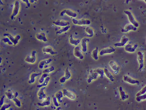
A wet mount of Naegleria fowleri trophozoites cultured from the CSF of a patient with primary amebic meningoencephalitis (PAM) viewed using phase contrast microscopy. Magnification: 600x. 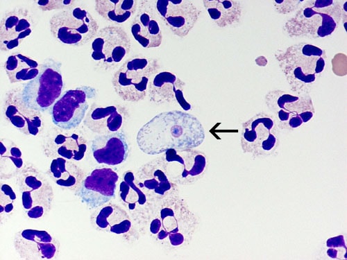
A cytospin of fixed CSF showing a Naegleria fowleri trophozoite (arrow) stained with Giemsa-Wright amidst polymorphonuclear leukocytes and a few lymphocytes. Within the trophozoite, the nucleus and nucleolus can be seen. Magnification: 1000x. 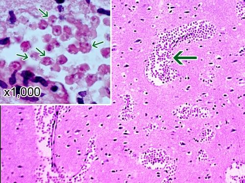
A section of the cerebral portion of the brain from a PAM patient, stained with hematoxylin and eosin, showing large clusters of Naegleria fowleri trophozoites and the destruction of the normal brain tissue architecture. Cysts are not seen. Magnification: 100x. Inset: Higher magnification (1000x) of Naegleria fowleri trophozoites (arrows) with the characteristic nuclear morphology. 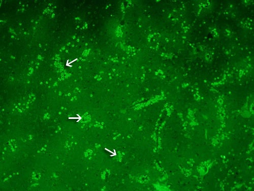
A section of the cerebral portion of the brain from a PAM patient reacted with the specific anti-Naegleria fowleri antibody which has been conjugated to a fluorescent antibody (immunofluorescent staining) viewed using microscopy with an exciter filter. Note the large numbers of Naegleria fowleri trophozoites staining bright green. No cysts are seen. Magnification: 100x. 
A section of the brain from a PAM patient stained with hemotoxylin and eosin showing a large cluster of Naegleria fowleri trophozoites surrounding capillaries. Magnification: 400x 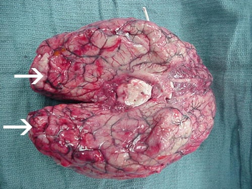
Extensive hemorrhage and necrosis is present in the brain, mainly in the frontal cortex. 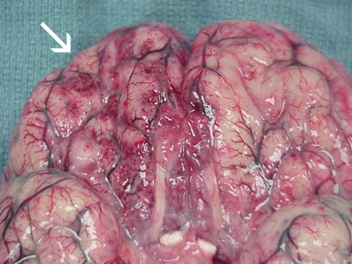
Extensive hemorrhage and necrosis is present in the brain, mainly in the frontal cortex. 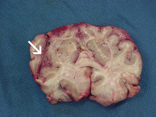
Focal hemorrhage and necrosis in frontal cortex due to Naegleria fowleri 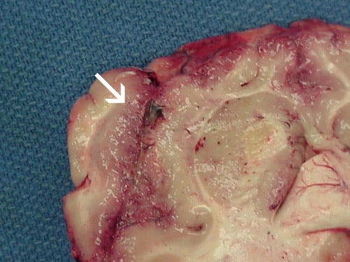
Focal hemorrhage and necrosis in frontal cortex due to Naegleria fowleri Syndicated Content Details:
Source URL: http://www.cdc.gov/parasites/naegleria/naegleria-fowleri-images.html Source Agency: Centers for Disease Control and Prevention (CDC) Captured Date: 2016-05-23 22:23:31.0
|