MERS-CoV PhotosCoronaviruses derive their name from the fact that under electron microscopic examination, each virion is surrounded by a “corona,” or halo. This is due to the presence of viral spike peplomers emanating from each proteinaceous envelope. Click on image to enlarge. 
Image source: Cynthia Goldsmith/Azaibi Tamin An electron micrograph of a thin section of MERS-CoV, showing the spherical particles within the cytoplasm of an infected cell. Click on image to enlarge. 
Image source: Jennifer L. Harcourt Human serum antibodies react with MERS-CoV-infected Vero cells, indicating the patient has been infected with MERS-CoV. Click on image to enlarge. 
Image source: Cynthia Goldsmith/Maureen Metcalfe/Azaibi Tamin MERS-CoV particles as seen by negative stain electron microscopy. Virions contain characteristic club-like projections emanating from the viral membrane. Click on image to enlarge. 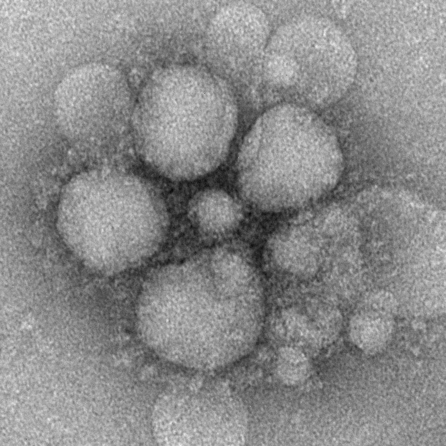
Image source: Cynthia Goldsmith/Maureen Metcalfe/Azaibi Tamin MERS-CoV particles as seen by negative stain electron microscopy. Virions contain characteristic club-like projections emanating from the viral membrane. Click on image to enlarge. 
Image source: Maureen Metcalfe/Azaibi Tamin An electron micrograph of a thin section of MERS-CoV, showing the spherical particles and cross-sections through the viral nucleocapsid. Click on image to enlarge. 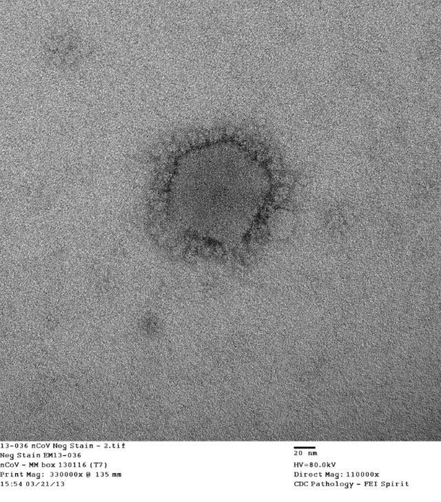
Image source: Cynthia Goldsmith/Azaibi Tamin Negative stain electron microscopy shows a MERS-CoV particle with club-shaped surface projections surrounding the periphery of the particle, a characteristic feature of coronaviruses. Syndicated Content Details:
Source URL: http://www.cdc.gov/coronavirus/mers/photos.html Source Agency: Centers for Disease Control and Prevention (CDC) Captured Date: 2016-05-23 22:24:10.0
|